User login
Microneedle system could replace blood draws, team says
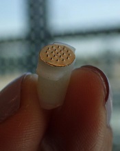
Photo courtesy of
Sahan Ranamukhaarachchi
A new microneedle drug monitoring system could one day replace invasive blood draws, according to researchers.
The system consists of a small, thin patch that is pressed against a patient’s arm during medical treatment and measures drugs in the bloodstream painlessly without drawing any blood.
The tiny projections on this patch resemble hollow cones and don’t pierce the skin like a standard hypodermic needle.
The researchers described this system in Scientific Reports.
“Many groups are researching microneedle technology for painless vaccines and drug delivery,” said study author Sahan Ranamukhaarachchi, a PhD student at the University of British Columbia (UBC) in Vancouver, British Columbia, Canada. “Using them to painlessly monitor drugs is a newer idea.”
The microneedle system Ranamukhaarachchi and his colleagues created was developed to monitor the antibiotic vancomycin. Patients taking vancomycin must be closely monitored because the drug can cause life-threatening side effects, so the patients undergo 3 to 4 blood draws per day.
The researchers discovered they could use fluid found just below the outer layer of skin, instead of blood, to monitor levels of vancomycin in the bloodstream.
The microneedle patch collects a tiny amount of the fluid, less than 1 nL, and a reaction occurs on the inside of the microneedles that can be detected using an optical sensor. This allows the user to quickly determine the concentration of vancomycin.
“This is probably one of the smallest probe volumes ever recorded for a medically relevant analysis,” said study author Urs Häfeli, PhD, of UBC.
This microneedle drug monitoring system was developed out of a research collaboration between Dr Häfeli and Boris Stoeber, PhD, also of UBC. The system is being commercialized by the UBC spin-off Microdermics Inc. ![]()
FDA approves reconstitution system for FVIII product
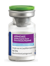
Photo courtesy of Baxalta
The US Food and Drug Administration (FDA) has approved the Baxject III reconstitution system for Adynovate, a pegylated recombinant factor VIII (FVIII) product.
The system is designed to mix a FVIII product with a diluent prior to infusion.
The Baxject III reconstitution system was previously FDA-approved for use with Advate, a recombinant FVIII product.
The latest FDA approval means the system will be available with Adynovate as well.
Adynovate and the diluent will come pre-packaged in the reconstitution system.
The Baxject III reconstitution system with Adynovate will be available to most customers in the fourth quarter of 2016, with a 2 mL diluent for the 250, 500, and 1000 IU potencies; and a 5 mL diluent for the 2000 IU potency.
Adynovate was approved by the FDA in 2015 for use in hemophilia A patients age 12 and older for on-demand treatment and control of bleeding and for prophylaxis to reduce the frequency of bleeding episodes. Full prescribing information is available here.
Advate was first approved by the FDA in 2003. The product is indicated for use in children and adults with hemophilia A for the control and prevention of bleeding episodes, perioperative management, and routine prophylaxis to prevent or reduce the frequency of bleeding episodes. Full prescribing information is available here.
The Baxject III reconstitution system, Adynovate, and Advate are all products of Baxalta, which is now a part of Shire. ![]()
HDAC inhibitor granted breakthrough designation

Image by Eric Smith
The US Food and Drug Administration (FDA) has granted breakthrough therapy designation for the histone deacetylase (HDAC) inhibitor pracinostat to be used in combination with azacitidine to treat newly diagnosed acute myeloid leukemia (AML) patients who are 75 and older or unfit for intensive chemotherapy.
The FDA’s breakthrough designation is intended to expedite the development and review of new therapies for serious or life-threatening conditions.
To earn the designation, a treatment must show encouraging early clinical results demonstrating substantial improvement over available therapies with regard to a clinically significant endpoint, or it must fulfill an unmet need.
The breakthrough therapy designation for pracinostat is supported by data from a phase 2 study of the HDAC inhibitor in combination with azacitidine in elderly patients with newly diagnosed AML who were not candidates for induction chemotherapy.
Detailed results from this trial were presented at the 20th Congress of the European Hematology Association last year. The research was sponsored by MEI Pharma, the company developing pracinostat.
The study included 50 AML patients who had a median age of 75 (range, 66-84).
The patients received pracinostat at 60 mg orally on days 1, 3, and 5 of each week for 21 days of each 28-day cycle. They received azacitidine subcutaneously or intravenously on days 1-7 or days 1-5 and 8-9 (per site preference) of each 28-day cycle.
According to updated data from MEI Pharma, the complete response rate was 42% (n=21), and the median overall survival was 19.1 months.
The company said these data compare favorably to a phase 3 study of azacitidine (AZA-AML-0011), which showed a median overall survival of 10.4 months with azacitidine alone and a complete response rate of 19.5% in a similar patient population.
The combination of pracinostat and azacitidine was thought to be well tolerated overall, with no unexpected toxicities. The most common grade 3-4 treatment-emergent adverse events included febrile neutropenia, thrombocytopenia, anemia, and fatigue. ![]()
Gene therapy shows promise for severe hemophilia A

Image by Spencer Phillips
ORLANDO—An investigational gene therapy can safely reduce bleeding in patients with severe hemophilia A, a phase 1/2 study suggests.
The therapy is BMN 270, a recombinant adeno-associated virus (AAV) vector coding for human coagulation factor VIII (FVIII).
Six of the 7 patients treated with the highest dose of BMN 270 had FVIII levels above 50%, and the number of bleeding events fell substantially from baseline.
None of the patients developed inhibitors to FVIII, there were no serious adverse events, and none of the patients discontinued the therapy due to safety reasons.
John Pasi, PhD, of Barts and the London School of Medicine and Dentistry in London, UK, presented the results of this study in a late-breaking oral presentation at the World Federation of Hemophilia 2016 World Congress.* The research was funded by BioMarin Pharmaceutical Inc.
This phase 1/2 dose-escalation study was designed to evaluate the safety and efficacy of BMN 270 in up to 12 patients with severe hemophilia A.
The primary endpoints are to assess the safety of a single dose of BMN 270 and the change from baseline of FVIII expression level at 16 weeks after infusion.
Secondary endpoints include assessing the impact of BMN 270 on the frequency of FVIII replacement therapy, the number of bleeding episodes requiring treatment, and any potential immune responses. Patients will be monitored for safety and durability of effect for 5 years.
Thus far, 9 patients with severe hemophilia A have received a single dose of BMN 270—1 at 6×10¹² vg/kg, 1 at 2×10¹³ vg/kg, and 7 at 6 x 10¹³ vg/kg.
As of the July 6 data cutoff, post-treatment follow-up ranges from 12 weeks to 28 weeks.
Safety
The most common adverse events were arthralgia (9 events in 6 subjects), contusion (6 events in 3 subjects), back pain (4 events in 3 subjects), and ALT elevation (6 events in 6 subjects).
No clinically relevant sustained rises in ALT levels or other markers of liver toxicity have been observed.
The maximum ALT levels were between 23 U/L and 82 U/L (less than 2 times the upper limit of normal, which is 43 U/L for the central laboratory in this study) approximately 12 weeks after gene delivery and generally declined over the next few weeks. ALT rises have not been associated with any decrease in FVIII levels.
A steroid regimen administered to all high-dose patients has been well-tolerated. Patients are successfully tapering off of steroids. Two patients have been off steroid therapy for up to 2.5 weeks, with no adverse impact on FVIII expression or ALT levels.
Efficacy
The patient treated at the lowest dose (6×10¹² vg/kg) had no change from baseline in FVIII levels. The patient treated at the mid-dose (2×10¹³ vg/kg) had a stable FVIII activity level greater than 2 IU/dL for more than 28 weeks.
All 7 patients treated at the highest dose (6×10¹³ vg/kg) had FVIII activity levels greater than 10 IU/dL after week 10.
As of each patient’s most recent reading, 6 of the 7 patients in the high-dose group had FVIII levels above 50%, as a percentage calculated based on the numbers of IU/dL. The seventh patient had levels above 10%.
Four patients who have been followed the longest had a mean FVIII level of 146% at their 20-week visit. Two patients with FVIII levels above 200% had no unexpected events or need for medical intervention.
For the 7 patients treated at the high dose, the median annualized bleeding rate measured from the day of gene transfer to the data cutoff fell from 20 to 5.
After week 7 post-infusion, there were no bleeds in 6 of the 7 patients. There were 10 bleeds from weeks 0 through 2 post-infusion, 7 bleeds from weeks 3 through 8, and 2 bleeds from weeks 9 through 28. From weeks 2 through 28, all but 1 bleed occurred in a single subject who is the lowest responder.
All of the patients in the high-dose cohort have switched to receiving FVIII therapy on-demand. Six of them were previously receiving FVIII therapy as prophylaxis.
“These data provide strong proof-of-concept evidence that restoration of clotting function may be achieved by gene therapy,” Dr Pasi said. “For the first time, patients have reason to hope to avoid bleeding and the opportunity to live a normal life.” ![]()
Molecule reverses effects of anticoagulants

A genetically engineered coagulation factor can reverse the effects of direct oral anticoagulants in vitro and in vivo, according to research published in Nature Medicine.
Researchers altered the shape of the coagulation factor, factor Xa (FXa), into a variant that appears to be more potent and longer-lasting than wild-type FXa.
The team found this variant, FXaI16L, could counteract the effects of rivaroxaban, apixaban, and dabigatran.
“This molecule holds the potential to fill an important unmet clinical need,” said study author Rodney A. Camire, PhD, of The Children’s Hospital of Philadelphia in Pennsylvania.
“There are limited treatment options to stop uncontrolled bleeding in patients who are using the newer anticoagulant medications.”
Dr Camire and his colleagues first found that FXaI16L could reverse the effects of rivaroxaban and apixaban in vitro, increasing peak thrombin generation to near-normal levels.
The team then showed that FXaI16L restores hemostasis in mice treated with rivaroxaban and significantly decreases blood loss in injured mice treated with the drug.
FXaI16L also significantly decreased blood loss in injured mice treated with dabigatran.
FXaI16L was more than 50 times more potent in the hemostasis models tested than andexanet alfa, a FXa inhibitor antidote currently in clinical development.
The researchers said FXaI16L’s ability to reverse the effects of anticoagulants depends, at least partly, on the ability of the active site inhibitor to hinder antithrombin-dependent FXa inactivation, which allows uninhibited FXa to persist in plasma.
“Our next steps will be to test this approach in large animals to help determine whether this variant is effective and safe and may progress to clinical trials,” Dr Camire said. “If so, we may be able to develop an important treatment to rapidly control bleeding in both children and adults.” ![]()
Targeted conjugate therapy kills ALL cells

Photo courtesy of the
University of California Davis
Researchers say they have developed a targeted conjugate therapy that harnesses a monoclonal antibody to deliver antisense DNA to acute lymphoblastic leukemia (ALL) cells.
Once delivered, the therapeutic DNA reduces levels of MXD3, a protein that helps cancer cells survive.
This conjugate therapy proved cytotoxic in ALL cell lines and showed promise in animal models, destroying ALL cells while limiting other damage.
“We’ve shown, for the first time, that anti-CD22 antibody-antisense conjugates are a potential therapeutic agent for ALL,” said Noriko Satake, MD, of the University of California Davis in Sacramento.
“This could be a new type of treatment that kills leukemia cells with few side effects.”
Dr Satake and her colleagues described the treatment in Molecular Medicine.
To create the therapy, the researchers attached antisense DNA that inhibits the MXD3 protein to an antibody that binds to CD22, a protein receptor expressed almost exclusively on ALL cells and normal B cells.
Once the antibody binds to CD22, the conjugate is drawn inside the cell, allowing the antisense molecule to prevent MXD3 production. Without this anti-apoptotic protein, cells are more prone to death.
The conjugate therapy was effective against ALL cell lines and primary ALL cells in a xenograft mouse model. Animals that received the therapy survived significantly longer than those in the control group.
While the conjugate therapy does target healthy B cells along with ALL cells, it is expected to leave hematopoietic stem cells and other tissues unharmed.
“Our novel conjugate is designed so that it does not harm hair, eyes, heart, kidneys, or other types of cells,” Dr Satake said.
She and her colleagues noted that, although this study shows the conjugate therapy can knock down MXD3, it is not clear exactly how this is accomplished. So the researchers plan to investigate the mechanism.
The team also plans to look into combining the conjugate therapy with other treatments. Because it hastens cell death, the conjugate could make traditional chemotherapy drugs more effective, and it might work against other cancers.
“You can see this as proof of principle,” Dr Satake said. “You could switch the target and substitute the antibody, which could be used to treat other cancers or even other diseases.”
This study was not industry-funded, but 4 study authors are employees and stockholders of Isis Pharmaceuticals. ![]()
Local Zika transmission and the US blood supply

Photo courtesy of
Muhammad Mahdi Karim
Officials have announced what is likely the first known occurrence of local mosquito-borne Zika virus transmission in the continental US.*
Fourteen cases of Zika virus in 2 Florida counties are believed to have been caused by bites of local Aedes aegypti mosquitoes.
The Florida Department of Health (DOH) believes that active transmission of the Zika virus is only occurring in a small area in Miami-Dade County.
The US Centers for Disease Control and Prevention (CDC) has recommended that women who are pregnant or thinking of becoming pregnant avoid unnecessary travel to the impacted area. The agency has also issued a guidance for people living in or traveling to the area.
“All the evidence we have seen indicates that this is mosquito-borne transmission that occurred several weeks ago in several blocks in Miami,” said Tom Frieden, MD, director of the CDC.
The exact location is within the boundaries of the following area: NW 5th Avenue to the west, US 1 to the east, NW/NE 38th Street to the north and NW/NE 20th Street to the south. This is about one square mile.
Protecting the blood supply
The US Food and Drug Administration (FDA) has requested that all blood centers in Miami-Dade and Broward counties stop collecting blood immediately.
Blood centers in these counties can resume blood collection once they begin testing each unit of blood with an available investigational donor screening test for Zika virus RNA or once they implement the use of an approved or investigational pathogen inactivation technology.
The FDA also recommended that blood centers in nearby counties implement the same precautions as soon as possible to help maintain the safety of the blood supply. The agency has encouraged screening of the blood supply in regions of the US at risk of local mosquito-borne Zika transmission.
The FDA said it is working with companies that are making blood screening tests available under an Investigational New Drug (IND) application to ensure these companies are ready to expand testing as needed. Blood collection centers may choose to participate in testing under an IND even in the absence of local mosquito-borne transmission of Zika virus.
Florida/CDC response
Florida state officials have implemented mosquito control measures and a community-wide search for additional Zika cases. Thus far, Florida’s DOH has conducted testing for the Zika virus in more than 2300 people statewide.
The DOH has activated the Joint Information Center within the State Emergency Operations Center to ensure the area impacted by local transmission of the Zika virus has coordinated access to information and resources.
The DOH has also begun the process of contracting with commercial pest control companies to enhance and expand mosquito mitigation and abatement, including increased spraying, in the impacted area.
Earlier this year, Florida’s governor, Rick Scott, directed the State Surgeon General to activate a Zika Virus Information Hotline for Florida residents and visitors. The number for this hotline is 1-855-622-6735.
The CDC said it is coordinating with Florida officials leading the ongoing investigation into local transmission of the Zika virus. At the state’s request, the CDC sent a medical epidemiologist to provide additional assistance.
Governor Scott has also asked the CDC to activate a CDC Emergency Response Team to assist the DOH and other partners in their investigation, sample collection, and mosquito control efforts.
To date, the CDC has provided Florida with more than $8 million in Zika-specific funding and about $27 million in emergency preparedness funding that can be used for Zika response efforts.
“We have been working with state and local governments to prepare for the likelihood of local mosquito-borne Zika virus transmission in the continental United States and Hawaii,” said Lyle Petersen, MD, incident manager for CDC’s Zika virus response.
“We anticipate that there may be additional cases of ‘homegrown’ Zika in the coming weeks. Our top priority is to protect pregnant women from the potentially devastating harm caused by Zika.”
For more information about the Zika virus, visit http://www.cdc.gov/zika/. ![]()
*This story was updated on August 1.
Photo courtesy of
Muhammad Mahdi Karim
Officials have announced what is likely the first known occurrence of local mosquito-borne Zika virus transmission in the continental US.*
Fourteen cases of Zika virus in 2 Florida counties are believed to have been caused by bites of local Aedes aegypti mosquitoes.
The Florida Department of Health (DOH) believes that active transmission of the Zika virus is only occurring in a small area in Miami-Dade County.
The US Centers for Disease Control and Prevention (CDC) has recommended that women who are pregnant or thinking of becoming pregnant avoid unnecessary travel to the impacted area. The agency has also issued a guidance for people living in or traveling to the area.
“All the evidence we have seen indicates that this is mosquito-borne transmission that occurred several weeks ago in several blocks in Miami,” said Tom Frieden, MD, director of the CDC.
The exact location is within the boundaries of the following area: NW 5th Avenue to the west, US 1 to the east, NW/NE 38th Street to the north and NW/NE 20th Street to the south. This is about one square mile.
Protecting the blood supply
The US Food and Drug Administration (FDA) has requested that all blood centers in Miami-Dade and Broward counties stop collecting blood immediately.
Blood centers in these counties can resume blood collection once they begin testing each unit of blood with an available investigational donor screening test for Zika virus RNA or once they implement the use of an approved or investigational pathogen inactivation technology.
The FDA also recommended that blood centers in nearby counties implement the same precautions as soon as possible to help maintain the safety of the blood supply. The agency has encouraged screening of the blood supply in regions of the US at risk of local mosquito-borne Zika transmission.
The FDA said it is working with companies that are making blood screening tests available under an Investigational New Drug (IND) application to ensure these companies are ready to expand testing as needed. Blood collection centers may choose to participate in testing under an IND even in the absence of local mosquito-borne transmission of Zika virus.
Florida/CDC response
Florida state officials have implemented mosquito control measures and a community-wide search for additional Zika cases. Thus far, Florida’s DOH has conducted testing for the Zika virus in more than 2300 people statewide.
The DOH has activated the Joint Information Center within the State Emergency Operations Center to ensure the area impacted by local transmission of the Zika virus has coordinated access to information and resources.
The DOH has also begun the process of contracting with commercial pest control companies to enhance and expand mosquito mitigation and abatement, including increased spraying, in the impacted area.
Earlier this year, Florida’s governor, Rick Scott, directed the State Surgeon General to activate a Zika Virus Information Hotline for Florida residents and visitors. The number for this hotline is 1-855-622-6735.
The CDC said it is coordinating with Florida officials leading the ongoing investigation into local transmission of the Zika virus. At the state’s request, the CDC sent a medical epidemiologist to provide additional assistance.
Governor Scott has also asked the CDC to activate a CDC Emergency Response Team to assist the DOH and other partners in their investigation, sample collection, and mosquito control efforts.
To date, the CDC has provided Florida with more than $8 million in Zika-specific funding and about $27 million in emergency preparedness funding that can be used for Zika response efforts.
“We have been working with state and local governments to prepare for the likelihood of local mosquito-borne Zika virus transmission in the continental United States and Hawaii,” said Lyle Petersen, MD, incident manager for CDC’s Zika virus response.
“We anticipate that there may be additional cases of ‘homegrown’ Zika in the coming weeks. Our top priority is to protect pregnant women from the potentially devastating harm caused by Zika.”
For more information about the Zika virus, visit http://www.cdc.gov/zika/.
*This story was updated on August 1.
mAb could change treatment of hemophilia A, doc says

Photo by Linda Bartlett
ORLANDO—Emicizumab, a bispecific monoclonal antibody (mAb), could potentially change the treatment paradigm of hemophilia A, according to a speaker at the World Federation of Hemophilia 2016 World Congress.
Results of a phase 1/2 study suggest that emicizumab can be safe and effective as once-weekly prophylaxis in patients with severe hemophilia A, whether or not they have factor VIII (FVIII) inhibitors.
There were no severe adverse events (AEs) or thromboembolic events associated with the mAb.
None of the patients developed neutralizing anti-drug antibodies, and the annualized bleeding rate (ABR) was low, with 8 of the 18 patients studied having no bleeds.
Keiji Nogami, MD, PhD, of Nara Medical University in Kashihara, Japan, presented these results at the congress.* This research was sponsored by Chugai Pharmaceutical Co., Ltd., which is co-developing emicizumab with Genentech.
The phase 1 part of this study included both healthy subjects and hemophilia A patients. Results in the healthy subjects were published in Blood, and early results in the patients were published in NEJM.
Patients and treatment
The 18 patients in this study had severe hemophilia A, and 11 had FVIII inhibitors. They were 12 to 58 years of age at baseline, and all were Japanese.
The patients received once-weekly subcutaneous injections of emicizumab at one of the following dose levels: 0.3 mg/kg (cohort 1), 1 mg/kg (cohort 2), or 3 mg/kg (cohort 3).
There were 6 patients in each cohort. Cohorts 1 and 2 each had 4 patients with inhibitors, and there were 3 patients with inhibitors in cohort 3.
For the phase 2 extension portion of the study, dose-escalation was allowed in cohorts 1 and 2. A total of 4 patients had their doses increased because of suboptimal bleeding control—3 in cohort 1 and 1 in cohort 2.
The mean follow-up was 32.6 months in cohort 1, 27.0 months in cohort 2, and 21.4 months in cohort 3.
One of the patients with inhibitors in cohort 2 discontinued emicizumab due to injection site erythema of mild intensity during the phase 1 portion of the study. And one of the patients without inhibitors in cohort 3 did not continue on to the phase 2 portion of the study because prior treatment was sufficiently effective.
Safety
The safety analysis included all 18 patients, and all of these patients experienced AEs—a total of 150 events. The most common AEs were nasopharyngitis (n=8), contusion (n=7), and injection site reactions (n=7).
The AEs were largely of mild or moderate intensity. There were 4 severe AEs, but none of them were related to emicizumab.
There were 14 treatment-related AEs in 7 patients, but all were resolved. There were no thromboembolic AEs or clinically significant abnormalities of coagulation tests.
There were no neutralizing anti-drug antibodies. Three patients developed anti-drug antibodies after starting emicizumab, but this did not affect the pharmacokinetics, pharmacodynamics, safety, or efficacy of the mAb.
Efficacy
In cohort 1, the median ABR was 32.5 at baseline, 4.4 at 12 weeks, and 1.4 at last follow-up (median of 32.6 months).
In cohort 2, the median ABR was 18.3 at baseline, 0.0 at 12 weeks, and 0.2 at last follow-up (median of 27.0 months).
In cohort 3, the median ABR was 15.2 at baseline, 0.0 at 12 weeks, and 0.0 at last follow-up (median of 21.4 months).
Eight patients did not experience any bleeds—1 inhibitor patient in cohort 1, 3 inhibitor patients in cohort 2, 2 inhibitor patients in cohort 3, and 2 non-inhibitor patients in cohort 3.
Breakthrough bleeds were successfully treated with standard episodic treatment, FVIII products, or bypassing agents.
Based on these results, Dr Nogami said emicizumab prophylaxis has the potential to change the treatment paradigm of hemophilia A, irrespective of inhibitor status.
How hemophilia B impacts education, work

ORLANDO—Even mild or moderate hemophilia B can have a negative impact on the education and careers of patients, according to the B-HERO-S study.
Ninety-four percent of the patients studied said their disease had a negative impact on their education, and 95% said hemophilia had a negative impact on their work.
More than half of patients who had stopped working said they stopped due to hemophilia-related financial issues and/or complications.
These findings were presented in a poster at the World Federation of Hemophilia 2016 World Congress.* The study was sponsored by Novo Nordisk.
The previous HERO study revealed career challenges for patients with hemophilia A or B, but it covered mostly males with moderate or severe disease.
The B-HERO-S study, on the other hand, included only patients with hemophilia B, some of whom were female and most of whom had moderate or mild disease. (This study also included caregivers of children with hemophilia, but data on those subjects will not be discussed here.)
In all, there were 299 patients, 86 of whom were women. The patients had a median age of 29 (range, 18-70). Twenty-five percent had mild hemophilia, 63% had moderate disease, and 11% had severe hemophilia.
Education
Most of the patients (71%) had at least some college education, and 17% had graduated from college.
A majority of patients (94%) said their disease had a negative impact on their education. Seventy-five percent of the patients, including those with mild or moderate disease, said hemophilia had a moderate or large impact on their education.
Patients reported difficulty concentrating at school due to bleeds or pain (69%), difficulty attending school or participating in activities due to mobility issues (44%), and hemophilia-related absences (32%).
Career
Most of the patients (81%) were working full- or part-time when surveyed. Fifty-eight percent had office-based jobs, and 39% had jobs involving manual labor. Only 10% of patients said their disease affected their career choice.
Of the 58 patients (19%) who were not working at the time of the study, 62% had never worked. Of those who worked previously, 59% said they stopped due to hemophilia-related financial issues, and 55% said they stopped due to the disease itself and/or its complications.
Nearly all patients (95%) said their disease had a negative impact on their work. Seventy percent said the impact was moderate, 20% said it was small, and 5% said it was large.
Having moderate disease, comorbidities, a higher education, and/or receiving routine infusions were all associated with a higher impact.
Thirty percent of patients said that treatment allowed them to work in most situations. This was more likely in patients with mild hemophilia (41%) and those treated on-demand (48%).
Twenty-seven percent of patients with moderate hemophilia and 28% with severe disease said they could work in most situations. Twenty-seven percent of patients receiving routine infusions said they were able to work in most situations.
“The B-HERO-S study brought to light many challenges faced by some patients with mild/moderate hemophilia B, including affected women and girls,” said Michelle Witkop, of Northern Regional Bleeding Disorder Center in Traverse City, Michigan.
“With hemophilia treatment centers (HTCs) and local hemophilia chapters focused on proactive education in people with severe hemophilia and their families, B-HERO-S data tells us we need to make sure that those with mild/moderate hemophilia B come to the HTC for routine visits so that we can proactively address issues that might come up in school, activities, and career choice.”
Light-based therapy may treat thrombocytopenia
in the bone marrow
A low-intensity type of laser therapy could provide a non-invasive, drug-free treatment option for thrombocytopenia, according to research published in Science Translational Medicine.
Researchers found that low-level laser (LLL) therapy increased the generation of platelets from megakaryocytes in vitro and had the same effect in mouse models of thrombocytopenia.
The team also identified the probable mechanism underlying this effect.
“Our study reveals, for the first time, that low-level laser therapy enhances platelet production in animals with thrombocytopenia but not in normal controls,” said study author Mei X. Wu, PhD, of Massachusetts General Hospital in Boston.
“This result suggests that a safe, drug-free method that does not depend on donated blood products can be developed for treating or preventing thrombocytopenia.”
LLLs emit low-powered laser light that does not heat its target tissue. LLLs are known to protect the function of mitochondria, and several conditions associated with impaired platelet production are characterized by abnormalities in the mitochondria of cells that give rise to platelets.
Dr Wu and her colleagues conducted a number of experiments to investigate whether LLLs’ ability to protect mitochondrial function could mitigate several forms of thrombocytopenia.
The team found that LLL treatment of megakaryocytes increased their size, accelerated the formation of proplatelets, and doubled the production of platelets.
Infusion of LLL-treated megakaryocytes into mice led to greater platelet production than did infusion of megakaryocytes treated with normal light.
One of the keys to determining the number of platelets generated from megakaryocytes was mitochondrial production of the energy molecule ATP.
LLL treatment greatly increased mitochondrial generation in polyploid megakaryocytes, but the increase was only slight in less mature megakaryocytes with only 2 copies of each chromosome.
Whole-body LLL treatment of mice with radiation-induced thrombocytopenia spurred the rapid maturation of megakaryocytes and restored platelet levels in a light-dose-dependent fashion.
Platelets from LLL-treated mice had normal structure and function. LLL treatment of normal mice did not raise levels of either megakaryocytes or platelets.
LLL treatment also restored platelet levels in mice with the autoimmune form of thrombocytopenia or with thrombocytopenia caused by chemotherapy.
In cultured human megakaryocytes, LLL treatment at dosage levels similar to those used in mice increased ATP production and platelet generation.
Dr Wu noted that LLL’s lack of an effect in animals without thrombocytopenia indicates it would probably avoid the potential complications of current drug treatments for thrombocytopenia, which act by increasing the production of megakaryocytes from their progenitors in the bone marrow.
“Directly stimulating the differentiation of [megakaryocytes] the way all current drugs do risks clotting if platelet levels rise too high,” Dr Wu said. “LLL appears to enhance [megakaryocytes’] inherent ability to produce platelets most effectively in response to low platelet levels in the circulation, a response that stops when platelet levels are normalized.”
“The fact that treatment only has an effect in polyploid cells, which are very rare, implies that it would not increase production of mitochondria in cancer cells or other cells. In fact, while LLL has been employed in research and in clinical treatment for decades, this is the first study reporting that it can promote mitochondrial biogenesis.”
Dr Wu added that the current primary obstacle to testing LLL in humans is the lack of a device large enough to treat either the entire body or enough bones to stimulate sufficient platelet production by megakaryocytes within the bone marrow, something her team plans to address.
She also noted that, while LLL will probably be beneficial for treatment of many forms of acquired thrombocytopenia, it may not be effective when the condition is caused by inborn genetic defects.