User login
Team creates database of autophagy-related proteins

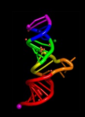
Image by Sarah Pfau
Scientists have identified previously undiscovered proteins related to autophagy and created a database containing them.
The team believes this database will enable the scientific community to find ways to activate autophagy inside human cells.
And it could aid the development of drugs to treat age-related conditions such as cancer.
The team described the creation of the database in the journal Autophagy.
The database is available here: https://ilir.warwick.ac.uk/.
“Our novel database resource will open a lot of new avenues in basic and translational science,” said Ioannis Nezis, PhD, of the University of Warwick in Coventry, UK.
“Identifying novel selective autophagy-related proteins will help for the development of novel pharmaceutical drug targets for a large variety of diseases like cancer, neurodegeneration, and other aging-related diseases, infections, diabetes, obesity, and Crohn’s disease.”
“Importantly, understanding the molecular mechanisms of selective autophagy will help researchers to find interventions to activate the autophagic pathway to prevent aging and promote healthy well-being during the life course.” ![]()
Drug can prevent bleeding in hemophilia A and B
ORLANDO—Results from an ongoing phase 1 study suggest fitusiran, a small interfering RNA therapeutic targeting antithrombin (AT), can restore hemostasis and prevent bleeding in patients with hemophilia A or B, with or without inhibitors.
In patients with inhibitors, fitusiran exhibited preliminary evidence of reduced bleeding. In patients without inhibitors, fitusiran reduced the median estimated annualized bleeding rate (ABR) to 0.
In addition, researchers said fitusiran was generally well tolerated, and none of the patients developed anti-drug antibodies.
These results were presented at the World Federation of Hemophilia 2016 World Congress.* The study was sponsored by Alnylam Pharmaceuticals, Inc.
Study design
This phase 1 trial consists of 4 parts. Part A enrolled healthy volunteers who were randomized 3:1 to fitusiran or placebo. This part of the study was completed after the first dose cohort received a single subcutaneous dose of fitusiran at 30 mcg/kg.
Part B, which is also complete, enrolled 12 patients with severe hemophilia A or B. Patients received 3 weekly subcutaneous injections of fitusiran at doses of 15, 45, or 75 mcg/kg.
Part C, in which dosing is complete, enrolled 18 patients with moderate or severe hemophilia A or B without inhibitors. Twelve patients received 3 monthly subcutaneous doses of fitusiran at 225, 450, 900, or 1800 mcg/kg. Six patients received 3 fixed monthly subcutaneous doses of fitusiran at 80 mg.
Part D is designed to enroll up to 18 patients with inhibitors. The first cohort of 6 patients received a 50 mg, fixed, once-monthly, subcutaneous dose. The second cohort has completed enrollment, with 6 patients receiving an 80 mg, fixed, once-monthly, subcutaneous dose.
Results from Parts C and D were presented at the meeting.
Part C results
Treatment with fitusiran resulted in a dose-dependent, statistically significant decrease in AT and increase in thrombin generation. At the 80 mg monthly dose, fitusiran achieved 87±1% mean maximal AT lowering with low inter-patient variability.
Researchers assessed the association between AT lowering and increased thrombin generation in a post-hoc exploratory analysis in which AT lowering was grouped by 25% increments for completed patients in Parts B (n=12) and C (n=17) of the study.
In the highest quartile of ≥75% AT lowering (n=16), fitusiran administration resulted in mean increases in thrombin generation of approximately 290% relative to baseline (P<0.001, as compared to the lowest AT-lowering quartile).
There was a significant, AT-lowering-dependent reduction in bleeding frequency as well.
To obtain a comprehensive assessment of fitusiran’s potential effects on bleeding, researchers performed a post-hoc analysis in evaluable patients from Part C (n=17).
The team compared bleeding events that occurred over the 6-month period prior to study entry, bleeding events that were assessed prospectively during days 0-28 following the initial fitusiran dose (the onset period), and bleeding events that occurred beyond day 29 up to day 112 (the observation period).
Prior to study entry, the estimated median ABR was 28 for patients receiving on-demand factor therapy (n=4) and 2 for patients receiving prophylactic factor therapy (n=13).
The estimated median ABR was 13 among all evaluable patients during the onset period and 0 during the observation period.
In all Part C dose cohorts during the observation period, 53% of patients (9/17) were bleed-free, and 82% of patients reported no spontaneous bleeds.
Part D results
Prior to study entry, all patients in Part D utilized bypass agents, including recombinant factor VIIa and activated prothrombin complex concentrate, to manage their bleeds. They had a pre-study ABR of up to 80.
The first cohort (n=6) of patients received a once-monthly, fixed subcutaneous dose of 50 mg. Fitusiran achieved a mean maximal AT lowering of 81±2% and a mean maximal thrombin generation increase of approximately 368%.
In addition, there was preliminary evidence of reduced bleeding, with a 49% to 100% reduction in estimated ABR during the observation period compared with pre-study values.
Safety results
As of July 11, 2016, fitusiran appears to be generally well tolerated in hemophilia patients, with or without inhibitors (n=31, with 5 patients participating in both Parts B and C).
There have been no serious adverse events related to the drug and no thromboembolic events or laboratory evidence of pathologic clot formation.
One non-inhibitor patient in Part C treated with the 80 mg fixed dose discontinued treatment due to an adverse event that was considered severe and possibly related to the study drug.
This event was described as non-cardiac chest pain and was accompanied by transient elevations of ALT (10x upper limit of normal), AST (8x upper limit of normal), C-reactive protein, and D-dimer, without an increase in total bilirubin. The event resolved with symptomatic management, including antacids and analgesics.
Eleven patients (35%) reported mild, drug-related injection site reactions, which were mostly pain or erythema at the injection site.
Additional adverse events reported in at least 10% of patients included upper respiratory tract infection (10%) and arthralgia (10%). The majority of these events were mild or moderate in severity. ![]()
Sickle cell trait doesn’t increase risk of death, study suggests
Results of a large study contradict the view that having sickle cell trait increases a person’s risk of death.
Health records of nearly 50,000 active-duty US Army soldiers showed no significant difference in the risk of death between soldiers who had sickle cell trait and those who did not.
The risk of exertional rhabdomyolysis (ER) was 54% higher among soldiers with sickle cell trait than those without it.
But the study suggested that tobacco use, obesity, and taking certain drugs also incur a heightened risk of ER.
Lianne Kurina, PhD, of Stanford University School of Medicine in California, and her colleagues reported these findings in NEJM.
Previous studies have suggested the health consequences of sickle cell trait might be dire, including higher mortality from ER. ER is characterized by the severe breakdown of skeletal-muscle tissue due to extreme physical exertion. The condition has been known to affect athletes and soldiers.
To assess the risk of ER and death among people with sickle cell trait, Dr Kurina and her colleagues reviewed the health records of 47,944 African-American soldiers who served on active duty between 2011 and 2014 and for whom sickle cell status was known.
The team found no significant difference in the risk of death among soldiers with sickle cell trait and those without. The hazard ratio (HR) was 0.99 (95% confidence interval [CI], 0.46 to 2.13; P=0.97).
Sickle cell trait was associated with a significantly higher adjusted risk of ER, with an HR of 1.54 (95% CI, 1.12 to 2.12; P=0.008).
However, the risk of ER was also higher for the following groups:
- Soldiers who used tobacco (HR=1.54, 95% CI, 1.23 to 1.94; P<0.001)
- Those with a body mass index of 30 or higher, as compared to 25 or lower (HR=1.39, 95% CI, 1.04 to 1.86; P=0.03)
- Those who recently used a statin (HR=2.89, 95% CI, 1.51 to 5.55; P=0.001)
- Those who recently used an antipsychotic agent (HR=3.02, 95% CI, 1.34 to 6.82; P=0.008).
Dr Kurina said the reason the results of this study differ from those of previous studies may be better safety for active-duty soldiers.
As of 2003, soldiers who are engaged in strenuous exercise are required to drink plenty of fluids, build up to strenuous exercise gradually, and take regular rests when it’s hot. All of these measures are known to reduce exercise-related fatality rates, regardless of whether individuals have sickle cell trait, the researchers said.
“Another critical difference between our study and the earlier, population-based studies is that, in our study, we knew the sickle cell status of everyone in the population,” Dr Kurina said.
She and her team looked only at soldiers whose sickle cell status was confirmed by blood tests taken during their years of service, instead of from self-reported sickle cell status or past medical history, as had been done in the other studies.
“The most important thing to come out of this study is the really reassuring news that, under conditions of universal precautions against dehydration and overheating, we don’t see an elevation in the risk of mortality in people with sickle cell trait,” Dr Kurina said.
She added that the study’s results call into question the need to screen service members with sickle cell trait, especially with better safety precautions during intense exertion. ![]()
Improving upon results with checkpoint inhibitors

Photo by Rob Press
Manipulating metabolic events might reinvigorate exhausted T cells and complement the effects of checkpoint inhibitors in the treatment of cancers, according to research published in Immunity.
When T cells are activated because of a tumor or microbe, they transition from a catabolic existence of slow metabolic burn to an anabolic one in which the body revs up to generate chemical intermediates to build new cells.
However, T cells are hard-wired to stop the anabolic mode at a certain point because functioning at that level is unsustainable.
PD-1, a cell surface receptor and target of checkpoint inhibitors, tells T cells to turn off the anabolic pathway, but other molecular signals attempt to keep this pathway turned on because growing tumors or chronic infection are still present.
This results in “metabolically confused” T cells, said study author E. John Wherry, PhD, of the University of Pennsylvania School of Medicine in Philadelphia.
To study this, Dr Wherry and his colleagues induced infection in mice using 2 different strains of the lymphocytic choriomeningitis virus, a model system for exploring T-cell biology.
“We found that, as early as the first week of a chronic viral infection, even before severe T-cell dysfunction becomes established, virus-specific T cells are already unable to match the bioenergetic demands of T cells generated during the height of fighting a well-contained viral infection in a mouse model,” Dr Wherry said.
In other words, these experiments revealed when PD-1 turns off the anabolic metabolism signal, and it’s earlier than previously thought.
The researchers said this finding is important because it identifies the altered metabolism as a distinct point in the development of exhausted T cells, rather than as a later consequence of exhausted T cells.
This research also revealed PD-1’s role as the metabolic switch in shutting down anabolic pathways and characterized downstream metabolic regulator targets of PD-1.
For example, restriction of glucose uptake and utilization, despite the upregulation of multiple backup metabolic pathways, was one metabolic defect in the exhausted T cells. PD-1 partially controls the development of this early defect in using glucose as a fuel, as well as the size and quality of mitochondria.
A second pathway repressed by PD-1 involved PGC-1α, a protein that regulates genes involved in metabolism. Correcting this PD-1-induced defect by overexpressing PGC-1α improved exhausted T-cell bioenergetics.
The researchers said these results have implications for therapeutic strategies aimed at the reinvigoration of exhausted T cells in chronic infections and cancer. And targeting exhausted T-cell metabolism could complement the effects of blocking PD-1 and other checkpoints. ![]()
Many pediatric trials go unfinished, unpublished

Photo by Logan Tuttle
Clinical trials in children too often go uncompleted or unpublished, according to a pair of researchers.
The duo evaluated nearly 560 pediatric trials and found that 19% were discontinued early. Of the trials that were completed, 30% remained unpublished several years later.
Industry-sponsored trials were more likely to be completed than trials sponsored by academic institutions. However, completed trials sponsored by industry were less likely to be published than trials sponsored by academia.
“Our findings are in line with previously published studies focusing on adult trials, which may speak to how commonplace discontinuation and non-publication are in medical research in general,” said study author Natalie Pica, MD, PhD, of Boston Children’s Hospital in Massachusetts.
She and Florence Bourgeois, MD, also of Boston Children’s Hospital, reported these findings in Pediatrics.
The researchers tracked 559 randomized, controlled pediatric trials registered on ClinicalTrials.gov from 2008 to 2010 and whose final status (completed or discontinued) was confirmed by the end of 2012.
The pair then searched for related peer-reviewed articles published through September 1, 2015. When no publication could be found, the researchers inquired with study investigators and sponsors via email.
Of the 559 trials, 104 (19%) were discontinued early. Two-thirds of these had enrolled participants.
Of the 455 completed trials, 136 (30%) remained unpublished after an average of 58 months post-completion. However, 42 of these (31%) did have results posted on ClinicalTrials.gov.
Of the 104 discontinued trials, 39% were sponsored by industry, and 55% were sponsored by academic institutions. (The rest were funded by other sources.)
Two years after trial completion, academia-sponsored trials accounted for 30% of unpublished trials, and industry-sponsored trials accounted for 63%.
Three years after trial completion, academia-sponsored trials accounted for 23% of unpublished trials, and industry-sponsored trials accounted for 70%.
In a multivariate analysis, the likelihood of non-publication was more than doubled for industry-sponsored trials 2 years after completion (odds ratio=2.21) and more than tripled 3 years after completion (odds ratio=3.12).
Overall, more than 8000 children were enrolled in trials that were never completed, and more than 69,000 children were enrolled in completed trials that were never published.
“This is the first study to look systematically at discontinuation and nonpublication of interventional pediatric clinical trials,” Dr Bourgeois said.
“A number of legislative initiatives have been implemented to increase the study of interventions in children. Now, we need to make sure that the proper resources are in place to ensure that information gleaned from these studies reaches the scientific community.”
One proposed initiative cited by Drs Bourgeois and Pica is RIAT (Restoring Invisible and Abandoned Trials), which is supported by some high-profile journals. RIAT invites researchers with unpublished trials to either commit to publish within a year or provide public access to their data.
“It’s hard to reanalyze others’ data,” Dr Pica noted, “but this may be a useful mechanism to make sure that findings from completed trials are disseminated in the medical literature.” ![]()
How iPSCs differentiate to blood cells

Image from Salk Institute
New research suggests the type of founder cell used to generate induced pluripotent stem cells (iPSCs) does not affect the iPSCs’ ability to differentiate into hematopoietic cells.
Instead, researchers found the expression of certain genes and DNA methylations were better indicators of the efficiency at which a cell line could be differentiated into the hematopoietic lineage.
The team reported these findings in Cell Stem Cell.
The researchers assessed the hematopoietic differentiation capacities of 35 iPSC lines derived from 4 types of somatic tissues—human dermal fibroblasts, hematopoietic cells such as cord blood and peripheral blood, dental pulp cells, and keratinocytes—from 15 donors.
The team also assessed 4 embryonic stem cell lines in early phase and late phase.
The researchers found that hematopoietic commitment capacity was associated with expression of IGF2 in undifferentiated iPSCs, but not with type of founder cell.
Higher expression of IFG2 was indicative of iPSCs initiating their conversion into hematopoietic cells. Even though IFG2 itself is not directly related to hematopoiesis, its uptake corresponded to an increase in the expression of genes that are.
Although IFG2 marked the beginnings of differentiation to hematopoietic lineage, the completion of differentiation was marked by the methylation profiles of the iPSC DNA.
“DNA methylation has an effect on a cell staying pluripotent or differentiating,” explained study author Yoshinori Yoshida, MD, PhD, of the Center for iPS Cell Research and Application at Kyoto University in Japan.
The completion of differentiation correlated with less aberrant methylation during the reprogramming process.
Hematopoietic founder cells showed a much lower propensity for aberrant methylation than did other founder cells, which could explain why, in the past, scientists attributed the founder cell to the effectiveness of differentiating iPSCs to the hematopoietic lineage.
Dr Yoshida and his colleagues said this research revealed molecular factors that can be used to evaluate the differentiation potential of different cell lines, which should expedite the progress of iPSCs to clinical use. ![]()
Method provides more accurate diagnosis of MDS, team says

Next-generation sequencing (NGS) of cell-free DNA should be the method of choice to confirm the diagnosis of myelodysplastic syndromes (MDS), according to researchers.
The team found that using NGS to analyze samples from MDS patients yielded more accurate results than Sanger sequencing.
And sequencing cell-free DNA rather than peripheral blood cell DNA increased the likelihood of detecting mutations associated with MDS.
The team reported these findings in Genetic Testing and Molecular Biomarkers. This research was funded by NeoGenomics Laboratories.
For this study, the researchers performed NGS on a panel of 14 target genes using total nucleic acid extracted from the plasma of 16 patients with early MDS (blasts <5%). The team also performed Sanger sequencing and NGS on peripheral blood cell DNA from the same patients.
The researchers found that NGS of cell-free DNA confirmed the diagnosis of MDS in all 16 patients.
In addition, NGS of cell-free DNA revealed abnormalities in 5 patients (31%) that were not detected by Sanger sequencing of peripheral blood cell DNA.
NGS of peripheral blood cell DNA produced the same results as NGS of cell-free DNA for 4 of the 5 patients. However, NGS of peripheral blood cell DNA did not detect a mutation in the RUNX1 gene that was evident in cell-free DNA from 1 patient.
Overall, the researchers found that mutant allele frequency was significantly higher in cell-free DNA than cellular DNA (P=0.008).
The team therefore concluded that cell-free DNA is more reliable than peripheral blood cell DNA for detecting molecular abnormalities in patients with MDS, and NGS is more accurate than Sanger sequencing. ![]()
Next-generation sequencing (NGS) of cell-free DNA should be the method of choice to confirm the diagnosis of myelodysplastic syndromes (MDS), according to researchers.
The team found that using NGS to analyze samples from MDS patients yielded more accurate results than Sanger sequencing.
And sequencing cell-free DNA rather than peripheral blood cell DNA increased the likelihood of detecting mutations associated with MDS.
The team reported these findings in Genetic Testing and Molecular Biomarkers. This research was funded by NeoGenomics Laboratories.
For this study, the researchers performed NGS on a panel of 14 target genes using total nucleic acid extracted from the plasma of 16 patients with early MDS (blasts <5%). The team also performed Sanger sequencing and NGS on peripheral blood cell DNA from the same patients.
The researchers found that NGS of cell-free DNA confirmed the diagnosis of MDS in all 16 patients.
In addition, NGS of cell-free DNA revealed abnormalities in 5 patients (31%) that were not detected by Sanger sequencing of peripheral blood cell DNA.
NGS of peripheral blood cell DNA produced the same results as NGS of cell-free DNA for 4 of the 5 patients. However, NGS of peripheral blood cell DNA did not detect a mutation in the RUNX1 gene that was evident in cell-free DNA from 1 patient.
Overall, the researchers found that mutant allele frequency was significantly higher in cell-free DNA than cellular DNA (P=0.008).
The team therefore concluded that cell-free DNA is more reliable than peripheral blood cell DNA for detecting molecular abnormalities in patients with MDS, and NGS is more accurate than Sanger sequencing.
Docs prescribe drugs despite possible interaction

Photo by Rhoda Baer
Physicians may still prescribe a controversial drug combination despite safety concerns, according to a study published in Pharmacology Research & Perspectives.
Regulatory agencies have warned against prescribing the antiplatelet agent clopidogrel with the proton pump inhibitors (PPIs) omeprazole and esomeprazole.
A PPI may be prescribed with clopidogrel to reduce the risk of gastrointestinal bleeding associated with antiplatelet therapy.
However, concomitant use of clopidogrel and esomeprazole/omeprazole is thought by some to reduce the pharmacological activity of clopidogrel.
In 2009 and 2010, regulatory agencies in Europe and the US published statements advising against concomitant use of clopidogrel and the aforementioned PPIs.
Willemien J. Kruik-Kolloffel, PharmD, of Medisch Spectrum Twente in Enschede, The Netherlands, and his colleagues wanted to determine if this recommendation was followed in The Netherlands.
The researchers studied data spanning the period from 2008 to 2011 and encompassing 39,496 patients. Forty percent of the patients did not use gastroprotective drugs at all during the study period.
Twenty-seven percent of patients were taking gastroprotective drugs before starting clopidogrel, 23% started gastroprotective drugs and clopidogrel concomitantly, and 10% started gastroprotective drugs at least 4 weeks after starting clopidogrel.
Among the patients who started a gastroprotective drug and clopidogrel concomitantly, an average of 40% started on esomeprazole/omeprazole before the first statement from a regulatory agency was released in January 2009.
This percentage decreased to around 20% after the statements were released. The percentage of patients starting on other PPIs rose from 60% to about 80%.
After the last statement was released in February 2010, there was an 11.9% decrease in dispensation of omeprazole and esomeprazole and an increase of 16.0% for other PPIs.
Results were similar among the patients who started taking a gastroprotective drug at least 4 weeks after starting clopidogrel.
These data suggest the regulatory agencies’ advice was followed, though not fully. The researchers said this may be, in part, because physicians doubt the suggested interaction between clopidogrel and esomeprazole/omeprazole.
“Regulatory agencies should base their advice on sound scientific data to convince prescribers,” Dr Kruik-Kolloffel said. “We, the authors, doubt the interaction, as do a lot of professionals all around the world.”
Overcoming drug resistance in malaria
infecting a red blood cell
Photo courtesy of St. Jude
Children’s Research Hospital
New research helps explain how one of Plasmodium falciparum’s best weapons against antimalarial drugs can actually be exploited to treat malaria.
Investigators believe the findings, published in PLOS Pathogens, might be used to stop the emergence and spread of drug-resistant malaria.
The team noted that mutations in the P falciparum chloroquine resistance transporter (PfCRT) confer resistance to
chloroquine and related antimalarial drugs by enabling the protein to transport the drugs away from their targets within the parasite’s digestive vacuole.
However, chloroquine resistance-conferring isoforms of PfCRT (PfCRTCQR) also render the parasite hypersensitive to a subset of structurally diverse drugs. And mutations in PfCRTCQR that suppress this hypersensitivity simultaneously reinstate sensitivity to chloroquine and related drugs.
With this study, the investigators uncovered 2 mechanisms by which PfCRT causes P falciparum to become hypersensitive to antimalarial drugs.
First, they found that quinine, which normally exerts its killing effect within the parasite’s digestive vacuole, can bind tightly to certain forms of PfCRT. This blocks the function of the protein, which is essential to the parasite’s survival.
Second, the team found that amantadine, which normally sequesters within the digestive vacuole as well, is leaked back into the cytosol via PfCRT.
The investigators noted that, in both of these cases, mutations that suppress hypersensitivity also revoke PfCRT’s ability to transport chloroquine, which explains why rescue from hypersensitivity restores the parasite’s sensitivity to chloroquine.
“[C]hanges that allow the protein to move chloroquine away from its antimalarial target simultaneously enable the protein to deliver other drugs to their antimalarial targets,” explained study author Rowena Martin, PhD, of Australian National University in Canberra.
“[W]hen the protein adapts itself to fend off one of these drugs, it is no longer able to deal with chloroquine and, hence, the parasite is re-sensitized to chloroquine. Essentially, the parasite can’t have its cake and eat it too. So if chloroquine or a related drug is paired with a drug that is super-active against the modified protein, no matter what the parasite tries to do, it’s ‘checkmate’ for malaria.”
Dr Martin and her colleagues believe their findings provide a foundation for understanding and exploiting the hypersensitivity of chloroquine-resistant parasites to several antimalarial drugs that are currently available.
“Health authorities could use our research to find ways to prolong the lifespan of antimalarial drugs,” said Sashika Richards, a PhD student at Australian National University.
“The current frontline antimalarial drug, artemisinin, is already failing in Asia, and we don’t have anything to replace it. It will be at least 5 years before the next new drug makes it to market. The low-hanging fruit is gone, and it’s now very costly and time-consuming to develop new treatments for malaria.”
Gene therapy reduces need for FIX prophylaxis

Image courtesy of NIGMS
ORLANDO—The gene therapy AMT-060 can reduce the need for factor IX (FIX) prophylaxis in patients with severe hemophilia B, results of a phase 1/2 study suggest.
All of the patients treated in the low-dose cohort of this study have had sustained improvements in their disease phenotype and continue to maintain durable levels of FIX gene activity for up to 39 weeks post-treatment.
Four of the 5 patients were able to discontinue prophylactic FIX infusions.
In addition, AMT-060 was considered well-tolerated. There were 2 serious adverse events, but both were temporary. And none of the patients developed FIX inhibitors.
These data were presented at the World Federation of Hemophilia 2016 World Congress.* The research is sponsored by uniQure.
“I am very encouraged by the stability of increased FIX activity of AMT-060 and the significant reduction in required infusions of factor replacement,” said study investigator Wolfgang Miesbach, MD, of the University of Frankfurt in Germany.
“This effect is particularly important because it is seen in severe patients with established joint disease who experienced a high frequency of joint bleeds despite intense use of prophylactic FIX prior to study entry.”
Patients and treatment
AMT-060 consists of a codon-optimized wild-type FIX gene cassette, the LP1 liver promoter, and an AAV5 viral vector manufactured by uniQure using its proprietary insect cell-based technology platform.
In this phase 1/2 trial, Dr Miesbach and his colleagues are testing AMT-060 in 10 patients. All patients had severe or moderately severe hemophilia at baseline, including documented FIX levels less than 1% to 2% of normal, and required chronic infusions of prophylactic or on-demand FIX therapy at the time of enrollment.
Each patient received a 1-time, 30-minute, intravenous dose of AMT-060, without the use of corticosteroids. Five patients received AMT-060 at 5x10¹² gc/kg, and 5 received AMT-060 at 2x10¹³ gc/kg.
Dr Miesbach presented results observed in the low-dose cohort. Patients in the high-dose cohort are still in the early stages of follow-up.
Most patients in the low-dose cohort were older than 50 years of age (range, 35-72). Four patients had severe hemophilia B, and 4 had advanced joint disease. All of the patients had frequent bleeding episodes, despite receiving once- or twice-weekly FIX prophylaxis.
Efficacy
For all 5 patients in the low-dose cohort, the mean annualized total FIX usage declined 75% after treatment with AMT-060.
“The majority of patients in this low-dose cohort of AMT-060 are showing FIX activity in the range of 5% of normal, and clinical experience has shown that patients in this range generally do not require prophylactic factor replacement and have a very low frequency of spontaneous joint bleeding episodes,” Dr Miesbach said.
Four patients discontinued prophylactic therapy. The 1 patient who remained on prophylactic therapy has sustained an improved disease phenotype and also required materially less FIX concentrate after treatment with AMT-060.
Through up to 9 months of follow-up, the mean steady-state FIX activity for the 4 patients who discontinued prophylactic FIX therapy was 5.4% of normal, with a range from 3.1% to 6.7% of normal. These patients had a mean reduction in annualized total FIX usage of 82%.
Safety and immunogenicity
Two patients experienced serious adverse events. One patient had self-limiting fever in the first 24 hours after receiving AMT-060.
The other patient had a transient elevation of alanine aminotransferase (ALT) that was responsive to tapering prednisolone (60 mg/day start dose) without loss of FIX activity. At baseline, this patient’s ALT was 26 IU/L. It hit a peak of 61 IU/L at week 10, but values returned to baseline levels within 2 weeks of treatment.
As expected, all of the patients developed anti-AAV5 antibodies after week 1. None of the patients developed inhibitory antibodies against FIX.
There was no evidence of sustained AAV5 capsid-specific T-cell activation, although 1 patient had transient T-cell activation slightly above the positive threshold at 1 time point. This patient did not have ALT elevation.
*Miesbach W et al, Updated results from a dose escalating study in adult patients with haemophilia B treated with AMT-060 (AAV5-hFIX) gene therapy, WFH 2016 World
Congress, July 2016.