User login
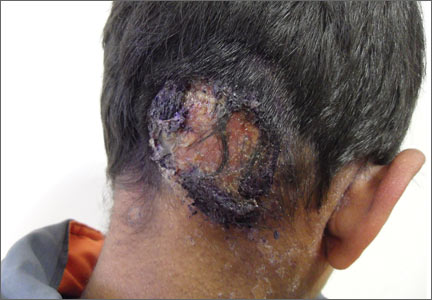
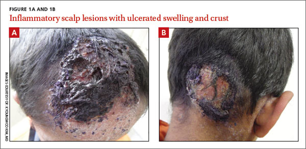
An 8-year-old boy was brought by his family to our clinic for treatment of 2 pruritic, inflammatory masses on his scalp. He’d had the masses for one month, and they hadn’t responded to an unknown treatment administered at a health center in Afghanistan. The edematous lesions were ulcerated with a crust, and had a diameter of approximately 6 cm and 4 cm on the frontal and occipital scalp, respectively (FIGURE 1A AND 1B).
The boy also had a well-demarcated, erythematous macule with scales on his face, but no other symptoms. The boy’s brother and sister also complained of pruritic, erythematous papules on their arms and faces. The family denied raising or having any recent contact with animals.
WHAT IS YOUR DIAGNOSIS?
HOW WOULD YOU TREAT THIS PATIENT?
Dx: Tinea capitis and erythema nodosum
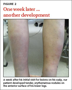
The patient was given a diagnosis of tinea capitis (ringworm of the scalp) based on the clinical presentation. (The patient’s brother and sister were told that they had tinea corporis and tinea faciei, which our patient also had on his face.) Our patient’s diagnosis was confirmed when he rapidly responded to treatment with the antifungal fluconazole. After the first week of this treatment, he complained of tender, erythematous nodules on the anterior surface of his lower legs, which we diagnosed as erythema nodosum (FIGURE 2).
Tinea capitis is a fungal infection of the scalp that usually starts as flaky and crusty patches of skin, broken-off hair, erythema, scaling, and pustules on the scalp. This can quickly deteriorate into a boggy and pruritic mass of inflamed tissue known as a kerion. The kerion can become severely inflamed and develop regional lymphadenopathy. Hypersensitive and highly inflammatory reactions that look similar to a bacterial infection may be found when the infection is caused by a zoophilic dermatophyte.1
Tinea capitis primarily affects children younger than age 10 years, with a peak incidence among African American boys.2 Because US public health agencies no longer require physicians to report cases of tinea capitis, its true incidence in the United States is unknown, but it is believed to be increasing.2
Differential diagnosis includes bacterial infections, psoriasis
Bacterial infections can cause abscesses or carbuncles on the scalp with tender and fluctuant changes that can also be accompanied by fever. However, because our patient was afebrile and relatively well, and the scalp lesions were nontender and without pus, a bacterial infection was unlikely.
Scalp psoriasis appears as raised, erythematous, dry and scaly patches, and not as inflammatory boggy masses (as was observed in our patient).
Skin cancer such as squamous cell carcinoma can present as erythematous, crusted, or scaly patches on sun-exposed skin. However, our patient’s lesions were too large to be malignant.4 In addition, skin cancer is rare in children.4
Oral antifungal medications are usually first-line treatment
Tinea capitis is treated with systemic antifungal medication. Oral antifungal agents, such as griseofulvin, itraconazole, terbinafine, and fluconazole, are effective.5-6
Erythema nodosum usually resolves without treatment, but should be observed until the underlying cause is treated.3
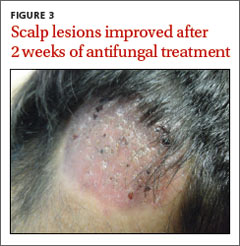
Our patient was treated with oral fluconazole 50 mg/d for 2 weeks and showed rapid improvement. After 2 weeks of treatment with oral fluconazole, he had hairless lesions on his scalp (FIGURE 3). The tender, erythematous nodules on his legs resolved spontaneously. Fluconazole was continued at 150 mg weekly for another 2 weeks, and our patient’s scalp lesions completely resolved after 6 weeks.
The patient’s siblings were initially treated with topical itraconazole, without effect. They were switched to oral fluconazole 50 mg/d and improved.
CORRESPONDENCE
Kyoungwoo Kim, MD, Department of Family Medicine, Inje University Seoul Paik Hospital, 9, Mareunnae-ro, Jung-gu, Seoul 100-032, Republic of Korea; [email protected]
1. Sohnle P. Dermatophytosis. In: Murphy J, Friedman H, Bendinelli M, eds. Fungal Infections and Immune Responses: Springer US;1993:2747.
2. Kao GF. Tinea capitis: Overview. Medscape Web site. Available at: http://emedicine.medscape.com/article/1091351-overview#showall. Accessed May 5, 2015.
3. Blake T, Manahan M, Rodins K. Erythema nodosum - a review of an uncommon panniculitis. Dermatol Online J. 2014;20:22376.
4. Christenson LJ, Borrowman TA, Vachon CM, et al. Incidence of basal cell and squamous cell carcinomas in a population younger than 40 years. JAMA. 2005;294:681-690.
5. Kakourou T, Uksal U; European Society for Pediatric Dermatology. Guidelines for the management of tinea capitis in children. Pediatr Dermatol. 2010;27:226-228.
6. González U, Seaton T, Bergus G, et al. Systemic antifungal therapy for tinea capitis in children. Cochrane Database Syst Rev. 2007;CD004685.
7. Gupta AK, Dlova N, Taborda P, et al. Once weekly fluconazole is effective in children in the treatment of tinea capitis: a prospective, multicentre study. Br J Dermatol. 2000;142:965-968.
8. Gupta AK, Adam P, Hofstader SL, et al. Intermittent short duration therapy with fluconazole is effective for tinea capitis. Br J Dermatol. 1999;141:304-306.
An 8-year-old boy was brought by his family to our clinic for treatment of 2 pruritic, inflammatory masses on his scalp. He’d had the masses for one month, and they hadn’t responded to an unknown treatment administered at a health center in Afghanistan. The edematous lesions were ulcerated with a crust, and had a diameter of approximately 6 cm and 4 cm on the frontal and occipital scalp, respectively (FIGURE 1A AND 1B).
The boy also had a well-demarcated, erythematous macule with scales on his face, but no other symptoms. The boy’s brother and sister also complained of pruritic, erythematous papules on their arms and faces. The family denied raising or having any recent contact with animals.
WHAT IS YOUR DIAGNOSIS?
HOW WOULD YOU TREAT THIS PATIENT?
Dx: Tinea capitis and erythema nodosum
The patient was given a diagnosis of tinea capitis (ringworm of the scalp) based on the clinical presentation. (The patient’s brother and sister were told that they had tinea corporis and tinea faciei, which our patient also had on his face.) Our patient’s diagnosis was confirmed when he rapidly responded to treatment with the antifungal fluconazole. After the first week of this treatment, he complained of tender, erythematous nodules on the anterior surface of his lower legs, which we diagnosed as erythema nodosum (FIGURE 2).
Tinea capitis is a fungal infection of the scalp that usually starts as flaky and crusty patches of skin, broken-off hair, erythema, scaling, and pustules on the scalp. This can quickly deteriorate into a boggy and pruritic mass of inflamed tissue known as a kerion. The kerion can become severely inflamed and develop regional lymphadenopathy. Hypersensitive and highly inflammatory reactions that look similar to a bacterial infection may be found when the infection is caused by a zoophilic dermatophyte.1
Tinea capitis primarily affects children younger than age 10 years, with a peak incidence among African American boys.2 Because US public health agencies no longer require physicians to report cases of tinea capitis, its true incidence in the United States is unknown, but it is believed to be increasing.2
Differential diagnosis includes bacterial infections, psoriasis
Bacterial infections can cause abscesses or carbuncles on the scalp with tender and fluctuant changes that can also be accompanied by fever. However, because our patient was afebrile and relatively well, and the scalp lesions were nontender and without pus, a bacterial infection was unlikely.
Scalp psoriasis appears as raised, erythematous, dry and scaly patches, and not as inflammatory boggy masses (as was observed in our patient).
Skin cancer such as squamous cell carcinoma can present as erythematous, crusted, or scaly patches on sun-exposed skin. However, our patient’s lesions were too large to be malignant.4 In addition, skin cancer is rare in children.4
Oral antifungal medications are usually first-line treatment
Tinea capitis is treated with systemic antifungal medication. Oral antifungal agents, such as griseofulvin, itraconazole, terbinafine, and fluconazole, are effective.5-6
Erythema nodosum usually resolves without treatment, but should be observed until the underlying cause is treated.3
Our patient was treated with oral fluconazole 50 mg/d for 2 weeks and showed rapid improvement. After 2 weeks of treatment with oral fluconazole, he had hairless lesions on his scalp (FIGURE 3). The tender, erythematous nodules on his legs resolved spontaneously. Fluconazole was continued at 150 mg weekly for another 2 weeks, and our patient’s scalp lesions completely resolved after 6 weeks.
The patient’s siblings were initially treated with topical itraconazole, without effect. They were switched to oral fluconazole 50 mg/d and improved.
CORRESPONDENCE
Kyoungwoo Kim, MD, Department of Family Medicine, Inje University Seoul Paik Hospital, 9, Mareunnae-ro, Jung-gu, Seoul 100-032, Republic of Korea; [email protected]
1. Sohnle P. Dermatophytosis. In: Murphy J, Friedman H, Bendinelli M, eds. Fungal Infections and Immune Responses: Springer US;1993:2747.
2. Kao GF. Tinea capitis: Overview. Medscape Web site. Available at: http://emedicine.medscape.com/article/1091351-overview#showall. Accessed May 5, 2015.
3. Blake T, Manahan M, Rodins K. Erythema nodosum - a review of an uncommon panniculitis. Dermatol Online J. 2014;20:22376.
4. Christenson LJ, Borrowman TA, Vachon CM, et al. Incidence of basal cell and squamous cell carcinomas in a population younger than 40 years. JAMA. 2005;294:681-690.
5. Kakourou T, Uksal U; European Society for Pediatric Dermatology. Guidelines for the management of tinea capitis in children. Pediatr Dermatol. 2010;27:226-228.
6. González U, Seaton T, Bergus G, et al. Systemic antifungal therapy for tinea capitis in children. Cochrane Database Syst Rev. 2007;CD004685.
7. Gupta AK, Dlova N, Taborda P, et al. Once weekly fluconazole is effective in children in the treatment of tinea capitis: a prospective, multicentre study. Br J Dermatol. 2000;142:965-968.
8. Gupta AK, Adam P, Hofstader SL, et al. Intermittent short duration therapy with fluconazole is effective for tinea capitis. Br J Dermatol. 1999;141:304-306.