User login
One in five female oncologists considering leaving academia, survey finds
More than half of respondents in academic medicine said they believe their gender adversely affects their likelihood for promotion, and 1 in 5 said they were considering leaving academia in the next 5 years.
Given the percentage of female oncologists planning to exit academia, “gender inequality is at high risk of continuing if the culture is not addressed,” write the authors in their study, published online Dec. 30 in JAMA Network Open.
Although women currently outnumber men in U.S. medical schools – a shift that first occurred in 2019 – female representation in academic oncology dwindles at more senior levels. Women represent 45% of hematology and oncology residents, only about 36% of academic faculty, and an even smaller percentage of leadership positions in academic medicine. Women, for instance, occupy about 31% of the chair positions in medical oncology, 17.4% in radiation oncology, and 11% in surgical oncology.
A team of researchers led by Emily C. Merfeld, MD, of the University of Wisconsin Hospitals and Clinics, Madison, set out to understand the factors influencing female oncologists’ decisions to pursue academic versus nonacademic career paths.
Dr. Merfeld and colleagues analyzed survey responses from 667 female oncologists between August 1 and Oct. 31, 2020 – 422 (63.2%) in academic medicine and 245 (36.8%) in nonacademic practice.
Overall, 1 in 4 oncologists said their spouse or partner and family “extremely or moderately” affected their decision to pursue academic practice.
Almost 43% of academic oncologists perceived time spent with loved ones as the biggest sacrifice related to pursuing a career in academic medicine. Approximately the same percentage (41.6%) of nonacademic oncologists perceived the pressure to achieve academic promotion as the most significant sacrifice associated with academic oncology, whereas only 22.4% perceived less time with loved ones as the biggest sacrifice.
“Although work-life balance was a concern for academic oncologists and may be a factor in female oncologists leaving academia, survey data suggested that women in nonacademic practice faced similar challenges,” the authors write.
More specifically, women in academic oncology reported working 2 more hours on the weekends compared to women not in academic medicine; however, both groups worked a similar number of hours during the week.
On the hiring front, almost 24% of academic oncologists said their gender had a “negative or somewhat negative” impact on their ability to get a job, compared with 21% of nonacademic oncologists. Conversely, nearly 28% of academic oncologists said their gender had a “positive or somewhat positive” influence on whether they were hired compared with 41.2% of nonacademic oncologists.
Respondents, however, perceived that gender strongly influenced promotion opportunities. More than half of the respondents – 54.6% of academic oncologists and 50.6% of nonacademic oncologists – believed they were less likely to be promoted than their male colleagues.
This perception aligns with findings from prior studies, which “found women were less likely than men to be promoted to associate professor, full professor, or department chair positions,” the authors write.
Overall, most respondents in each group – 71.3% in academic medicine and 68.6% in nonacademic practice – said they would choose the same career path again. But almost 22% of those in academia said they were “likely or very likely” to leave academic oncology in the next 5 years. Of these women, 28.2% said they would switch to industry employment and 25% would move to community practice.
“Contrary to popular assumptions,” the researchers note, “a spouse or partner and/or family were not a major factor in female oncologists favoring nonacademic careers, because this factor was similarly important to both academic and nonacademic oncologists.”
However, they note, “the increased financial compensation in nonacademic oncology may play a large role in some women’s career decisions.”
Making headway on gender equality?
In 2013, oncologist Katherine Reeder-Hayes, MD, MBA, now an associate at the University of North Carolina, Chapel Hill, published a study on gender equality in oncology in which she concluded that despite “an increasingly significant presence in the oncology physician workforce” women remained “under-represented in leadership positions and at the senior levels of academic medicine.”
Since then, Dr. Reeder-Hayes says that she has seen progress but recognizes the need for more.
“To some extent, I think that representation is improving over time due to factors outside the workplace – women are entering medical school in large numbers and may have more supportive partners and more social support for pursuing a professional career in general, [compared with] a decade or two ago,” Dr. Reeder-Hayes told this news organization.
On a personal level, she noted, “I do see many midcareer women assuming key leadership roles in my own institution.” However, she added, “I think the translation of those good candidates into increased representation in leadership probably varies widely across different institutions.”
In a 2019 editorial, researchers highlighted this variation while calling attention to the “notable progress” made by the American Association for Cancer Research (AACR). Specifically, the editorialists reported that women represent 40% of AACR members, 45% of the AACR Board of Directors, and half of the last 10 association presidents.
Editorial coauthor Elizabeth Jaffee, MD, deputy director of the Sidney Kimmel Comprehensive Cancer Center at Johns Hopkins, Baltimore, and former AACR president, told this news organization that she attributes this progress to “concrete measures to ensure equality throughout the organization,” which include gender balance on nominating and program committees as well as research meetings and providing opportunities for mentoring, leadership training, and networking.
Despite this positive change, the COVID-19 pandemic threatens to widen the gender imbalance. In a recent article, Julie Silver, MD, an expert in gender equity in medicine, told this news organization that she anticipates trouble ahead.
“There are many indications that women are leaving medicine in disproportionately high numbers,” said Dr. Silver, associate chair and director of cancer rehabilitation in the department of physical medicine and rehabilitation at Harvard Medical School, Boston. “A lack of fair pay and promotion opportunities that were present before COVID-19 are now combined with a host of pandemic-related challenges.”
In addition to salary and promotion disparities, the U.S. continues to suffer from “a chronic shortage of available, affordable, and high-quality childcare and a lack of federal-level policy initiatives or employer initiatives to broaden paid family leave and develop childcare infrastructure and workforce,” Dr. Reeder-Hayes said. Providing extended leave for new parents and on-site childcare could go a long way to improving this problem, she said.
However, Dr. Reeder-Hayes noted that perhaps the “leaky pipeline” problem in oncology highlights the fact that women “are making good decisions that reflect balanced life priorities, [and that] if we don’t structure job responsibilities, childcare, and pacing of promotion and tenure in ways that allow people to nurture other parts of their lives, employees will feel they’re being asked to sacrifice key things.”
In other words, she said, “it’s the workplace that needs to change if we’re going to convince [women], and many men with similar values, to stay.”
A version of this article first appeared on Medscape.com.
FROM JAMA NETWORK OPEN
Clinical Edge Journal Scan Commentary: Breast Cancer February 2022

Breast cancer diagnosis and treatment in young women can present unique challenges based on their life stage, including potential impact on fertility and future pregnancy. The role of GnRH analogues for ovarian protection during chemotherapy has been shown in both the POEMS-SWOG S0230 and PROMISE-GIM6 studies. Zong and colleagues conducted a phase 3 trial in China among premenopausal women with stage I-III breast cancer receiving cyclophosphamide-containing chemotherapy, with randomization to GnRHa + chemotherapy vs chemotherapy alone. Among 301 patients eligible for primary endpoint analysis, the premature ovarian insufficiency rate at 12 months was 10.3% for the GnRHa group vs 44.5% for the control group (odds ratio 0.23; P < 0.001). The rate of ovarian function recovery was also 46.4% higher in the GnRHa group. Furthermore, although survival outcomes were similar between groups, in patients <35 years of age, the tumor-free survival was higher in the GnRHa group vs control (93% vs 62%, P = 0.004) (Zong et al). These data reinforce the role of GnRHa as a means to reduce POI risk and support ovarian function recovery in young women undergoing chemotherapy for breast cancer. Measures of fertility and timing of pregnancy after breast cancer diagnosis continue to be areas of active research.
The treatment landscape for early-stage HER2-positive breast cancer continues to rapidly evolve with efforts to enhance efficacy and minimize toxicity for patients. The phase 3 KAITLIN study included 1846 patients with early-stage HER2-positive breast cancer (node-positive or node-negative, hormone receptor-negative and ≥T2 primary tumor) with randomization after surgery to adjuvant AC followed by taxane + trastuzumab + pertuzumab (AC-THP) or AC followed by T-DM1 + pertuzumab (AC-KP). In both the overall and node-positive populations, there was no significant difference in IDFS between the arms (stratified HR 0.98 and 0.97, respectively). In the overall population, the 3-year IDFS was 93.1% for AC-KP and 94.2% for AC-THP. Treatment completion rates were lower for AC-KP vs AC-THP (65.0% vs 88.4%), with T-DM1 discontinuation driven mostly by lab abnormalities (elevated liver function tests and thrombocytopenia) (Krop et al). Many patients diagnosed with early HER2-positive breast cancer (specifically those with tumors >2cm or node-positive) are treated with neoadjuvant chemotherapy + HER2-targeted therapy with subsequent tailoring of adjuvant treatment pending response, including use of T-DM1 if residual disease present. Future escalation and de-escalation strategies are being explored to further optimize outcomes and decrease side effects.
The addition of CDK 4/6 inhibitors to endocrine therapy has led to improved survival outcomes for patients diagnosed with advanced HR-positive-HER2-negative breast cancer. Lu and colleagues presented exploratory updated OS results among 672 patients with extended follow-up (median 53.5 months) from MONALEESA-7, which was a phase 3 randomized trial of ribociclib + endocrine therapy vs endocrine therapy alone among peri/pre-menopausal patients with HR-positive/HER2-negative advanced breast cancer. Median OS was 58.7 months vs 48.0 months for the ribociclib and placebo arms, respectively (HR 0.76), and a more pronounced benefit was seen in patients <40 years of age (median OS 51.3 months vs 40.5 months for ribociclib vs placebo arm; HR 0.65) (Lu et al). Furthermore, there was a significant delay in time to chemotherapy with ribociclib vs placebo (50.9 months vs 36.8 months; HR 0.69) which can certainly impact quality of life. A prior pooled analysis of the various MONALEESA trials demonstrated consistent PFS benefit with ribociclib across all intrinsic breast cancer subtypes, with the exception of basal-like and a more pronounced favorable impact in HER2-enriched. Future research to elucidate differences among CDK 4/6 inhibitors, influence of breast cancer subtype on their effect and how this can be translated to routine clinical practice are warranted.
Dairy intake may increase risk of Parkinson’s disease in men
according to investigators. Men of European ancestry with a genetic marker predicting dairy consumption had significantly greater risk of Parkinson’s disease than individuals without the marker, suggesting a causal relationship between dairy intake and Parkinson’s disease, lead author Cloé Domenighetti, MSc, a PhD student at UVSQ, Université Paris Sud, and colleagues reported.
“Previous studies highlighted dairy intake as a risk factor of Parkinson’s disease,” the investigators wrote in Movement Disorders. “A meta-analysis of prospective studies reported a 40% increased Parkinson’s disease risk in participants with the highest intake. It is unclear whether the association is causal or explained by confounding or reverse causation, given the long prodromal phase of Parkinson’s disease.”
A Mendelian randomization study
The investigators evaluated this link by comparing 9,823 cases of Parkinson’s disease with 8,376 controls, all individuals of European ancestry from the Courage-Parkinson’s disease consortium, comprising 23 studies. Data were analyzed by two-sample Mendelian randomization, a technique that uses genotype to predict behavior, thereby replacing conventional methods of capturing behavior, such as questionnaires. In this case, the investigators screened all participants for rs4988235, a single-nucleotide polymorphism (SNP) upstream of the lactase gene that is well documented to predict dairy intake among individuals of European ancestry.
“Mendelian randomization uses genetic variants associated with exposures as instrumental variables to estimate causal relationships between exposures and outcomes,” the investigators wrote. “Mendelian randomization analyses are less likely to be biased by confounding or reverse causation than observational studies if a set of assumptions are met.”
The approach uncovered a significant association between rs4988235 and Parkinson’s disease, with a 70% increase in disease risk per one serving of dairy per day (odds ratio, 1.70; 95% confidence interval, 1.12-2.60; P = .013). Further analysis revealed that this finding was driven by men, who had a 2.5-fold increased risk of Parkinson’s disease per one serving per day (OR, 2.50; 95% CI, 1.37-4.56; P = .003) versus women, among whom there was no significant association (OR, 1.04; 95% CI, 0.56-1.92; P = .91). No significant associations were observed among individuals grouped by age or Parkinson’s disease duration.
“Our findings suggest that dairy intake increases Parkinson’s disease risk,” the investigators concluded. “Therefore, diets with limited milk intake (e.g., Mediterranean diet) may be beneficial with respect to Parkinson’s disease.”
Further evidence supporting a link between diet and Parkinson’s disease
According to Silke Appel-Cresswell, MD, Marg Meikle Professor for Parkinson’s Research at the University of British Columbia, Vancouver, the findings align with previous prospective cohort studies demonstrating an increased risk of Parkinson’s disease with greater consumption of dairy.

“What the current study adds,” Dr. Appel-Cresswell said, “is a complementary approach to assess the association where the risk of reverse causation and of confounding are minimized. Like in some of the previous studies, the authors find sex differences with an increased risk for men but not women.”
Dr. Appel-Cresswell noted that an increasing body of evidence supports a link between diet and Parkinson’s disease, including a study of her own published last year, which showed later onset of Parkinson’s disease among individuals with a Mediterranean-style diet.
“We are accumulating evidence for a role of diet (or more broadly, the food exposome) for the risk to develop Parkinson’s disease,” Dr. Appel-Cresswell said, noting that “key pieces are still missing, including mechanisms underlying associations, clinical trials in individuals with established Parkinson’s disease and – eventually – preventive interventions. This research is urgently needed and analyses will need to take sex differences and a large range of potential other factors into account.”
A ‘modest’ contributing factor?
Vikas Kotagal, MD, associate professor of neurology at the University of Michigan, Ann Arbor, offered a perspective on the study methodology, and suggested that a causal link between dairy intake and Parkinson’s disease, if present, is likely minimal.
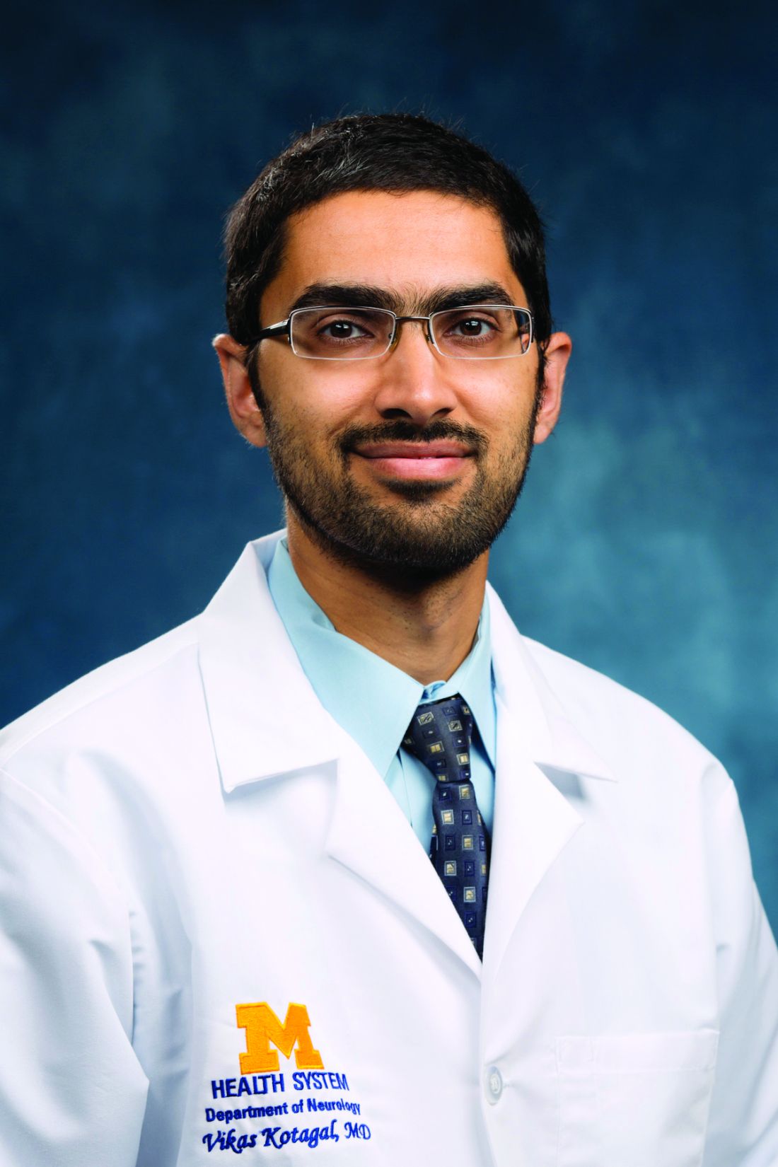
“Limitations to the study include the fact that participants weren’t actually asked or tested for how much dairy they truly consumed,” Dr. Kotagal said*. “Their dairy intake was estimated based on their genetic background – there are certainly many assumptions baked into this analytic approach which may or may not be true. It is also worth noting the fact that this causal association was seen in men and not women, suggesting that even if dairy intake was truly causal, it is likely to be a modest contributing factor and not a significant cause of Parkinson’s disease in the broader population in general.”
Still, Dr. Kotagal agreed with Dr. Appel-Cresswell that underlying mechanisms need further investigation.
“The biggest takeaway here is to heighten the urgency for researchers and funders to explore whether factors that might cluster with dairy intake – including pesticide exposure in milk or even the make-up of bacterial populations in different peoples’ intestines – might deserve closer scrutiny as a missing link connecting dairy consumption to increased Parkinson’s disease risk,” Dr. Kotagal said.
Dietary advice
Considering all available evidence, Dr. Appel-Cresswell offered some dietary advice with benefits that may extend beyond prevention of Parkinson’s disease.
“From a clinical point of view, I suggest to limit dairy intake to a moderate amount,” she said. “Mediterranean diets so far have the best supporting evidence for a lower Parkinson’s disease risk, although data is lacking for benefits in established Parkinson’s disease. Given the low risk of the Mediterranean diet and the established benefits for a host of other medical conditions, this is generally a safe and delicious recommendation whether one is living with Parkinson’s or not.”
The study was supported by the European Union Joint Program for Neurodegenerative Disease Research, the National Centre of Excellence in Research on Parkinson’s Disease, the National Institutes of Health, and others. The investigators disclosed additional relationships with Astellas Pharma, Sanofi, Pfizer, and others. Dr. Kotagal and Dr. Appel-Cresswell reported no relevant conflicts of interest.
*Correction, 2/10/22: An earlier version of this article misstated Dr. Kotagal's name in certain instances, including a photo caption.
according to investigators. Men of European ancestry with a genetic marker predicting dairy consumption had significantly greater risk of Parkinson’s disease than individuals without the marker, suggesting a causal relationship between dairy intake and Parkinson’s disease, lead author Cloé Domenighetti, MSc, a PhD student at UVSQ, Université Paris Sud, and colleagues reported.
“Previous studies highlighted dairy intake as a risk factor of Parkinson’s disease,” the investigators wrote in Movement Disorders. “A meta-analysis of prospective studies reported a 40% increased Parkinson’s disease risk in participants with the highest intake. It is unclear whether the association is causal or explained by confounding or reverse causation, given the long prodromal phase of Parkinson’s disease.”
A Mendelian randomization study
The investigators evaluated this link by comparing 9,823 cases of Parkinson’s disease with 8,376 controls, all individuals of European ancestry from the Courage-Parkinson’s disease consortium, comprising 23 studies. Data were analyzed by two-sample Mendelian randomization, a technique that uses genotype to predict behavior, thereby replacing conventional methods of capturing behavior, such as questionnaires. In this case, the investigators screened all participants for rs4988235, a single-nucleotide polymorphism (SNP) upstream of the lactase gene that is well documented to predict dairy intake among individuals of European ancestry.
“Mendelian randomization uses genetic variants associated with exposures as instrumental variables to estimate causal relationships between exposures and outcomes,” the investigators wrote. “Mendelian randomization analyses are less likely to be biased by confounding or reverse causation than observational studies if a set of assumptions are met.”
The approach uncovered a significant association between rs4988235 and Parkinson’s disease, with a 70% increase in disease risk per one serving of dairy per day (odds ratio, 1.70; 95% confidence interval, 1.12-2.60; P = .013). Further analysis revealed that this finding was driven by men, who had a 2.5-fold increased risk of Parkinson’s disease per one serving per day (OR, 2.50; 95% CI, 1.37-4.56; P = .003) versus women, among whom there was no significant association (OR, 1.04; 95% CI, 0.56-1.92; P = .91). No significant associations were observed among individuals grouped by age or Parkinson’s disease duration.
“Our findings suggest that dairy intake increases Parkinson’s disease risk,” the investigators concluded. “Therefore, diets with limited milk intake (e.g., Mediterranean diet) may be beneficial with respect to Parkinson’s disease.”
Further evidence supporting a link between diet and Parkinson’s disease
According to Silke Appel-Cresswell, MD, Marg Meikle Professor for Parkinson’s Research at the University of British Columbia, Vancouver, the findings align with previous prospective cohort studies demonstrating an increased risk of Parkinson’s disease with greater consumption of dairy.
“What the current study adds,” Dr. Appel-Cresswell said, “is a complementary approach to assess the association where the risk of reverse causation and of confounding are minimized. Like in some of the previous studies, the authors find sex differences with an increased risk for men but not women.”
Dr. Appel-Cresswell noted that an increasing body of evidence supports a link between diet and Parkinson’s disease, including a study of her own published last year, which showed later onset of Parkinson’s disease among individuals with a Mediterranean-style diet.
“We are accumulating evidence for a role of diet (or more broadly, the food exposome) for the risk to develop Parkinson’s disease,” Dr. Appel-Cresswell said, noting that “key pieces are still missing, including mechanisms underlying associations, clinical trials in individuals with established Parkinson’s disease and – eventually – preventive interventions. This research is urgently needed and analyses will need to take sex differences and a large range of potential other factors into account.”
A ‘modest’ contributing factor?
Vikas Kotagal, MD, associate professor of neurology at the University of Michigan, Ann Arbor, offered a perspective on the study methodology, and suggested that a causal link between dairy intake and Parkinson’s disease, if present, is likely minimal.
“Limitations to the study include the fact that participants weren’t actually asked or tested for how much dairy they truly consumed,” Dr. Kotagal said*. “Their dairy intake was estimated based on their genetic background – there are certainly many assumptions baked into this analytic approach which may or may not be true. It is also worth noting the fact that this causal association was seen in men and not women, suggesting that even if dairy intake was truly causal, it is likely to be a modest contributing factor and not a significant cause of Parkinson’s disease in the broader population in general.”
Still, Dr. Kotagal agreed with Dr. Appel-Cresswell that underlying mechanisms need further investigation.
“The biggest takeaway here is to heighten the urgency for researchers and funders to explore whether factors that might cluster with dairy intake – including pesticide exposure in milk or even the make-up of bacterial populations in different peoples’ intestines – might deserve closer scrutiny as a missing link connecting dairy consumption to increased Parkinson’s disease risk,” Dr. Kotagal said.
Dietary advice
Considering all available evidence, Dr. Appel-Cresswell offered some dietary advice with benefits that may extend beyond prevention of Parkinson’s disease.
“From a clinical point of view, I suggest to limit dairy intake to a moderate amount,” she said. “Mediterranean diets so far have the best supporting evidence for a lower Parkinson’s disease risk, although data is lacking for benefits in established Parkinson’s disease. Given the low risk of the Mediterranean diet and the established benefits for a host of other medical conditions, this is generally a safe and delicious recommendation whether one is living with Parkinson’s or not.”
The study was supported by the European Union Joint Program for Neurodegenerative Disease Research, the National Centre of Excellence in Research on Parkinson’s Disease, the National Institutes of Health, and others. The investigators disclosed additional relationships with Astellas Pharma, Sanofi, Pfizer, and others. Dr. Kotagal and Dr. Appel-Cresswell reported no relevant conflicts of interest.
*Correction, 2/10/22: An earlier version of this article misstated Dr. Kotagal's name in certain instances, including a photo caption.
FROM MOVEMENT DISORDERS
Can immunotherapy replace surgery for stomach cancer?
GERCOR NEONIPIGA was a phase 2 study with no comparator group and only 32 patients, but even so, after a 6-cycle course of nivolumab and ipilimumab, there was no sign of tumor in 17 of the 29 patients (59%) who had surgery specimens evaluable by pathology.
Indeed, two patients refused surgery after their preop endoscopic biopsies came back clear with no tumor cells. Surgery was called off in a third patient who developed metastases beforehand.
After a median of 12 months follow-up, there’s was no recurrence or progression in 30 patients (94%). The remaining two included the metastatic patient and one who died 3 days after surgery from cardiovascular complications.
If the findings pan out with additional research, the approach could be a boon for people who respond. “Avoiding surgery is a dream for these patients,” said lead investigator Thierry Andre, MD, a medical oncology professor at Sorbonne University, Paris, when he presented the findings at the American Society of Clinical Oncology Gastrointestinal Cancers Symposium.
The trial “raises the question whether surgery can be delayed or avoided in some patients with localized” disease. Given the findings, “it seems possible not for all but probably for half, maybe more.” As in the two subjects who opted out of surgery, preop endoscopic biopsies could be used to identify complete responders with active surveillance afterwards, he said.
The study included 16 patients with gastric cancer and 16 with esophagogastric adenocarcinoma. They were mismatch repair deficient, which Dr. Andre said predicts response to immunotherapy.
At baseline, 22 had stage T3 disease and four had stage T2 disease, and stage was not evaluable by echo-endoscopy in 6. Nodal status was unknown, but the patients had no metastases at baseline.
They underwent six nivolumab 240-mg infusions and two ipilimumab 1–mg/kg infusions over 12 weeks, followed by R0 resections a median of 5 weeks after the last nivolumab injection.
Surgical specimens from 17 patients (59%) showed a complete pathological response to neoadjuvant immunotherapy (Becker tumor regression grade (TRG) 1a, ypT0N0). TRG was 1b – less than 10% residual tumor in tumor bed in four patients. TRG was 2 in two patients with 10%-50% of residual tumor remaining, and six had a TRG of 3 with more than half of the tumor remaining after immunotherapy.
Based on tumor response, 25 patients had nine additional nivolumab infusions after surgery with 480 mg infused monthly.
Dr. Andre explained that people want to avoid surgery because of the substantial morbidity that was shown in the study, plus 54% of patients had complications, including anastomotic leaks, pancreatitis, pneumonia, and other problems.
There were no new safety signals with neoadjuvant therapy; 25% of patients had grade 3 or 4 events.
The study was conducted in 10 centers in France. About three-quarters of the subjects were men and the median age was 65 years.
Bristol Meyers Squibb supplied the nivolumab and ipilimumab and partially funded the work. Many of the investigators had ties to the company, including Dr. Andre, who is a consultant for BMS and reported payments from the company.
FROM GI CANCERS SYMPOSIUM 2022
New combo therapy for breast implant–associated lymphoma
The immediate treatment is surgical removal of the implant, which is sometimes followed with chemotherapy.
New data show that women who develop breast implant–associated anaplastic large-cell lymphoma (BIA-ALCL) who require chemotherapy can achieve excellent results with a combination of chemotherapy (cyclophosphamide, doxorubicin, and prednisone) and the antibody–drug conjugate brentuximab vedotin.
The findings were published in Blood.
The authors, led by Fabien Le Bras, MD, from the Henri Mondor Hospital, Créteil, France, note that despite BIA-ALCL being recently recognized as a provisional entity by the World Health Organization, its pathogenesis has yet to be fully elucidated, and a standard of care has not been established.
Results from the ECHELON 2 trial established brentuximab vedotin plus cyclophosphamide, doxorubicin, and prednisone (BV-CHP) as a new standard of care in CD30-positive peripheral T-cell lymphoma.
That trial included 316 patients with ACLC, although none of these cases were associated with breast implants.
The principal investigator on that trial, Steven Horwitz, MD, from Memorial Sloan Kettering Center, New York, told this news organization that although BIA-ALCL is “incredibly rare,” it causes “distress” to patients, as “many of them made a choice for reconstruction ... that they thought was safe.”
He said that the latest data from France is “interesting” and that the application of the ECHELON-2 findings to BIA-ALCL is “very logical.”
“For the people who need systemic therapy,” it appears from the current results that BV-CHP “is a very good option,” he said.
The “difficulty” in interpreting the data, however, is that “perhaps 80% of people with BIA-ALCL don’t need any systemic therapy” and are “cured with surgery alone.”
Dr. Horwitz said that while patients with infiltrative disease have a “higher risk of recurrence ... many of those are still cured with surgery alone.”
The main outstanding question he has is how many of the patients who received BV-CHP “might have been okay with observation.”
Details of the new data from France
For their study, Dr. Le Bras and colleagues analyzed data from the Lymphoma Study Association registry between 2009 and 2021 and identified 85 patients with BIA-ALCL, including 73 in France and 12 in Belgium.
Most of these patients (whose median age was 57 years) had unilateral lymphoma (94.1%), and only a few patients (5.9%) had bilateral disease.
The team notes that 41.2% of these women had received breast implants once, 41.2% received implants twice, and 17.6% received them three times or more.
In 45.9% of cases, the first implant followed mastectomy for breast cancer.
All patients had at least one textured implant. These have been associated with more cases of BIA-ALCL than smooth implants, and in 2019, Allergan recalled all BioCell textured breast implant products from the United States and around the world, due to the risk for BIA-ALCL, as reported, at the time, by this news organization.
For the women in this registry, the median time from the last implant to BIA-ALCL diagnosis was 7 years.
The most common presentation was seroma, which occurred in 75.3% of patients, while 21.2% of had a breast tumor mass with or without seroma.
Stage I-II disease was identified in 76.5% of patients, and 21.2% of cases were stage IV. Infiltrative disease was present in 24.7%.
Implant removal with total capsulectomy was performed in 77.6%; 29.4% of women also received chemotherapy, with 11.8% receiving BV-CHP.
A complete response was achieved in 84% of patients who received chemotherapy, while 8% failed to respond. Among the patients who received BV-CHP, 80% achieved a complete response.
After a median follow-up of 28.6 months, 91.8% patients were alive and progression free. All patients treated with BV-CHP were alive and progression free after a median follow-up of 1 year.
Patients with infiltrative disease had a significantly worse 2-year progression-free survival than those with in situ/mixed disease, at 73.8% versus 96.7%, or a hazard ratio for progression of 5.3 (P = .0039).
They also had worse 2-year overall survival, at 78.7% versus 100%, or a hazard ratio for death of 8.5 (P = .0022).
The authors note that these patients with infiltrative disease had significantly worse survival outcomes and may benefit most from BV-CHP.
No funding for the study was declared. Dr. Le Bras reports relationships with Novartis, Celgene, BMS, Takeda, Kite, and Gilead. Other authors declare numerous relevant financial relationships.
A version of this article first appeared on Medscape.com.
COVID brain fog is a ‘true neurologic condition’
early research suggests. Investigators found abnormalities in cerebrospinal fluid (CSF) and other risk factors, including diabetes and hypertension, present in individuals with mild COVID-19 experiencing persistent cognitive problems, often referred to as “brain fog.”
“We’re seeing changes to the [CSF] in the brain of most people who report cognitive changes,” said Joanna Hellmuth, MD, assistant professor of neurology, Memory and Aging Center, University of California, San Francisco. “We’re just in the beginning stages, but I hope this study will provide some legitimacy to this being a true neurologic condition.”
The study was published online Jan. 18, 2022, in Annals of Clinical and Translational Neurology.
No guidance
There is currently no guidance on how to identify patients with COVID-related cognitive changes, said Dr. Hellmuth. “The term ‘brain fog’ is not based in science or medicine, but that’s the most common term we use to describe this.”
The analysis included adults with confirmed SARS-CoV-2 infection not requiring hospitalization who were enrolled in the Long-term Impact of Infection with Novel Coronavirus study.
Participants underwent a structured interview that covered COVID-19 illness, past medical history, preexisting cognitive risk factors, medications, and cognitive symptoms following onset of COVID-19. They also completed an in-person battery of cognitive tests.
The analysis included 22 participants with at least one new cognitive symptom who had cognitive post-acute sequelae of SARS-CoV-2 infection (PASC). Ten cognitive controls reported no new cognitive symptoms after acute infection.
Participants were a median age of 41 years, had a median of 16 years of education, and were assessed a median of 10.1 months from their first COVID-19 symptom. There were no group differences in terms of age, gender, years of education, or distribution of race/ethnicity (all P > .05).
Among those with cognitive PASC, 43% reported cognitive symptoms starting 1 or more months after the first COVID symptom. About 29% reported cognitive changes started 2 or more months after their first COVID symptom.
“The immune system could be altered in some way after the infection, and perhaps that’s what’s contributing to these delayed onset cognitive changes,” said Dr. Hellmuth.
Compared with controls, participants with cognitive PASC had more preexisting cognitive risk factors (a median of 2.5 vs. 0; P = .03). These included hypertension and diabetes, which increase the risk of stroke, mild cognitive impairment, vascular dementia, traumatic brain injury, (TBI), learning disabilities, anxiety, depression, stimulant use, and ADHD, which may make the brain more vulnerable to executive functioning problems.
Dr. Hellmuth noted that the study wasn’t powered to determine whether any individual risk factor was associated with risk of cognitive changes.
As there are no published neuropsychological testing criteria for cognitive PASC, the researchers applied the equivalent criteria for HIV-associated neurocognitive disorder (HAND), a similar, virally associated cognitive disorder. Only 59% of those with cognitive PASC met equivalent HAND criteria for objective cognitive impairment versus 70% of cognitive controls. This, the investigators noted, highlights “the challenges and incongruities of using subjective, versus objective cognitive assessments for diagnosis.”
Is self-report enough?
While there is currently “nothing objective doctors can hang their hats on to say ‘you do’ or ‘you don’t’ have cognitive changes related to COVID,” using the HAND criteria is “not particularly helpful,” said Dr. Hellmuth. “Comparing an individual to a population-based norm in this case is really nuanced, and we shouldn’t rely on this solely to determine whether they do, or don’t, have cognitive changes.”
Perhaps self-reports in this case are “enough” said Dr. Hellmuth. “People know their brains better than anyone else, better than any doctor will.”
A total of 13 in the cognitive PASC group and 4 in the control group consented to a lumbar puncture. Cognitive PASC participants were older than controls (median of 47 vs. 28 years; P = .03) with no other between-group differences.
Overall, 77% of participants with cognitive PASC had a CSF abnormality, compared with 0% of cognitive controls (P = .01). CSF abnormalities included elevated protein levels with no other explainable cause in 2 of the 13 subjects with PASC, which Dr. Hellmuth said is typically a marker of inflammation.
Researchers also noted abnormal oligoclonal banding, a collection of antibodies, in the blood or brain fluid. These were identified in 69% of participants with cognitive PASC, compared with 0% of cognitive controls (P = .03).
“When we find this pattern in both blood and brain, it suggests a systemic inflammatory disorder,” although “we have no idea what these antibodies are targeting,” said Dr. Hellmuth.
The study represents “the very beginning stages” of PASC becoming a medical diagnosis “where doctors know what to call it, how to treat it, and how to do blood and cerebrospinal fluid tests to diagnose it,” said Dr. Hellmuth.
She hopes PASC will receive medical legitimacy just as TBI has. In years past, a player was hit on the head or had their “bell rung,” simply returned to the field. “Now that we understand the science, we call it a mild TBI or concussion, and we have a very different medical approach to it.”
A limitation of the study was the small sample size, which may hinder the results’ validity. In addition, the study demographics may not reflect the broader population of those impacted by PASC.
‘A first substantial step’
Commenting on the research, William Schaffner, MD, professor, division of infectious diseases, Vanderbilt University Medical Center, Nashville, Tenn., said the new results represent “a first substantial step on the road to trying to find out what’s going on” with COVID patients dealing with cognitive issues.
Dr. Schaffner noted that elevated protein levels, identified in some study subjects, “is usually a consequence of previous inflammation” and is “a very interesting” finding. “In people who are otherwise normal, if you do a lumbar puncture, you don’t find elevated proteins.”
However, he noted the “diversity of results” from CSF examinations. “A single pattern does not leap out.”
What the researchers are observing “is not just a phenomenon of the mind or just something psychological,” said Dr. Schaffner. “Something physical is going on here.”
The study was funded by grants from the National Institute of Mental Health and the National Institute of Neurological Disorders and Stroke. Dr. Hellmuth received grant support from the National Institutes of Health/NIMH supporting this work and personal fees for medical-legal consultation outside of the submitted work. Dr. Schaffner has disclosed not relevant financial relationships.
A version of this article first appeared on Medscape.com.
FROM ANNALS OF CLINICAL AND TRANSLATIONAL NEUROLOGY
Medicare NCDs hinder access to cancer biomarker testing for minorities
of data from patients with advanced non–small cell lung cancer (aNSCLC), metastatic colorectal cancer, metastatic breast cancer, or advanced melanoma. The finding was reported in JAMA Network Open.
Biomarker testing has become an essential tool in cancer care over the last decade. In 2011, for example, less than 1% of patients with aNSCLC, metastatic colorectal cancer, metastatic breast cancer, and advanced melanoma underwent NGS testing, but by 2019, 40% of patients with these cancers received the testing.
“Next-generation sequencing testing has become increasingly important because it enables identification of multiple biomarkers simultaneously and efficiently while minimizing the number of biopsies required,” wrote the authors, led by William B. Wong, PharmD, of Genentech.
It has been unknown whether for Medicare beneficiaries and the overall population, if the NCD affected health equity issues, the authors wrote. While increased use of appropriate targeted therapies facilitated by NGS testing is associated with improved survival rates in patients with advanced or metastatic cancer, variability in health care coverage policies has posed a significant barrier to obtaining NGS testing for cancer patients, specifically through policy coverage limitations. It has remained unclear if the NCD has influenced NGS testing coverage in insurance types (for example, Medicaid) encompassing a larger population of minority racial and ethnic groups often experiencing poorer care and outcomes.
The retrospective cohort analysis compared EHR data from 280 U.S. cancer clinics in the (800 sites of care) pre- versus post-NCD period for patients with aNSCLC, metastatic colorectal cancer, metastatic breast cancer, or advanced melanoma (January 2011–March 2020). Nearly 70% of all patients in the study were Medicare recipients who needed NCD approval to cover the cost of testing.
Among 92,687 patients (mean age, 66.6 years; 55.7% women), compared with Medicare beneficiaries, changes in pre- to post-NCD NGS testing trends were similar in commercially insured patients (odds ratio, 1.03; 95% CI, 0.98-1.08; P = .25). Pre- to post-NCD NGS testing trends increased at a slower rate among patients in assistance programs (OR, 0.93; 95% CI, 0.87-0.99; P = .03), compared with Medicare beneficiaries. The rate of increase for patients receiving Medicaid was not significantly different statistically compared with those receiving Medicare (OR, 0.92; 95% CI, 0.84-1.01; P = .07). Also, the NCD was not associated with racial and ethnic groups within Medicare beneficiaries alone or across all insurance types.
Compared with non-Hispanic White individuals, increases in average NGS use from the pre-NCD to post-NCD period were 14% lower (OR, 0.86; 95% CI, 0.74-0.99; P = .04) among African American and 23% lower (OR, 0.77; 95% CI, 0.62-0.96; P = .02) among Hispanic/Latino individuals; increases were similar, however, among Asian individuals and other races and ethnicities.
The authors observed that the post-NCD trend of increasing NGS testing seen in Medicare beneficiaries was similarly observed in those with commercial insurance. Testing rate differences, however, widened or were maintained after versus before the NCD in PAP (personal assistance program) and Medicaid beneficiaries relative to Medicare beneficiaries, suggesting that access to NGS testing did not improve equally across insurance types. Since Medicare coverage is determined at the state level, the authors urged research examining individual state coverage policies to further elucidate factors slowing uptake among Medicaid beneficiaries. “Additional efforts beyond coverage policies,” the authors concluded, “are needed to ensure equitable access to the benefits of precision medicine.”
The study was supported by Genentech.
FROM JAMA NETWORK OPEN
Does COVID-19 induce type 1 diabetes in kids? Jury still out
Two new studies from different parts of the world have identified an increase in the incidence of type 1 diabetes in children since the COVID-19 pandemic began, but the reasons still aren’t clear.
The findings from the two studies, in Germany and the United States, align closely, endocrinologist Jane J. Kim, MD, professor of pediatrics and principal investigator of the U.S. study, told this news organization. “I think that the general conclusion based on their data and our data is that there appears to be an increased rate of new type 1 diabetes diagnoses in children since the onset of the pandemic.”
Dr. Kim noted that because her group’s data pertain to just a single center, she is “heartened to see that the [German team’s] general conclusions are the same as ours.” Moreover, she pointed out that other studies examining this question came from Europe early in the pandemic, whereas “now both they [the German group] and we have had the opportunity to look at what’s happening over a longer period of time.”
But the reason for the association remains unclear. Some answers may be forthcoming from a database designed in mid-2020 specifically to examine the relationship between COVID-19 and new-onset diabetes. Called CoviDiab, the registry aims “to establish the extent and characteristics of new-onset, COVID-19–related diabetes and to investigate its pathogenesis, management, and outcomes,” according to the website.
The first new study, a multicenter German diabetes registry study, was published online Jan. 17 in Diabetes Care by Clemens Kamrath, MD, of Justus Liebig University, Giessen, Germany, and colleagues.
The other, from Rady Children’s Hospital of San Diego, was published online Jan. 24 in JAMA Pediatrics by Bethany L. Gottesman, MD, and colleagues, all with the University of California, San Diego.
Mechanisms likely to differ for type 1 versus type 2 diabetes
Neither the German nor the U.S. investigators were able to directly correlate current or prior SARS-CoV-2 infection in children with the subsequent development of type 1 diabetes.
Earlier this month, a study from the U.S. Centers for Disease Control and Prevention did examine that issue, but it also included youth with type 2 diabetes and did not separate out the two groups.
Dr. Kim said her institution has also seen an increase in type 2 diabetes among youth since the COVID-19 pandemic began but did not include that in their current article.
“When we started looking at our data, diabetes and COVID-19 in adults had been relatively well established. To see an increase in type 2 [diabetes] was not so surprising to our group. But we had the sense we were seeing more patients with type 1, and when we looked at our hospital that was very much the case. I think that was a surprise to people,” said Dr. Kim.
Although a direct effect of SARS-CoV-2 on pancreatic beta cells has been proposed, in both the German and San Diego datasets the diagnosis of type 1 diabetes was confirmed with autoantibodies that are typically present years prior to the onset of clinical symptoms.
The German group suggests possible other explanations for the link, including the lack of immune system exposure to other common pediatric infections during pandemic-necessitated social distancing – the so-called hygiene hypothesis – as well as the possible role of psychological stress, which several studies have linked to type 1 diabetes.
But as of now, Dr. Kim said, “Nobody really knows.”
Is the effect direct or indirect?
Using data from the multicenter German Diabetes Prospective Follow-up Registry, Dr. Kamrath and colleagues compared the incidence of type 1 diabetes in children and adolescents from Jan. 1, 2020 through June 30, 2021 with the incidence in 2011-2019.
During the pandemic period, a total of 5,162 youth were newly diagnosed with type 1 diabetes at 236 German centers. That incidence, 24.4 per 100,000 patient-years, was significantly higher than the 21.2 per 100,000 patient-years expected based on the prior decade, with an incidence rate ratio of 1.15 (P < .001). The increase was similar in both males and females.
There was a difference by age, however, as the phenomenon appeared to be limited to the preadolescent age groups. The incidence rate ratios (IRRs) for ages below 6 years and 6-11 years were 1.23 and 1.18 (both P < .001), respectively, compared to a nonsignificant IRR of 1.06 (P = .13) in those aged 12-17 years.
Compared with the expected monthly incidence, the observed incidence was significantly higher in June 2020 (IRR, 1.43; P = .003), July 2020 (IRR, 1.48; P < 0.001), March 2021 (IRR, 1.29; P = .028), and June 2021 (IRR, 1.39; P = .01).
Among the 3,851 patients for whom data on type 1 diabetes-associated autoantibodies were available, the adjusted rates of autoantibody negativity did not differ from 2018-2019 during the entire pandemic period or during the year 2020 or the first half of 2021.
“Therefore, the increase in the incidence of type 1 diabetes in children appears to be due to immune-mediated type 1 diabetes. However, because autoimmunity and progressive beta-cell destruction typically begin long before the clinical diagnosis of type 1 diabetes, we were surprised to see the incidence of type 1 diabetes followed the peak incidence of COVID-19 and also the pandemic containment measures by only approximately 3 months,” Dr. Kamrath and colleagues write.
Taken together, they say, the data suggest that “the impact on type 1 diabetes incidence is not due to infection with SARS-CoV-2 but rather a consequence of environmental changes resulting from the pandemic itself or pandemic containment measures.”
Similar findings at a U.S. children’s hospital
In the cross-sectional study in San Diego, Dr. Gottesman and colleagues looked at the electronic medical records (EMRs) at Rady Children’s Hospital for patients aged younger than 19 years with at least one positive type 1 diabetes antibody titer.
During March 19, 2020 to March 18, 2021, a total of 187 children were admitted for new-onset type 1 diabetes, compared with just 119 the previous year, a 57% increase.
From July 2020 through February 2021, the number of new type 1 diabetes diagnoses significantly exceeded the number expected based on a quarterly moving average of each of the preceding 5 years.
Only four of the 187 patients (2.1%) diagnosed during the pandemic period had a COVID-19 infection at the time of presentation. Antibody testing to assess prior infection wasn’t feasible, and now that children are receiving the vaccine – and therefore most will have antibodies – “we’ve lost our window of opportunity to look at that question,” Dr. Kim noted.
As has been previously shown, there was an increase in the percentage of patients presenting with diabetic ketoacidosis during the pandemic compared with the prior 5 years (49.7% vs. 40.7% requiring insulin infusion). However, there was no difference in mean age at presentation, body mass index, A1c, or percentage requiring admission to intensive care.
Because these data only go through March 2021, Dr. Kim noted, “We need to see what’s happening with these different variants. We’ll have a chance to look in a month or two to see the effects of Omicron on the rates of diabetes in the hospital.”
Will CoviDiab answer the question?
Data from CoviDiab will include diabetes type in adults and children, registry coprincipal investigator Francesco Rubino, MD, of King’s College London, told this news organization.
“We aimed at having as many as possible cases of new-onset diabetes for which we can have also a minimum set of clinical data including type of diabetes and A1c. By looking at this information we can infer whether a role of COVID-19 in triggering diabetes is clinically plausible – or not – and what type of diabetes is most frequently associated with COVID-19 as this also speaks about mechanisms of action.”
Dr. Rubino said that the CoviDiab team is approaching the data with the assumption that, at least in adults diagnosed with type 2 diabetes, the explanation might be that the person already had undiagnosed diabetes or that the hyperglycemia may be stress-induced and temporary.
“We’re looking at this question with a skeptical eye ... Is it just an association, or does the virus have a role in inducing diabetes from scratch, or can the virus advance pathophysiology in a way that it ends up in full-blown diabetes in predisposed individuals?”
While no single study will prove that SARS-CoV-2 causes diabetes, “combining observations from various studies and approaches we may get a higher degree of certainty,” Dr. Rubino said, noting that the CoviDiab team plans to publish data from the first 800 cases “soon.”
Dr. Kim has reported no relevant financial relationships. Dr. Rubino has reported receiving grants from Ethicon and Medtronic, personal fees from GI Dynamic, Keyron, Novo Nordisk, Ethicon, and Medtronic.
A version of this article first appeared on Medscape.com.
Could probiotics reduce ‘chemo brain’ in breast cancer patients?
compared with a control group taking placebo capsules, reports the first study of its kind.
“Our finding[s] provide a simple, inexpensive, and effective prevention strategy for chemotherapy-related side effects, including cognitive impairment,” senior author Jianbin Tong, MD, PhD, of the department of anesthesiology, Third Xiangya Hospital, Central South University, Changsha, Hunan, China, said in an interview.
The research “is the first study showing that probiotics supplementation during chemotherapy can prevent chemotherapy-related brain impairment,” he noted.
The double-blind, randomized study was published in the European Journal of Cancer. It involved 159 patients in China with stage I-III breast cancer who required adjuvant chemotherapy between 2018 and 2019. These patients were randomized to receive a regimen of three capsules twice per day containing either probiotics (n = 80) or placebo (n = 79) during their chemotherapy.
The probiotic capsule (Bifico, Sine Pharmaceuticals) contained Bifidobacterium longum, Lactobacillus acidophilus, and Enterococcus faecalis (210 mg of each).
The reductions in symptoms seen with the supplementation “exceed our expectations,” Dr. Tong said in an interview.
He speculated that this may have longer-term effects, with the prevention of initial cognitive impairment potentially “changing the neurodegenerative trajectory of patients after chemotherapy.”
“Patients don’t need to take probiotics continuously, but it’s better to take probiotics intermittently,” he said.
Approached for comment, Melanie Sekeres, PhD, Canada Research Chair and assistant professor at the University of Ottawa, said the improvements, such as those seen in delayed recall, are especially of interest.
“This is particularly notable because one of the brain regions that is critically involved in long-term memory processing, the hippocampus, is known to be highly sensitive to chemotherapy-induced neurotoxicity,” she said in an interview.
“The finding that probiotic treatment given alongside chemotherapy is sufficient to, in part, protect against memory disturbances in these patients suggests that there may be some neuroprotection conferred by the probiotic treatment,” she said.
A key question is whether similar results would be seen with other chemotherapy regimens, Dr. Sekeres added. “To better understand the effectiveness of these probiotics in preventing CRCI, they should be tested using other classes of chemotherapies before any broad conclusions can be made.”
Measuring the effect on ‘chemo brain’
“Chemo brain” is commonly reported after chemotherapy, and some 35% of patients report having long-term effects. Key symptoms include deficits in memory, attention, and executive and processing speed skills.
In their study, Dr. Tong and colleagues assessed patients on their cognitive status with a number of validated neuropsychological battery tests 1 day prior to initiating chemotherapy and 21 days after the last cycle of chemotherapy. Tests included the Hopkins Verbal Learning Test–Revised for verbal memory, the Brief Visuospatial Memory Test–Revised for visuospatial memory, and various others.
The team reports that, after adjustment for confounding factors, the total incidence of CRCI was significantly lower in the probiotics group versus the placebo group 21 days post chemotherapy (35% vs. 81%; relative risk, 0.43).
Rates of mild cognitive impairment were also lower in the probiotics group (29% vs 52%; RR, 0.55), as were rates of moderate cognitive impairment (6% vs. 29%; RR, 0.22).
The improvements with probiotics were observed across most other neuropsychological domains, including instantaneous verbal memory and delayed visuospatial memory (for both, P = .003) and visuospatial interference and verbal fluency (for both, P < .001).
The greater improvements in the probiotics group were seen regardless of use of other medications or the type of chemotherapy regimen received, which could have included epirubicin or docetaxel and/or cyclophosphamide.
CRCI was more common in patients who were older and had lower education or a higher body mass index; however, the improvements in the probiotics group were observed regardless of those factors, the authors commented.
In addition to the reduction in cognitive impairment that was seen, the treatment with probiotics was also associated with lower blood glucose (mean, 4.96 vs. 5.30; P = .02) and lower LDL cholesterol (2.61 vs. 2.89; P = .03) versus placebo, while there were no significant differences between the groups prior to chemotherapy.
There were no reports of severe emesis or constipation (grade 3 or higher) in either group; however, the probiotics group did have a significantly lower incidence of both, the authors note.
How does it work?
The potential benefits with probiotics are theorized to result from stabilizing the colonic and bacterial disruptions that are caused by chemotherapy, potentially offsetting the neuroinflammation that is linked to the cancer treatment, the authors speculated.
A subanalysis of 78 stool samples from 20 patients in the study showed no differences in alpha diversity or beta diversity before or after chemotherapy; however, there were significant reductions in the abundance of Streptococcus and Tyzzerella (P = .023 and P = .033, respectively) in the probiotics group after chemotherapy.
Further analysis showed that probiotics supplement modulated the levels of nine plasma metabolites in patients with breast cancer, with the results suggesting that metabolites (including p-mentha-1,8-dien-7-ol) “may be modulators in preventing CRCI by probiotics,” the authors noted.
Benefits reported beyond breast cancer
A subsequent trial conducted by Dr. Tong and colleagues following the CRCI study further showed similar protective benefits with probiotics in the prevention of chemotherapy-related hand-foot syndrome and oral mucositis.
And in a recent study, the research team found evidence of probiotic supplements protecting against cognitive impairment in the elderly following surgery.
The study received support from the National Natural Science Foundation of China, Subproject of the National Key Research and Development Program Project of China, science and technology innovation platform and talent plan of Hunan province and Natural Science Foundation of Hunan Province.
A version of this article first appeared on Medscape.com.
FROM THE EUROPEAN JOURNAL OF CANCER
Radiologist fatigue affects breast imaging interpretation
, based on data from more than 97,000 screening mammograms.
Psychology literature has shown the impact of fatigue on performance in a range of settings, and previous studies have shown that radiologists’ performances are more accurate earlier in their shifts compared to later-shift performance, write Michael H. Bernstein, PhD, and colleagues at Brown University, Providence, R.I., in a study published online Jan. 11 in Radiology.
The effect of time of day on performance may be greater for more detailed imaging modalities that are more “cognitively taxing,” and the effect may be greater in less-experienced radiologists, but the impact of time and experience on overall patient recall and false-positive rates has not been well-studied, the researchers said.
In the retrospective review, the researchers identified 97,671 screening mammograms read by 18 radiologists at one of 12 community sites between Jan. 2018 and Dec. 2019. The researchers analyzed the results by type of image, either standard digital mammography (DM) or the more complex digital breast tomosynthesis (DBT). The researchers separated radiologists into two groups: those with at least 5 post-training years of experience and those with less than 5 post-training years of experience. A total of nine radiologists fell into each category.
Overall, the recall rates were significantly different and higher for DM versus DBT (10.2% vs. 9.0%; P = .006). The false-positive (FP) rate also differed significantly and was higher for DM versus DBT (9.8% vs. 8.6%; P = .004).
The odds of recall increased by 11.5% with each hour of reading time for radiologists with less than 5 post-training years of experience for both DBT (odds ratio, 1.12) and DM (OR, 1.09). For the more experienced radiologists, the odds of recall increased by 1.6% for each hour of reading time for DBT but decreased by 0.1% for DM, with no significant difference.
Similarly, the odds of an FP result increased by 12.1% for DBT and 9% for DM per hour of reading time for radiologists with less experience. For more experienced radiologists, the odds of an FP increased by 1.6% for DBT but decreased by 1.1% for DM per hour of reading time.
Cancer detection (defined as true-positive, or TP) was not higher for DM across time, the researchers note. However, “DBT achieved a higher TP rate than DM regardless of the time of day; this shows that for DBT to maintain a constant and superior TP rate relative to DM, radiologists’ FP rates had to go up as the day went on,” they write. “That is, although DBT achieves a superior TP rate, more junior radiologists appeared to compensate for their fatigue later in the day when using DBT by recalling a broader range of mammograms, more of which were FP findings.”
The researchers caution that their findings were limited by several factors, including the study’s retrospective design and the lack of randomization of the imaging technology, patients, and time of day, which prohibit conclusions regarding causality. Other limitations included the consideration of time of day without the ability to use hours since the start of a clinical shift and the use of a 5-year mark to indicate experience without accounting for work volume.
However, the stronger impact of a time-of-day effect for more junior radiologists agrees with findings from other studies, the researchers add. More empirical research is needed, and the researchers propose a longitudinal study of how time of day affects radiologists as they gain experience, as well as experimental studies to test strategies for mitigating the time-of-day effect observed in the current study.
Scheduled breaks may reduce impact of fatigue
“Digital breast tomosynthesis is increasingly used in clinical practice and takes significantly longer to interpret compared with digital mammography,” said corresponding author Ana P. Lourenco, MD, in an interview. “Radiologists interpret hundreds of images for each screening digital breast tomosynthesis exam, compared with four images for each screening digital mammogram exam; this may certainly contribute to radiologist fatigue.”
“I found it interesting that there was a difference based on years of experience of the radiologist, but I was not surprised that recall rate increased later in the day, as some of us had anecdotally noted this in our clinical practice,” Dr. Lourenco said. In fact, the idea to conduct the study was prompted by a conversation with her statistician colleagues “about how I subjectively felt like my own recall rate increased at the end of the day.”
Ways to counteract the impact of fatigue could include intermittent breaks to refocus attention, said Dr. Lourenco. “Potential barriers would include imaging volumes and attending to patients in the breast imaging center,” she said. “If we can show that decreasing fatigue improves mammography performance metrics, then this may encourage practices to support such interventions.”
However, “more research that includes a larger number of radiologists, wider range of imaging interpretation experience, perhaps even experimental studies comparing metrics for radiologists reading with scheduled breaks versus without such breaks would be of interest,” Dr. Lourenco said.
Fatigue in health care goes beyond radiology
“Due primarily to staffing shortages and increased volume and complexity of patients, burnout and fatigue of all medical personnel, not just physicians, have become hallmarks of modern health care delivery in the United States, and this has been exacerbated by COVID-19 and other societal factors,” said Jeffrey C. Weinreb, MD, professor of radiology and biomedical imaging at Yale University, New Haven, Conn., in an interview.
Previous studies have documented the fact that radiologists are among the specialists most affected by burnout and fatigue, and it has an impact on their performance, Dr. Weinreb said. The current study is important because it tries to pinpoint the key variables that are responsible for fatigue, so resources can be directed to effect change, he said.
Dr. Weinreb said he was not particularly surprised by the study findings. “Diagnostic mammography is a high-volume repetitive enterprise, so it would have been surprising if radiologist experience and time of day had no effect on performance and recall rate,” he said. “As most radiologists will attest based on personal experience, human beings get tired and lose some level of cognition over the course of a long, intense workday,” he added.
“I am a bit surprised that less experienced radiologists were more likely to recommend additional imaging at a higher rate when interpreting DBT but not for DM and only later in the day,” Dr. Weinreb noted. “The authors suggest that this could be due to the increased number of images that are viewed with DBT and the different ways experienced and less experienced radiologists process the information. However, there could be other explanations, such as differences in volumes or differences in ages.”
“Reducing the study volumes per radiologist is one obvious solution to reducing fatigue, but it will not be practical in many practices,” said Dr. Weinreb. “The important work of interpreting diagnostic mammograms needs to continue and grow. Without an increase in radiologist mammographers in the labor pool, this is not going to happen any time soon.”
Instead, “more immediate obvious solutions to radiologist fatigue in clinical practice include more frequent breaks during the workday, which would include walking around and not looking at a computer or cell phone screen, fewer images per study, report templates, streamlined workflow, more variety in daily work, and AI assistance for interpretation and reporting,” said Dr. Weinreb. Using nonradiologists when possible to relieve some of the burden could be considered, “but this is a complex and politically charged issue,” he noted.
Radiology is a well-compensated specialty, but further increasing compensation would help to mitigate burnout, said Dr. Weinreb. However, “perhaps even more important is making certain that the efforts of individual radiologists are appreciated and recognized,” he said.
As for additional research needs, “mammographers are not the only radiologists experiencing fatigue, but the most critical contributing factors for other types of imaging exams and subspecialities may not be identical,” Dr. Weinreb emphasized. “Data for other radiologists, similar to that provided by this study for diagnostic mammography, could be useful.
“An additional area of research could address the issue of individual radiologist circadian rhythms,” said Dr. Weinreb. “Perhaps we could rigorously determine whom amongst us is a ‘morning person’ versus one who performs equally well or better later in the day and use this information for radiologist scheduling,” he said. “Finally, once we know the key factors affecting performance for each type of exam and subspecialty, studies of possible incremental and combined benefits of various interventions would be needed.”
The study received no outside funding. The researchers and Dr. Weinreb have disclosed no relevant financial relationships.
A version of this article first appeared on Medscape.com.