User login
JULIET: CAR T cells go the distance in r/r DLBCL
SAN DIEGO – Two-thirds of adults with relapsed or refractory diffuse large B-cell lymphoma who had early responses to chimeric antigen receptor T-cell (CAR T) therapy with tisagenlecleucel (Kymriah) remain in remission with no evidence of minimal residual disease, according to an updated analysis of the JULIET trial.
In the single-arm, open-label trial, the overall response rate after 19 months of follow-up was 54%, including 40% complete remissions and 14% partial remissions. The median duration of response had not been reached at the time of data cutoff, and the median overall survival had not been reached for patients with a complete remission. Overall survival in this heavily pretreated population as a whole (all patients who received CAR T-cell infusions) was 11.1 months.
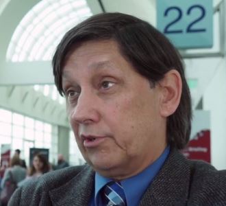
Adverse events were similar to those previously reported and were manageable, according to investigator Richard Thomas Maziarz, MD, from the Oregon Health & Science Knight Cancer Institute in Portland.
In this video interview at the annual meeting of the American Society of Hematology, Dr. Maziarz discusses the promising results using CAR T cells in this difficult to treat population.
SAN DIEGO – Two-thirds of adults with relapsed or refractory diffuse large B-cell lymphoma who had early responses to chimeric antigen receptor T-cell (CAR T) therapy with tisagenlecleucel (Kymriah) remain in remission with no evidence of minimal residual disease, according to an updated analysis of the JULIET trial.
In the single-arm, open-label trial, the overall response rate after 19 months of follow-up was 54%, including 40% complete remissions and 14% partial remissions. The median duration of response had not been reached at the time of data cutoff, and the median overall survival had not been reached for patients with a complete remission. Overall survival in this heavily pretreated population as a whole (all patients who received CAR T-cell infusions) was 11.1 months.
Adverse events were similar to those previously reported and were manageable, according to investigator Richard Thomas Maziarz, MD, from the Oregon Health & Science Knight Cancer Institute in Portland.
In this video interview at the annual meeting of the American Society of Hematology, Dr. Maziarz discusses the promising results using CAR T cells in this difficult to treat population.
REPORTING FROM ASH 2018
JULIET: CAR T cells keep trucking against DLBCL
SAN DIEGO – Chimeric antigen receptor T-cell therapy with tisagenlecleucel (Kymriah) is associated with a high rate of durable responses in adults with relapsed or refractory diffuse large B-cell lymphoma, an updated analysis of the JULIET trial showed.
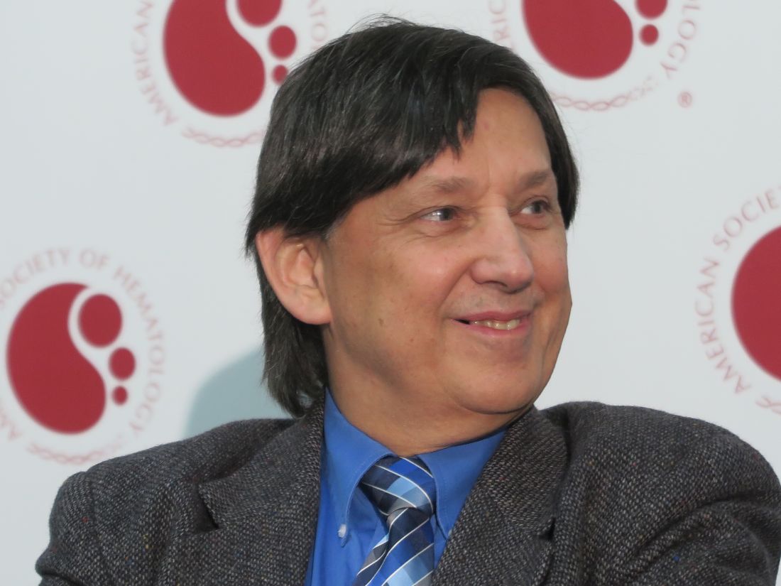
After a median follow-up of 19 months, two-thirds of adults with relapsed or refractory diffuse large B-cell lymphoma (DLBCL) who had early responses to chimeric antigen receptor (CAR) T-cell therapy with tisagenlecleucel remained in remission with no evidence of minimal residual disease, reported Richard Thomas Maziarz, MD, from the Oregon Health & Science Knight Cancer Institute in Portland, at the annual meeting of the American Society of Hematology.
“Since the previous report, no new deaths have been reported due to any cause other than patient disease progression. No treatment-related mortality was seen throughout the study, and there were three early deaths, all related to lymphoma that progressed,” he said in a briefing prior to presentation of the data in a scientific poster.
The updated study results were published simultaneously online in the New England Journal of Medicine.
JULIET then
In the phase 2, single-arm trial, investigators enrolled adults with DLBCL that had relapsed or was refractory after two or more prior lines of therapy and who were either ineligible for hematopoietic stem cell transplant or who experienced disease progression after transplant.
Interim results of the study were previously reported at the European Hematology Association Congress in 2017.
At that meeting, Gilles Salles, MD, PhD, from the University of Lyon (France), presented results of an analysis of available efficacy data on 51 patients with at least 3 months of follow-up. In this population, the best overall response rate was 59%. The 3-month overall response rate was 45%, consisting of 37% complete responses and 8% partial responses. Relapse-free survival at 6 months was 79% and all patients who had responses at 3 months continued to have responses at the time of data cutoff.
JULIET now
In the most recent analysis, completed after a median time from infusion to data cutoff of 14 months, the investigators reported on efficacy in 93 patients who received CAR T-cell infusions.
The best overall response rate, the primary endpoint, was 52%, comprising 40% complete responses and 12% partial responses. The response rates were consistent across all prognostic subgroups, including age, sex, previous response status, International Prognostic Index score at enrollment, prior therapy, molecular subtype, and other factors.
Estimated relapse-free survival 12 months after documentation of an initial response was 65%, and was 79% among patients who had complete responses.
The median duration of response had not been reached at the time of data cutoff; the median overall survival had not been reached for patients with a complete remission. Overall survival in this heavily pretreated population as a whole (all patients who received CAR T-cell infusions) was 11.1 months.
Adverse events of special interest included grade 3 or 4 cytokine release syndrome (CRS) in 23% of patients, prolonged cytopenia in 34%, infections in 19%, neurologic events in 11%, febrile neutropenia in 15%, and tumor lysis syndrome in 2%.
There were no deaths attributable to CRS or to cerebral edema, a complication of CAR T-cell therapy that appears to be related to the costimulatory molecule used in various constructs.
“Patients with relapsed or refractory DLBCL who are not eligible for high-dose therapy and hematopoietic cell transplantation or for whom such therapy was not successful have very few treatment options. For these patients, tisagenlecleucel shows promise that will need to be confirmed through larger studies with longer follow-up,” the investigators wrote in the New England Journal of Medicine.
The JULIET Trial is supported by Novartis. Dr. Maziar reported personal fees from Incyte, Kite Therapeutics, and Athersys.
SOURCE: Maziarz RT et al. N Engl J Med. 2018 Dec 1. doi: 10.1056/NEJMoa1804980.
SAN DIEGO – Chimeric antigen receptor T-cell therapy with tisagenlecleucel (Kymriah) is associated with a high rate of durable responses in adults with relapsed or refractory diffuse large B-cell lymphoma, an updated analysis of the JULIET trial showed.
After a median follow-up of 19 months, two-thirds of adults with relapsed or refractory diffuse large B-cell lymphoma (DLBCL) who had early responses to chimeric antigen receptor (CAR) T-cell therapy with tisagenlecleucel remained in remission with no evidence of minimal residual disease, reported Richard Thomas Maziarz, MD, from the Oregon Health & Science Knight Cancer Institute in Portland, at the annual meeting of the American Society of Hematology.
“Since the previous report, no new deaths have been reported due to any cause other than patient disease progression. No treatment-related mortality was seen throughout the study, and there were three early deaths, all related to lymphoma that progressed,” he said in a briefing prior to presentation of the data in a scientific poster.
The updated study results were published simultaneously online in the New England Journal of Medicine.
JULIET then
In the phase 2, single-arm trial, investigators enrolled adults with DLBCL that had relapsed or was refractory after two or more prior lines of therapy and who were either ineligible for hematopoietic stem cell transplant or who experienced disease progression after transplant.
Interim results of the study were previously reported at the European Hematology Association Congress in 2017.
At that meeting, Gilles Salles, MD, PhD, from the University of Lyon (France), presented results of an analysis of available efficacy data on 51 patients with at least 3 months of follow-up. In this population, the best overall response rate was 59%. The 3-month overall response rate was 45%, consisting of 37% complete responses and 8% partial responses. Relapse-free survival at 6 months was 79% and all patients who had responses at 3 months continued to have responses at the time of data cutoff.
JULIET now
In the most recent analysis, completed after a median time from infusion to data cutoff of 14 months, the investigators reported on efficacy in 93 patients who received CAR T-cell infusions.
The best overall response rate, the primary endpoint, was 52%, comprising 40% complete responses and 12% partial responses. The response rates were consistent across all prognostic subgroups, including age, sex, previous response status, International Prognostic Index score at enrollment, prior therapy, molecular subtype, and other factors.
Estimated relapse-free survival 12 months after documentation of an initial response was 65%, and was 79% among patients who had complete responses.
The median duration of response had not been reached at the time of data cutoff; the median overall survival had not been reached for patients with a complete remission. Overall survival in this heavily pretreated population as a whole (all patients who received CAR T-cell infusions) was 11.1 months.
Adverse events of special interest included grade 3 or 4 cytokine release syndrome (CRS) in 23% of patients, prolonged cytopenia in 34%, infections in 19%, neurologic events in 11%, febrile neutropenia in 15%, and tumor lysis syndrome in 2%.
There were no deaths attributable to CRS or to cerebral edema, a complication of CAR T-cell therapy that appears to be related to the costimulatory molecule used in various constructs.
“Patients with relapsed or refractory DLBCL who are not eligible for high-dose therapy and hematopoietic cell transplantation or for whom such therapy was not successful have very few treatment options. For these patients, tisagenlecleucel shows promise that will need to be confirmed through larger studies with longer follow-up,” the investigators wrote in the New England Journal of Medicine.
The JULIET Trial is supported by Novartis. Dr. Maziar reported personal fees from Incyte, Kite Therapeutics, and Athersys.
SOURCE: Maziarz RT et al. N Engl J Med. 2018 Dec 1. doi: 10.1056/NEJMoa1804980.
REPORTING FROM ASH 2018
Key clinical point: Chimeric antigen receptor T-cell therapy produced durable responses in patients with heavily pretreated diffuse large B-cell lymphoma.
Major finding: The best overall response rate, the primary endpoint, was 52%, comprising 40% complete responses and 12% partial responses.
Study details: A single-arm, open-label study of tisagenlecleucel in adults with relapsed or refractory diffuse large B-cell lymphoma.
Disclosures: The JULIET trial is supported by Novartis. Dr. Maziarz reported personal fees from Incyte, Kite Therapeutics, and Athersys.
Source: Maziarz RT et al. N Engl J Med. 2018 Dec 1. doi: 10.1056/NEJMoa1804980.
Ibrutinib outperforms bendamustine and rituximab in older CLL patients
SAN DIEGO – Ibrutinib alone or in combination with rituximab resulted in superior progression-free survival (PFS) when compared with bendamustine plus rituximab in the randomized, phase 3 Alliance A041202 trial of older patients with previously untreated chronic lymphocytic leukemia (CLL).
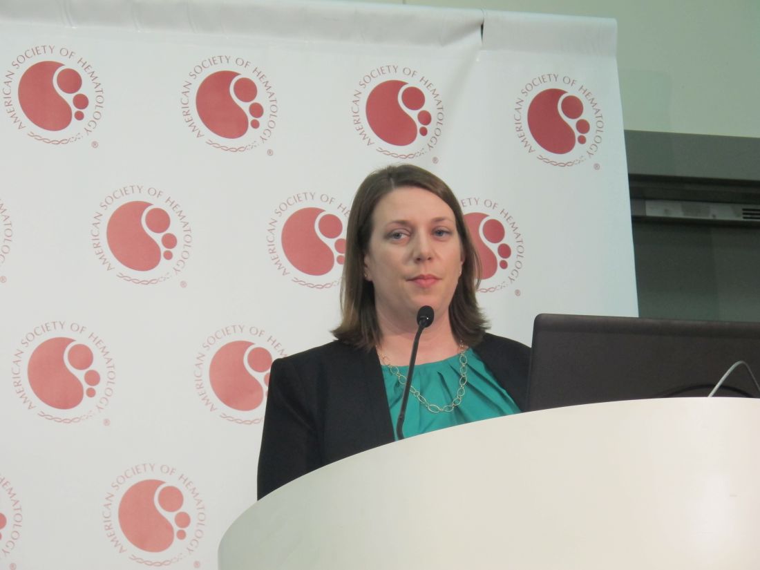
“There was no difference in progression-free survival between ibrutinib and ibrutinib plus rituximab,” said Dr. Woyach of the Ohio State University, Columbus.
Median PFS in this study was 43 months in the BR arm, and was not reached in either of the ibrutinib-containing arms, she said. No significant differences in overall survival (OS) were seen among the treatment arms, which may have been because of short follow-up and the fact that patients in the BR arm were allowed to cross over to ibrutinib if they progressed on treatment.
Participants in the international, multicenter trial – a project of the National Cancer Institute National Clinical Trials Network – were 547 adults aged 65 years or older (median, 71 years) with previously untreated, symptomatic CLL. They were enrolled from 219 sites across the United States and Canada between Dec. 9, 2013, and May 16, 2016.
The three arms were well matched with respect to baseline characteristics except for a slightly higher number of patients with complex karyotypes in the IR arm, Dr. Woyach said.
Treatment in the BR arm included bendamustine 90 mg/m2 on days 1 and 2 of each 28-day cycle plus rituximab at a dose of 375 mg/m2 on day 0 of cycle 1 then 500 mg/m2 on day 1 of cycles 2-6. Patients in the ibrutinib arms received 420 mg daily until disease progression either with or without rituximab at 375 mg/m2 weekly for 4 weeks starting at cycle 2 day 1 and then given on day 1 of cycles 3-6.
Grade 3-5 treatment-emergent hematologic adverse events (AEs) occurred in 61%, 41%, and 38% of patients in the BR, ibrutinib, and IR arms, respectively. Neutropenia and thrombocytopenia occurred more often in the BR than in the ibrutinib arms. Nonhematologic AEs occurred in 63%, 74%, and 74%, respectively, with an overall greater incidence in the ibrutinib arms. Infections and sudden deaths were numerically but not significantly higher in the ibrutinib arms.
“We undertook this study to determine the most effective therapy for older patients with CLL,” Dr. Woyach said, explaining that while older patients make up the majority of patients with CLL, they are typically underrepresented in trials.
At the start of the study, BR was widely used and the Bruton’s tyrosine kinase inhibitor ibrutinib was “just entering the clinic,” she noted.
“Despite now-widespread use in the [front-line setting] following FDA approval for this indication in 2016, the efficacy of ibrutinib versus standard chemoimmunotherapy has not previously been investigated,” she said.
Since adding rituximab has been shown to improve PFS and OS when added to chemotherapy in CLL, she and her colleagues also looked at whether this was the case with ibrutinib as well.
“This is the only phase 3 trial designed to answer this question,” she noted, adding that the findings justify the use of ibrutinib as a standard-of-care treatment for CLL patients aged 65 years and older.
David P. Steensma, MD, of Dana-Farber Cancer Institute in Boston, who moderated the press briefing, agreed. “I think this really does indicate that ibrutinib as front-line therapy, which many clinicians have been doing, is a very reasonable practice.”
Dr. Woyach added, however, that while ibrutinib represents a major therapeutic advance, its cost and its toxicities in older patients are a concern that warrant close monitoring and development of strategies to reduce the need for long-term continuous treatment.
Additional phase 3 studies set to open soon will compare ibrutinib in combination with venetoclax and obinutuzumab with standard ibrutinib
Dr. Woyach reported having no disclosures. Dr. Steensma reported receiving research funding from, and/or serving as a consultant, board member, or adviser for Takeda Pharmaceutical, Syros Pharmaceuticals, Otsuka Pharmaceutical, Onconova Therapeutics, Novartis, Kura Oncology, Janssen, H3 Biosciences, Celgene, Amphivena Therapeutics, and Acceleron Pharma.
SOURCE: Woyach JA et al. ASH 2018, Abstract 6.
SAN DIEGO – Ibrutinib alone or in combination with rituximab resulted in superior progression-free survival (PFS) when compared with bendamustine plus rituximab in the randomized, phase 3 Alliance A041202 trial of older patients with previously untreated chronic lymphocytic leukemia (CLL).
“There was no difference in progression-free survival between ibrutinib and ibrutinib plus rituximab,” said Dr. Woyach of the Ohio State University, Columbus.
Median PFS in this study was 43 months in the BR arm, and was not reached in either of the ibrutinib-containing arms, she said. No significant differences in overall survival (OS) were seen among the treatment arms, which may have been because of short follow-up and the fact that patients in the BR arm were allowed to cross over to ibrutinib if they progressed on treatment.
Participants in the international, multicenter trial – a project of the National Cancer Institute National Clinical Trials Network – were 547 adults aged 65 years or older (median, 71 years) with previously untreated, symptomatic CLL. They were enrolled from 219 sites across the United States and Canada between Dec. 9, 2013, and May 16, 2016.
The three arms were well matched with respect to baseline characteristics except for a slightly higher number of patients with complex karyotypes in the IR arm, Dr. Woyach said.
Treatment in the BR arm included bendamustine 90 mg/m2 on days 1 and 2 of each 28-day cycle plus rituximab at a dose of 375 mg/m2 on day 0 of cycle 1 then 500 mg/m2 on day 1 of cycles 2-6. Patients in the ibrutinib arms received 420 mg daily until disease progression either with or without rituximab at 375 mg/m2 weekly for 4 weeks starting at cycle 2 day 1 and then given on day 1 of cycles 3-6.
Grade 3-5 treatment-emergent hematologic adverse events (AEs) occurred in 61%, 41%, and 38% of patients in the BR, ibrutinib, and IR arms, respectively. Neutropenia and thrombocytopenia occurred more often in the BR than in the ibrutinib arms. Nonhematologic AEs occurred in 63%, 74%, and 74%, respectively, with an overall greater incidence in the ibrutinib arms. Infections and sudden deaths were numerically but not significantly higher in the ibrutinib arms.
“We undertook this study to determine the most effective therapy for older patients with CLL,” Dr. Woyach said, explaining that while older patients make up the majority of patients with CLL, they are typically underrepresented in trials.
At the start of the study, BR was widely used and the Bruton’s tyrosine kinase inhibitor ibrutinib was “just entering the clinic,” she noted.
“Despite now-widespread use in the [front-line setting] following FDA approval for this indication in 2016, the efficacy of ibrutinib versus standard chemoimmunotherapy has not previously been investigated,” she said.
Since adding rituximab has been shown to improve PFS and OS when added to chemotherapy in CLL, she and her colleagues also looked at whether this was the case with ibrutinib as well.
“This is the only phase 3 trial designed to answer this question,” she noted, adding that the findings justify the use of ibrutinib as a standard-of-care treatment for CLL patients aged 65 years and older.
David P. Steensma, MD, of Dana-Farber Cancer Institute in Boston, who moderated the press briefing, agreed. “I think this really does indicate that ibrutinib as front-line therapy, which many clinicians have been doing, is a very reasonable practice.”
Dr. Woyach added, however, that while ibrutinib represents a major therapeutic advance, its cost and its toxicities in older patients are a concern that warrant close monitoring and development of strategies to reduce the need for long-term continuous treatment.
Additional phase 3 studies set to open soon will compare ibrutinib in combination with venetoclax and obinutuzumab with standard ibrutinib
Dr. Woyach reported having no disclosures. Dr. Steensma reported receiving research funding from, and/or serving as a consultant, board member, or adviser for Takeda Pharmaceutical, Syros Pharmaceuticals, Otsuka Pharmaceutical, Onconova Therapeutics, Novartis, Kura Oncology, Janssen, H3 Biosciences, Celgene, Amphivena Therapeutics, and Acceleron Pharma.
SOURCE: Woyach JA et al. ASH 2018, Abstract 6.
REPORTING FROM ASH 2018
Key clinical point: In chronic lymphocytic leukemia patients aged 65 years and older, progression-free survival is better with ibrutinib than with bendamustine and rituximab.
Major finding: The 2-year progression-free survival was 74%, 87%, and 88% with bendamustine and rituximab, ibrutinib, and ibrutinib and rituximab, respectively.
Study details: A randomized, phase 3 study of 547 previously untreated patients with CLL.
Disclosures: Dr. Woyach reported having no disclosures. Dr. Steensma reported receiving research funding from, and/or serving as a consultant, board member, or adviser for Takeda Pharmaceutical, Syros Pharmaceuticals, Otsuka Pharmaceutical, Onconova Therapeutics, Novartis, Kura Oncology, Janssen, H3 Biosciences, Celgene, Amphivena Therapeutics, and Acceleron Pharma.
Source: Woyach JA et al. ASH 2018, Abstract 6.
Stroke, arterial dissection events reported with Lemtrada, FDA says
Instances of stroke and arterial dissection in the head and neck have been reported in some multiple sclerosis patients soon after an infusion of alemtuzumab (Lemtrada), according to a safety announcement issued by the Food and Drug Administration on Nov. 29.

Since the FDA approved alemtuzumab in 2014 for relapsing forms of MS, 13 cases of ischemic and hemorrhagic stroke or arterial dissection have been reported worldwide via the FDA Adverse Event Reporting System, but “additional cases we are unaware of may have occurred,” the FDA said in the announcement.
Most of the patients who developed stroke or arterial lining tears showed symptoms within a day of taking the medication, although one patient reported symptoms three days after treatment. The drug is given via intravenous infusion and is generally reserved for patients with relapsing MS who have not responded adequately to other approved MS medications, according to the FDA.
Symptoms include sudden onset of the following: severe headache or neck pain; numbness or weakness in the arms or legs, especially on only one side of the body; confusion or trouble speaking or understanding speech; vision problems in one or both eyes; and dizziness, loss of balance, or difficulty walking.
As a result of the reports, the FDA has updated the drug label prescribing information and the patient Medication Guide to reflect these risks, and added the risk of stroke to the medication’s existing boxed warning.
Health care providers should remind patients of the potential for stroke and arterial dissection at each treatment visit and advise them to seek immediate medical attention if they experience any of the symptoms reported in previous cases. “The diagnosis is often complicated because early symptoms such as headache and neck pain are not specific,” according to the agency, but patients complaining of such symptoms should be evaluated immediately.
Alemtuzumab was also approved in May 2001 for treating B-cell chronic lymphocytic leukemia (B-CLL) under the brand name Campath. The FDA will update the Campath label to reflect the new warnings and risks.
Instances of stroke and arterial dissection in the head and neck have been reported in some multiple sclerosis patients soon after an infusion of alemtuzumab (Lemtrada), according to a safety announcement issued by the Food and Drug Administration on Nov. 29.
Since the FDA approved alemtuzumab in 2014 for relapsing forms of MS, 13 cases of ischemic and hemorrhagic stroke or arterial dissection have been reported worldwide via the FDA Adverse Event Reporting System, but “additional cases we are unaware of may have occurred,” the FDA said in the announcement.
Most of the patients who developed stroke or arterial lining tears showed symptoms within a day of taking the medication, although one patient reported symptoms three days after treatment. The drug is given via intravenous infusion and is generally reserved for patients with relapsing MS who have not responded adequately to other approved MS medications, according to the FDA.
Symptoms include sudden onset of the following: severe headache or neck pain; numbness or weakness in the arms or legs, especially on only one side of the body; confusion or trouble speaking or understanding speech; vision problems in one or both eyes; and dizziness, loss of balance, or difficulty walking.
As a result of the reports, the FDA has updated the drug label prescribing information and the patient Medication Guide to reflect these risks, and added the risk of stroke to the medication’s existing boxed warning.
Health care providers should remind patients of the potential for stroke and arterial dissection at each treatment visit and advise them to seek immediate medical attention if they experience any of the symptoms reported in previous cases. “The diagnosis is often complicated because early symptoms such as headache and neck pain are not specific,” according to the agency, but patients complaining of such symptoms should be evaluated immediately.
Alemtuzumab was also approved in May 2001 for treating B-cell chronic lymphocytic leukemia (B-CLL) under the brand name Campath. The FDA will update the Campath label to reflect the new warnings and risks.
FDA approves biosimilar rituximab for NHL

The U.S. Food and Drug Administration (FDA) has approved a biosimilar rituximab product for the treatment of non-Hodgkin lymphoma (NHL).
Celltrion’s Truxima (rituximab-abbs) is a biosimilar of Genentech’s Rituxan and the first biosimilar approved in the United States to treat NHL.
Truxima (formerly CT-P10) is approved to treat adults with CD20-positive, B-cell NHL, either as a single agent or in combination with chemotherapy.
Specifically, Truxima is approved as a single agent to treat relapsed or refractory, low grade or follicular, CD20-positive, B-cell NHL.
Truxima is approved in combination with first-line chemotherapy to treat previously untreated follicular, CD20-positive, B-cell NHL.
Truxima is approved as single-agent maintenance therapy in patients with follicular, CD20-positive, B-cell NHL who achieve a complete or partial response to a rituximab product in combination with chemotherapy.
And Truxima is approved as a single agent to treat non-progressing, low-grade, CD20-positive, B-cell NHL after first-line treatment with cyclophosphamide, vincristine, and prednisone.
The label for Truxima contains a boxed warning detailing the risk of fatal infusion reactions, severe skin and mouth reactions (some with fatal outcomes), hepatitis B virus reactivation that may cause serious liver problems (including liver failure and death), and progressive multifocal leukoencephalopathy.
The FDA said its approval of Truxima is “based on a review of evidence that included extensive structural and functional characterization, animal study data, human pharmacokinetic data, clinical immunogenicity data, and other clinical data that demonstrates Truxima is biosimilar to Rituxan.”
A phase 3 trial recently published in The Lancet Haematology suggested that Truxima is equivalent to the reference product in patients with low-tumor-burden follicular lymphoma.
For more details on Truxima, see the prescribing information.
FDA approves rituximab biosimilar for lymphoma
(NHL).

Celltrion’s Truxima (rituximab-abbs) is a biosimilar of Genentech’s Rituxan (rituximab) and the first biosimilar approved in the United States to treat NHL.
Truxima (formerly CT-P10) is approved to treat adults with CD20-positive, B-cell NHL, either as a single agent or in combination with chemotherapy. Truxima is approved as a single agent to treat relapsed or refractory, low grade or follicular, CD20-positive, B-cell NHL. Truxima is approved in combination with first-line chemotherapy to treat previously untreated follicular, CD20-positive, B-cell NHL.
Truxima is approved as single-agent maintenance therapy in patients with follicular, CD20-positive, B-cell NHL who achieve a complete or partial response to a rituximab product in combination with chemotherapy. Truxima also is approved as a single agent to treat nonprogressing, low-grade, CD20-positive, B-cell NHL after first-line treatment with cyclophosphamide, vincristine, and prednisone.The label for Truxima contains a boxed warning detailing the risk of fatal infusion reactions, severe skin and mouth reactions (some with fatal outcomes), hepatitis B virus reactivation that may cause serious liver problems (including liver failure and death), and progressive multifocal leukoencephalopathy.
The FDA said its approval of Truxima is “based on a review of evidence that included extensive structural and functional characterization, animal study data, human pharmacokinetic data, clinical immunogenicity data, and other clinical data that demonstrates Truxima is biosimilar to Rituxan.”
Findings from a phase 3 trial suggested that Truxima is equivalent to the reference product in patients with low-tumor-burden follicular lymphoma (Lancet Haematol. 2018 Nov;5[11]:e543-53).
(NHL).
Celltrion’s Truxima (rituximab-abbs) is a biosimilar of Genentech’s Rituxan (rituximab) and the first biosimilar approved in the United States to treat NHL.
Truxima (formerly CT-P10) is approved to treat adults with CD20-positive, B-cell NHL, either as a single agent or in combination with chemotherapy. Truxima is approved as a single agent to treat relapsed or refractory, low grade or follicular, CD20-positive, B-cell NHL. Truxima is approved in combination with first-line chemotherapy to treat previously untreated follicular, CD20-positive, B-cell NHL.
Truxima is approved as single-agent maintenance therapy in patients with follicular, CD20-positive, B-cell NHL who achieve a complete or partial response to a rituximab product in combination with chemotherapy. Truxima also is approved as a single agent to treat nonprogressing, low-grade, CD20-positive, B-cell NHL after first-line treatment with cyclophosphamide, vincristine, and prednisone.The label for Truxima contains a boxed warning detailing the risk of fatal infusion reactions, severe skin and mouth reactions (some with fatal outcomes), hepatitis B virus reactivation that may cause serious liver problems (including liver failure and death), and progressive multifocal leukoencephalopathy.
The FDA said its approval of Truxima is “based on a review of evidence that included extensive structural and functional characterization, animal study data, human pharmacokinetic data, clinical immunogenicity data, and other clinical data that demonstrates Truxima is biosimilar to Rituxan.”
Findings from a phase 3 trial suggested that Truxima is equivalent to the reference product in patients with low-tumor-burden follicular lymphoma (Lancet Haematol. 2018 Nov;5[11]:e543-53).
Cortactin expression aids in CLL-MCL differential
The presence or absence in tumor cells of cortactin, a cytoskeleton-remodeling adapter protein, may be a marker to help pathologists distinguish between chronic lymphocytic leukemia (CLL) and mantle cell lymphoma (MCL), investigators suggest.
A study of cortactin expression in tumor samples from patients with B-cell CLL, MCL, and other hematologic malignancies showed that while cortactin was present in 14 of 17 CLL samples, it was not expressed on any of 16 MCL samples, reported Marco Pizzi, MD, PhD, from the University of Padova (Italy) and his colleagues.
“In particular, cortactin may contribute to the differential diagnosis between CLL and MCL, two neoplasms with similar histological features but very different clinical outcome. Further studies are needed to clarify the molecular mechanisms of deranged cortactin expression in MCL and CLL and to investigate any possible relationship between cortactin status and the biological features of these lymphomas,” they wrote in Human Pathology.
Overexpression of cortactin has been reported in several solid tumors, and increased expression of CTTN, the gene encoding for cortactin, has been associated with aggressive, poor prognosis disease, the investigators noted.
To characterize cortactin expression in lymphoid and hematopoietic cells and detect potential associations between cortactin and virulence of hematologic malignancies, the investigators performed immunohistochemical analysis on samples from 131 patients treated at their center. The samples included 17 cases of CLL, 16 of MCL, 25 of follicular lymphoma (FL), 30 of marginal zone lymphoma (MZL), 10 of hairy cell leukemia, three of splenic diffuse red pulp small B-cell lymphomas (SDRPBL), and 30 of diffuse large B-cell lymphoma (DLBCL).
They found that cortactin was expressed in 14 of the 17 CLL samples, all 10 of the HCL samples, and 22 of the 30 DLBCL samples. In contrast, there was no cortactin expression detected in any of either 16 MCL or three SDRPBL samples. The researchers found that 13 of 30 MZL samples had low-level staining. In FL, cortactin was expressed in 2 of the samples but in the remaining 23 cases the researchers found only scattered cortactin-positive lymphoid elements of non–B-cell lineage.
The investigators also found that cortactin expression in CLL correlated with other CLL-specific markers, and found that expression of two or more of the markers had 89.1% sensitivity, 100% specificity, a 100% positive predictive value, and 90.5% negative predictive value for a diagnosis of CLL.
In addition, they saw that the immunohistochemical results were similar to those for CTTN gene expression assessed by in silico analysis.
The investigators noted that CLL and MCL are challenging to differentiate from one another because of morphologic similarities and partially overlapping immunophenotypes.
“In this context, cortactin expression would strongly sustain a diagnosis of CLL over MCL, particularly in association with other CLL markers (i.e., LEF1 and CD200),” they wrote.
The study was internally supported. The authors declared no conflicts of interest.
SOURCE: Pizzi M et al. Hum Pathol. 2018 Nov 17. doi: 10.1016/j.humpath.2018.10.038.
FROM HUMAN PATHOLOGY
Key clinical point:
Major finding: Cortactin was expressed on 14 of 17 CLL samples vs. none of 16 MCL samples.
Study details: Immunohistochemistry analysis of samples from 131 patients with B-cell lineage non-Hodgkin lymphomas.
Disclosures: The study was internally supported. The authors reported having no conflicts of interest.
Source: Pizzi M et al. Hum Pathol. 2018 Nov 17. doi: 10.1016/j.humpath.2018.10.038.
Fatigue in MS: Common, often profound, tough to treat
BERLIN – In addition to the pain, motor and sensory impairments, and cognitive dysfunction that can stalk multiple sclerosis (MS) patients, for many, there’s also the challenge of an invisible, tough-to-quantify entity: fatigue.

“Approximately 80% of patients suffer from fatigue, so it’s an immense problem in MS. There’s no real clear relationship with disease severity,” Vincent de Groot, MD, said at the annual congress of the European Committee for Treatment and Research in Multiple Sclerosis. “Despite what a lot of people think, there’s no clear or strong relationship between fatigue and the amount of physical activity people undertake daily,” he noted.
“Patients all know what we are talking about when we ask about fatigue,” but there are a variety of definitions of fatigue used in research, a fact that has limited progress in the field, said Dr. de Groot.
Primary fatigue is related to the pathophysiology of MS itself, while secondary fatigue can result from MS symptoms, such as poor sleep from spasms. Secondary fatigue can also be a side effect of MS medications; baclofen, used for spasticity, is a good example, said Dr. de Groot. “We must not underestimate how many problems these drugs can give people.”
What’s the mechanism by which MS causes primary fatigue? “The simple answer is that we do not know,” said Dr. de Groot, a physiatrist and researcher at Vrije University, Amsterdam.
Though immune-mediated fatigue had been proposed as a factor for patients with MS, Dr. de Groot said that his own lab’s work has not found any connection between fatigue levels and any immune-related biomarkers. “So I don’t think the immune hypothesis has a lot of evidence.”
Similarly, though there might be mechanistic reasons to suspect the hypothalamic-pituitary-adrenal (HPA) axis as a culprit for MS fatigue, no consistent association has been found between any markers for HPA axis disruption and fatigue ratings, Dr. de Groot said.
Newer theories center on MS-related disruptions in brain connectivity, with imaging studies now able to detect some of these disruptions in functional connectivity that correlate with fatigue. “Right now, I think this is the hypothesis to bet money on,” Dr. de Groot said.
Many factors come into play, including environmental and psychological factors, he said.
“What can we do to treat MS-related fatigue?” Though several drugs have been used, “if you carefully look at the systematic reviews, the evidence is very, very disappointing,” Dr. de Groot said. For both amantadine and modafinil, “there is no evidence that these drugs are effective,” he said, citing a systematic review and meta-analysis that found a pooled effect size of 0.07 (95% confidence interval, –0.22 to 0.37) for medications (Mult Scler Int. 2014 May; doi: 10.1155/2014/798285).
Only two trials have looked at multidisciplinary rehabilitation for MS-related fatigue, Dr. de Groot said. Two studies that looked at multidisciplinary strategies that pulled in a variety of disciplines to help develop tailored fatigue management strategies saw no between-group effect when the multidisciplinary intervention was compared with nurse-provided information or with non–fatigue-related rehabilitation.
In an effort to determine whether MS-related fatigue is truly refractory to treatment, Dr. de Groot said that he and his colleagues decided to take “three steps back” to look at the individual interventions that make up a multidisciplinary approach to tackling fatigue. “So, we looked at exercise therapy, energy conservation management, and cognitive-behavioral therapy,” beginning with a literature review, he said.
Members of his research group found that effect sizes ranged from small to moderate for the three approaches, but there were methodologic problems with many of the studies; in the case of cognitive-behavioral therapy (CBT), the effect size waned over time, Dr. de Groot said. A newer randomized, controlled trial showed a relatively robust effect size of 0.52 for Internet-delivered CBT, which may provide a promising and practical approach (J Neurol Neurosurg Psychiatry. 2018 Sep;89[9]:970-6. doi: 10.1136/jnnp-2017-317463).
Looking at fatigue and societal participation, Dr. de Groot and his colleagues examined what effect aerobic training, energy conservation management, and CBT had on the two outcome measures. The three interventions were studied in three stand-alone trials, he said.
Patients were assessed at baseline, and at 8, 16, 26, and 52 weeks. The assessments were performed by a blinded researcher and were the same across trials: For fatigue, researchers used the Checklist Individual Strength–fatigue (CIS20R-fatigue), and for societal participation, they administered the Impact on Participation and Autonomy (IPA).
Each trial included 90 patients, randomized 1:1 to receive high- or low-intensity treatment. Patients had to have MS with no exacerbations within the prior 6 months and an Expanded Disability Status Scale score of 6 or less. However, the included patients had severe fatigue, with a CIS20R-fatigue subscore of 35 or higher, and the fatigue could not be attributable to such secondary causes as infection, depression, or thyroid or sleep problems. Finally, patients could not have been treated for fatigue in the 3 months prior to enrollment.
Those in the high-intensity treatment group received 12 sessions focused on the particular intervention over 4 months, provided by an expert therapist. Each type of intervention had a treatment protocol that was followed over the 4 months. Patients receiving low-intensity treatment saw an MS-specialized nurse three times over 4 months.
The aerobic training intervention had patients performing high-intensity exercise on a cycle ergometer for 30 minutes, three times weekly for 16 weeks. In addition to the 12 supervised sessions, patients also completed 36 home-based sessions. The level of intensity for each patient was personalized based on their baseline cardiopulmonary exercise test, Dr. de Groot said.
At the end of 1 year, patients in the high-intensity group and those in the low-intensity group reported virtually the same fatigue scores. Though there was an initial drop in fatigue for those in the high-intensity group, compared with baseline and with the low-intensity participants, values on the CIS20R never dropped below 35, the “severe fatigue” cutoff.
And, Dr. de Groot said, there was no effect on societal participation or in other fatigue scores. In sum, the effect size was barely significant at –0.54 (95% CI, –1.00 to –0.06), with a number needed to treat of 9.
Adherence to attempting the workouts was fairly good for participants in the high-intensity group; 74% completed the sessions, with 71% doing so at the prescribed workload. The median rate of perceived exertion on a 1-20 scale was 14.
However, the thrice-weekly exercise bouts didn’t improve aerobic fitness parameters: Neither V02peak, V02peak adjusted for body mass, nor anaerobic threshold changed for those in the high-intensity group. Peak power did increase by 11.7 watts (P = .048).
Energy conservation education, whether delivered in high- or low-intensity format, had almost no effect on fatigue scores, with a number needed to treat of 158 – a figure that is “neither significant nor clinically meaningful,” Dr. de Groot said. Other fatigue scores and societal participation levels also went unchanged.
However, CBT delivered in a series of 10 modules to address various beliefs and coping mechanisms about MS, fatigue, pain, and activity regulation did have a positive effect on fatigue. Here, the effect size was –0.79 (95% CI, –1.26 to –0.32). The number needed to treat was 3, and CIS20R values did dip below the “severe fatigue” threshold during treatment. A similar effect, Dr. de Groot said, was seen for other fatigue and quality of life measures, though societal participation scores didn’t change. No significant improvement was seen for the low-intensity CBT group.
“Severe MS-related fatigue can be reduced effectively with CBT in the short term. More research is needed on how to maintain this effect in the long term,” Dr. de Groot said. Still, “it’s currently the best treatment option,” he said.
The fact that patients reverted to their preintervention fatigue levels regardless of the intervention shows that effective treatment for MS-related fatigue should probably be ongoing, viewed more as a process than an occurrence, Dr. de Groot said.
To that end, Dr. de Groot and his colleagues are conducting a randomized, controlled trial that includes 166 MS patients with fatigue. The study has two arms: The first is a noninferiority trial comparing face-to-face CBT with e-learning delivery of the content, and the second looks at the efficacy of ongoing booster sessions after initial CBT.
An online database of randomized, controlled trials of rehabilitation for MS patients can be found at www.appeco.net.
The study was funded by Fonds NutsOhra, a private Dutch foundation. Dr. de Groot reported no relevant conflicts of interest.
SOURCE: de Groot V. Mult Scler. 2018;24(S2):83, Abstract 225.
BERLIN – In addition to the pain, motor and sensory impairments, and cognitive dysfunction that can stalk multiple sclerosis (MS) patients, for many, there’s also the challenge of an invisible, tough-to-quantify entity: fatigue.
“Approximately 80% of patients suffer from fatigue, so it’s an immense problem in MS. There’s no real clear relationship with disease severity,” Vincent de Groot, MD, said at the annual congress of the European Committee for Treatment and Research in Multiple Sclerosis. “Despite what a lot of people think, there’s no clear or strong relationship between fatigue and the amount of physical activity people undertake daily,” he noted.
“Patients all know what we are talking about when we ask about fatigue,” but there are a variety of definitions of fatigue used in research, a fact that has limited progress in the field, said Dr. de Groot.
Primary fatigue is related to the pathophysiology of MS itself, while secondary fatigue can result from MS symptoms, such as poor sleep from spasms. Secondary fatigue can also be a side effect of MS medications; baclofen, used for spasticity, is a good example, said Dr. de Groot. “We must not underestimate how many problems these drugs can give people.”
What’s the mechanism by which MS causes primary fatigue? “The simple answer is that we do not know,” said Dr. de Groot, a physiatrist and researcher at Vrije University, Amsterdam.
Though immune-mediated fatigue had been proposed as a factor for patients with MS, Dr. de Groot said that his own lab’s work has not found any connection between fatigue levels and any immune-related biomarkers. “So I don’t think the immune hypothesis has a lot of evidence.”
Similarly, though there might be mechanistic reasons to suspect the hypothalamic-pituitary-adrenal (HPA) axis as a culprit for MS fatigue, no consistent association has been found between any markers for HPA axis disruption and fatigue ratings, Dr. de Groot said.
Newer theories center on MS-related disruptions in brain connectivity, with imaging studies now able to detect some of these disruptions in functional connectivity that correlate with fatigue. “Right now, I think this is the hypothesis to bet money on,” Dr. de Groot said.
Many factors come into play, including environmental and psychological factors, he said.
“What can we do to treat MS-related fatigue?” Though several drugs have been used, “if you carefully look at the systematic reviews, the evidence is very, very disappointing,” Dr. de Groot said. For both amantadine and modafinil, “there is no evidence that these drugs are effective,” he said, citing a systematic review and meta-analysis that found a pooled effect size of 0.07 (95% confidence interval, –0.22 to 0.37) for medications (Mult Scler Int. 2014 May; doi: 10.1155/2014/798285).
Only two trials have looked at multidisciplinary rehabilitation for MS-related fatigue, Dr. de Groot said. Two studies that looked at multidisciplinary strategies that pulled in a variety of disciplines to help develop tailored fatigue management strategies saw no between-group effect when the multidisciplinary intervention was compared with nurse-provided information or with non–fatigue-related rehabilitation.
In an effort to determine whether MS-related fatigue is truly refractory to treatment, Dr. de Groot said that he and his colleagues decided to take “three steps back” to look at the individual interventions that make up a multidisciplinary approach to tackling fatigue. “So, we looked at exercise therapy, energy conservation management, and cognitive-behavioral therapy,” beginning with a literature review, he said.
Members of his research group found that effect sizes ranged from small to moderate for the three approaches, but there were methodologic problems with many of the studies; in the case of cognitive-behavioral therapy (CBT), the effect size waned over time, Dr. de Groot said. A newer randomized, controlled trial showed a relatively robust effect size of 0.52 for Internet-delivered CBT, which may provide a promising and practical approach (J Neurol Neurosurg Psychiatry. 2018 Sep;89[9]:970-6. doi: 10.1136/jnnp-2017-317463).
Looking at fatigue and societal participation, Dr. de Groot and his colleagues examined what effect aerobic training, energy conservation management, and CBT had on the two outcome measures. The three interventions were studied in three stand-alone trials, he said.
Patients were assessed at baseline, and at 8, 16, 26, and 52 weeks. The assessments were performed by a blinded researcher and were the same across trials: For fatigue, researchers used the Checklist Individual Strength–fatigue (CIS20R-fatigue), and for societal participation, they administered the Impact on Participation and Autonomy (IPA).
Each trial included 90 patients, randomized 1:1 to receive high- or low-intensity treatment. Patients had to have MS with no exacerbations within the prior 6 months and an Expanded Disability Status Scale score of 6 or less. However, the included patients had severe fatigue, with a CIS20R-fatigue subscore of 35 or higher, and the fatigue could not be attributable to such secondary causes as infection, depression, or thyroid or sleep problems. Finally, patients could not have been treated for fatigue in the 3 months prior to enrollment.
Those in the high-intensity treatment group received 12 sessions focused on the particular intervention over 4 months, provided by an expert therapist. Each type of intervention had a treatment protocol that was followed over the 4 months. Patients receiving low-intensity treatment saw an MS-specialized nurse three times over 4 months.
The aerobic training intervention had patients performing high-intensity exercise on a cycle ergometer for 30 minutes, three times weekly for 16 weeks. In addition to the 12 supervised sessions, patients also completed 36 home-based sessions. The level of intensity for each patient was personalized based on their baseline cardiopulmonary exercise test, Dr. de Groot said.
At the end of 1 year, patients in the high-intensity group and those in the low-intensity group reported virtually the same fatigue scores. Though there was an initial drop in fatigue for those in the high-intensity group, compared with baseline and with the low-intensity participants, values on the CIS20R never dropped below 35, the “severe fatigue” cutoff.
And, Dr. de Groot said, there was no effect on societal participation or in other fatigue scores. In sum, the effect size was barely significant at –0.54 (95% CI, –1.00 to –0.06), with a number needed to treat of 9.
Adherence to attempting the workouts was fairly good for participants in the high-intensity group; 74% completed the sessions, with 71% doing so at the prescribed workload. The median rate of perceived exertion on a 1-20 scale was 14.
However, the thrice-weekly exercise bouts didn’t improve aerobic fitness parameters: Neither V02peak, V02peak adjusted for body mass, nor anaerobic threshold changed for those in the high-intensity group. Peak power did increase by 11.7 watts (P = .048).
Energy conservation education, whether delivered in high- or low-intensity format, had almost no effect on fatigue scores, with a number needed to treat of 158 – a figure that is “neither significant nor clinically meaningful,” Dr. de Groot said. Other fatigue scores and societal participation levels also went unchanged.
However, CBT delivered in a series of 10 modules to address various beliefs and coping mechanisms about MS, fatigue, pain, and activity regulation did have a positive effect on fatigue. Here, the effect size was –0.79 (95% CI, –1.26 to –0.32). The number needed to treat was 3, and CIS20R values did dip below the “severe fatigue” threshold during treatment. A similar effect, Dr. de Groot said, was seen for other fatigue and quality of life measures, though societal participation scores didn’t change. No significant improvement was seen for the low-intensity CBT group.
“Severe MS-related fatigue can be reduced effectively with CBT in the short term. More research is needed on how to maintain this effect in the long term,” Dr. de Groot said. Still, “it’s currently the best treatment option,” he said.
The fact that patients reverted to their preintervention fatigue levels regardless of the intervention shows that effective treatment for MS-related fatigue should probably be ongoing, viewed more as a process than an occurrence, Dr. de Groot said.
To that end, Dr. de Groot and his colleagues are conducting a randomized, controlled trial that includes 166 MS patients with fatigue. The study has two arms: The first is a noninferiority trial comparing face-to-face CBT with e-learning delivery of the content, and the second looks at the efficacy of ongoing booster sessions after initial CBT.
An online database of randomized, controlled trials of rehabilitation for MS patients can be found at www.appeco.net.
The study was funded by Fonds NutsOhra, a private Dutch foundation. Dr. de Groot reported no relevant conflicts of interest.
SOURCE: de Groot V. Mult Scler. 2018;24(S2):83, Abstract 225.
EXPERT ANALYSIS FROM ECTRIMS 2018

Particular lesions early after CIS predict long-term MS disability
BERLIN – The presence of infratentorial and deep white matter lesions early in the course of relapse-onset multiple sclerosis was associated with high levels of disability 30 years later in a study looking at MRI predictors.
Univariate predictors of an Expanded Disability Status Scale (EDSS) score of more than 3.5, which is indicative of impaired mobility after 30 years, were the presence of an IT lesion at baseline, with an odds ratio (OR) of 12.4 (95% confidence interval [CI], 3.35-3.46; P less than .001), the presence of a deep white matter (DWM) lesion 1 year after presenting with clinically-isolated syndrome (CIS; OR = 10.65; 95% CI, 2.84-38.84; P less than .001), and the presence of an infratentorial (IT) lesion 1 year post CIS presentation (OR = 11.1; 95% CI, 3.31-37.22; P less than .001). At 5 years after a CIS presentation, the EDSS score, EDSS score change, and a DWM lesion score of more than 5 were indicative of worse disability after 30 years.
There was no significant association with age of onset, gender, CIS type, baseline EDSS, or disease duration, study investigator Karen Chung, MBBS, reported at the annual congress of the European Committee for Treatment and Research in Multiple Sclerosis.
“As we know, approximately 85% of people with MS initially present with a clinically isolated syndrome,” said Dr. Chung, a clinical research associate at the Queen Square Multiple Sclerosis Centre in the UCL Institute of Neurology in London. She added that the long-term prognosis after CIS is very variable, with some patients developing little detectable disability over time, while others may experience considerable decline.
There have been few studies examining whether there are any MRI parameters that might help predict patients’ long-term outcomes, so the aim of the study Dr. Chung presented was to see if there were any MRI parameters that might be predictive of clinical outcome 30 years after the onset of CIS.
Dr. Chung and her coauthors examined data on 120 of 132 individuals from the First London CIS Cohort who were prospectively recruited between 1984 and 1987 and had known outcomes. They looked at prospectively gathered MRI data and EDSS data at baseline, 5, 10, 14, 20, and 30 years. MRI data were obtained for 1 year after the CIS event, and data on the lowest EDSS score after the CIS event were retrospectively determined from patient notes or recall. The cohort was predominantly female (61%), with a mean of 31.5 years at CIS onset. Around half (52%) presented with optic neuritis, 27% with a spinal cord syndrome, and 20% with a brainstem syndrome. The high percentage of patients presenting with optic neuritis may be due to the fact that one of the recruiting centers was a specialist eye hospital, Dr. Chung noted later during discussion.
The 2010 McDonald Criteria were used to determine whether patients had CIS, relapsing-remitting MS (RRMS), secondary progressive MS (SPMS), or death related to MS.
“We looked at all the MRIs available to us and quantified the T2 lesion count for whole brain as well as by location,” Dr. Chung explained. The locations considered were juxtacortical, periventricular, infratentorial, and deep white matter.
“I think it is important to remind ourselves that we have come a long way with MRI technology in the 30 years timespan,” she added, noting that there was “clearly a difference in the quality.”
Clinical outcomes at 30 years were as follows: 80 patients (67%) developed MS, of whom 35 (44%) had RRMS, 26 (33%) had SPMS, 15 (20%) had died as a result of MS, and 3 (4%) had died for unknown or unrelated causes. Of the 40 patients (33%) who remained with CIS, 10 (25%) died without developing MS.
“This is a largely untreated cohort where, within the 80 people with MS, 11 (14%) were treated with a DMT [disease-modifying treatment] at some point,” Dr. Chung reported. All DMTs used were first-line injectable agents, she observed.
EDSS scores could be obtained for 107 patients. At 30 years, people with low EDSS scores were those who remained with CIS or RRMS, and as EDSS scores increased, the severity of MS increased.
“Overall, T2 lesions were better predictors of 30-year outcome than EDSS,” Dr. Chung said. For combinations of predictors, patients who had at least one IT and one DWM lesion within 1 year of a CIS had a higher probability (94%) of having an EDSS score of more than 3.5 at 30 years than when compared with those with neither lesion (13%) and those with one DWM but no IT lesions (49%).
Looking at the best independent predictors up to 5 years, the predicted probability of an EDSS score of more than 3.5 if there were no IT lesions and fewer than five DWM lesions was 18%. But if there were no IT but more than 5 DWM lesions, the probability of disability at 30 years rose to 52%. The probabilities rose even higher to 63% if there was one or more IT and five or less DWM lesions and 90% if there was one or more IT and more than five DWM lesions.
“In this cohort, the presence of early infratentorial and deep white matter lesions following a CIS are associated with higher level of disability 30 years later,” Dr. Chung concluded. “Early predictive models can add information to risk-benefit stratification.”
During discussion, one delegate expressed concerns that these data were “not generalizable to the current situation.” This was a cohort of patients that largely wasn’t treated or if they were, treatment was delayed by more than 10 years. These data were interesting “from a historical perspective,” he argued, “but I don’t understand, how in the absence of contemporary therapies this is applicable in a way that will allow us to use this information to make prognoses for the future.”
Dr. Chung agreed, noting that this was more of a natural history study. “However, I think it is applicable in clinical practice in my opinion.
“When you have a patient presenting with a CIS, at the time of diagnosis, especially now when we can diagnose patients earlier with the new 2017 criteria, it will be helpful for the patient and ourselves to apply some of the information I presented to help perhaps in aiding decisions regarding treatment.”
The study was funded by a grant from the MS Society of Great Britain. Dr. Chung disclosed receiving honoraria from Teva, Biogen, and Roche.
SOURCE: Chung K et al. Mult Scler. 2018;24(S2):58-59, Abstract 157.
REPORTING FROM ECTRIMS 2018
Key clinical point: Early magnetic resonance parameters can provide information that can help risk-stratify patients.
Major finding: Infratentorial and deep white matter lesions early in the course of relapse-onset multiple sclerosis were associated with high levels of disability 30 years later.
Study details: Data on 120 patients with clinically isolated syndrome recruited as part of the First London CIS Cohort between 1984 and 1987.
Disclosures: The MS Society of Great Britain funded the study. Dr. Chung disclosed receiving honoraria from Teva, Biogen, and Roche.
Source: Chung K et al. Mult Scler. 2018;24(S2):58-9, Abstract 157.
Cancer Survivors’ Risk of Mood Disorders
Cancer survivors have a higher risk of depression within 2 years after the diagnosis, according to a meta-analysis. But is that true of survivors of all types of cancer? In fact, risk is multifactorial because patients, cancers, comorbidities, and impacts of treatments are all different, say researchers who conducted a study to compare the risk of mood disorders longitudinally.
They matched 190,748 survivors with controls from the Taiwan National Health Insurance Research Database. The median follow-up times were 8.13 and 8.49 years, respectively. The 3 most common cancers were breast, colorectal, and head and neck. Surgery alone was the main treatment, followed by combinations of surgery, chemotherapy, and radiation.
Survivors had a significantly higher risk of mood disorders: 8.38 per 1,000 person-years, compared with 7.21 in the control patients. Major depression and depression disorder were the most common subtypes.
However, the risk of mood disorders (1.13-fold) peaked during the year after the index date and declined thereafter. Moreover, 2 and 5 years later, the risk was similar between the 2 groups. And after 5 years, the risk was even lower in the survivor group than in the control group.
The researchers found patients fell into 3 main categories: persistently increasing risk, higher risk in the first few years and after 5 years of follow-up, and higher risk in the first few years but no difference thereafter. Patients with head and neck cancer, nasopharyngeal cancer, and esophageal cancer were in the first group, with distinct longitudinal patterns. Their risk at 5 years was greater than that of the general population.
Being female, aged 40-59 years, having > 2 primary cancers, having ≥ 2 treatment modalities, Charlson comorbidity index scores > 3, higher urbanization level, and lower income levels were independent risk factors for mood disorders.
The researchers say their findings highlight the importance of taking follow-up time, cancer types, and cancer-related treatment into consideration when evaluating mood disorders in cancer survivors. They also emphasize the need for better psychological management not only in the early postdiagnosis years, but in late follow-up for patients with a “persistent” risk.
Source:
Huang WK, Juang YY, Chung CC, et al. J Affect Disord. 2018;236:80-87.