User login
Women survive more often than men do when hospitalized with cirrhosis
Women hospitalized with cirrhosis are less likely to die in the hospital than are men, according to a retrospective analysis of more than half a million patients.
Although women more often had infections and comorbidities, men more often had liver decompensation, which contributed most significantly to their higher mortality rate, reported lead author Jessica Rubin, MD, of the University of California, San Francisco, and her colleagues.
Their findings add to an existing body of knowledge about sex-related differences in chronic liver disease. Women are less likely to develop chronic liver disease; however, when women do develop disease, it often follows a unique clinical course, with milder early disease followed by more severe end-stage disease, meaning many women are too sick for a transplant, or die on the waiting list.
“The reasons behind this ‘reversal’ in [sex] disparities is unknown,” the investigators wrote in Journal of Clinical Gastroenterology.
Considering recent findings that showed a correlation between hospitalization and mortality rates in chronic liver disease, the investigators believed that a comparison of hospital-related outcomes in men and women could explain why women apparently fare worse when dealing with end-stage disease.
The retrospective, cross-sectional study involved 553,017 patients (median age, 57 years) who were hospitalized for cirrhosis between 2009 and 2013. Data were drawn from the National Inpatient Sample (NIS). Inpatient mortality was the primary outcome.
In agreement with previous findings, the minority of patients were women (39%). Against expectations, however, women had a significantly lower mortality rate than that of men (5.7% vs. 6.4%; multivariable analysis odds ratio, 0.86). Better survival was associated with lower rates of decompensation (Baveno IV criteria; 34% vs. 38.8%) and other cirrhosis complications, such as hepatorenal syndrome, variceal bleeding, ascites, and spontaneous bacterial peritonitis. The only cirrhosis complication more common in women than men was hepatic encephalopathy (17.8% vs. 16.8%). Owing to fewer complications, fewer women required liver-related interventions, including transjugular intrahepatic portosystemic shunt (0.8% vs. 1.0%), upper endoscopy (12.8% vs. 13.0%), or paracentesis (17.6% vs. 20.6%).
While less frequent complications and a lower mortality rate might suggest that women were admitted with better overall clinical pictures, not all data supported this conclusion. For instance, women were more likely to have noncirrhosis comorbidities, including diabetes, hypertension, heart failure, stroke, and cancer. Furthermore, women had a higher rate of acute bacterial infection than that of men (34.9% vs. 28.2%), although this disparity should be considered in light of urinary tract infections (UTIs), which were significantly more common among women (18.8% vs. 8.0%).
“Interestingly, infections were a stronger predictor of inpatient mortality in women than men,” the investigators wrote. “Despite this, women in our cohort were less likely to die in the hospital than men.”
Additional analysis revealed etiological differences that may have contributed to differences in mortality rates. For instance, women less often had liver disease due to viral hepatitis (27.6% vs. 35.2%) or alcohol (24.1% vs. 38.7%). In contrast, women more often had autoimmune hepatitis (2.5% vs. 0.4%) or cirrhosis due to unspecified or miscellaneous reasons (45.7% vs. 25.7%).
“Our data suggest that differential rates of ongoing liver injury – including by cofactors such as active alcohol use – explain some but not all of the [sex] difference we observed in hepatic decompensation,” the investigators wrote, before redirecting focus to a clearer clinical finding. “The poor prognosis of decompensated cirrhosis ... provides a reasonable explanation for the higher rates of in-hospital mortality seen among men versus women,” they concluded.
Considering the surprising findings and previously known sex disparities, Dr. Rubin and her colleagues suggested that more research in this area is needed, along with efforts to deliver sex-appropriate care.
“The development of [sex]-specific cirrhosis management programs – focused on interventions to manage the interaction between cirrhosis and other common comorbidities, improving physical function both before and during hospitalization, and postacute discharge programs to facilitate resumption of independent living – would target differential needs of women and men living with cirrhosis, with the ultimate goal of improving long-term outcomes in these patients,” the investigators wrote.
The study was funded by a National Institute on Aging Paul B. Beeson Career Development Award in Aging and a National Institute of Diabetes and Digestive and Kidney Diseases National Research Service Award hepatology training grant. The investigators declared no conflicts of interest.
SOURCE: Rubin et al. J Clin Gastroenterol. 2019 Feb 22. doi: 10.1097/MCG.0000000000001192.
FROM JOURNAL OF CLINICAL GASTROENTEROLOGY
FDA Expanded Access benefits heavily pretreated patients, especially children
Single-patient use (SPU) of investigational therapies via the Food and Drug Administration’s Expanded Access program is an option worth considering for heavily pretreated cancer patients, according to a retrospective analysis of SPUs at Memorial Sloan Kettering Cancer Center.
Although approximately 2% of cancer cases at Kettering are pediatric, 34.1% of SPUs were for children, reported lead author Noah Z. Feit of Cornell University, New York, and his colleagues.
Therefore, “SPUs may provide an important means of pediatric drug access,” the investigators wrote in a JAMA Oncology letter.
The analysis involved 179 patients with 43 cancer types; these were more often solid tumors than hematologic malignancies (57.9% vs. 42.1%). The most common solid tumor type was neuroblastoma (15.3%), followed by lung (7.9%), primary brain (7.9%), and breast (5.9%). Sixty-six investigational products were given; the top three types were kinase inhibitors (28.8%), naked antibodies (12.5%), and allogeneic cell therapy (12.0%). Therapies were in various stages of development, including phase 3 (39.4%), phase 2 (36.1%), and phase 1 (18.8%). SPU approval was most often based on previous clinical experience (61.5%), although genomic data (38.0%) and preclinical evidence (30.8%) were also cited. The median number of prior treatments was four, suggesting a heavily pretreated patient population.
Analysis showed that the overall response rate to SPU agents was 20.1%, and patients with hematologic cancers responded more often than did those with solid tumors (30.4% vs. 12.2%). Median progression-free survival and overall survival were 3.9 months and 11.4 months, respectively. About one-third of patients (29.7%) had at least one serious treatment-related adverse event, with adults more often affected than children (35.3% vs. 19.1%). No treatment-related deaths occurred.
“In summary, our data provide an initial evidence basis to evaluate the FDA Expanded Access mechanism. We find its use is broad, involving a wide variety of patients and products, and clinical benefit was observed,” the investigators concluded. “Routine prospective collection of key safety and efficacy metrics should be considered moving forward.”
The study was funded by National Institutes of Health, the St. Baldrick’s Foundation, and the Nonna’s Garden Foundation Initiative in Precision Oncology. The investigators reported financial relationships with Mylan, Atara Biotherapeutics, Chugai Pharma, Boehringer Ingelheim, and others.
SOURCE: Feit et al. JAMA Onc. 2019 Feb 28. doi: 10.1001/jamaoncol.2018.7002.
FROM JAMA ONCOLOGY
Gene therapy in hemophilia is just version 1.0
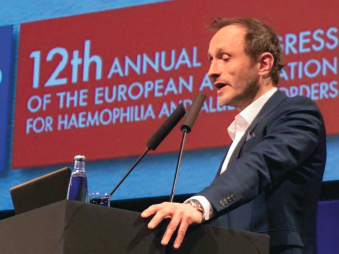
PRAGUE – Adeno-associated virus (AAV)–based gene therapy is probably not the “endgame” in gene therapy for hemophilia, according to John Pasi, MD, PhD, director of the Haemophilia Centre at the Royal London Hospital.

“Gene therapy today is essentially gene therapy version 1.0,” Dr. Pasi said, kicking off the third day at the annual congress of the European Association for Haemophilia and Allied Disorders.
Interwoven with a summary of recent research and upcoming trends, Dr. Pasi reflected upon the medical community’s expectations for gene therapy, both past and present.
“For years,” Dr. Pasi said, “gene therapy has been regarded, essentially, as the Holy Grail of treatment for hemophilia.” This sentiment has been supported by the fact that hemophilia “is a single-gene disorder with a cause and effect relationship that is extremely clear and straightforward for us to recognize.”
A small increase in clotting factor can significantly reduce bleeding while providing a measurable efficacy outcome, making it a strong research candidate.
Looking back, however, when gene therapy research began in the early 1990s, it “really did go through a peak of inflated expectations,” Dr. Pasi said. “We thought for years it was just around the corner. But there were abject failures; there were learning curves we had to go through to understand many of the issues that gene therapy threw up, which we didn’t understand at the beginning.”
When this early excitement was met with tough realities, a period of disillusionment began and persisted through the early 2000s. Dr. Pasi suggested that this period of disillusionment may be reaching an end because of a “huge amount of steady work” that has ushered in a new period of productivity.
Dr. Pasi cited two 2017 studies published in the New England Journal of Medicine by Savita Rangarajan, MBBS, and colleagues and Lindsey A. George, MD, and colleagues for hemophilia A and B, respectively (N Engl J Med. 2017; 377:2519-30; N Engl J Med. 2017; 377:2215-27).
In contrast with previous studies showing factor levels in the single digits, recent studies have achieved normal-range values. Seeing such improvements in hemophilia A, is a particular source of optimism; historically, gene therapy for hemophilia A has lagged behind hemophilia B because of a larger gene that is more difficult to work with.
“This has stepped up the game hugely,” Dr. Pasi said.
The elements of gene therapy for hemophilia may change over time. For instance, lentiviruses could be used instead of AAV-based methods, and ex vivo techniques could make a comeback, potentially using different tissue sources. These changes are likely on the distant horizon, however.
Gene modification is also not coming anytime soon.
“Gene replacement, gene editing, and gene repair are something that we hear a lot about in the general field of gene therapy,” Dr. Pasi said, “But in practical terms for our patients, we probably are a significant way off from this at the moment, and this is because we know and we all recognize that there are a wide range of mutations causing hemophilia, and many of these are highly specific; we will need specific gene therapies to address specific mutations.”
Dr. Pasi likened the current state of gene therapy to that of the self-driving car, suggesting that “we still have strides to make, and there are still things we can improve on beyond what we have today.
“The biggest question for gene therapy today is durability,” Dr. Pasi said. “How long are these things going to last?”
Patients from earlier studies are now crossing the half-decade mark, albeit with relatively low factor levels, compared with recent techniques. One such study, presented at the 2018 annual meeting of the American Society of Hematology by Amit C. Nathwani, MD, PhD, and colleagues, is “very critical to our understanding,” Dr. Pasi said, referring to patients who are now 6-8 years post treatment. “What we see is … continued, stable expression of factor IX,” he said.
Alongside questions of durability, safety remains paramount in the quest for better methods of gene therapy.
“We are seeing liver function abnormalities,” Dr. Pasi said, noting that these tend to be transient elevations of ALT. “We know that many patients now have to receive steroid treatment, which very effectively reduces the immune response, but it is something that we are increasingly having to bear in mind.”
The latest techniques are using liver-specific promoters in novel synthetic capsids, and more capsids are under development.
Dr. Pasi also emphasized safety and caution. “We must never forget that gene therapy is a completely new approach to treatment, and we’ve got to think about safety. It is the number one priority when we are investigating new treatments.”
Safety remains untested in several key patient subpopulations, including children and those with comorbidities or inhibitors.
“For gene therapy in 2019,” he said, “we’ve made massive strides, but we’re not quite there yet.”
PRAGUE – Adeno-associated virus (AAV)–based gene therapy is probably not the “endgame” in gene therapy for hemophilia, according to John Pasi, MD, PhD, director of the Haemophilia Centre at the Royal London Hospital.
“Gene therapy today is essentially gene therapy version 1.0,” Dr. Pasi said, kicking off the third day at the annual congress of the European Association for Haemophilia and Allied Disorders.
Interwoven with a summary of recent research and upcoming trends, Dr. Pasi reflected upon the medical community’s expectations for gene therapy, both past and present.
“For years,” Dr. Pasi said, “gene therapy has been regarded, essentially, as the Holy Grail of treatment for hemophilia.” This sentiment has been supported by the fact that hemophilia “is a single-gene disorder with a cause and effect relationship that is extremely clear and straightforward for us to recognize.”
A small increase in clotting factor can significantly reduce bleeding while providing a measurable efficacy outcome, making it a strong research candidate.
Looking back, however, when gene therapy research began in the early 1990s, it “really did go through a peak of inflated expectations,” Dr. Pasi said. “We thought for years it was just around the corner. But there were abject failures; there were learning curves we had to go through to understand many of the issues that gene therapy threw up, which we didn’t understand at the beginning.”
When this early excitement was met with tough realities, a period of disillusionment began and persisted through the early 2000s. Dr. Pasi suggested that this period of disillusionment may be reaching an end because of a “huge amount of steady work” that has ushered in a new period of productivity.
Dr. Pasi cited two 2017 studies published in the New England Journal of Medicine by Savita Rangarajan, MBBS, and colleagues and Lindsey A. George, MD, and colleagues for hemophilia A and B, respectively (N Engl J Med. 2017; 377:2519-30; N Engl J Med. 2017; 377:2215-27).
In contrast with previous studies showing factor levels in the single digits, recent studies have achieved normal-range values. Seeing such improvements in hemophilia A, is a particular source of optimism; historically, gene therapy for hemophilia A has lagged behind hemophilia B because of a larger gene that is more difficult to work with.
“This has stepped up the game hugely,” Dr. Pasi said.
The elements of gene therapy for hemophilia may change over time. For instance, lentiviruses could be used instead of AAV-based methods, and ex vivo techniques could make a comeback, potentially using different tissue sources. These changes are likely on the distant horizon, however.
Gene modification is also not coming anytime soon.
“Gene replacement, gene editing, and gene repair are something that we hear a lot about in the general field of gene therapy,” Dr. Pasi said, “But in practical terms for our patients, we probably are a significant way off from this at the moment, and this is because we know and we all recognize that there are a wide range of mutations causing hemophilia, and many of these are highly specific; we will need specific gene therapies to address specific mutations.”
Dr. Pasi likened the current state of gene therapy to that of the self-driving car, suggesting that “we still have strides to make, and there are still things we can improve on beyond what we have today.
“The biggest question for gene therapy today is durability,” Dr. Pasi said. “How long are these things going to last?”
Patients from earlier studies are now crossing the half-decade mark, albeit with relatively low factor levels, compared with recent techniques. One such study, presented at the 2018 annual meeting of the American Society of Hematology by Amit C. Nathwani, MD, PhD, and colleagues, is “very critical to our understanding,” Dr. Pasi said, referring to patients who are now 6-8 years post treatment. “What we see is … continued, stable expression of factor IX,” he said.
Alongside questions of durability, safety remains paramount in the quest for better methods of gene therapy.
“We are seeing liver function abnormalities,” Dr. Pasi said, noting that these tend to be transient elevations of ALT. “We know that many patients now have to receive steroid treatment, which very effectively reduces the immune response, but it is something that we are increasingly having to bear in mind.”
The latest techniques are using liver-specific promoters in novel synthetic capsids, and more capsids are under development.
Dr. Pasi also emphasized safety and caution. “We must never forget that gene therapy is a completely new approach to treatment, and we’ve got to think about safety. It is the number one priority when we are investigating new treatments.”
Safety remains untested in several key patient subpopulations, including children and those with comorbidities or inhibitors.
“For gene therapy in 2019,” he said, “we’ve made massive strides, but we’re not quite there yet.”
EXPERT ANALYSIS FROM EAHAD 2019
Think duration, not dose, when managing bleeding with non–factor replacements
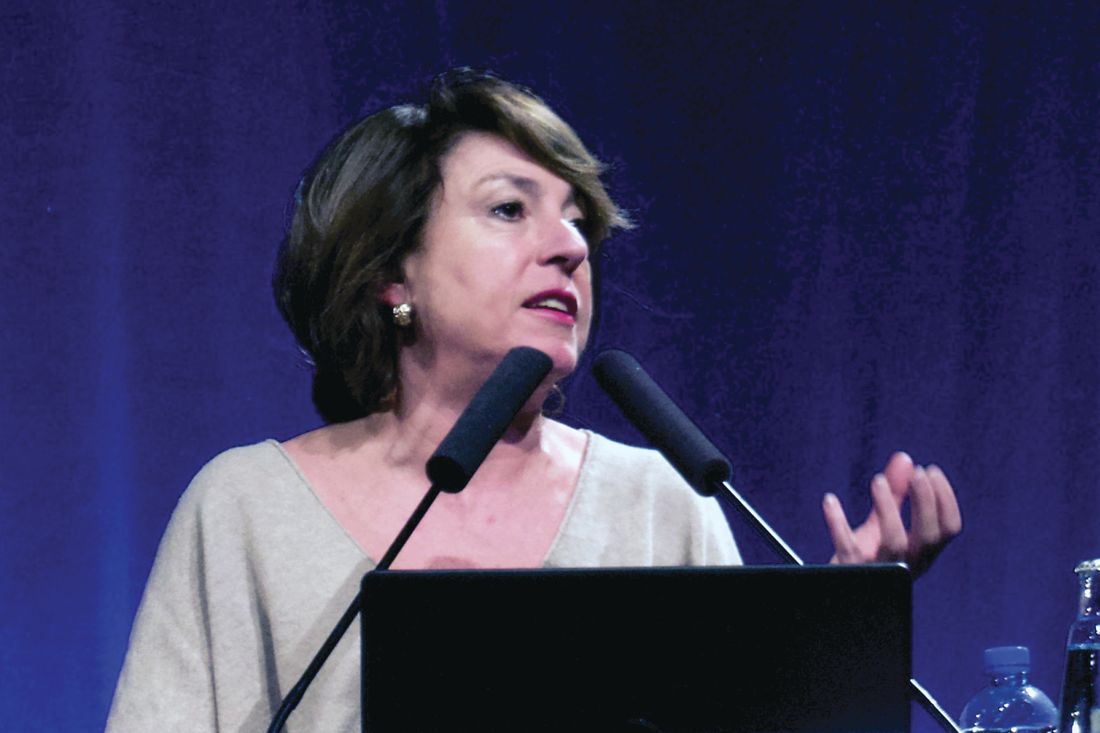
PRAGUE – Clinicians should prioritize treatment duration with factors or bypassing agents – not dose level – when managing breakthrough bleeds in patients with hemophilia who are on non–factor replacement therapy, according to a leading expert.
Duration of treatment is more strongly associated with thromboembolism than dose magnitude, said Andreas Tiede, MD, PhD, head of hemostaseology at Hannover (Germany) Medical School in Germany, noting that recommendations vary by non–factor replacement agent.
These remarks were part of a presentation about novel agents for treatment of hemophilia with inhibitors delivered at the annual congress of the European Association for Haemophilia and Allied Disorders.
“Concomitant use of factor products, both factor VIII and IX, and the bypassing agents, have usually preceded thromboembolic events in clinical trials [for non–factor replacement therapies] so this [topic] is crucial,” Dr. Tiede said.
Other experts recommend lower doses and shorter treatment durations. “I think that’s reasonable, but with some question mark behind the low doses,” he said. “I think it depends a little bit on the interaction between the non–factor replacement therapy and the bypassing agent in your patient.”
With a busy pipeline of non–factor replacement agents for hemophilia, such interactions are becoming increasingly relevant for clinicians and their patients.
Emicizumab, for instance, which is now approved for hemophilia with or without inhibitors, has synergistic activity with activated prothrombin complex concentrates (APCC). This was demonstrated by an emicizumab prophylaxis trial in which five out of eight patients with breakthrough bleeding who were treated with APPC at a dose higher than 100 IU/kg per day for more than 24 hours developed thrombotic microangiopathy. (N Engl J Med. 2017;377:809-18).
Other patients who received multiple infusions of APCC developed skin necrosis, cavernous vein thrombosis, and thrombophlebitis. Consequently, it is now recommended that APCC be avoided in patients taking emicizumab, and if unavoidable, given at the lowest dose possible. However, Dr. Tiede advised that this recommendation for APCC should not be extrapolated to encompass all factors and bypassing agents, based on existing data.
“Regarding higher or lower doses for initial treatment, I would be a little bit more careful,” he said. “That obviously depends on [whether] there is a synergistic effect with the non–factor replacement therapy and the bypassing agent. Synergistic effects have clearly been shown for the interaction of emicizumab and APCC, but when it comes to the interaction between emicizumab and VIIa, I’m not so sure. I don’t think that we have enough evidence to recommend lower doses of VIIa.”
Dr. Tiede also suggested that lower doses of factor VIII are probably unnecessary. “At high doses or high concentrations of factor VIII, emicizumab’s low affinity to the targets will not result in any significant action anymore,” he said. “So I think we have to wait for more data from basic research and also more clinical data.”
Regarding concern for duration of therapy, Dr. Tiede explained that, when treating breakthrough bleeding in a patient on non–factor replacement therapy, “the patient’s hemostatic protection level will never fall to zero, as it would have done in a patient treated previously, on demand with bypassing agents only.” Since hemostatic protection levels never return to zero, it is easier to enter the thromboembolic danger zone.
This risk was recently demonstrated by an emerging non-factor replacement therapy. In a phase 3 trial for fitusiran – a small interfering RNA therapy that targets antithrombin – a patient with hemophilia A developed a breakthrough bleed and 31-46 IU/kg of factor VIII was given, resulting in fatal cerebral sinus thrombosis. After a temporary hold, the study restarted with new limits on factor and bypassing agent doses.
Dr. Tiede reported financial relationships with Bayer, Biotest, CSL Behring, Novo Nordisk, Pfizer, and other companies.
EXPERT ANALYSIS FROM EAHAD 2019
Emicizumab performs well in surgical setting
PRAGUE – Emicizumab appears safe and effective for patients with hemophilia A undergoing surgical procedures, based on experience with a subpopulation of HAVEN 3 trial participants.
Out of 28 minor procedures performed without preventive factor VIII (FVIII), only 2 were associated with postoperative bleeds requiring treatment, reported lead author Elena Santagostino, MD, PhD, of Fondazione IRCCS Ca’ Granda Ospedale Maggiore Policlinico in Milan, and her colleagues.
All events requiring bleeding treatment were associated with dental procedures, highlighting an area where clinicians and dentists may need to exercise caution. Still, overall results supported emicizumab in a surgical setting.
“There were no thrombotic complications or other unexpected events, including inhibitor development,” Dr. Santagostino said at the annual congress of the European Association for Haemophilia and Allied Disorders.
The findings were drawn from 30 patients who underwent 50 surgeries (46 minor, 4 major) during HAVEN 3, a previously reported phase 3 trial investigating the use of emicizumab, a humanized bispecific monoclonal antibody for patients with hemophilia A without inhibitors.
The minor surgeries included dental or orthopedic procedures, esophagogastroduodenoscopy, or colonoscopy. The four major procedures were all orthopedic (knee arthroscopic synovectomy, biceps femoris tear repair, total ankle arthroplasty, and total hip replacement). The investigators analyzed surgery-related bleeds and the nature of FVIII usage.
Preventive FVIII was used in 18 procedures; infusion duration was 24 hours or less in 14 procedures, between 25 hours and 48 hours in 2 procedures, and more than 72 hours in 2 procedures. The median cumulative preventive FVIII dose per procedure was 30 IU/kg.
Of the 46 minor procedures, 28 (61%) were performed without preventive FVIII, and 2 (7.1%) were associated with bleeding requiring treatment, both after dental procedures. Two other participants who received preventive FVIII also needed postoperative bleeding treatment. Of note, these events were also after dental procedures, meaning all four instances of bleeding requiring treatment during the trial were associated with dentistry.
“[I]n this experience, dental procedures were somewhat tricky because the bleeding complications were mainly there,” Dr. Santagostino said.
When asked by an audience member if this trend was unique to mucosal bleeding, Dr. Santagostino said it was too early to draw such a conclusion but offered some insight. “To control and prevent bleeding during a dental procedure is not trivial, because … sometimes if you stop factor VIII treatment quite early, you may have late bleeding, mainly due to local reasons, because … dental procedures are very heterogenous.”
Among three other participants who had postoperative bleeding but did not require treatment, two underwent dental procedures, further supporting this association. Although the study numbers are relatively small, the findings may at least support caution, if not preventive FVIII in the dental setting, Dr. Santagostino said.
The four major procedures – all orthopedic – were knee arthroscopic synovectomy, biceps femoris tear repair, total ankle arthroplasty, and total hip replacement. Along with preoperative preventive FVIII, three of four patients undergoing major surgery received preventive FVIII for 14-18 days postoperatively. Doses ranged from 99-522 IU/kg. No postoperative bleeds occurred in this subgroup.
Study funding was provided by F. Hoffmann–La Roche and Chugai Pharmaceutical. The investigators reported financial relationships with Bayer, Shire, Pfizer, Novo Nordisk, and others.
SOURCE: Santagostino E et al. EAHAD 2019, Abstract OR15.
REPORTING FROM EAHAD 2019
Mucinous ovarian tumor survival rates stress correct diagnosis
Women with invasive, well-differentiated mucinous ovarian cancer are more likely to die from their disease within 10 years of diagnosis than women with mucinous borderline ovarian tumors, according to a retrospective study of more than 2,700 cases.
The analysis also revealed different clinical characteristics, reported lead author Koji Matsuo, MD, PhD of the University of Southern California, Los Angeles, and his colleagues. For example, compared with borderline ovarian tumors (BOT), patients with ovarian cancer (OC) were usually older and had undergone hysterectomy.
“Our study endorses the importance of making the proper diagnosis for invasive cancer when the ovarian tumor is of mucinous histology,” the investigators wrote in Gynecologic Oncology.
Using Surveillance, Epidemiology, and End Results data from 1988-2000, the analysis included 581 cases of stage I, invasive, well-differentiated mucinous OC and 2,130 cases of stage I mucinous BOT. The investigators noted “histological misclassification of BOT as OC or OC as BOT is not uncommon,” because of similar histopathologic characteristics.
Multivariable analysis showed that, compared with cases of BOT, women with OC were more often from the eastern United States (22.0% vs. 13.6%), older (51.9 vs. 48.0 years), and had a history of lymphadenectomy (47.0% vs. 23.2%) or hysterectomy (64.4% vs. 35.8%). Women with OC were also more likely to have tumors smaller than 4 cm (12.9% vs. 8.9%) and stage T1c disease (15.7% vs. 7.3%). Rates of OC declined over time, from 34.7% during 1988-1991 to 22.0% during 1997-2000. Following propensity score matching, multivariable analysis showed that 10-year cause-specific survival rates for OC and BOT were 92.7% and 97.5%, respectively, giving OC a hazard ratio of 2.03 (P = .007). Overall survival showed a similar disparity, of 76.1% for OC, compared with 83.6% for BOT. The investigators concluded that “survival of these two diseases is completely different.”
Regarding histopathologic misclassification, the investigators proposed “a standardized specimen sectioning protocol and diagnostic criteria for mucinous ovarian tumors.”
The study was funded by Ensign Endowment for Gynecologic Cancer Research. The investigators reported financial relationships with KIYATEC, BioPath, M-Trap, and Chugai.
SOURCE: Matsuo K et al. Gynecol Oncol. 2019 Feb 20. doi: 10.1016/j.ygyno.2019.02.003.
FROM GYNECOLOGIC ONCOLOGY
Subcutaneous FVIIa marzeptacog alfa reduced bleeding days
PRAGUE – Patients with hemophilia A or B and inhibitors may have reduced bleeding days when given subcutaneous factor VIIa (FVIIa) marzeptacog alfa, according to early results from a phase 2/3 trial.

The ongoing study is evaluating pharmacokinetics, pharmacodynamics, efficacy, and safety of marzeptacog alfa, which has four engineered amino acid substitutions within the FVIIa protein to increase catalytic activity, reported lead author Howard Levy, MBBCh, PhD, chief medical officer of Catalyst Biosciences in San Francisco, and his colleagues.
“[Marzeptacog alfa] is about nine times as potent as NovoSeven [RT],” Dr. Levy said, referring to the FVIIa product from Novo Nordisk. “This allows for subcutaneous dosing. One of the advantages of subcutaneous dosing is that it further prolongs the half-life.”
Dr. Levy presented the findings at the annual congress of the European Association for Haemophilia and Allied Disorders.
The study involved 12 patients with hemophilia A or B and inhibitors, all of whom began with an annualized bleeding rate (ABR) of more than 12 days per year; of these, 7 have completed dosing.
Following pharmacokinetic analysis, subcutaneous dosing began at an initial dose of 30 mcg/kg daily. As-needed dose escalations were allowed at regular intervals. Specifically, if spontaneous bleeding occurred, then at the next 50-day interval, an individual patient’s dose would be increased to the next level. Dose levels were 30 mcg/kg, 60 mcg/kg, 90 mcg/kg, and 120 mcg/kg.
Any patient completing a 50-day interval without bleeding was finished with the study and proceeded to a 30-day follow-up period, during which time resumption of bleeding was monitored. The primary endpoint was a reduction in the ABR at the final dose level. Secondary endpoints were safety, tolerability, and a lack of inhibitor formation.
As Dr. Levy discussed results, he expressed concern that ABR is an inadequate measure of efficacy. “There is a significant amount hidden by the raw statistic of an annualized bleed rate.” He elaborated further by describing two trial participants who each had an ABR of about 23 days per year but had very different proportions of days with bleeding in the 6-month lead-in period (22% vs. 9%), suggesting that this proportion may provide a clearer impression of efficacy.
Prior to treatment, the mean ABR among all patients was 19 days per year and the median proportion of days with bleeding was 11%. This latter value dropped from 11% to 1% with treatment. The former value, ABR, was reduced in a “clinically significant” fashion, although exact values were not provided (P = .009).
Two patients required dose escalation to 60 mcg/kg, and five out of seven patients had no bleeding (traumatic or spontaneous), at their final dose levels.
Subcutaneous half-life was 13.1 hours, compared with intravenous half-life of 3.9 hours. After more than 260 cumulative days of subcutaneous injections, no thromboses or antidrug antibodies were detected. Two subjects had a total of six injection site reactions, with mild to moderate redness and moderate swelling that resolved without sequelae.
One patient died on day 11 of the trial because of fatal hemorrhagic stroke, but this patient had uncontrolled hypertension and the death was not considered drug related.
More clinical data from this trial will be reported in July 2019 at the annual congress of the International Society on Thrombosis and Haemostasis in Melbourne.
The study was funded by Catalyst Biosciences. Dr. Levy and multiple coauthors are employees, shareholders, or consultants for Catalyst Biosciences.
SOURCE: Levy H et al. EAHAD 2019, Abstract OR11.
PRAGUE – Patients with hemophilia A or B and inhibitors may have reduced bleeding days when given subcutaneous factor VIIa (FVIIa) marzeptacog alfa, according to early results from a phase 2/3 trial.
The ongoing study is evaluating pharmacokinetics, pharmacodynamics, efficacy, and safety of marzeptacog alfa, which has four engineered amino acid substitutions within the FVIIa protein to increase catalytic activity, reported lead author Howard Levy, MBBCh, PhD, chief medical officer of Catalyst Biosciences in San Francisco, and his colleagues.
“[Marzeptacog alfa] is about nine times as potent as NovoSeven [RT],” Dr. Levy said, referring to the FVIIa product from Novo Nordisk. “This allows for subcutaneous dosing. One of the advantages of subcutaneous dosing is that it further prolongs the half-life.”
Dr. Levy presented the findings at the annual congress of the European Association for Haemophilia and Allied Disorders.
The study involved 12 patients with hemophilia A or B and inhibitors, all of whom began with an annualized bleeding rate (ABR) of more than 12 days per year; of these, 7 have completed dosing.
Following pharmacokinetic analysis, subcutaneous dosing began at an initial dose of 30 mcg/kg daily. As-needed dose escalations were allowed at regular intervals. Specifically, if spontaneous bleeding occurred, then at the next 50-day interval, an individual patient’s dose would be increased to the next level. Dose levels were 30 mcg/kg, 60 mcg/kg, 90 mcg/kg, and 120 mcg/kg.
Any patient completing a 50-day interval without bleeding was finished with the study and proceeded to a 30-day follow-up period, during which time resumption of bleeding was monitored. The primary endpoint was a reduction in the ABR at the final dose level. Secondary endpoints were safety, tolerability, and a lack of inhibitor formation.
As Dr. Levy discussed results, he expressed concern that ABR is an inadequate measure of efficacy. “There is a significant amount hidden by the raw statistic of an annualized bleed rate.” He elaborated further by describing two trial participants who each had an ABR of about 23 days per year but had very different proportions of days with bleeding in the 6-month lead-in period (22% vs. 9%), suggesting that this proportion may provide a clearer impression of efficacy.
Prior to treatment, the mean ABR among all patients was 19 days per year and the median proportion of days with bleeding was 11%. This latter value dropped from 11% to 1% with treatment. The former value, ABR, was reduced in a “clinically significant” fashion, although exact values were not provided (P = .009).
Two patients required dose escalation to 60 mcg/kg, and five out of seven patients had no bleeding (traumatic or spontaneous), at their final dose levels.
Subcutaneous half-life was 13.1 hours, compared with intravenous half-life of 3.9 hours. After more than 260 cumulative days of subcutaneous injections, no thromboses or antidrug antibodies were detected. Two subjects had a total of six injection site reactions, with mild to moderate redness and moderate swelling that resolved without sequelae.
One patient died on day 11 of the trial because of fatal hemorrhagic stroke, but this patient had uncontrolled hypertension and the death was not considered drug related.
More clinical data from this trial will be reported in July 2019 at the annual congress of the International Society on Thrombosis and Haemostasis in Melbourne.
The study was funded by Catalyst Biosciences. Dr. Levy and multiple coauthors are employees, shareholders, or consultants for Catalyst Biosciences.
SOURCE: Levy H et al. EAHAD 2019, Abstract OR11.
REPORTING FROM EAHAD 2019
Padua variant factor IX gene shines in AMT-061
PRAGUE – AMT-061, a gene therapy consisting of an adeno-associated virus serotype 5 (AAV5) vector and a Padua variant human factor IX (hFIX) gene, provides clinically meaningful FIX activity, based on early results from an ongoing, phase 2b study.
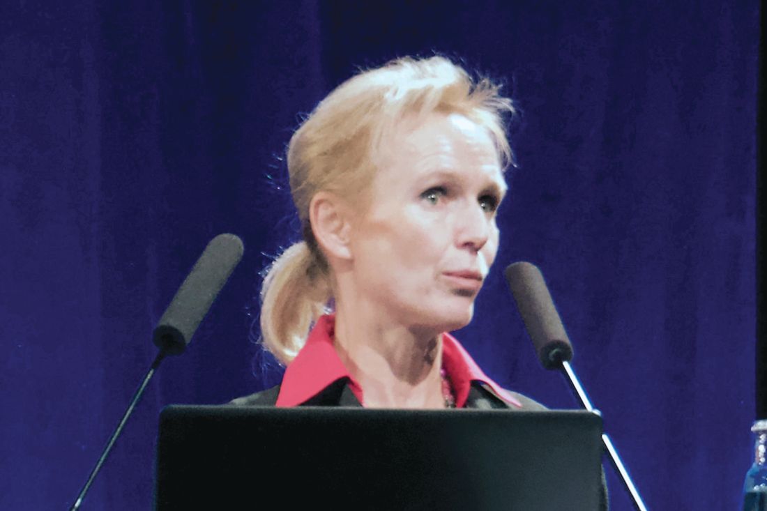
The findings provide traction for a phase 3 trial (HOPE-B; NCT03569891) which is currently enrolling, according to lead author Annette Von Drygalski, MD, PharmD, director of the Hemophilia and Thrombosis Treatment Center at the University of California, San Diego.
Dr. Von Drygalski said that the results also demonstrate the positive impact of switching from a wild-type hFIX gene in AMT-060 to a Padua hFIX variant in AMT-061, achieved by replacing arginine with leucine at R338L.
Speaking at the annual congress of the European Association for Haemophilia and Allied Disorders, Dr. Von Drygalski referred to AMT-061 as the “enhanced version” of AMT-060. Prior to human trials, nonhuman primate studies comparing AMT-060 with AMT-061 demonstrated a 550% increase in factor IX activity, similar to previous reports of 600%-800% boosts from the Padua variant.
The current findings revealed early outcomes for three men with severe hemophilia B who were given AMT-061. The patients were between 43 years and 50 years of age and had been taking clotting factors prophylactically. Among them, annualized bleed rates ranged from one to five events per year. Two of the three men were HIV-positive, one had IgM antibodies against AAV5, and all three had neutralizing antibodies against AAV5.
“Of note,” Dr. Von Drygalski said, “two of the participants had previously been excluded from participation in gene therapy due to preexisting antibodies against AAV.”
Each man was given a single dose of 2 x 10¹³ gene copies/kg of AMT-061 intravenously. The primary endpoint was efficacy at 6 weeks after administration. Ongoing secondary endpoints include patient-reported bleeding and FIX concentrate use, joint health, patient-reported quality of life, and laboratory parameters. Patients will be followed for 1 year.
Within 2 weeks, all three men achieved meaningful levels of FIX. Two of three patients demonstrated an upward trend of FIX activity that peaked at about 50% of normal. The third patient’s levels plateaued sooner, with the most recent reading at week 14 showing 25%. Mean factor IX activity level was 38% of normal 12 weeks after gene therapy.
“These clotting factor activity levels translated into complete control [of bleeding] with no requirement for factor IX replacement after infusion,” Dr. Von Drygalski said. For comparison, AMT-060, the wild-type version, only achieved levels of 3%-13%, which are less clinically meaningful.
Along with demonstrating efficacy, AMT-061 was well tolerated. One patient had two adverse events that resolved without treatment: very mild C-reactive protein elevation and transient headache. One patient had a very mild and transient AST elevation (week 2, 43 U/L; week 4, 48 U/L) that resolved without treatment. No patients had ALT elevations and immunosuppression was not needed.
These findings provide a strong foundation for the phase 3 HOPE-B trial, which will include about 50 patients with FIX activity no greater than 2%. Dr. Von Drygalski noted that patients with AAV5-neutralizing antibodies will not be excluded.
The investigators reported financial relationships with Bayer, UniQure, Pfizer, Novo Nordisk, Shire, and others.
SOURCE: Von Drygalski A et al. EAHAD 2019, Abstract OR10.
PRAGUE – AMT-061, a gene therapy consisting of an adeno-associated virus serotype 5 (AAV5) vector and a Padua variant human factor IX (hFIX) gene, provides clinically meaningful FIX activity, based on early results from an ongoing, phase 2b study.
The findings provide traction for a phase 3 trial (HOPE-B; NCT03569891) which is currently enrolling, according to lead author Annette Von Drygalski, MD, PharmD, director of the Hemophilia and Thrombosis Treatment Center at the University of California, San Diego.
Dr. Von Drygalski said that the results also demonstrate the positive impact of switching from a wild-type hFIX gene in AMT-060 to a Padua hFIX variant in AMT-061, achieved by replacing arginine with leucine at R338L.
Speaking at the annual congress of the European Association for Haemophilia and Allied Disorders, Dr. Von Drygalski referred to AMT-061 as the “enhanced version” of AMT-060. Prior to human trials, nonhuman primate studies comparing AMT-060 with AMT-061 demonstrated a 550% increase in factor IX activity, similar to previous reports of 600%-800% boosts from the Padua variant.
The current findings revealed early outcomes for three men with severe hemophilia B who were given AMT-061. The patients were between 43 years and 50 years of age and had been taking clotting factors prophylactically. Among them, annualized bleed rates ranged from one to five events per year. Two of the three men were HIV-positive, one had IgM antibodies against AAV5, and all three had neutralizing antibodies against AAV5.
“Of note,” Dr. Von Drygalski said, “two of the participants had previously been excluded from participation in gene therapy due to preexisting antibodies against AAV.”
Each man was given a single dose of 2 x 10¹³ gene copies/kg of AMT-061 intravenously. The primary endpoint was efficacy at 6 weeks after administration. Ongoing secondary endpoints include patient-reported bleeding and FIX concentrate use, joint health, patient-reported quality of life, and laboratory parameters. Patients will be followed for 1 year.
Within 2 weeks, all three men achieved meaningful levels of FIX. Two of three patients demonstrated an upward trend of FIX activity that peaked at about 50% of normal. The third patient’s levels plateaued sooner, with the most recent reading at week 14 showing 25%. Mean factor IX activity level was 38% of normal 12 weeks after gene therapy.
“These clotting factor activity levels translated into complete control [of bleeding] with no requirement for factor IX replacement after infusion,” Dr. Von Drygalski said. For comparison, AMT-060, the wild-type version, only achieved levels of 3%-13%, which are less clinically meaningful.
Along with demonstrating efficacy, AMT-061 was well tolerated. One patient had two adverse events that resolved without treatment: very mild C-reactive protein elevation and transient headache. One patient had a very mild and transient AST elevation (week 2, 43 U/L; week 4, 48 U/L) that resolved without treatment. No patients had ALT elevations and immunosuppression was not needed.
These findings provide a strong foundation for the phase 3 HOPE-B trial, which will include about 50 patients with FIX activity no greater than 2%. Dr. Von Drygalski noted that patients with AAV5-neutralizing antibodies will not be excluded.
The investigators reported financial relationships with Bayer, UniQure, Pfizer, Novo Nordisk, Shire, and others.
SOURCE: Von Drygalski A et al. EAHAD 2019, Abstract OR10.
REPORTING FROM EAHAD 2019
SABR response rate falls short in early NSCLC
For patients with resectable stage I non–small cell lung cancer (NSCLC), stereotactic ablative radiotherapy (SABR) may lack the efficacy needed to replace surgical intervention, according to a recent phase II trial.
SABR provided a pathologic complete response rate (pCR) of 60%, which was “lower than hypothesized,” reported lead author David A. Palma, MD, PhD, of London (Ont.) Health Sciences Centre, and his colleagues.
“In patients with cancer who are fit for resection ... the role of SABR is controversial,” the investigators wrote in JAMA Oncology. “Although some recent studies suggest that SABR may achieve outcomes similar to surgery, others do not, and randomized clinical trials are currently under way to compare these 2 modalities.”
The present trial involved 40 adult patients with stage I NSCLC, good pulmonary function, and good performance status. Of these, 35 were evaluable for the primary endpoint, which was tumor pCR rate after SABR. Secondary endpoints were toxic effects, quality of life, distant control, regional control, and local control. Patients with tumors no larger than 3 cm in diameter were given 54 Gy in 3 fractions, while bigger tumors received 55 Gy in 5 fractions. Patients with tumors 2 cm or closer to the brachial plexus or mediastinum were given 60 Gy in 8 fractions. Ten weeks after SABR, sublobar resection or lobectomy was performed, via an open approach or with video-assisted thoracoscopic surgery (VATS).
Analysis revealed a pCR rate of 60%, which was lower than anticipated. One-month and 3-month survival rates were both 100%. After a median follow-up of 19 months, local control rate was 100%, but only three out of four patients (76%) had distant control, and half (53%) had regional control. After 2 years, three out of four patients were still alive (77%). Eighteen percent of patients experienced grade 3 or higher toxic effects.
“[T]he pCR rate of 60% at 10 weeks suggests that practitioners should be cautious in the use of SABR in patients with cancers who are fit for resection,” the investigators concluded.
The Ontario Institute for Cancer Research funded the study. Dr. Palma and Dr. Ward are patent holders for a computed tomography technology that assesses responses after radiotherapy. Dr. Louie reported honoraria from AstraZeneca and Varian Medical Systems Inc.
SOURCE: Palma et al. 2019 Feb 21. doi: 10.1001/jamaoncol.2018.6993.
FROM JAMA ONCOLOGY
Haplo-HCT shows viability in DLBCL
For patients with diffuse large B-cell lymphoma (DLBCL) who need allogeneic hematopoietic cell transplantation (allo-HCT), a haploidentical family member could be a viable donor, according to a retrospective study of 1,438 patients.
When combined with nonmyeloablative/reduced intensity conditioning (NMC/RIC) and posttransplant cyclophosphamide (PTCy), patients treated with haploidentical HCT (haplo-HCT) had outcomes similar to those seen in patients with matched donors, reported Peter Dreger, MD, of the University of Heidelberg (Germany) and his colleagues.
“Using well-matched sibling donors (MSDs) or unrelated donors (MUDs), allo-HCT can result in sustained disease control in 30% to 45% of patients with DLBCL who have early disease recurrence after standard chemoimmunotherapy or have failed auto-HCT [autologous HCT],” the investigators wrote in Blood Advances. “However, the search for a well-matched unrelated donor could be time-consuming and unsuccessful in up to 50% of the patients in need.”
But the present findings suggest that haplo-HCT may one day improve these odds by providing a larger pool of potential donors.
The patients in the study were divided into four treatment groups: haplo-HCT (n = 132), MSD (n = 525), MUD with T-cell depletion (n = 403), and MUD without T-cell depletion (n = 378). For graft-versus-host disease (GVHD) prophylaxis, patients in the haplo-HCT group received PTCy, with or without a calcineurin inhibitor and mycophenolate mofetil, whereas all patients with matched donors received a calcineurin inhibitor. T-cell depletion was accomplished by in vivo antithymocyte globulin and alemtuzumab.
The primary end point was overall survival (OS). Secondary end points were progression-free survival (PFS), progression/relapse, and nonrelapse mortality (NRM).
After a median follow-up of 4.1 years, all groups had similar outcomes, without statistical differences in multivariable analysis.
In the haplo-HCT group, the 3-year OS rate was 46%, the NRM rate was 22%, the PFS rate was 38%, and the relapse/progression rate was 41%.
Of note, patients receiving haplo-HCT did have a lower cumulative incidence of chronic GVHD, at 15% after 1 year and 18% after 2 years. These rates were significantly lower than the other groups’ 1- and 2-year GVHD rates, which were as follows: MSD, 41% and 48%; MUD with T-cell depletion, 23% and 27%; and MUD without T-cell depletion, 48% and 57%.
The investigators noted that these disparities may actually be caused by the use of bone marrow grafts in the haplo-HCT group instead of peripheral blood grafts, which were used in most of the patients in the other groups.
Overall, the findings were encouraging, but the investigators cautioned that “additional studies are needed before haploidentical donors can be considered as equivalent to well-matched related or unrelated donors in patients with DLBCL.”
The study was funded by the Center for International Blood & Marrow Transplant Research (CIBMTR) and the European Society for Blood and Marrow Transplantation. CIBMTR is supported by grants from the U.S. government and the pharmaceutical industry. The authors reported having no competing financial interests.
SOURCE: Dreger P et al. Blood Adv. 2019 Feb 12;3(3):360-9.
FROM BLOOD ADVANCES