User login
HDL hypothesis: New trial expected to show why prior ones failed
NATIONAL HARBOR, MD. – If positive, a major ongoing phase 3 trial of CSL112, an agent designed to promote efflux of cholesterol from macrophages, is positioned to prove the HDL hypothesis, according to an outline of the rationale of the trial at CRT 2020 sponsored by MedStar Heart & Vascular Institute.

“Twenty papers now show better efflux means better outcomes independent of standard risk factors” and “we know this drug improves efflux,” explained C. Michael Gibson, MD, an interventional cardiologist at Beth Israel Deaconess Hospital, Boston.
The HDL hypothesis was derived from the Framingham Heart Study, which correlated high levels of HDL cholesterol with a reduced risk of adverse cardiovascular (CV) outcomes, according to Dr. Gibson. Just as elevated LDL proved to be a treatable risk factor for CV events, reduced HDL was the target of numerous trials to achieve the same types of benefits.
All have failed.
The problem has been in seeing HDL as a number without addressing its function, Dr. Gibson said. In essence, he believes “the HDL hypothesis not been really tested to date.”
CSL112 is a novel formulation of apolipoprotein A-1 (apoA-1) that has been purified from human plasma and reconstituted to form HDL. In the experimental and clinical setting, including the AEGIS I pilot study, weekly infusions of CSL112 have been associated with a degree of cholesterol efflux that predicts major CV risk reductions.
At the same time that the multinational event-driven AEGIS II trial will determine whether cholesterol efflux with CSL112 does translate into protection from CV events, it will also examine the HDL side of the lipid equation. Dr. Gibson said that it is specifically designed to circumvent the weaknesses of previous efforts to target HDL for reducing CV risk.
“The previous studies were conducted in the wrong patients with the wrong drugs given in the wrong doses at the wrong times,” said Dr. Gibson, who is also professor of medicine at Harvard Medical School, Boston.
One major difference from previous trials is that AEGIS II is enrolling patients with an acute coronary syndrome rather than stable atherosclerosis. Many of those being enrolled have had a recent event. Also, rather than raising HDL, the goal of CSL112 is to increase cholesterol efflux, which is now considered to be the key function of HDL. Furthermore, the time frame for the primary outcome, which is a composite of major adverse cardiac outcomes (MACE), is 90 days rather than several years.
In patients with ACS, “it is the early period of vulnerability where efflux of cholesterol really appears to have the greatest influence on outcomes,” Dr. Gibson explained.
The failure of previous efforts to treat HDL now appears to be based on an incomplete understanding of the goals, according to Dr. Gibson. The doomed cholesteryl ester transfer protein (CETP) drugs, for example, effectively increased HDL levels, but generated a form of HDL that “was not all that functional.”
He noted that niacin raises HDL but has off-target effects. Apo-A1 Milano, a mutant variation of apo-A1, is now understood to reduce the endogenous form, which Dr. Gibson said might explain its counterproductive effect on CV protection.
Using a garbage truck analogy to explain the growing appreciation of factors involved in cholesterol accumulation in the macrophage, Dr. Gibson characterized ABCA1, a transporter protein sitting on the surface of the macrophage, as the loader. He described LCAT (lecithin-cholesterol acyltransferase), an enzyme that converts cholesterol into cholesteryl ester, as the compactor. He sees CRL112 as an empty garbage truck sent into the macrophage to reverse the process.
“We are moving beyond thinking of HDL as a number to try to better appreciate its function,” Dr. Gibson said.
The AEGIS II trial was opened in March of 2018. It has a planned enrollment of 17,400 patients, with an estimated completion date of October 2021.
Dr. Gibson reports financial relationships with Bayer, Janssen, Johnson & Johnson, and CSL Behring, the sponsor of the AEGIS II trial.
NATIONAL HARBOR, MD. – If positive, a major ongoing phase 3 trial of CSL112, an agent designed to promote efflux of cholesterol from macrophages, is positioned to prove the HDL hypothesis, according to an outline of the rationale of the trial at CRT 2020 sponsored by MedStar Heart & Vascular Institute.
“Twenty papers now show better efflux means better outcomes independent of standard risk factors” and “we know this drug improves efflux,” explained C. Michael Gibson, MD, an interventional cardiologist at Beth Israel Deaconess Hospital, Boston.
The HDL hypothesis was derived from the Framingham Heart Study, which correlated high levels of HDL cholesterol with a reduced risk of adverse cardiovascular (CV) outcomes, according to Dr. Gibson. Just as elevated LDL proved to be a treatable risk factor for CV events, reduced HDL was the target of numerous trials to achieve the same types of benefits.
All have failed.
The problem has been in seeing HDL as a number without addressing its function, Dr. Gibson said. In essence, he believes “the HDL hypothesis not been really tested to date.”
CSL112 is a novel formulation of apolipoprotein A-1 (apoA-1) that has been purified from human plasma and reconstituted to form HDL. In the experimental and clinical setting, including the AEGIS I pilot study, weekly infusions of CSL112 have been associated with a degree of cholesterol efflux that predicts major CV risk reductions.
At the same time that the multinational event-driven AEGIS II trial will determine whether cholesterol efflux with CSL112 does translate into protection from CV events, it will also examine the HDL side of the lipid equation. Dr. Gibson said that it is specifically designed to circumvent the weaknesses of previous efforts to target HDL for reducing CV risk.
“The previous studies were conducted in the wrong patients with the wrong drugs given in the wrong doses at the wrong times,” said Dr. Gibson, who is also professor of medicine at Harvard Medical School, Boston.
One major difference from previous trials is that AEGIS II is enrolling patients with an acute coronary syndrome rather than stable atherosclerosis. Many of those being enrolled have had a recent event. Also, rather than raising HDL, the goal of CSL112 is to increase cholesterol efflux, which is now considered to be the key function of HDL. Furthermore, the time frame for the primary outcome, which is a composite of major adverse cardiac outcomes (MACE), is 90 days rather than several years.
In patients with ACS, “it is the early period of vulnerability where efflux of cholesterol really appears to have the greatest influence on outcomes,” Dr. Gibson explained.
The failure of previous efforts to treat HDL now appears to be based on an incomplete understanding of the goals, according to Dr. Gibson. The doomed cholesteryl ester transfer protein (CETP) drugs, for example, effectively increased HDL levels, but generated a form of HDL that “was not all that functional.”
He noted that niacin raises HDL but has off-target effects. Apo-A1 Milano, a mutant variation of apo-A1, is now understood to reduce the endogenous form, which Dr. Gibson said might explain its counterproductive effect on CV protection.
Using a garbage truck analogy to explain the growing appreciation of factors involved in cholesterol accumulation in the macrophage, Dr. Gibson characterized ABCA1, a transporter protein sitting on the surface of the macrophage, as the loader. He described LCAT (lecithin-cholesterol acyltransferase), an enzyme that converts cholesterol into cholesteryl ester, as the compactor. He sees CRL112 as an empty garbage truck sent into the macrophage to reverse the process.
“We are moving beyond thinking of HDL as a number to try to better appreciate its function,” Dr. Gibson said.
The AEGIS II trial was opened in March of 2018. It has a planned enrollment of 17,400 patients, with an estimated completion date of October 2021.
Dr. Gibson reports financial relationships with Bayer, Janssen, Johnson & Johnson, and CSL Behring, the sponsor of the AEGIS II trial.
EXPERT ANALYSIS FROM CRT 2020
FDA promises rigorous review of new renal denervation trials
NATIONAL HARBOR, MD. – Just a month before results from the first of several new pivotal trials with a renal denervation device are to be presented, a Food and Drug Administration medical officer speaking at CRT 2020 sponsored by MedStar Heart & Vascular Institute explained which data will most attract the scrutiny of regulators.
“The FDA is very interested in these devices. We recognize that there is a clinical need, but a reasonable benefit-to-risk relationship has to be established,” said Meir Shinnar, MD, PhD, who works in the division of cardiac devices in the FDA’s Office of Device Evaluation.
The field of renal denervation is expected to heat up again if the results of the SPYRAL HTN OFF MED pivotal trial, planned as a late-breaking presentation at the annual meeting of the American College of Cardiology in March 2020, are positive. However, long-term safety will remain a concern, and positive results will not diminish the rigor with which the relative safety and efficacy of other devices in late stages of clinical testing are evaluated.
“The safety profile is unique to the device design and the procedural technique,” Dr. Shinnar said. For example, vascular injury from the energy employed for denervation, whether radiofrequency or another modality, is an important theoretical risk. A minor initial injury might have no immediate consequences but pose major risks if it leads to altered kidney function over time.
“Most of the follow-up data we have now [with renal denervation devices] is about 1-3 years, but I think long-term safety requires a minimum of 5 years of safety data,” Dr. Shinnar said. “We do not expect all that data to be available at the time of approval, but postmarketing studies will be needed.”
Almost 6 years after the SYMPLICITY HTN-3 trial failed to show a significant reduction in blood pressure among patients with resistant hypertension treated with renal denervation rather than a sham procedure (N Engl J Med 2014;370:1393-401), this treatment is again considered promising. The surprising SYMPLICITY HTN-3 result led to several revisions in technique based on the suspicion that denervation was inadequate.
However, the basic principles remain unchanged. For renal denervation, SPYRAL HTN OFF MED, like the SYMPLICITY HTN 3 study, is employing the Symplicity (Medtronic) device, which has been approved in 50 countries but not in the United States, Canada, or Japan.
SPYRAL HTN OFF MED is designed to provide a very straightforward test of efficacy. Unlike SYMPLICITY HTN-3, which permitted patients to remain on their antihypertensive medications, patients in SPYRAL HTN OFF MED will be tested in the absence of drug therapy (a trial with adjunctive antihypertensive drugs, SPYRAL HTN ON MED, is ongoing). This is a design feature that is relevant to regulatory evaluation.
Although not speaking about the SPYRAL HTN OFF MED trial specifically, Dr. Shinnar noted that “the bar is considered to be higher for a first-line indication than when a device is used as an adjunctive to drug therapy.”
Whether used with or without medications, devices are not likely to receive approval without showing a durable benefit. Dr. Shinnar, citing the surgical studies in which blood pressure control was lost 1-2 years after denervation, said 12 months is now considered a “preferred” length of follow-up to confirm efficacy.
If renal denervation moves forward as a result of the new wave of phase 3 trials, there will still be many unanswered questions, according to Dr. Shinnar, who noted that the FDA convened an advisory committee in December 2018 to gather expert opinion about meaningful safety as well as efficacy endpoints for this modality. One will be determining which populations, defined by age, gender, or phenotype, most benefit.
It also remains unclear whether the first approval will create a standard to which subsequent devices should be compared, according to Dr. Shinnar. Although the FDA recognizes blood pressure reductions as an acceptable endpoint, he believes that documentation of the impact on clinical events will be sought in postmarketing analyses.
“All of the denervation modalities involve class 3 devices that require significant data,” Dr. Shinnar cautioned.
Even if the SPYRAL HTN OFF MED trial is positive on the basis of efficacy, it does not guarantee regulatory approval. Dr. Shinnar described a multifaceted approach to defining an acceptable risk-to-benefit ratio from approved devices, and warned that several points regarding the evaluation of renal denervation devices by the FDA are still being debated internally.
Dr. Shinnar reported no potential financial conflicts of interest.
EXPERT ANALYSIS FROM CRT 2020
Key to denervation response for hypertension may be in carotid body
NATIONAL HARBOR, MD. – Of characteristics that might predict which patients with resistant hypertension will respond to carotid body ablation, the relative activity of the carotid body organ itself might be critical for moving this technology forward, an investigator on a first-in-man study suggests.
The study, first presented in 2018 at the European Society of Cardiology congress, showed carotid body ablation resulted in significant but modest reductions in blood pressure in patients with resistant hypertension.
For many reasons, the carotid body is an attractive target for sustained or indefinite control of resistant hypertension, but median systolic blood pressure reductions following ultrasound ablation were highly variable in the first-in-man study, according to Felix Mahfoud, MD, of Saarland University Hospital in Homburg, Germany, a coinvestigator on the study who discussed the findings at CRT 2020 sponsored by MedStar Heart & Vascular Institute.
Of several strategies being pursued to separate those most likely to gain a major benefit, simply measuring carotid body activity is now emerging as particularly promising.
“Patients with a high degree of carotid body activity had a significantly larger fall in blood pressure versus all other methods of grouping patients,” Dr. Mahfoud reported.
The carotid body is a “grain-size” organ of about 2 mm in size that sits on the carotid bifurcation. It communicates directly with the brain to alter sympathetic activity in response to changing levels of such physiologic variable as oxygen, carbon dioxide, and pH, according to Dr. Mahfoud.
In a 2016 proof-of-principle study conducted in resistant hypertension patients, surgical resection of the carotid body was associated with a median 18–mm Hg reduction in systolic blood pressure (SBP) on 24-hour ambulatory monitoring that was sustained through 24 months (Narkiewicz K et al. JACC Basic Transl Sci. 2016;29:313-24).
Subsequently, ultrasound ablation of the carotid body was by way of a transcatheter approach. Dr. Mahfoud’s unpublished first-in-man study, conducted in 2018 enrolled 38 patients with resistant hypertension. The median reductions from baseline of 7-8 mm Hg at 1, 3, and 6 months were significant (P less than .01), but the benefit was disappointingly modest.
However, the variability was large. Some patients achieved SBP reductions of up to 20 mm Hg at 6 months, prompting additional analyses to understand if the best responders could be identified. When compared to the mean reduction of 7 mm Hg at 6 months, this represented a 13–mm Hg additional reduction. There are now several potential approaches being considered.
In addition to carotid body activity, which is readily measured and has substantial potential to serve as a routine selection criterion, isolated systolic hypertension (ISH) was also found to be a discriminator for response. For those with ISH, which Dr. Mahfoud noted is also characterized as “stiff arteries,” SBP reductions at 6 months were negligible, but in those without ISH, the median reduction from baseline was 11 mm Hg.
Further investigations are now planned to evaluate potential predictors of response, according to Dr. Mahfoud. He believes carotid body ablation might have advantages over alternatives, including other experimental therapies, in at least some patients.
To deliver ultrasound ablation in the first-in-man study, a propriety catheter (Cibiem transvenous system) was advanced through the jugular vein guided with intravascular ultrasound. When the carotid body was reached, two to three ultrasound ablations of 8-12 seconds each were applied. The procedure time was 20-30 minutes.
Initially, an arterial approach to the carotid body was used. However, after a transient ischemic attack early in the series, the approach was switched to the jugular vein. There have been no serious subsequent procedural-related complications since.
Although the median SBP reductions were modest, they were not insignificant in a population selected for severe resistant hypertension. The median SBP at entry was 180 mm Hg in patients taking a median of 4.5 antihypertensive drugs, according to Dr. Mahfoud.
In other words, this approach still retains promise for selected patients if larger studies demonstrate that response can be predicted, and the data continue to support tolerability.
A venous approach to the carotid body with intravascular ultrasound guidance and therapeutic ultrasound appears “to offer a safe and effective treatment option in resistant hypertension,” according to Dr. Mahfoud. “A companion diagnostic test is being developed to determine whether patients are likely to respond to this therapy.”
Dr. Mahfoud reports financial relationships with Medtronic, St. Jude, and ReCor.
This article was updated to clarify the study details and correct misspellings of the presenter's name.
SOURCE: CRT 2020.
REPORTING FROM CRT 2020
Poor OR posture a key cause of vascular burnout
NEW YORK – Career burnout is common is common among physicians and surgeons, but vascular surgeons might be able to lower their risk simply by taking steps to improve their posture in the operating room, according to data presented at a symposium on vascular and endovascular issues on an evolution that is already underway.
“We looked at physical pain and we were able to demonstrate a correlation with burnout. More pain, more burnout,” said Samuel R. Money, MD, division of vascular surgery, Mayo Clinic, Phoenix, Arizona.
Pain was a reasonable focus for efforts to identify causes of burnout because it is so common among vascular surgeons. In data recently published by Dr. Money and his coinvestigators, 78.3% reported moderate to severe physical pain at the end of a day of surgery (J Vasc Surg 2018;70:913-920).
“Forty percent of vascular surgeons have chronic pain,” Dr. Money said at the symposium sponsored by the Cleveland Clinic Foundation.
Physical pain is not the only cause of burnout, which affects 30% of vascular surgeons, according to data recently presented at the annual meeting of the Society of Vascular Surgery (J Vasc Surg 2019;69[6]:e97.). In that survey, physical pain was joined by work hours, documentation tasks, on-call frequency, and conflicts between work and personal life as significant factors.
“The average vascular surgeon in North America works 63 hours per week,” noted Dr. Money, adding that his survey found nearly 90% of surgeons operate on 3 or more days of every week. This amount of time in the operating room is relevant because almost all surgeons report some degree of pain after a procedure. In the survey, the proportion was greater than 95%.
Yet, risk of pain is modifiable.
“Body position matters,” said Dr. Money, citing studies showing that open procedures are most closely associated with neck pain whereas endovascular procedures are more likely to produce back pain. Although there is a high risk of either type of pain with these procedures, the types of predominant pain are consistent with the demands on body positioning.
“The more you lean forward, the more stress is placed on your neck and back. When standing straight, your head weighs 10-12 pounds, but leaning forward, it can put 60 pounds of pressure on your neck,” he said.
The relative stress can be measured objectively. Dr. Money cited work with a device that measures the body force in inertial measurement units (IMU). According to Dr. Money, the neck is in a high stress position about 75% of the time spent performing typical vascular surgery.
“The trunk is placed in a high stress position approximately 40% of the time, while the other parts of the body that were measured were not generally that bad,” Dr. Money said.
To avoid postural pain, which is not often stressed in surgical training, Dr. Money had specific recommendations. Some are obvious, such as positioning the operating table to minimize the amount of time the head is inclined. He also recommended positioning display monitors no more than 10-20 degrees below and no higher than eye level.
“If you sit down to perform tasks during the procedure, use an adjustable chair so that you can optimize the height,” he said.
He identified loupes as a risk factor for bad posture, and he stressed the importance of wearing lead garments only when necessary and adjusted properly.
“Padded floor mats? They really help,” Dr. Money said. He also recommended appropriate footwear and support stocking.
“Microbreaks are being used in a lot of professions. This means stopping for a moment to stretch every 15-30 minutes,” Dr. Money said.
As a first step, Dr. Money recommended simply developing posture awareness. Many surgeons are simply ignoring the risk and failing to optimize the ways they can increase their comfort during surgery.
Even before entering the surgical suite, regular exercise, yoga, and stretching are all strategies that have the potential to make a difference, according to Dr. Money.
The immediate goal is to reduce the physical pain that is an important occupational hazard for vascular surgeons, but the ultimate goal is to improve job satisfaction, an important defense against professional burnout.
REPORTING FROM VEITHsymposium
Differences in U.S. and European aneurysm guidelines called unavoidable
NEW YORK – Published 12 months apart, guidelines on management of abdominal aortic aneurysm (AAA) from the European Society for Vascular Surgery are similar but diverged in instructive ways from those of the Society for Vascular Surgery, according to a critical review at a symposium on vascular and endovascular issues sponsored by the Cleveland Clinic Foundation. “Some of the differences were almost unavoidable in the sense that the ESVS guidelines represent multiple idiosyncratic health care systems across Europe,” reported Ronald L. Dalman, MD, chief of vascular surgery, Stanford (Calif.) University.
As a result, the ESVS guidelines provide very little specificity about pharmacologic options because of the differences in availability of these treatments within specific health systems. In addition, both open and endovascular aneurysm repair (EVAR) are given similar emphasis because of the limited availability of EVAR in some parts of Europe.
“The ESVS guidelines specifically recommend repair of an aneurysm within 8 weeks when repair is indicated, but there are not many aneurysms that go 8 weeks in the U.S. without being fixed by a fee-for-service surgeon,” Dr. Dalman observed.
The SVS AAA guidelines were published in January 2018 (J Vasc Surg 2018;67:2-77) and the ESVS guidelines followed 1 year later (Eur J Vasc Surg 2019;57:8-93).
The differences in the guidelines, although modest, are interesting because each set of guidelines was based largely on the same set of trials and published studies, according to Dr. Dalman, who was a coauthor of the SVS guidelines and an external reviewer for the ESVS guidelines.
In the lag between completion of the two guidelines, new information led to three ESVS additions not found in the SVS guidelines, according to Dr. Dalman. They involved the importance of considering aneurysm diameter as a prognostic factor, new understanding of the limitations on endovascular aneurysm sealing (EVAS), and new information about how aneurysm size should affect frequency of surveillance.
Overall, the U.S. guidelines contain 111 recommendations based on 177 references, while the ESVS guidelines contain 125 guidelines based on 189 references. In retrospect, Dr. Dalman believes both sets of guidelines omitted some clinically meaningful information, such as the risk of large-diameter devices for causing endoleaks.
The authors of the ESVS guidelines did have an opportunity to review of a draft of the SVS guidelines, so differences can be interpreted as intentional. For example, the SVS guidelines recommend risk calculators, but Dr. Dalman suggested that the authors of the ESVS guidelines were less convinced that their utility was established.
The decision not to recommend a door-to-treatment time for ruptured aneurysms, as in the SVS recommendations, might have been in deference to disparate practice across European countries, Dr. Dalman suggested.
Ultimately, the guidelines are “substantially similar,” according to Dr. Dalman, but he expressed concerned that neither guideline is accompanied by a specific mechanism or recommended strategy to ensure implementation.
Many of the SVS recommendations are likely to be translated into quality metrics at U.S. institutions, but “there are implementation issues” for ensuring that each guideline is applied, Dr. Dalman said.
Given the agreement on the vast majority of the recommendations, Dr. Dalman suggested that “it might be time to consider global guidelines” for management of AAA and other vascular diseases. Some type of language might be required to accommodate divergent resources or practices across borders, but Dr. Dalman questioned the need to review the same literature to arrive at mostly the same conclusions.
REPORTING FROM VEITHsymposium
Key clinical point:
Major finding: Less emphasis on endovascular repair and specific drugs in Europe reflects accommodation of nationalized health systems.
Study details: Expert review.
Disclosures: Dr. Dalman reports no potential financial conflicts of interest relevant to this topic.
Source: Dalman RL et al. 46th VEITHsymposium.
Challenges outlined for teaching open surgery to Gen Z in endovascular era
NEW YORK – The dual challenges of teaching open vascular surgery techniques when few are performed and reaching a generation that has a different attitude to absorbing information requires new and innovative approaches, according to an academic surgeon speaking at symposium on vascular and endovascular issues sponsored by the Cleveland Clinic Foundation.
“The see one, do one, teach one approach to surgical training is no longer possible,” explained R. Clement Darling III, MD, chief of the division of vascular surgery at Albany (N.Y.) Medical Center Hospital.
For open procedures, the problem is a rapidly declining number of cases in the era of endovascular surgery, but Dr. Darling also recommended adjusting training to the outlook and expectations of a new generation. First observed in the millennial generation, an attitude of firm work-related boundaries is also being seen in generation Z. Generation Z, characterized by birth after 1995, is just now beginning to reach residency programs.
“The approach to work-life balance is extremely different for these individuals than it was for my generation,” Dr. Darling said. While previous generations were often motivated by fear and pressure, newer generations appear to respond less well to anxiety.
“People learn differently. Some from their tactile sense, some intellectually, and some from fear or pressure, but mostly, particularly those who are younger, now learn from positive reinforcement,” Dr. Darling said.
For teaching open procedures at his own institution, Dr. Darling has switched from the traditional model of one-on-one instruction undertaken in the surgical suite to a group approach. The limited number of open cases was the impetus, but group instruction now extends beyond the operating room.
“We have a meeting before the case, when we go over the technical aspects,” Dr. Darling explained. Fellows are asked to envision and describe potential problems and potential solutions.
“We try to make them visualize as well as verbalize exactly what will be done in the operating room,” Dr. Darling said. The plans are outlined carefully “so no one does any thinking in the OR. All the thinking is done in advance.”
Videos and simulators are teaching aids, but a great deal of learning can be accomplished independent of doing, according to Dr. Darling. Moreover, understanding the anatomy, which comes before developing surgical skills, is the same for open and endovascular procedures, so each is relevant to the other.
After witnessing an open case, all of the trainees along with the nurses and attending physicians go through a debriefing to consider the potential lessons. At Dr. Darling’s center, open procedures increasingly involve sicker and older patients, conferring case analysis with a particularly vital learning function in the curriculum.
Because of the diminishing number of open cases and the diminishing open skills, even among experienced vascular surgeons, residents in an increasing number of training programs “graduate without any open experience, which is a little shocking,” Dr. Darling said.
Importantly, group instruction, although valuable and necessary for exposing residents and fellows to open vascular surgery, has its own lessons to impart even if it was born out of necessity.
“We always emphasize that it is not the sewing that counts, it is the setup that counts,” said Dr. Darling, indicating that this is a clear message when the group is assembled for case planning. The group planning also emphasizes that surgery is a team sport.
“All of us is smarter than one of us,” said Dr. Darling, articulating the implicit message of group training.
Although this is a departure from a bygone era where infallible surgeons ruled the OR, it is fits nicely with changing attitudes about the best attributes of a competent surgeon.
REPORTING FROM VEITHsymposium
Independent vascular surgery board called key to self-determination
NEW YORK – Numerous experts, including a past president of the Society for Clinical Vascular Surgery, lined up at a symposium on vascular and endovascular issues sponsored by the Cleveland Clinic Foundation to explain why there is a critical need for a separate vascular surgery board with membership on the American Board of Medical Specialties.
Not least of the reasons is, “We need to control our destiny by training more residents and fellows in vascular surgery,” said Alan M. Dietzek, MD, who served as SCVS president in 2018 and is an active vascular surgeon in Danbury, Conn.
According to Dr. Dietzek, the American Board of Surgeons recently rejected an application for a vascular resident review committee (RRC), which develops and controls training within a specialty.
“A vascular surgery RRC would facilitate more applications [for training] as well as innovations in specialized programs, which we could certainly use in aortic and venous fellowships,” Dr. Dietzek said. “All ABMS-recognized boards have their own RRC.”
Other speakers, including Timothy M. Sullivan, MD, a professor of surgery at the University of Minnesota, Minneapolis, made the same point. He also believes that a vascular surgery RRC is pivotal in establishing recognition for the specialty and what it offers.
From his perspective, O. William Brown, MD, a vascular surgeon from Bingham Falls, Minn., believes that creating a vascular surgery board will increase recognition in general. Like the others, he maintained that an independent board could draw attention to the specific skills of vascular surgeons, creating a basis for attracting patients, advocating for their needs, and lobbying for resources.
Many experts, including Dr. Brown, believe that the specialty of vascular surgery already meets the qualifications for creating an independent board. However, Dr. Dietzek said that membership in ABMS is dependent on support from the Society of Vascular Surgeons, “and we don’t have that yet.”
In the title of his talk on creating a vascular surgery board, Dr. Brown called for the SVS Executive Committee to “recognize this need and go after it with full force.”
Dr. Dietzek believes the SVS should survey the membership. If there is support for an independent board, it should move ahead with the appropriate support.
“Can we afford it? Other small boards have done just fine,” said Dr. Dietzek, citing the American Board of Colorectal Surgery and the American Board of Thoracic Surgery. He said both are doing well financially, and he provided estimates suggesting that a vascular surgery board would also achieve firm financial footing.
The value of an independent board in exercising control over training programs is part of a larger issue of self-determination, according to Dr. Dietzek. For example, vascular surgeons have “little or no control over the priorities or the budget” at most institutions where they work. An established and recognized vascular surgery board could help these specialists define their identity and separate from other surgical specialties to create their own divisions or departments.
Others who spoke on this topic agreed. Many expressed concern about marginalization by hospital administrators who are often unclear on what vascular surgeons do. A vascular surgery board has the potential to provide a degree of stature that is now lacking.
“We need to build relationships with hospital administrators, politicians, and the insurance industry. This is critical,” Dr. Dietzek said. He believes a vascular surgery board offers an opportunity to achieve these goals and “help us control our own destiny.”
Dr. Dietzek and the other participants report no financial conflicts of interest relevant to this topic.
REPORTING FROM VEITHsymposium
Telehealth consults for vascular surgery reimbursed at par with office visits
NEW YORK – Telehealth should be embraced by vascular surgeons for their own self-interest independent of the evidence that it is well accepted and more convenient for patients, according to an update on an evolution that is already underway.
“One of the great advantages of telehealth is the efficacy of time for the clinician,” John W. Hallett, MD, professor of vascular surgery at the Medical University of South Carolina, Charleston, said at a symposium on vascular and endovascular issues sponsored by the Cleveland Clinic Foundation.
This efficiency is purchased with no loss of revenue, he added. He said that many clinicians are unaware of the opportunity this affords.
“Almost every payer reimburses telehealth visit at the same rate as that of an office visit,” Dr. Hallett explained. The only additional step is adding a “GT” modifier when billing Medicare or a “95” modifier when billing private payers.
Telemedicine is not a new concept. Published studies date back decades, but this interaction is increasingly understood to be the future. Along with an increasing array of sensors employing smartphone technology to allow physicians remote access to vital signs and other clinical data, patient attitudes have changed.
“Patients like telemedicine. It is convenient for them,” said Dr. Hallett, who noted that many providers are recognizing telemedicine as a potential marketing tool.
“On my way in from the airport yesterday, there was an advertisement for telemedicine from NYU on the television in the cab,” said Dr. Hallett, referring to the New York University health system.
The data supporting the benefits of telemedicine even include studies undertaken in vascular surgery patients. In one recent retrospective study cited by Dr. Hallett, substantial time and travel costs were saved for every vascular surgery consult conducted by telemedicine rather than in an office visit (Paquette S et al. Ann Vasc Surg. 2019;59:167-172).
“There was no difference in the rate of complications, and 94% of the patients considered the telehealth consultation adequate,” Dr. Hallett said.
He said there is urgency for vascular surgeons to pursue telemedicine. With the number of individuals over the age of 65 growing by thousands in the United States every day, there will be increasing pressure on the relatively fixed pool of vascular surgeons to improve their efficiency.
In addition, telemedicine is coming whether vascular surgeons like it or not.
“Patients are becoming more interested in looking at an app on their smartphone than coming to the office,” said Tony S. Das, MD, an interventional cardiologist who practices in Dallas. Dr. Das also spoke about the value of telemedicine for the vascular and cardiovascular surgeon at the VIETHsymposium.
In his overview, Dr. Das spoke about telehealth in the context of the estimated $12 billion dollars that will be spent on digital health in vascular medicine by 2021. The growth in digital health in vascular medicine is a reflection of a global change in clinical care. According to Dr. Das, there were more than 600 vendors of wearable sensors to monitor disease and health at a recent consumer electronics convention.
“This technology is here to stay,” said Dr. Das, who, appropriately, was not present at the symposium but delivered his presentation remotely.
Both the Centers for Medicare and Medicaid Services and the Food and Drug Administration have digital health action plans, according to Dr. Das. The CMS has already developed reimbursement codes to pay for remote monitoring services and more are expected.
Calling this type of telehealth “untethered vascular care,” Dr. Das agreed with Dr. Hallett that an evolution is coming whether vascular surgeons choose to get on board now or are forced to take action later.
SOURCE: VIETHsymposium
REPORTING FROM VIETH SYMPOSIUM
Pulmonary embolism treatment teams adopted widely for complex disease
NEW YORK – Seven years after the formation of the first pulmonary embolism response team (PERT), more than 100 institutions have joined the PERT Consortium, which was created to guide care and research for this thrombotic complication, according to a status report at a symposium on vascular and endovascular issues sponsored by the Cleveland Clinic Foundation.
“Why are PERTs needed? Pulmonary embolism patients are like snowflakes. No two are the same,” explained Richard Channick, MD, director of the pulmonary vascular disease program, University of California, Los Angeles.
Patient variability is an issue because algorithms for pulmonary embolism (PE) often differ at the point of diagnosis, such as the emergency department or intensive are unit, according to Dr. Channick, who was present when the first PERT was created in 2012 at Massachusetts General Hospital (MGH) in Boston. In addition, treatment algorithms can seem complex at a time when patients are deteriorating quickly.
“The treatment algorithms always say consider this or consider that, and then you get a recommendation with a 2B grade of evidence. So what do you do?” Dr. Channick asked, “This has really been crying for an organized approach.”
PERTs were created to fill this need. In most centers, PERTs are organized to respond to a diagnosis of PE wherever it occurs in the hospital. The goal is rapid activation of a team of experts who can reach a single consensus recommendation.
At MGH and UCLA, a similar relatively simple scheme has been created to guide physicians on how to activate the PERT and which situations make this appropriate.
“A big part of the PERT value has been our ability to conduct a real-time virtual consultation where we leverage online technology to look at images together in order to agree on a strategy,” Dr. Channick explained.
Although frequently asked what specialists are needed for an effective PERT, Dr. Channick said it depends on institutional structures, the types of specialists available, and, in some cases, the specific characteristics of the patient. In many situations, a pulmonary vascular specialist and an interventional radiologist might be sufficient. In others, team members might include some combination of an interventional cardiologist, a cardiac surgeon, and a hematologist.
It is also appropriate to include clinicians likely to participate in care following acute treatment of the PE. “One of the most critical values to PERT is the ability to systematically follow patients” after the PE is treated, Dr. Channick said.
So far, there are no data to confirm patients managed with PERT achieve better outcomes than those who are not. Reductions in mortality, length of stay, and costs are reasonably anticipated and might eventually be demonstrated, but Dr. Channick said that PERTs already have value.
“I think the efficiency of care is important,”he said. He called PERT a “one-stop shopping” approach to ensuring that multiple strategies are considered systematically.
There are many anecdotal examples of the benefits of shared decision-making for PE treatment. In one, a pulmonary specialist in a PERT team narrowly averted a planned thrombolysis in a patient diagnosed with PE who was actually found to have severe pulmonary fibrosis, according to Dr. Channick.
Not least important, the shared decision-making of a PERT could relieve the burden of difficult choices in complex situations. Bad outcomes in PE can be unavoidable even with optimal therapy.
“To me personally, a very important benefit of being part of a PERT is the feeling that we are all in it together,” Dr. Channick said. “Patients can go from being pretty stable to being dead very quickly.”
The PERT Consortium has sponsored an annual meeting on PE since 2015. It also maintains an ongoing registry for PE data from member institutions. These data are expected to have increasing value for comparing the impact of patient characteristics, treatment strategies, and other variables on outcomes.
For clinicians who are uncertain whether the PE incidence at their institution justifies a PERT, Dr. Channick had some advice. “If you build it, they will clot,” he said, meaning that due to the frequency of PE, a PERT will generally have plenty of work once created.
SOURCE: VEITHSYMPOSIUM
REPORTING FROM THE VEITHSYMPOSIUM
Balloon pulmonary angioplasty beats riociguat in randomized CTEPH trial
MADRID – according to a multicenter trial presented at the annual congress of the European Respiratory Congress.
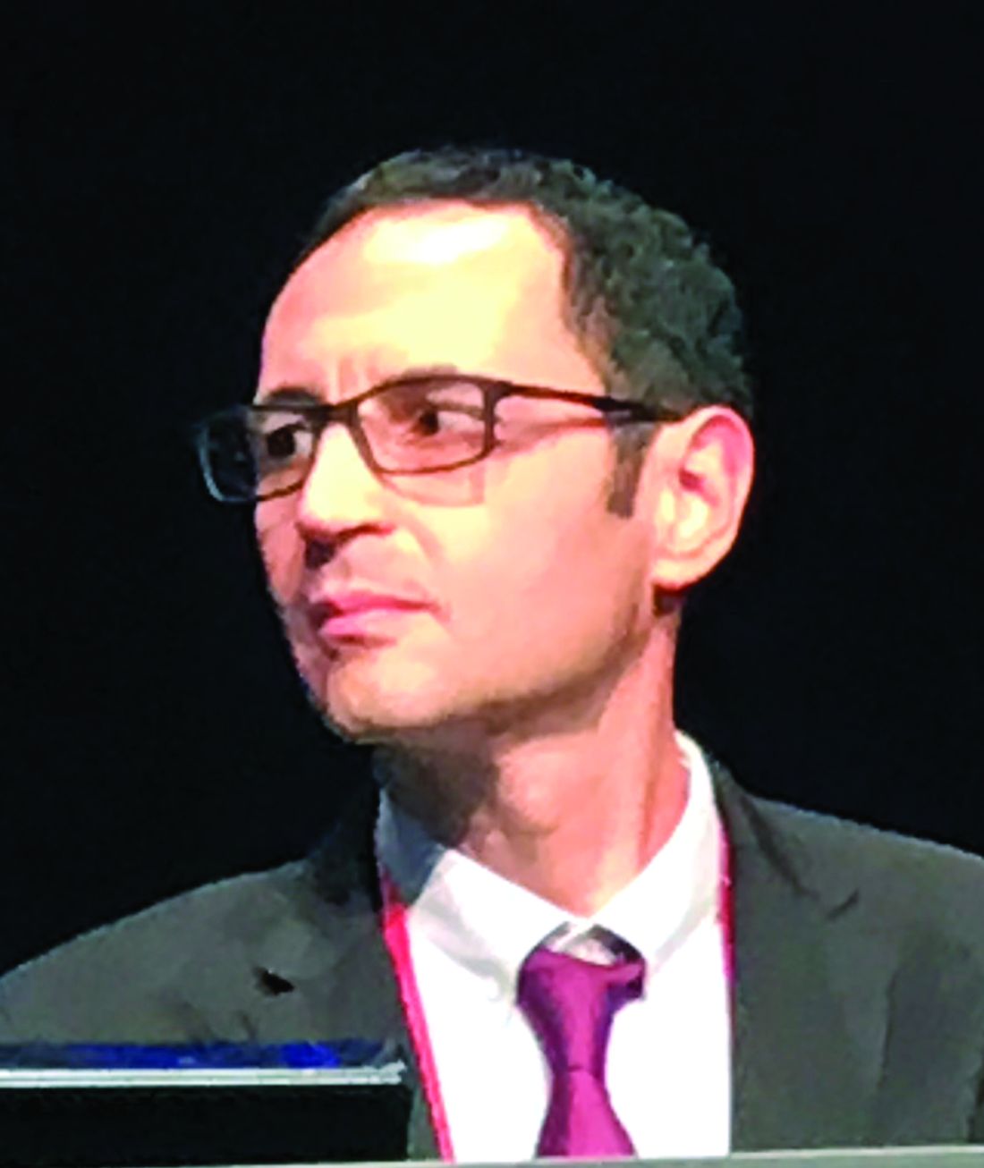
Both therapies are widely used in the treatment of CTEPH, but this is the first controlled trial in which they were directly compared, according to Xavier Jaïs, MD, of the Pulmonology Service, Kremlin-Bicêtre Hospital, University of Paris-Sud.
In this randomized trial, called RACE, newly diagnosed and previously untreated patients with nonoperable CTEPH were enrolled. The key eligibility criteria included PVR greater than 320 dynes/sec per cm–5 and a pulmonary capillary wedge pressure of 15 mm Hg or less.
The patients were randomized to BPA or riociguat and followed for 26 weeks. The primary endpoint was relative change in PVR from baseline. The 6-minute walk distance, change in functional class, time to clinical worsening and safety were among secondary endpoints.
As calculated by geometric mean from baseline, PVR was reduced by nearly 60% in the BPA group and by 32% in the riociguat group, providing a 40% (P less than .0001) relative advantage of BPA.
Although there was a small relative advantage in the 6-minute walk distance for the BPA group at the end of the study, it did not reach statistical significant. However, 88% of those randomized to BPA versus 49% of those treated with riociguat (P less than .0001) improved by at least one WHO class by the end of the study.
Clinical worsening events over the course of the trial were uncommon. All three of these events occurred in the riociguat group, but the difference was not significant.
The end-of-study reduction in brain natriuretic peptide, which was another secondary endpoint, was 67% greater in the BPA group (P less than .0001).
There was a safety cost for the greater efficacy of BPA. This included a higher proportion of patients in the BPA group with at least one serious adverse event (50% vs. 26%) and at least one serious treatment-related adverse event (14% vs. 9%). No patient in either arm discontinued therapy because of treatment-related adverse events, and there were no deaths over the course of the study in either arm.
The study has included a 6-month extension to allow patients symptomatic on their originally assigned therapy to switch to the opposite treatment. Results of the extension are not yet available, but Dr. Jaïs said that these data might provide insight about which therapy to start first.
“It was very important to do this trial,” according to the ERS-invited discussant, Martin Kolb, MD, of the Firestone Institute of Respiratory Health, McMaster University, Hamilton, Ont. The most recent World Symposium on Pulmonary Hypertension identified BPA and medical therapy as reasonable choices in inoperable CTEPH, but Dr. Kolb said there has been an unmet need for comparative data.
“This was a very strong study that demonstrated a powerful impact for both interventions on pulmonary vascular resistance,” Dr. Kolb said, adding that, although BPA proved to be more effective, clinicians consider the greater risk of adverse events. He believes further work needs to be done in identifying the best candidates for each and to explore hybrid approaches.
“What do you think about doing these sequentially so that you lower the pressure first with medical therapy and then go in with the balloon?” Dr. Kolb asked Dr. Jaïs during a discussion that followed presentation of the RACE results.
Dr. Jaïs conceded this point, noting that the treatments have different targets and might be complementary.
“We plan to do a study like this in the future,” Dr. Jaïs said.
Dr. Jaïs reported no potential conflicts of interest.
MADRID – according to a multicenter trial presented at the annual congress of the European Respiratory Congress.
Both therapies are widely used in the treatment of CTEPH, but this is the first controlled trial in which they were directly compared, according to Xavier Jaïs, MD, of the Pulmonology Service, Kremlin-Bicêtre Hospital, University of Paris-Sud.
In this randomized trial, called RACE, newly diagnosed and previously untreated patients with nonoperable CTEPH were enrolled. The key eligibility criteria included PVR greater than 320 dynes/sec per cm–5 and a pulmonary capillary wedge pressure of 15 mm Hg or less.
The patients were randomized to BPA or riociguat and followed for 26 weeks. The primary endpoint was relative change in PVR from baseline. The 6-minute walk distance, change in functional class, time to clinical worsening and safety were among secondary endpoints.
As calculated by geometric mean from baseline, PVR was reduced by nearly 60% in the BPA group and by 32% in the riociguat group, providing a 40% (P less than .0001) relative advantage of BPA.
Although there was a small relative advantage in the 6-minute walk distance for the BPA group at the end of the study, it did not reach statistical significant. However, 88% of those randomized to BPA versus 49% of those treated with riociguat (P less than .0001) improved by at least one WHO class by the end of the study.
Clinical worsening events over the course of the trial were uncommon. All three of these events occurred in the riociguat group, but the difference was not significant.
The end-of-study reduction in brain natriuretic peptide, which was another secondary endpoint, was 67% greater in the BPA group (P less than .0001).
There was a safety cost for the greater efficacy of BPA. This included a higher proportion of patients in the BPA group with at least one serious adverse event (50% vs. 26%) and at least one serious treatment-related adverse event (14% vs. 9%). No patient in either arm discontinued therapy because of treatment-related adverse events, and there were no deaths over the course of the study in either arm.
The study has included a 6-month extension to allow patients symptomatic on their originally assigned therapy to switch to the opposite treatment. Results of the extension are not yet available, but Dr. Jaïs said that these data might provide insight about which therapy to start first.
“It was very important to do this trial,” according to the ERS-invited discussant, Martin Kolb, MD, of the Firestone Institute of Respiratory Health, McMaster University, Hamilton, Ont. The most recent World Symposium on Pulmonary Hypertension identified BPA and medical therapy as reasonable choices in inoperable CTEPH, but Dr. Kolb said there has been an unmet need for comparative data.
“This was a very strong study that demonstrated a powerful impact for both interventions on pulmonary vascular resistance,” Dr. Kolb said, adding that, although BPA proved to be more effective, clinicians consider the greater risk of adverse events. He believes further work needs to be done in identifying the best candidates for each and to explore hybrid approaches.
“What do you think about doing these sequentially so that you lower the pressure first with medical therapy and then go in with the balloon?” Dr. Kolb asked Dr. Jaïs during a discussion that followed presentation of the RACE results.
Dr. Jaïs conceded this point, noting that the treatments have different targets and might be complementary.
“We plan to do a study like this in the future,” Dr. Jaïs said.
Dr. Jaïs reported no potential conflicts of interest.
REPORTING FROM ERS 2019