User login
Sharon Worcester is an award-winning medical journalist for MDedge News. She has been with the company since 1996, first as the Southeast Bureau Chief (1996-2009) when the company was known as International Medical News Group, then as a freelance writer (2010-2015) before returning as a reporter in 2015. She previously worked as a daily newspaper reporter covering health and local government. Sharon currently reports primarily on oncology and hematology. She has a BA from Eckerd College and an MA in Mass Communication/Print Journalism from the University of Florida. Connect with her via LinkedIn and follow her on twitter @SW_MedReporter.
Don’t miss these features of dermatomyositis
DESTIN, FLA. – Dermatomyositis doesn’t always present with the violaceous upper eyelid erythema and associated edema seen in textbook cases.
Rather, it often presents without muscle disease and with pink eyelid erythema, mid-facial erythema that hugs the nasolabial folds and extends onto the forehead, and subtle scaly pink plaques on the knuckles, Dr. Ruth Ann Vleugels said at the annual Congress of Clinical Rheumatology.
Dr. Vleugels described an unfortunate case involving a patient with this type of amyopathic presentation who presented to the emergency department with shortness of breath and what was thought to be a pulmonary infection. The patient was admitted to the intensive care unit, and dermatology was not consulted until the third day.
Unfortunately, the patient died in the ICU, said Dr. Vleugels of the department of dermatology at Harvard Medical School and director of the Autoimmune Skin Diseases Program at Brigham & Women’s Hospital, Boston.

The patient had skin-limited dermatomyositis; his family reported that his rash had been present for more than a year, she said.
Even Gottron’s papules, which are pathognomonic for dermatomyositis, don’t always present as pink to violaceous papules on the knuckles as expected. In some cases they can be very subtle – especially in darker skin types – and sometimes there is involvement between the knuckles as well, Dr. Vleugels said.
She described another patient who had been misdiagnosed with flat warts on the back of her hands before being diagnosed with dermatomyositis. In some cases, the papules are very scaly and can be mistaken for psoriasis, she noted.
"We have to recognize these clinical features," she said.
One helpful tip: Examine the nail folds. Nearly all dermatomyositis patients with active disease "have nice-looking nail folds," explained Dr. Vleugels.
"So the feature we will be looking for is cuticular hypertrophy," she said, noting that women tend to be acutely aware of this symptom and may want to push their cuticles back.
Dilated capillary loops, dropout capillaries, and hemosiderin deposition of the nail fold are also common.
"Really look at those nail folds. Lupus patients do not have nail folds like this," Dr. Vleugels emphasized.
Another clue to diagnosing dermatomyositis is poikiloderma in sun-exposed areas, characterized by redness, dispigmentation, and atrophy or thinning of the skin.
Poikiloderma becomes permanent, which is one reason why photoprotection is critical in these patients, said Dr. Vleugels. It can also involve the scalp (sometimes in addition to scaling), and in fact is "probably an underrecognized feature in our dermatomyositis patients," she said.
The condition is very pruritic and detrimental to quality of life, and hair loss can occur in association with the disease activity on the scalp.
"We treat this as a component of the rash, so if we don’t get their scalp better we increase or change their immunosuppressive therapy," Dr. Vleugels noted.
Keep in mind that poikiloderma can also occur on the lateral thighs (the "holster sign"), even though this is not typically a sun-exposed area, she added.
A recent study found that the MDA5 autoantibody, in particular, is associated with certain cutaneous findings in dermatomyositis. The most common was ulcerations in Gottron’s papules, and this is representative of vasculopathy (J. Am. Acad. Dermatol. 2011; 25-34).
Such ulcerations are not a sign of infection, but a sign that the disease is active and required more aggressive treatment.
Calcinosis is another concern, and occurs more often in children than adults.
However, "good data from the pediatric literature say if we treat these patients early and aggressively, they get less calcinosis," Dr. Vleugels said.
In an adult who is developing calcinosis, consider changing their overall treatment regimen, as this is a sign of active disease, she added.
"Mechanic’s hands" can be another sign of dermatomyositis, particularly if there is scaling and fissures on the lateral and palmar aspects of the fingers (as opposed to scaly Gottron’s papules elsewhere on the hands); this cutaneous phenotype is associated with high risk of antisynthetase syndrome or lung involvement, she said.
Recognizing these signs and symptoms of dermatomyositis can help prevent outcomes such as the death of the patient in the ICU after presenting with unrecognized fulminant lung disease associated with dermatomyositis, Dr. Vleugels emphasized.
"Our skin-limited patients are the ones who are often missed, and these are the ones who have approximately equal risk of lung disease and malignancy," she said.
Data suggest these conditions occur in about 25% of patients with skin-limited dermatomyositis
"So even the patients with amyopathic disease need to have regular screening with pulmonary function tests with diffusion capacity," she said.
Data from the Mayo Clinic, Rochester, Minn., suggest that about 20% of dermatomyositis patients have clinically amyopathic disease, and most (76%) of these cases are in women (Arch. Dermatol. 2010;146:26-30).
"The take-home point here is that these skin-limited patients are much harder to diagnose than the ones who come in with the classic muscle weakness, because the rash can be subtle," Dr. Vleugels said, noting that the diagnosis must be made clinically, because there is no confirmatory test.
She described a dermatomyositis patient who had experienced a rash for more than 2 years, and who was referred for lupus – even a biopsy was read as lupus.
"We need to say, ‘Oh, that could also be dermatomyositis,’" she said, adding: "If you see a patient with skin-limited disease, you’re still going to screen them for pulmonary disease, and you’re still going to screen them for cancer."
DESTIN, FLA. – Dermatomyositis doesn’t always present with the violaceous upper eyelid erythema and associated edema seen in textbook cases.
Rather, it often presents without muscle disease and with pink eyelid erythema, mid-facial erythema that hugs the nasolabial folds and extends onto the forehead, and subtle scaly pink plaques on the knuckles, Dr. Ruth Ann Vleugels said at the annual Congress of Clinical Rheumatology.
Dr. Vleugels described an unfortunate case involving a patient with this type of amyopathic presentation who presented to the emergency department with shortness of breath and what was thought to be a pulmonary infection. The patient was admitted to the intensive care unit, and dermatology was not consulted until the third day.
Unfortunately, the patient died in the ICU, said Dr. Vleugels of the department of dermatology at Harvard Medical School and director of the Autoimmune Skin Diseases Program at Brigham & Women’s Hospital, Boston.
The patient had skin-limited dermatomyositis; his family reported that his rash had been present for more than a year, she said.
Even Gottron’s papules, which are pathognomonic for dermatomyositis, don’t always present as pink to violaceous papules on the knuckles as expected. In some cases they can be very subtle – especially in darker skin types – and sometimes there is involvement between the knuckles as well, Dr. Vleugels said.
She described another patient who had been misdiagnosed with flat warts on the back of her hands before being diagnosed with dermatomyositis. In some cases, the papules are very scaly and can be mistaken for psoriasis, she noted.
"We have to recognize these clinical features," she said.
One helpful tip: Examine the nail folds. Nearly all dermatomyositis patients with active disease "have nice-looking nail folds," explained Dr. Vleugels.
"So the feature we will be looking for is cuticular hypertrophy," she said, noting that women tend to be acutely aware of this symptom and may want to push their cuticles back.
Dilated capillary loops, dropout capillaries, and hemosiderin deposition of the nail fold are also common.
"Really look at those nail folds. Lupus patients do not have nail folds like this," Dr. Vleugels emphasized.
Another clue to diagnosing dermatomyositis is poikiloderma in sun-exposed areas, characterized by redness, dispigmentation, and atrophy or thinning of the skin.
Poikiloderma becomes permanent, which is one reason why photoprotection is critical in these patients, said Dr. Vleugels. It can also involve the scalp (sometimes in addition to scaling), and in fact is "probably an underrecognized feature in our dermatomyositis patients," she said.
The condition is very pruritic and detrimental to quality of life, and hair loss can occur in association with the disease activity on the scalp.
"We treat this as a component of the rash, so if we don’t get their scalp better we increase or change their immunosuppressive therapy," Dr. Vleugels noted.
Keep in mind that poikiloderma can also occur on the lateral thighs (the "holster sign"), even though this is not typically a sun-exposed area, she added.
A recent study found that the MDA5 autoantibody, in particular, is associated with certain cutaneous findings in dermatomyositis. The most common was ulcerations in Gottron’s papules, and this is representative of vasculopathy (J. Am. Acad. Dermatol. 2011; 25-34).
Such ulcerations are not a sign of infection, but a sign that the disease is active and required more aggressive treatment.
Calcinosis is another concern, and occurs more often in children than adults.
However, "good data from the pediatric literature say if we treat these patients early and aggressively, they get less calcinosis," Dr. Vleugels said.
In an adult who is developing calcinosis, consider changing their overall treatment regimen, as this is a sign of active disease, she added.
"Mechanic’s hands" can be another sign of dermatomyositis, particularly if there is scaling and fissures on the lateral and palmar aspects of the fingers (as opposed to scaly Gottron’s papules elsewhere on the hands); this cutaneous phenotype is associated with high risk of antisynthetase syndrome or lung involvement, she said.
Recognizing these signs and symptoms of dermatomyositis can help prevent outcomes such as the death of the patient in the ICU after presenting with unrecognized fulminant lung disease associated with dermatomyositis, Dr. Vleugels emphasized.
"Our skin-limited patients are the ones who are often missed, and these are the ones who have approximately equal risk of lung disease and malignancy," she said.
Data suggest these conditions occur in about 25% of patients with skin-limited dermatomyositis
"So even the patients with amyopathic disease need to have regular screening with pulmonary function tests with diffusion capacity," she said.
Data from the Mayo Clinic, Rochester, Minn., suggest that about 20% of dermatomyositis patients have clinically amyopathic disease, and most (76%) of these cases are in women (Arch. Dermatol. 2010;146:26-30).
"The take-home point here is that these skin-limited patients are much harder to diagnose than the ones who come in with the classic muscle weakness, because the rash can be subtle," Dr. Vleugels said, noting that the diagnosis must be made clinically, because there is no confirmatory test.
She described a dermatomyositis patient who had experienced a rash for more than 2 years, and who was referred for lupus – even a biopsy was read as lupus.
"We need to say, ‘Oh, that could also be dermatomyositis,’" she said, adding: "If you see a patient with skin-limited disease, you’re still going to screen them for pulmonary disease, and you’re still going to screen them for cancer."
AT CCR 14

Recognize and treat discoid lupus early to prevent scarring, expert says
DESTIN, FLA. – Recognizing and treating discoid lupus early is imperative for preventing permanent scarring, according to Dr. Ruth Ann Vleugels.
When a patient presents with a "textbook case" of discoid lupus with the classic hyperpigmentation around the border with central atrophy and scarring that is characteristic of unchecked disease, it’s too late, she said at the annual Congress of Clinical Rheumatology.

"When I see a case like this, I unfortunately can do nothing for them. There is no cream I can put on this, there is no laser I can give them ... all of this is permanent damage and scarring," she said, noting that even hair transplants in those with scalp scarring won’t work, because of the scar tissue.
For this reason, it is important to look for signs and symptoms – typically erythema and/or scaling, said Dr. Vleugels of Harvard Medical School, Boston.
Remember that erythema can look very different in different skin types, she advised, explaining that in light skin, the erythema will be red and obvious, but in darker skin it often looks violaceous and can be subtle.
"So we really, in our cutaneous lupus patients, need to pick up that activity in our darker skin patients to prevent scarring," added Dr. Vleugels, director of the autoimmune skin disease program at Brigham and Women’s Hospital, Boston.
Scaling is also an important feature that can help in diagnosing discoid lupus, and it may present with or without erythema.
Dr. Vleugels described one patient with scarring who clearly had existing disease, but who also had plaques with some erythema and scaling at the borders. While it was too late to do anything about the existing scars, the erythema and scaling represented disease activity that would lead to additional scarring without rapid treatment.
On the dorsal hands, discoid lupus tends to spare the knuckles, and on the scalp, the extensive scaling associated with discoid lupus can look like tinea capitis. Conchal bowl involvement is common, as is follicular plugging with excess keratin.
Another patient – a 19-year-old male with severe systemic lupus erythematosus who had been treated with cyclophosphamide and rituximab for renal disease, had impressive skin involvement, and at first glance, his skin disease may have appeared "a lost cause" under the circumstances. However, faint erythema was present throughout his scalp, and with aggressive treatment it was possible to bring back a lot of his hair.
"So in addition to maximizing the systemic regimen, something we do right away in these patients is give intralesional steroid injections to the scalp," Dr. Vleugels said, noting that the injections are very simple and easily learned.
She said she uses 10 mg/cc of intralesional triamcinolone diluted to 5 mg/cc with normal saline for scalp disease (and to a more cautious 3 mg/cc for facial disease), giving 0.1 cc per injection to avoid atrophy. The injections are given in the dermis, about 1 cm apart. The procedure can be completed in about 3 minutes in the office setting, and can be repeated every 4-5 weeks as needed.
Dr. Vleugels noted that she often gets referrals involving alopecia areata that has been confused with discoid lupus.
"Any patient with a circular area of hair loss, erythema, dyspigmentation, or scaling has discoid lupus," she said.
Both conditions can be treated with the intralesional injections, but the patient with signs and symptoms of discoid lupus requires a work-up for cutaneous lupus.
Scalp psoriasis can also be mistaken as discoid lupus because of the prominent scaling, but other characteristics of discoid lupus can help in making the diagnosis, she said, describing one such patient who also had the erythema and conchal bowl involvement that helped distinguish the two conditions.
Knowing the signs and symptoms of discoid lupus is important, because recent research suggests that a higher percentage of patients with cutaneous disease will go on to develop systemic disease than was previously believed, Dr. Vleugels said.
It was long held and taught that about 5%-10% of adult patients would go on to develop systemic disease, but 18% of more than 1,000 patients in a Swedish epidemiologic study who were followed for 3 years developed systemic disease (Br. J. Dermatol. 2011;164:1335-41), and 20% of those in a Mayo Clinic study who were followed for a mean of 8 years developed systemic disease.
"So this changes how we think about skin-only patients," she said, explaining that she follows adult patients a little longer than she used to, and screens them annually for systemic involvement.
Children, however, are more likely to develop systemic disease. "About half will develop systemic involvement, so we comanage them with rheumatology," she said.
Dr. Vleugels reported having no disclosures.
DESTIN, FLA. – Recognizing and treating discoid lupus early is imperative for preventing permanent scarring, according to Dr. Ruth Ann Vleugels.
When a patient presents with a "textbook case" of discoid lupus with the classic hyperpigmentation around the border with central atrophy and scarring that is characteristic of unchecked disease, it’s too late, she said at the annual Congress of Clinical Rheumatology.
"When I see a case like this, I unfortunately can do nothing for them. There is no cream I can put on this, there is no laser I can give them ... all of this is permanent damage and scarring," she said, noting that even hair transplants in those with scalp scarring won’t work, because of the scar tissue.
For this reason, it is important to look for signs and symptoms – typically erythema and/or scaling, said Dr. Vleugels of Harvard Medical School, Boston.
Remember that erythema can look very different in different skin types, she advised, explaining that in light skin, the erythema will be red and obvious, but in darker skin it often looks violaceous and can be subtle.
"So we really, in our cutaneous lupus patients, need to pick up that activity in our darker skin patients to prevent scarring," added Dr. Vleugels, director of the autoimmune skin disease program at Brigham and Women’s Hospital, Boston.
Scaling is also an important feature that can help in diagnosing discoid lupus, and it may present with or without erythema.
Dr. Vleugels described one patient with scarring who clearly had existing disease, but who also had plaques with some erythema and scaling at the borders. While it was too late to do anything about the existing scars, the erythema and scaling represented disease activity that would lead to additional scarring without rapid treatment.
On the dorsal hands, discoid lupus tends to spare the knuckles, and on the scalp, the extensive scaling associated with discoid lupus can look like tinea capitis. Conchal bowl involvement is common, as is follicular plugging with excess keratin.
Another patient – a 19-year-old male with severe systemic lupus erythematosus who had been treated with cyclophosphamide and rituximab for renal disease, had impressive skin involvement, and at first glance, his skin disease may have appeared "a lost cause" under the circumstances. However, faint erythema was present throughout his scalp, and with aggressive treatment it was possible to bring back a lot of his hair.
"So in addition to maximizing the systemic regimen, something we do right away in these patients is give intralesional steroid injections to the scalp," Dr. Vleugels said, noting that the injections are very simple and easily learned.
She said she uses 10 mg/cc of intralesional triamcinolone diluted to 5 mg/cc with normal saline for scalp disease (and to a more cautious 3 mg/cc for facial disease), giving 0.1 cc per injection to avoid atrophy. The injections are given in the dermis, about 1 cm apart. The procedure can be completed in about 3 minutes in the office setting, and can be repeated every 4-5 weeks as needed.
Dr. Vleugels noted that she often gets referrals involving alopecia areata that has been confused with discoid lupus.
"Any patient with a circular area of hair loss, erythema, dyspigmentation, or scaling has discoid lupus," she said.
Both conditions can be treated with the intralesional injections, but the patient with signs and symptoms of discoid lupus requires a work-up for cutaneous lupus.
Scalp psoriasis can also be mistaken as discoid lupus because of the prominent scaling, but other characteristics of discoid lupus can help in making the diagnosis, she said, describing one such patient who also had the erythema and conchal bowl involvement that helped distinguish the two conditions.
Knowing the signs and symptoms of discoid lupus is important, because recent research suggests that a higher percentage of patients with cutaneous disease will go on to develop systemic disease than was previously believed, Dr. Vleugels said.
It was long held and taught that about 5%-10% of adult patients would go on to develop systemic disease, but 18% of more than 1,000 patients in a Swedish epidemiologic study who were followed for 3 years developed systemic disease (Br. J. Dermatol. 2011;164:1335-41), and 20% of those in a Mayo Clinic study who were followed for a mean of 8 years developed systemic disease.
"So this changes how we think about skin-only patients," she said, explaining that she follows adult patients a little longer than she used to, and screens them annually for systemic involvement.
Children, however, are more likely to develop systemic disease. "About half will develop systemic involvement, so we comanage them with rheumatology," she said.
Dr. Vleugels reported having no disclosures.
AT CCR 14

Data support suture vs. staple skin closure after cesarean delivery
CHICAGO – Suture closure of the skin incision after cesarean delivery was associated with significantly improved patient satisfaction, compared with staple closure, according to findings in a prospective, randomized, multicenter study of 746 women.
Suture closure also was associated with improved patient and physician assessments of cosmesis, compared with staple closure, Dr. Jonah Fleisher reported at the annual meeting of the American Congress of Obstetricians and Gynecologists.
The median interquartile range of overall "satisfaction with method of closure" scores on the validated Patient and Observer Scar Assessment Scale (POSAS) differed significantly for 299 suture patients and 307 staple patients for whom the data were available (scores of 10 vs. 9, respectively), as did median "satisfaction with appearance of skin incision" scores (also 10 vs. 9, respectively), said Dr. Fleisher of Geisinger Health System, Danville, Pa.

The cosmetic-specific question was included to help adjust for the multifactorial nature of the overall satisfaction score, which could be affected by the patient’s degree of pain, healing time, need for extra office visits, and frequency of complications, among other factors.
The two measures correlated positively, and both subjective and objective components of the POSAS favored suture closure, he noted.
Study participants were women undergoing cesarean delivery via low-transverse skin incisions at any of three participating centers. After fascia closure, the women were allocated to either the suture (type was surgeon’s choice) or metal staple closure group. Staples were removed between postoperative days 4 and 10.
About 1.3 million cesarean deliveries are performed each year in the United States, Dr. Fleisher said.
"Those scars that are formed as a result are very important to patients, both as far as cosmetic and general satisfaction ... and it turns out they are important to obstetricians, too," he added, noting that data to guide decision making are lacking.
In the current study, the differences between the suture and staple groups were significant, but patients in both groups had high rates of satisfaction.
"Given these considerations, I would suggest that we ought to incorporate wound complications rates into the decision of which method to use," he said.
An article that addresses the wound complication rates in this study is currently in press and scheduled for publication in the American Journal of Obstetrics & Gynecology in June. The data show a 57% lower complication rate with suture closure vs. staple closure, Dr. Fleisher said.
"The bottom line is, for all these reasons, we have moved to recommending suture closure in all transverse C-sections," he said.
As for whether the findings apply to other types of surgical operations, such as abdominal hysterectomy, Dr. Fleisher said there are many changes in pregnancy, including immunologic changes, that don’t necessarily apply to nonobstetric situations.
"There is some literature in the general surgery area about this, and some literature in gynecologic surgery, but given the immunologic changes – I think we’re seeing that even the adhesion data is not necessarily the same in those two contexts. ... I wouldn’t want to generalize from this to nonobstetric indications," he said.
This study was supported by Ethicon. Dr. Fleisher reported having no disclosures.
CHICAGO – Suture closure of the skin incision after cesarean delivery was associated with significantly improved patient satisfaction, compared with staple closure, according to findings in a prospective, randomized, multicenter study of 746 women.
Suture closure also was associated with improved patient and physician assessments of cosmesis, compared with staple closure, Dr. Jonah Fleisher reported at the annual meeting of the American Congress of Obstetricians and Gynecologists.
The median interquartile range of overall "satisfaction with method of closure" scores on the validated Patient and Observer Scar Assessment Scale (POSAS) differed significantly for 299 suture patients and 307 staple patients for whom the data were available (scores of 10 vs. 9, respectively), as did median "satisfaction with appearance of skin incision" scores (also 10 vs. 9, respectively), said Dr. Fleisher of Geisinger Health System, Danville, Pa.
The cosmetic-specific question was included to help adjust for the multifactorial nature of the overall satisfaction score, which could be affected by the patient’s degree of pain, healing time, need for extra office visits, and frequency of complications, among other factors.
The two measures correlated positively, and both subjective and objective components of the POSAS favored suture closure, he noted.
Study participants were women undergoing cesarean delivery via low-transverse skin incisions at any of three participating centers. After fascia closure, the women were allocated to either the suture (type was surgeon’s choice) or metal staple closure group. Staples were removed between postoperative days 4 and 10.
About 1.3 million cesarean deliveries are performed each year in the United States, Dr. Fleisher said.
"Those scars that are formed as a result are very important to patients, both as far as cosmetic and general satisfaction ... and it turns out they are important to obstetricians, too," he added, noting that data to guide decision making are lacking.
In the current study, the differences between the suture and staple groups were significant, but patients in both groups had high rates of satisfaction.
"Given these considerations, I would suggest that we ought to incorporate wound complications rates into the decision of which method to use," he said.
An article that addresses the wound complication rates in this study is currently in press and scheduled for publication in the American Journal of Obstetrics & Gynecology in June. The data show a 57% lower complication rate with suture closure vs. staple closure, Dr. Fleisher said.
"The bottom line is, for all these reasons, we have moved to recommending suture closure in all transverse C-sections," he said.
As for whether the findings apply to other types of surgical operations, such as abdominal hysterectomy, Dr. Fleisher said there are many changes in pregnancy, including immunologic changes, that don’t necessarily apply to nonobstetric situations.
"There is some literature in the general surgery area about this, and some literature in gynecologic surgery, but given the immunologic changes – I think we’re seeing that even the adhesion data is not necessarily the same in those two contexts. ... I wouldn’t want to generalize from this to nonobstetric indications," he said.
This study was supported by Ethicon. Dr. Fleisher reported having no disclosures.
AT THE ACOG ANNUAL CLINICAL MEETING
Key clinical point: Patients and physicians preferred sutures over staples for skin closure after cesarean section.
Major finding: Satisfaction scores were 10 and 9 (interquartile range) for sutures vs. staples.
Data source: A prospective, multicenter, randomized trial involving 746 women.
Disclosures: This study was supported by Ethicon. Dr. Fleisher reported having no disclosures.

Barrier film during repeat C-section confers no clinical benefit
CHICAGO – The use of a sodium hyaluronate–carboxycellulose adhesion barrier during primary cesarean delivery was not associated with decreased delivery time or improved adhesion scores during a first repeat cesarean delivery in a cohort study involving 97 women.
The incision to delivery time among 71 women who did not receive sodium hyaluronate–carboxycellulose (HA-CC) barrier film during their primary cesarean delivery was 9.5 minutes, compared with 10.6 minutes in 26 women who did receive HA-CC barrier film, Dr. Maria Gaspar-Oishi of the University of Hawaii, Honolulu, reported at the annual meeting of the American Congress of Obstetricians and Gynecologists.
Average blood loss and adhesion scores also were similar in the two groups. Blood loss was 564 mL and 563 mL, in the groups, respectively; mean fascia adhesion scores were 1.45 and 1.31; and mean intraperitoneal adhesion scores were 1.11 and 0.92, said Dr. Gaspar-Oishi, whose paper received a Donald F. Richardson Memorial Prize Paper award as one of the best papers by a junior fellow at the meeting.
Patients included in the combined prospective/retrospective cohort study were women who gave birth at a single center. Intraoperative data were collected prospectively at the time of first repeat cesarean delivery, and a retrospective chart study was performed for each subject to determine whether HA-CC barrier film was used during the primary cesarean delivery. The two study groups were similar with respect to age, body mass index, parity, and gestational age.
HA-CC barrier film is commonly used in gynecologic and other surgeries in an effort to reduce the risk of postoperative adhesions, which can result in significant morbidity – including pain, infertility, and bowel obstruction, Dr. Gaspar-Oishi said.
Data with respect to the use of HA-CC barrier film for cesarean delivery, however, are limited. In fact, this is the first prospective study to show that barrier film use does not improve delivery time or adhesion severity in subsequent cesarean deliveries, she noted.
The findings support a recent commentary in Obstetrics & Gynecology, which stated that the routine use of HA-CC barrier film is ill advised because of a lack of evidence regarding clinical benefit, she noted.
Although limited by the fact that about 80% of the women in the study were Asian American (an accurate reflection of the population served by the study hospital), the findings are nonetheless important, because an estimated 90% of all primary cesarean deliveries will result in a repeat cesarean delivery, and because HA-CC barrier film use can be costly, she said.
Additional study in a more diverse population is needed to allow for more generalized conclusions, she noted.
Dr. Gaspar-Oishi reported having no relevant financial disclosures.
AT THE ACOG ANNUAL CLINICAL MEETING
Key clinical point: HA-CC barrier film doesn’t reduce adhesions when used during a first repeat C-section.
Major finding: Incision to delivery time in those who did not receive HA-CC barrier film was 9.5 minutes vs. 10.6 minutes in those who did.
Data source: A combined prospective/retrospective cohort study of 97 women.
Disclosures: Dr. Gaspar-Oishi reported having no relevant financial disclosures.
Mesh erosion less likely in prior vaginal prolapse repair
CHICAGO – The presence of scar tissue may protect against synthetic mesh erosion following vaginal prolapse repair, according to findings from a retrospective case-control study.
The findings provide further support for the existing American College of Obstetricians and Gynecologists recommendation that vaginal mesh repair should be reserved for women with recurrent prolapse, and may be helpful for preoperative counseling in these patients, according to Dr. Nicholas Kongoasa of Saint Peter’s University Hospital, New Brunswick, N.J.
"Women are four times less likely to have mesh erosion if they have previously undergone prolapse repair, and that mesh should be reserved for those who fail primary repair," Dr. Kongoasa said in an interview.
Dr. Kongoasa was unable to make his presentation at the scheduled session at the annual meeting of the American Congress of Obstetricians and Gynecologists.
Patients in the study included all those who underwent mesh-augmented pelvic floor repair between June 1, 2008, and Dec. 31, 2011. All surgeries were performed by a single surgeon using polypropylene mesh. Of 810 mesh-augmented pelvic floor repair surgeries, 688 involved no prior vaginal prolapse surgery and 142 were in patients with a prior vaginal prolapse repair.
The two groups did not differ with respect to mean operating time, intraoperative complications, postoperative complications, hospital stay, or prolapse recurrence. Also, they were similar in terms of demographics and pelvic organ prolapse quantification staging with the exception of prior hysterectomy in the prior surgery group.
During follow-ups of 1-4.5 years, mesh erosion occurred in 10.6% of those with no prior vaginal prolapse surgery, and in 2.8% of those with prior vaginal prolapse surgery (odds ratio, 4.1).
Among those with mesh erosion, no differences were seen between those with and without prior surgery with respect to time to diagnosis or location and size of erosion.
"The large number of cases and the high odds ratio warrant that this subject be further investigated by future prospective studies," he said.
The study was limited by its retrospective nature and the fact that some patients who experienced complications may have sought care elsewhere.
As for why previous surgery may protect against mesh erosion, "my personal theory is that there is generally less blood supply to the previously operated area, and this may lead to less inflammation and reaction to the foreign body," he said.
Dr. Kongoasa reported having no disclosures.
AT THE ACOG ANNUAL CLINICAL MEETING
Key clinical point: Vaginal mesh repair should be reserved for women with recurrent prolapse.
Major finding: Mesh erosion occurred in 10.6% of those without and 2.8% of those with prior vaginal prolapse surgery (OR, 4.10).
Data source: A retrospective case-control study involving 810 repairs.
Disclosures: Dr. Kongoasa reported having no disclosures.
PAMG-1 is cost effective in questionable preterm PROM
CHICAGO – The placental alpha-microglobulin-1 test is superior from a cost-benefit perspective to the combined traditional diagnostic test of pooling, nitrazine, and ferning for screening in the setting of questionable preterm premature rupture of membranes, Dr. Nelson C. Echebiri reported at the annual meeting of the American Congress of Obstetricians and Gynecologists.
The PAMG-1 also should be used if the traditional test result is equivocal or inconclusive, he said.
The cost of PAMG-1 is about $85, compared with $14 for the traditional diagnostic test. Because of the added expense, testing all patients who present with possible preterm premature rupture of membranes (PROM) and those whose diagnosis is obvious would be counterproductive, Dr. Echebiri said.
The PAMG-1 test is easy to use and has been shown in multiple studies to have high sensitivity and specificity for detecting PAMG-1 – a marker for amniotic fluid. Dr. Echebiri said he felt the test would be a good noninvasive alternative to the invasive standard test of indigo carmine dye injection into the amniotic cavity.
For the study, a decision analysis was constructed to estimate the cost benefit of the PAMG-1 test as compared with the traditional test with respect to preterm delivery costs, which average $47,000. The analysis was based on the setting of potential preterm PROM at 34 weeks to less than 37 weeks of gestation as applied to 1,000 theoretical patients, 1,000 times each. Cost estimates reflected 2013 dollars.
When used according to the decision analysis, the estimated annual net benefit with the noninvasive placental alpha-microglobulin-1 (PAMG-1) test was $12,215/person tested, compared with $4,737/person tested using the traditional test. The PAMG-1 test was cost-beneficial in cases in which the probability of preterm PROM was less than 42% (net savings of $5,000-$32,000 per person tested vs. $5,000-$9,000 per person with the traditional test.
The PAMG-1 test was not associated with any savings in cases with 44% or greater probability of preterm PROM, said Dr. Echebiri of the University of Buffalo, N.Y., whose paper received a Donald F. Richardson Memorial Prize Paper Award as one of the best papers by a junior fellow at the meeting.
Dr. Echebiri reported having no disclosures.
AT THE ACOG ANNUAL CLINICAL MEETING
Uterine perforation rare with levonorgestrel and copper IUDs
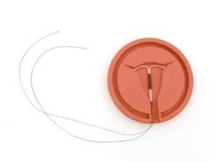
CHICAGO – Uterine perforation rates are low for both the levonorgestrel intrauterine device and copper devices, and do not differ significantly between the two, according to the multinational EURAS-IUD study of more than 61,400 women.
Between 2006 and 2013, the rate of perforation among new users of levonorgestrel IUDs was 1.4/1,000 insertions and the rate for copper IUDs was 1.1/1,000 insertions, according to Dr. Klaas Heinemannn of ZEG-Berlin, who presented his prize-winning abstract at the annual meeting of the American Congress of Obstetricians and Gynecologists.
However, a striking difference was seen in the perforation rate among first-ever vs. repeat IUD users: 2.5/1,000 levonorgestrel IUDs and 1.4/1,000 for copper IUDs among first-time users, compared with 0.5/1,000 for both types among repeat users.
Of the 81 perforations, 64 were associated with known potential risk factors. For example, 35 women who experienced perforation were breastfeeding at the time of insertion, and 25 of those were breastfeeding at least six times daily. Seven women were treated with prostaglandin prior to insertion.
Breastfeeding was associated with a sixfold increase in risk of perforation with both the levonorgestrel and copper IUDs, Dr. Heinemann said.
None of the perforations in this study led to serious illness or injury to an internal organ, he noted.
Levonorgestrel IUD use was associated with a 10-fold decrease in the contraceptive failure rate, compared with copper IUDs. The rate of contraceptive failure did not differ significantly between the different types of copper IUDs used in the study. Similarly, the ectopic pregnancy rate was lower in the levonorgestrel IUD group, he noted.
The prospective, noninterventional EUSAR-IUD cohort study included 61,448 women from Germany, Austria, United Kingdom, Finland, Poland, and Sweden. The women completed follow-up questionnaires at 12 months after enrollment, and all patient-reported outcomes of interest were validated by the treating physician.
Most (about 70%) of the women used levonorgestrel IUDs, and about 30% used copper IUDs. Half were first-time users.
Only 2% of participants were lost to follow-up, with no significant difference between the two groups.
Differences between the groups remained significant after adjustment for a number of variables, including age, body mass index, time since last delivery, and breastfeeding.
The most striking difference in use was age. Women in their 20s and 30s used copper IUDs at more than twice the rate of levonorgestrel IUDs, while those aged 40 years and older used levonorgestrel IUDs at twice the rate of copper IUDs.
Although this study is limited by its observational nature, the results suggest that there is no clinically relevant increase in the risk of uterine perforation with either type of IUD, Dr. Heinemann said.
"However, we found a striking difference in the contraceptive failure rate," he said.
Based on the findings, the Safety Monitoring and Advisory Council that oversaw the study concluded that "the health benefits of IUDs outweigh the rare risk of perforation, including for women postpartum and those breastfeeding. No new restrictions in IUD use are warranted based on this study."
This study was supported by an unconditional grant from Bayer Healthcare, which markets Mirena (levonorgestrel-releasing intrauterine system). Dr. Heinemann reported having no disclosures.
AT THE ACOG ANNUAL CLINICAL MEETING
Major finding: The perforation rates were 1.4/1,000 insertions and 1.1/1,000 insertions for levonorgestrel and copper IUDs, respectively.
Data source: The prospective, multinational EURAS-IUD study of 61,448 women.
Disclosures: This study was supported by an unconditional grant from Bayer Healthcare, which markets Mirena (levonorgestrel-releasing intrauterine system). Dr. Heinemann reported having no disclosures.
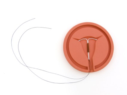
Perinatal loss dramatically affects maternal mental health
Women who experience stillbirth or infant death have extraordinarily high rates of depression and anxiety, which are inadequately addressed, according to findings from the Michigan Mother’s Study.
The effects were particularly pronounced among African American women, according to Dr. Katherine J. Gold who will present the findings of her award-winning abstract on Wednesday at the annual meeting of the American Congress of Obstetricians and Gynecologists.
At 9 months after delivery, the rates of positive screens for a number of mental health issues were dramatically and statistically significantly higher in 377 bereaved women who experienced stillbirth or infant death in the first month, compared with 232 control mothers with live birth who participated in the 2-year longitudinal population-based cohort study. The rates were 23% vs. 8% for depression, 41% vs. 12% for posttraumatic stress disorder, 19% vs. 7% for general anxiety disorder, 19% vs. 6% for social phobia, and 12% vs. 6% for panic disorder for the groups, respectively, said Dr. Gold of the University of Michigan, Ann Arbor.
"These high rates of symptoms were significant even when controlling for demographic factors, prior mental health problems, social support, and interpersonal violence," Dr. Gold said in an interview.
Also, the rates of these mental health issues were similar among mothers who experienced stillbirth and those who experienced infant death, suggesting that both losses can be powerful and traumatic experiences.
Although bereaved African American women had similar levels of distress as other bereaved women, they were significantly less likely to have received treatment, Dr. Gold noted.
This study is the first population-based study to look at mental health outcomes among women who experience perinatal death.
"The findings suggest that bereaved mothers have dramatically higher levels of persistent distress, which may be underrecognized by health care providers. It is important for physicians and midwives to assess for mental health symptoms after a loss," she said adding that because it has been shown that depression during a subsequent pregnancy poses substantial risk to fetal and infant outcomes, identifying and treating depression in women before and during subsequent pregnancy is critical for improving outcomes.
Dr. Gold reported having no disclosures.
The findings by Dr. Gold and her colleagues highlight the need to enhance efforts to identify women with mental health sequelae after the loss of an infant, according to Dr. C. Neill Epperson.
"As a reproductive psychiatrist for over 20 years, the main findings of this study ring true in my clinical experience. The loss of a child at birth or during infancy can be as traumatic as the loss of an older child," she said.
While the results of the study are from mailed surveys and not clinical interviews, the degree of self-reported distress is considerable, and the fact that African American women were in greater distress and less likely to be receiving treatment is quite disturbing.
Dr. Epperson is director of the Penn Center for Women’s Behavioral Wellness and is in the departments of psychiatry and obstetrics and gynecology at the University of Pennsylvania, Philadelphia. She reported having no disclosures.
Women who experience stillbirth or infant death have extraordinarily high rates of depression and anxiety, which are inadequately addressed, according to findings from the Michigan Mother’s Study.
The effects were particularly pronounced among African American women, according to Dr. Katherine J. Gold who will present the findings of her award-winning abstract on Wednesday at the annual meeting of the American Congress of Obstetricians and Gynecologists.
At 9 months after delivery, the rates of positive screens for a number of mental health issues were dramatically and statistically significantly higher in 377 bereaved women who experienced stillbirth or infant death in the first month, compared with 232 control mothers with live birth who participated in the 2-year longitudinal population-based cohort study. The rates were 23% vs. 8% for depression, 41% vs. 12% for posttraumatic stress disorder, 19% vs. 7% for general anxiety disorder, 19% vs. 6% for social phobia, and 12% vs. 6% for panic disorder for the groups, respectively, said Dr. Gold of the University of Michigan, Ann Arbor.
"These high rates of symptoms were significant even when controlling for demographic factors, prior mental health problems, social support, and interpersonal violence," Dr. Gold said in an interview.
Also, the rates of these mental health issues were similar among mothers who experienced stillbirth and those who experienced infant death, suggesting that both losses can be powerful and traumatic experiences.
Although bereaved African American women had similar levels of distress as other bereaved women, they were significantly less likely to have received treatment, Dr. Gold noted.
This study is the first population-based study to look at mental health outcomes among women who experience perinatal death.
"The findings suggest that bereaved mothers have dramatically higher levels of persistent distress, which may be underrecognized by health care providers. It is important for physicians and midwives to assess for mental health symptoms after a loss," she said adding that because it has been shown that depression during a subsequent pregnancy poses substantial risk to fetal and infant outcomes, identifying and treating depression in women before and during subsequent pregnancy is critical for improving outcomes.
Dr. Gold reported having no disclosures.
FROM THE ACOG ANNUAL CLINICAL MEETING
Major finding: Bereaved mothers had significantly more depression (23% vs. 8%), PTSD (41% vs. 12%), general anxiety disorder (19% vs. 7%), social phobia (19% vs. 6%), and panic disorder (12% vs. 6%) than did 232 control mothers who experienced live birth.
Data source: A longitudinal, population-based cohort study involving 609 women (the Michigan Mother’s Study).
Disclosures: Dr. Gold reported having no disclosures.
Topical lidocaine reduces menopausal dyspareunia
Applying topical liquid lidocaine to the vulvar vestibule prior to penetration allows for comfortable intercourse in breast cancer survivors with severe menopausal dyspareunia, according to findings from a randomized, controlled study involving 46 women.
During a double-blind phase of the study, patients randomized to apply 4% aqueous lidocaine had less intercourse pain than those who applied saline (median pain scores of 1.0 and 5.3 out of 10, respectively), according to Dr. Martha F. Goetsch of Oregon Health and Science University, Portland, who will report the finding at the annual meeting of the American Congress of Obstetricians and Gynecologists.
During an open-label phase of the study in which all patients were allowed to apply lidocaine, 37 of 41 (90%) reported comfortable penetration, and sexual distress scores decreased from a median of 30.5 to a median of 14. Additionally, 17 of 20 women (85%) who were abstaining from intercourse because of the discomfort had resumed penetrative intimacy, said Dr. Goetsch, whose abstract was awarded first prize among oral presentations by ACOG.
Patients included in the study were estrogen-deficient breast cancer survivors with severe penetrative dyspareunia not associated with pelvic muscle or organ pain. All had severe vulvovaginal atrophy. During the 1-month blinded phase of the study, the women applied either the lidocaine or the saline to the vulvar vestibule for 3 minutes prior to penetration. Effects of twice-weekly tampon insertion or intercourse were documented in a diary. No partners complained of numbness resulting from the lidocaine.
The findings are notable, because breast cancer survivors number in the millions in the United States alone.
"They often suffer from severe dyspareunia and are urged to refrain from using estrogen, which is the therapy most effective for dyspareunia in menopause," Dr. Goetsch said in an interview.
Furthermore, prior research has focused primarily on vaginal atrophy as the cause of dyspareunia in postmenopausal women.
"This study showed that pain could be prevented even though atrophy was unchanged," she said, noting that this suggests that perhaps atrophy is the wrong therapeutic focus.
"Success came with therapy to the vestibule, not the vagina," she said.
Dr. Goetsch reported having no disclosures.
FROM THE ACOG ANNUAL CLINICAL MEETING
Key clinical point: Consider lidocaine as an option for women with severe menopausal dyspareunia for whom estrogen therapy is not recommended.
Major finding: 37 of 41 patients (90%) reported comfortable penetration during intercourse.
Data source: A randomized, controlled, double-blind study of 46 women.
Disclosures: Dr. Goetsch reported having no disclosures.
Inadequate weight gain ups preterm birth risk in twin pregnancies
Women with twin pregnancies who have inadequate gestational weight gain during the second trimester have a more than twofold increase in the risk of preterm birth before 32 weeks, compared with those with adequate weight gain, findings from a retrospective cohort study suggest.
Inadequate weight gain between 20 and 28 weeks’ of gestation was the strongest predictor of preterm birth before 32 weeks in the study of 489 women with twin pregnancies, according to Dr. Kate E. Pettit, who will report the findings on Monday at the annual meeting of the American Congress of Obstetricians and Gynecologists in Chicago.
The rate of preterm birth prior to 32 weeks was 37.6% among women with inadequate weight gain during weeks 20-28, compared with 15.2% among those with adequate weight gain during that time period. Inadequate weight gain prior to 20 weeks was not associated with preterm birth, said Dr. Pettit of the University of California, San Diego, and her associates.
Other factors found to be associated with preterm birth in this study were monochorionicity and short cervical length, she noted.
All viable twin pregnancies delivered in the UCSD Health System between 2001 and 2013 were included in the study. Adequacy of weight gain was determined by dividing the minimum Institute of Medicine weight gain recommendation by 37 weeks.
"This study affirms the association between poor total gestational weight gain and preterm birth, and highlights the likely crucial period of time of 20-28 weeks of gestation that may be a target for future intervention trials," Dr. Pettit said in an interview.
The higher rate of preterm birth prior to 32 weeks’ of gestation among those with inadequate weight gain was attributable to spontaneous preterm birth, as there was no difference in the rates of indicated preterm birth among those with and without adequate weight gain, she explained, noting that "the biologic plausibility of the effect of gestational weight gain on the rate of spontaneous preterm birth is unknown."
"However, there is much room for improvement in gestational weight gain counseling, and a need for additional interventional trials to determine if adequacy of gestational weight gain at 20-28 weeks’ will, in fact, be one of the modifiable factors in the quest to reduce preterm birth in twin pregnancies," she said in an interview.
Interventions could include early goal-setting, nutritional assessment, and activity modification, she added.
Dr. Pettit reported having no disclosures.
FROM THE ACOG ANNUAL CLINICAL MEETING
Major finding: The rate of preterm birth prior to 32 weeks was 37.6% among women with inadequate weight gain during gestational weeks 20-28 and 15.2% for those with adequate weight gain.
Data source: A retrospective cohort study of 489 women with twin pregnancies.
Disclosures: Dr. Pettit reported having no disclosures.