User login
Ultrasound offers noninvasive option for body contouring, skin tightening
SAN DIEGO – Ultrasound treatments are an alternative for patients looking to avoid the pain and prolonged recovery associated with surgical body contouring or skin tightening procedures, according to Dr. Elizabeth L. Tanzi. Currently, several types of ultrasound devices are available for such procedures, she said at the annual meeting of the American Society for Dermatologic Surgery.
High-intensity, focused ultrasound and pulsed focused ultrasound devices are used for body contouring, and the micro-focused high-intensity ultrasound device is used for skin tightening, Dr. Tanzi explained. Body contouring has rapidly gained popularity in the United States and elsewhere, but surgical contouring procedures can be painful and can require prolonged recovery times, she noted.

Ultrasound for body contouring works via two noninvasive mechanisms, both of which cause selective damage of adipose tissue, said Dr. Tanzi, codirector of the Washington (D.C.) Institute of Dermatologic Laser Surgery.
First, adipose tissue directly absorbs ultrasonic energy during treatment, leading to immediate thermal coagulation and necrosis of fat cells, she said. Mechanical processes also break down tissue, such as when shear forces are created at high levels of pressure during treatment. Circulating macrophages then absorb the damaged adipocytes and the triglycerides they release, leading to a gradual reduction of the fat layer at the treatment site, she added.
When using ultrasound for body contouring, “patient selection is paramount,” Dr. Tanzi emphasized. Although the procedure carries no risk of infection or scarring, ultrasound yields subtler cosmetic results than would liposuction, and patients should understand that difference up front, she said. “This is body contouring, not weight loss,” she added. Patients can typically expect to see a slow reduction in adipose tissue at the treatment area about 1-4 months after treatment, and they also should understand that they might need multiple treatments to achieve their cosmetic goals, Dr. Tanzi said.
“Discuss side effects with patients,” Dr. Tanzi added. When used for body contouring, ultrasound can cause temporary erythema, bruising at the treatment site, soreness, and contour defects, although the latter are rare, she noted. Dr. Tanzi said she now treats patients with lower levels of ultrasonic energy to improve their comfort. “We are much more aggressive now with the overall number of lines we place on the body,” she added. “Go over the areas again and again.”
For example, in one clinical study, high-intensity focused ultrasound yielded statistically significant decreases in waist circumference at 12 weeks with treatment of 150-180 Joules per square centimeter, she noted. However, six passes at just 30 J/cm2 was more comfortable and gave similar results, she said (Dermatol. Surg. 2014;40:641-51).
Micro-focused high-intensity ultrasound can be used for skin tightening, particularly on the face. The procedure targets precise areas of adipose tissue within the skin to create small foci of thermal necrosis, and also stimulates localized collagen production for about 3-6 months afterward, Dr. Tanzi said. As with body contouring, patients should expect subtler results than would be expected from a surgical procedure, but the absence of scarring and the opportunity to immediately return to regular activities can bolster their satisfaction, she said.
Dr. Tanzi is a consultant for Ulthera, Solta, Cynosure/Palomar, Syneron & Candela, and Cutera, and has received financial support from Zeltiq, miraDry, and Clarisonic.
SAN DIEGO – Ultrasound treatments are an alternative for patients looking to avoid the pain and prolonged recovery associated with surgical body contouring or skin tightening procedures, according to Dr. Elizabeth L. Tanzi. Currently, several types of ultrasound devices are available for such procedures, she said at the annual meeting of the American Society for Dermatologic Surgery.
High-intensity, focused ultrasound and pulsed focused ultrasound devices are used for body contouring, and the micro-focused high-intensity ultrasound device is used for skin tightening, Dr. Tanzi explained. Body contouring has rapidly gained popularity in the United States and elsewhere, but surgical contouring procedures can be painful and can require prolonged recovery times, she noted.
Ultrasound for body contouring works via two noninvasive mechanisms, both of which cause selective damage of adipose tissue, said Dr. Tanzi, codirector of the Washington (D.C.) Institute of Dermatologic Laser Surgery.
First, adipose tissue directly absorbs ultrasonic energy during treatment, leading to immediate thermal coagulation and necrosis of fat cells, she said. Mechanical processes also break down tissue, such as when shear forces are created at high levels of pressure during treatment. Circulating macrophages then absorb the damaged adipocytes and the triglycerides they release, leading to a gradual reduction of the fat layer at the treatment site, she added.
When using ultrasound for body contouring, “patient selection is paramount,” Dr. Tanzi emphasized. Although the procedure carries no risk of infection or scarring, ultrasound yields subtler cosmetic results than would liposuction, and patients should understand that difference up front, she said. “This is body contouring, not weight loss,” she added. Patients can typically expect to see a slow reduction in adipose tissue at the treatment area about 1-4 months after treatment, and they also should understand that they might need multiple treatments to achieve their cosmetic goals, Dr. Tanzi said.
“Discuss side effects with patients,” Dr. Tanzi added. When used for body contouring, ultrasound can cause temporary erythema, bruising at the treatment site, soreness, and contour defects, although the latter are rare, she noted. Dr. Tanzi said she now treats patients with lower levels of ultrasonic energy to improve their comfort. “We are much more aggressive now with the overall number of lines we place on the body,” she added. “Go over the areas again and again.”
For example, in one clinical study, high-intensity focused ultrasound yielded statistically significant decreases in waist circumference at 12 weeks with treatment of 150-180 Joules per square centimeter, she noted. However, six passes at just 30 J/cm2 was more comfortable and gave similar results, she said (Dermatol. Surg. 2014;40:641-51).
Micro-focused high-intensity ultrasound can be used for skin tightening, particularly on the face. The procedure targets precise areas of adipose tissue within the skin to create small foci of thermal necrosis, and also stimulates localized collagen production for about 3-6 months afterward, Dr. Tanzi said. As with body contouring, patients should expect subtler results than would be expected from a surgical procedure, but the absence of scarring and the opportunity to immediately return to regular activities can bolster their satisfaction, she said.
Dr. Tanzi is a consultant for Ulthera, Solta, Cynosure/Palomar, Syneron & Candela, and Cutera, and has received financial support from Zeltiq, miraDry, and Clarisonic.

Dihydroergotamine has a key role in migraine and daily headache, despite its underuse
SCOTTSDALE, ARIZ. – Dihydroergotamine remains underused in the treatment of migraine and daily severe headaches, especially in inpatient settings, according to Dr. Priyanka Chaudhry.
The drug offers several clinical advantages, said Dr. Chaudhry, a neurologist specializing in headache treatment at the University of Texas Southwestern Medical Center in Dallas.

When used in combination with an antiemetic such as metoclopramide, dihydroergotamine (DHE) has been found to be as effective as opiates, valproate, and ketorolac in relieving migraine headache and preventing relapses, Dr. Chaudhry said. Its bioavailability is 100% when given intramuscularly or intravenously, and about 40% when given as a nasal spray, she added.
With its relatively long half-life of 10-13 hours, DHE also poses less risk of physical dependence than does ergotamine, Dr. Chaudhry noted. And in a recent uncontrolled retrospective study, its cumulative positive effects persisted for up to 1 month after patient discharge, she said at a symposium sponsored by the American Headache Society.
Dr. Chaudhry typically starts patients on an infusion of 0.3 mg DHE given over a period of 45 minutes, and if the patient tolerates this dose, increases it to 0.5 mg, then 0.75 mg, and then 1 mg spaced every 6-8 hours, she said. When the patient is free of headaches, she reduces the dose to 0.5 mg. She prefers that patients be headache free for 24 hours before discontinuing DHE, although this goal is not always achievable, she noted.
Treatment with DHE for 5 days seems to be more effective than shorter courses, Dr. Chaudhry added. She, therefore, will admit patients to the infusion clinic for 5 days, and then readmit them later for follow-up treatment with lidocaine, she said.
Dihydroergotamine was associated with several potentially serious adverse effects. “It can be very harsh on veins, so if you’re admitting somebody in an inpatient setting, it is preferable to order a midline catheter or a PICC [peripherally inserted central catheter] line,” said Dr. Chaudhry. Patients also can experience chest pain or a feeling of warmth or pressure in the neck during DHE infusion, especially the first time they are exposed to a given dose of the drug, she noted. “This side effect is not a contraindication for use,” she emphasized. “But consider a slow infusion, and do not bolus.” She orders electrocardiograms for such patients but sees no need to monitor cardiac enzymes, she said.
Nausea is the most common side effect of DHE, and can be treated with 8 mg odansetron, said Dr. Chaudhry. She adds a second antiemetic such as promethazine on an as-needed basis, decreases the rate of DHE infusion, and cuts the dose if nothing else works, she said. Clinicians might also consider adding 50 mg hydroxyzine orally or IM every 6 hours, starting at 25 mg if patients are relatively small or sensitive to medications, she said.
Dihydroergotamine also can cause light-headedness, diarrhea, abdominal cramping, and leg cramps, Dr. Chaudhry noted. Patients who are pregnant or have renal or hepatic failure, uncontrolled hypertension, sepsis, or vascular disease, should not take DHE, she cautioned.
She said she had no relevant financial disclosures.
SCOTTSDALE, ARIZ. – Dihydroergotamine remains underused in the treatment of migraine and daily severe headaches, especially in inpatient settings, according to Dr. Priyanka Chaudhry.
The drug offers several clinical advantages, said Dr. Chaudhry, a neurologist specializing in headache treatment at the University of Texas Southwestern Medical Center in Dallas.
When used in combination with an antiemetic such as metoclopramide, dihydroergotamine (DHE) has been found to be as effective as opiates, valproate, and ketorolac in relieving migraine headache and preventing relapses, Dr. Chaudhry said. Its bioavailability is 100% when given intramuscularly or intravenously, and about 40% when given as a nasal spray, she added.
With its relatively long half-life of 10-13 hours, DHE also poses less risk of physical dependence than does ergotamine, Dr. Chaudhry noted. And in a recent uncontrolled retrospective study, its cumulative positive effects persisted for up to 1 month after patient discharge, she said at a symposium sponsored by the American Headache Society.
Dr. Chaudhry typically starts patients on an infusion of 0.3 mg DHE given over a period of 45 minutes, and if the patient tolerates this dose, increases it to 0.5 mg, then 0.75 mg, and then 1 mg spaced every 6-8 hours, she said. When the patient is free of headaches, she reduces the dose to 0.5 mg. She prefers that patients be headache free for 24 hours before discontinuing DHE, although this goal is not always achievable, she noted.
Treatment with DHE for 5 days seems to be more effective than shorter courses, Dr. Chaudhry added. She, therefore, will admit patients to the infusion clinic for 5 days, and then readmit them later for follow-up treatment with lidocaine, she said.
Dihydroergotamine was associated with several potentially serious adverse effects. “It can be very harsh on veins, so if you’re admitting somebody in an inpatient setting, it is preferable to order a midline catheter or a PICC [peripherally inserted central catheter] line,” said Dr. Chaudhry. Patients also can experience chest pain or a feeling of warmth or pressure in the neck during DHE infusion, especially the first time they are exposed to a given dose of the drug, she noted. “This side effect is not a contraindication for use,” she emphasized. “But consider a slow infusion, and do not bolus.” She orders electrocardiograms for such patients but sees no need to monitor cardiac enzymes, she said.
Nausea is the most common side effect of DHE, and can be treated with 8 mg odansetron, said Dr. Chaudhry. She adds a second antiemetic such as promethazine on an as-needed basis, decreases the rate of DHE infusion, and cuts the dose if nothing else works, she said. Clinicians might also consider adding 50 mg hydroxyzine orally or IM every 6 hours, starting at 25 mg if patients are relatively small or sensitive to medications, she said.
Dihydroergotamine also can cause light-headedness, diarrhea, abdominal cramping, and leg cramps, Dr. Chaudhry noted. Patients who are pregnant or have renal or hepatic failure, uncontrolled hypertension, sepsis, or vascular disease, should not take DHE, she cautioned.
She said she had no relevant financial disclosures.
AT A SYMPOSIUM SPONSORED BY THE AMERICAN HEADACHE SOCIETY

Take these five key steps to limit reimbursement headaches
SCOTTSDALE, ARIZ. – Headache specialists can take five key steps related to coding and billing that can protect their financial investment in their practice, said Stacy Girard, M.B.A., a certified professional coder and revenue analyst at Mayo Clinic Arizona in Phoenix.
Those steps include reviewing annual changes in Current Procedural Terminology (CPT) codes, understanding code descriptions and how to use modifiers, consistently tracking reimbursement denials and intervening if they trend out of control, reviewing medical and reimbursement policies at least once 1 year, and determining their financial liability when services are not covered, Ms. Girard said at a symposium sponsored by the American Headache Society.
The CPT codes for neurologic procedures will not change in the upcoming year, Ms. Girard said. In headache and migraine treatment, some of the most common codes include 64405, for unilateral occipital nerve block; 64450, for unilateral block of another peripheral nerve; 64612, for unilateral facial nerve chemodenervation; and 64615, for bilateral chemodenervation of the facial nerve, trigeminal nerve, cervical spinal nerve, and accessory nerves, she added. Code 64615 was introduced in January 2013 and has “greatly simplified billing for a number of procedures for chronic migraine,” she said.
Payers routinely deny reimbursement claims, Ms. Girard noted. To avoid a reimbursement denial, clinicians and coders must use modifiers correctly when appending claims, she said. For example, they should use modifier 25 only if the same physician performed a completely separate evaluation and management (E/M) service for a patient on the same day of a procedure. And CPT code 64615 already signifies bilateral chemodenervation, so coders should not add modifier 50 to indicate that the service was bilateral, she added.
“You cannot expect what you don’t inspect,” said Ms. Girard. “You cannot expect to be paid for everything if you are not on the back end, inspecting your denials.” She creates a chart to track reimbursement denials by type and month, and reports the results to her practice, highlighting areas “that are really getting out of control,” she said.
Several denial reason codes worth tracking include CO50, CO55, and CO56, for treatments that are deemed “not a medical necessity,” “experimental/investigational,” or “not proven to be effective,” and CO151, in which the payer decides that there is insufficient support for the number or frequency of services provided, Ms. Girard said. “Take action on denials,” she emphasized. “Work with your payers and make a connection with them. We meet with our payers on a regular basis and show them what’s routinely being denied. Maybe you need to understand where they are coming from, and maybe you need to say to them that I really don’t agree with this, and here’s why.”
To minimize denials, practices also need to understand reimbursement policies and who is responsible for paying for services, she said. Information on coverage determinations can be found on payer websites under “medical policies,” or “reimbursement policies,” she noted. Medical policies list the payer’s criteria for covering specific services and diagnosis codes, while reimbursement policies specify how the payer handles issues such as bundling rules, global surgery, and multiple procedures for a single patient.
Practices can further limit their financial liability by obtaining signed advanced beneficiary notices (ABNs) from Medicare patients, Ms. Girard said. Without a signed ABN, patients cannot be billed for services that Medicare does not cover, leaving the practice holding the bill, she said. “Are you going to give away your services for free, or is it something the patient is going to pay for because they’ve signed an ABN?”
Ms. Girard declared no conflicts of interest.
FROM A SYMPOSIUM SPONSORED BY THE AMERICAN HEADACHE SOCIETY
Use PEARLS to build relationships with patients
SCOTTSDALE – Reasoning with patients can be challenging when they are demanding, have unrealistic expectations about their treatment, are emotionally reactive, or are seeking prescriptions for narcotics, Dr. David Gullen said. “These patients tax our patience and often our clinical skills, because they often don’t respond to the usual logic or presentation of treatment options,” Dr. Gullen said at a symposium sponsored by the American Headache Society.
But by switching the focus from logic to empathy, clinicians can transform relationships, even with very difficult patients who are dealing with migraine and other types of chronic pain, said Dr. Gullen, an internist who teaches communication skills to faculty, fellows, and residents at Mayo Clinic Arizona, Phoenix. Using empathy and other skills to improve relationships enhances physician and patient satisfaction, increases compliance, and improves clinical outcomes, he said.

“The question is, can the patient join you on a cognitive level?” Dr. Gullen concluded. “Often they can’t, but they may be able to join you on another level. It’s a noncognitive way of responding to them.”
Relationship building can also help clinicians heal the patient when they “don’t have a lot of medical options,” Dr. Gullen added. “I think that’s something our forebears knew 200 and 300 years ago,” he said. “Healers in the past had to use this approach because they didn’t have a lot of the tools we have today.”
To build relationships with patients, clinicians can use the mnemonic PEARLS – which stands for partnership, empathy, apology/acknowledgment, respect, legitimation, and support, Dr. Gullen said. For example, when seeing a new patient with migraine who has already tried and “failed” numerous medications, the clinician can use a partnership statement, such as, “I really want us to work on this together.” This approach increases the odds of creating a working relationship with the patient, he said.
“Empathy is probably the most important of all, because it’s your ability to convey to the patient that you understand their position,” Dr. Gullen emphasized. “Showing empathy for a patient’s perspective is not the same as endorsing it,” he added. “They may misunderstand what you’re telling them, or you may disagree with them. By showing that you understand their perspective, you’re not saying it’s correct, but you are saying you understand.”
An apology or acknowledgement could simply mean apologizing for running behind schedule or acknowledging a patient’s frustration with a treatment outcome.
Respecting a patient’s efforts to cope with the pain, and legitimizing the emotional response by letting the patient know it is normal and understandable also helps build trust. “Be aware of when you’re emotionally activated, so that you can use some of the PEARLS or respond with empathy, and take time to calm yourself down,” Dr. Gullen added.
How something is said to a patient matters as much as the words used, he said. “Think of a time in your life when you got a piece of advice that was really important for you to hear, but when you first heard it, it landed on you really badly,” he added. “Now think about getting that advice from someone you thought did not understand you or did not have your best interests at heart, compared with someone who you thought got you and really understood you. That’s why we emphasize the importance of relationship.”
Dr. Gullen declared no financial disclosures.
SCOTTSDALE – Reasoning with patients can be challenging when they are demanding, have unrealistic expectations about their treatment, are emotionally reactive, or are seeking prescriptions for narcotics, Dr. David Gullen said. “These patients tax our patience and often our clinical skills, because they often don’t respond to the usual logic or presentation of treatment options,” Dr. Gullen said at a symposium sponsored by the American Headache Society.
But by switching the focus from logic to empathy, clinicians can transform relationships, even with very difficult patients who are dealing with migraine and other types of chronic pain, said Dr. Gullen, an internist who teaches communication skills to faculty, fellows, and residents at Mayo Clinic Arizona, Phoenix. Using empathy and other skills to improve relationships enhances physician and patient satisfaction, increases compliance, and improves clinical outcomes, he said.
“The question is, can the patient join you on a cognitive level?” Dr. Gullen concluded. “Often they can’t, but they may be able to join you on another level. It’s a noncognitive way of responding to them.”
Relationship building can also help clinicians heal the patient when they “don’t have a lot of medical options,” Dr. Gullen added. “I think that’s something our forebears knew 200 and 300 years ago,” he said. “Healers in the past had to use this approach because they didn’t have a lot of the tools we have today.”
To build relationships with patients, clinicians can use the mnemonic PEARLS – which stands for partnership, empathy, apology/acknowledgment, respect, legitimation, and support, Dr. Gullen said. For example, when seeing a new patient with migraine who has already tried and “failed” numerous medications, the clinician can use a partnership statement, such as, “I really want us to work on this together.” This approach increases the odds of creating a working relationship with the patient, he said.
“Empathy is probably the most important of all, because it’s your ability to convey to the patient that you understand their position,” Dr. Gullen emphasized. “Showing empathy for a patient’s perspective is not the same as endorsing it,” he added. “They may misunderstand what you’re telling them, or you may disagree with them. By showing that you understand their perspective, you’re not saying it’s correct, but you are saying you understand.”
An apology or acknowledgement could simply mean apologizing for running behind schedule or acknowledging a patient’s frustration with a treatment outcome.
Respecting a patient’s efforts to cope with the pain, and legitimizing the emotional response by letting the patient know it is normal and understandable also helps build trust. “Be aware of when you’re emotionally activated, so that you can use some of the PEARLS or respond with empathy, and take time to calm yourself down,” Dr. Gullen added.
How something is said to a patient matters as much as the words used, he said. “Think of a time in your life when you got a piece of advice that was really important for you to hear, but when you first heard it, it landed on you really badly,” he added. “Now think about getting that advice from someone you thought did not understand you or did not have your best interests at heart, compared with someone who you thought got you and really understood you. That’s why we emphasize the importance of relationship.”
Dr. Gullen declared no financial disclosures.
EXPERT ANALYSIS FROM A SYMPOSIUM SPONSORED BY THE AMERICAN HEADACHE SOCIETY

Biopsy can underestimate diversity, aggressiveness of basal cell carcinomas
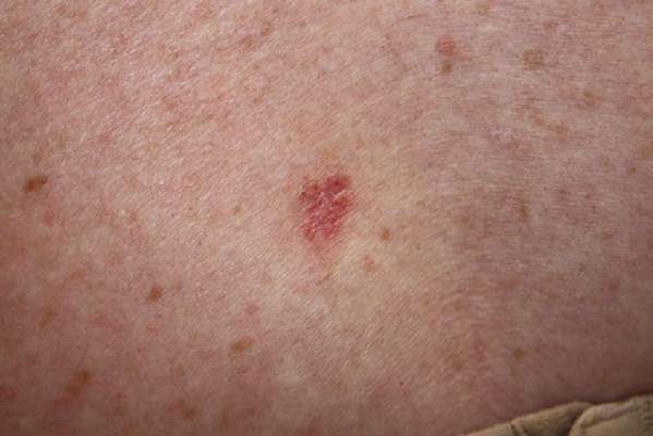
SAN DIEGO – Histology of basal cell carcinomas removed by Mohs micrographic surgery showed that presurgical biopsies had not revealed all tumor subtypes in 64% of cases, and had underestimated the aggressiveness of the tumors 24% of the time, according to data from a large, multicenter, retrospective study.
“Unfortunately, while cheap and cost-effective, biopsies are a subsample of the full malignancy,” said Dr. Murad Alam, professor of dermatology, otolaryngology, and surgery at Northwestern University in Chicago. “Skin biopsy of basal cell carcinoma [BCC] may fail to detect all BCC subtypes, and as such may underestimate the aggressiveness of an individual BCC tumor.”

Basal cell carcinoma is the most common skin cancer worldwide, and can broadly be grouped into aggressive and indolent types, Dr. Alam said at the annual meeting of the American Society for Dermatologic Surgery. But tumors often show mixed histology, and cancer treatment needs to target the most aggressive subtype present in the tumor, he added. Results of past studies suggested that biopsies of BCCs could miss tumor subtypes, but the current research is the first large, multicenter study to confirm these findings, he and his associates said.
For the study, the investigators compared biopsy reports and microscopic slides of Mohs micrographic surgery (MMS) specimens from 871 consecutive cases of BCC treated at three hospitals in Illinois from 2013 to 2014. Patients first underwent biopsies, followed by complete excision of their tumors during MMS. Almost 59% of patients were male, and tumors were most commonly removed from the nose or cheek. In all, 78% of biopsies were obtained by the shave technique, but punch and excisional biopsies also were performed, the researchers noted.
Using standard definitions of BCC subtypes, the investigators compared levels of concordance between biopsy and MMS histology findings, Dr. Alam said. They also grouped tumor specimens as high risk (that is, infiltrative, morpheic, micronodular, basosquamous) or low risk (superficial or nodular), and determined whether tumor biopsy and MMS histology yielded the same or discordant risk assessments, he added.
Biopsies identified only 18% of tumors as being of mixed histology, compared with 57% of MMS specimens, said Dr. Alam. Biopsy results matched MMS histologies in only 31% of cases, while in 64% of cases, the MMS specimen yielded more tumor subtypes than the biopsy specimen. The researchers noted that biopsy yielded more subtypes than did MMS in 4% of cases, and that MMS and biopsy subtypes were fully discordant in only four cases.
Dr. Alam and his associates declared no external funding sources or conflicts of interest.
SAN DIEGO – Histology of basal cell carcinomas removed by Mohs micrographic surgery showed that presurgical biopsies had not revealed all tumor subtypes in 64% of cases, and had underestimated the aggressiveness of the tumors 24% of the time, according to data from a large, multicenter, retrospective study.
“Unfortunately, while cheap and cost-effective, biopsies are a subsample of the full malignancy,” said Dr. Murad Alam, professor of dermatology, otolaryngology, and surgery at Northwestern University in Chicago. “Skin biopsy of basal cell carcinoma [BCC] may fail to detect all BCC subtypes, and as such may underestimate the aggressiveness of an individual BCC tumor.”
Basal cell carcinoma is the most common skin cancer worldwide, and can broadly be grouped into aggressive and indolent types, Dr. Alam said at the annual meeting of the American Society for Dermatologic Surgery. But tumors often show mixed histology, and cancer treatment needs to target the most aggressive subtype present in the tumor, he added. Results of past studies suggested that biopsies of BCCs could miss tumor subtypes, but the current research is the first large, multicenter study to confirm these findings, he and his associates said.
For the study, the investigators compared biopsy reports and microscopic slides of Mohs micrographic surgery (MMS) specimens from 871 consecutive cases of BCC treated at three hospitals in Illinois from 2013 to 2014. Patients first underwent biopsies, followed by complete excision of their tumors during MMS. Almost 59% of patients were male, and tumors were most commonly removed from the nose or cheek. In all, 78% of biopsies were obtained by the shave technique, but punch and excisional biopsies also were performed, the researchers noted.
Using standard definitions of BCC subtypes, the investigators compared levels of concordance between biopsy and MMS histology findings, Dr. Alam said. They also grouped tumor specimens as high risk (that is, infiltrative, morpheic, micronodular, basosquamous) or low risk (superficial or nodular), and determined whether tumor biopsy and MMS histology yielded the same or discordant risk assessments, he added.
Biopsies identified only 18% of tumors as being of mixed histology, compared with 57% of MMS specimens, said Dr. Alam. Biopsy results matched MMS histologies in only 31% of cases, while in 64% of cases, the MMS specimen yielded more tumor subtypes than the biopsy specimen. The researchers noted that biopsy yielded more subtypes than did MMS in 4% of cases, and that MMS and biopsy subtypes were fully discordant in only four cases.
Dr. Alam and his associates declared no external funding sources or conflicts of interest.
Key clinical point: Definitive excision by Mohs micrographic surgery reveals more information about basal cell carcinoma subtypes and tumor behavior than does biopsy.
Major finding: Compared with Mohs specimens, biopsy underestimated the diversity of tumor subtypes in 64% of cases, and underestimated tumor aggressiveness in 24% of cases.
Data source: Multicenter retrospective study of 871 basal cell carcinomas that were biopsied and then removed by Mohs micrographic surgery.
Disclosures: The investigators declared no external funding sources or conflicts of interest.
Collagen filler succeeds against acne scars
SAN DIEGO – An injectable collagen-based filler significantly outperformed saline placebo for treating acne scars, with durable effects at 12 months, according to a randomized, double-blind crossover trial of 147 adults.
The study “successfully demonstrates the effectiveness and safety of polymethylmethacrylate in atrophic acne scars,” said Dr. James Spencer, who conducted the research while at Mount Sinai School of Medicine in New York. Dr. Spencer is now in private practice in St. Petersburg, Fla.
Six months after treatment, 64% of patients who received the polymethylmethacrylate-collagen filler (or PMMA) had at least half their scars improve by at least 2 points on an acne rating scale, Dr. Spencer and his associates said at the annual meeting of the American Society for Dermatologic Surgery. Only 32% of the control group achieved that result (P = .0005). Response rates for the filler were 61% at 9 months and 70% at 12 months, and “crossover subjects subsequently treated with PMMA collagen showed similar response levels,” they said.
The PMMA-collagen filler is easy to administer; “works well on deep, severe scars; and should also work very well on shallow scars,” the researchers noted. The treatment “may enable practitioners to effectively treat acne scarring with no need for capital equipment expenditure or the risks associated with resurfacing procedures,” they said.
About 61% of patients in the study were female, participants averaged 44 years of age, and 20% were Fitzpatrick skin type V or VI. To enter the study, participants had to have at least four facial acne scars that were soft contoured, rolling, distensible, and rated moderate to severe (3-4) on a 4-point acne rating scale.
The patients were treated every 2 weeks for a month, and again at months 3 and 6. At 6 months, patients in the placebo group crossed over and received the filler, and all patients were followed for another 6 months.
Adverse effects were uncommon and included mild transient pain at the injection site, swelling, bruising, and acne, the researchers said. “There was no evidence of granulomas, changes in pigmentation, or hypertrophic scarring,” they added.
No funding source was reported for the study. Dr. Spencer and his coauthors reported financial relationships with Photomedex, Genentech, and Leo Pharma.
Key clinical point: A collagen-based filler significantly improved the appearance of acne scars in adults, with persistent improvement at 12 months.
Major finding: At 6-month evaluation, 64% of the intervention group were considered responders, compared with 32% of the control group (P = .0005).
Data source: A prospective, randomized, double-blind, controlled, multicenter crossover study of 147 adults with acne scars.
Disclosures: The researchers did not report funding sources. Dr. Spencer and his coauthors reported financial relationships with Photomedex, Genentech, and Leo Pharma.
Mixed results seen for imaging skin tumors before Mohs surgery
SAN DIEGO – Dermoscopy was no better than visual clinical inspection for delineating skin tumor margins before Mohs micrographic surgery, based on data from a pooled analysis, according to Dr. Syril Que.
But her review showed that confocal microscopy and optical coherence tomography devices “may be of potential diagnostic value – both for defining BCC [basal cell carcinoma] tumor margins prior to excision and for minimizing the number of stages needed subsequently,” said Dr. Que, a dermatology resident at the University of Connecticut Health Center in Farmington. “Nevertheless, there are limitations to both imaging modalities, and adjustments that need to be made before they are incorporated as a standard of practice in Mohs surgery,” she emphasized.
In a search of PubMed, Dr. Que found 27 original research studies that compared noninvasive imaging devices for delineating skin tumor margins with Mohs histology results. The studies were published between 2004 and 2014; 26 studies included BCC specimens, 3 included squamous cell carcinomas, and only 1 looked at lentigo maligna and melanoma. When broken down by type of imaging device, 17 of the studies focused on confocal microscopy, 5 were of dermoscopy, and 5 were of optical coherence tomography, she added. The number of specimens per study ranged from 2 to 115.
Only one study found an added diagnostic benefit from dermoscopy of skin tumor margins, compared with visual inspection, and that was a report of only two cases, said Dr. Que. The larger studies of 40-60 patients showed no significant difference between clinical inspection and dermoscopy for delineating margins. “Currently available evidence shows that dermoscopy has no advantage over clinical inspection when used prior to Mohs surgery,” she concluded.
Studies of confocal microscopy of BCCs were more promising, Dr. Que noted. These papers reported sensitivities of 73%-100%, and specificities of 89%-99%. “Sensitivity and specificity tended to be lower for micronodular or infiltrative BCCs,” she added. The papers did not report sensitivity or specificity values for squamous cell carcinomas, but did state that it was difficult to use confocal microscopy alone to distinguish between these tumors and actinic keratoses, she said at the annual meeting of the American Society for Dermatologic Surgery. Few studies addressed the use of confocal microscopy for melanoma or lentigo maligna, she added.
Finally, the papers on optical coherence tomography reported that the device “showed excellent correlation with histopathology, and was 84% accurate in predicting surgical margins,” said Dr. Que. “Optical coherence tomography appropriately assessed the subclinical spread of tumor, based on the close proximity of optical coherence tomography margins to the final Mohs defect,” she added.
Dr. Que said she had no relevant financial disclosures.
Vismodegib offers promise for basal cell carcinoma, with caveats
SAN DIEGO – Patients treated with vismodegib for locally advanced or metastatic basal cell carcinoma went a median of 15 months before their disease progressed or they stopped treatment because of side effects, according to a 30-month update of the pivotal ERIVANCE basal cell carcinoma study.
Median progression-free survival on the first-in-class oral hedgehog-pathway inhibitor was 9 months, reported Dr. Seaver Soon at the annual meeting of the American Society for Dermatologic Surgery.
Data from two other trials of vismodegib resemble results from ERIVANCE, added Dr. Soon, a dermatologist in private practice in La Jolla, Calif. An expanded access study (J. Am. Acad. Dermatol 2014;70:60-9) of 119 patients with advanced basal cell carcinoma (BCC) reported comparable objective response rates (46.4% for patients with locally advanced BCC and 30.8% for patients with metastatic disease), and an interim analysis of data from the STEVIE trial had findings that were “very similar” to ERIVANCE, he said.

Thus far, vismodegib “offers a hope in treating otherwise difficult to manage, unresectable basal cell carcinoma tumors,” said Dr. Iren Kossintseva, a dermatologist in Vancouver, B.C. But the drug “may not be as tissue sparing as promised, she added. In a patient with chronic lymphocytic leukemia who had a large BCC on his lower eyelid and cheek, 7.5 months of vismodegib reduced the exophyticity and erosiveness of the tumor, but “likely did not substantially reduce the overall extent of necessary reconstruction,” she reported.
Vismodegib can cause potentially severe side effects. All seven patients who Dr. Kossintseva treated with 150 mg vismodegib per day during 2013-2014 developed “notable” adverse effects – including polycyclic rash, sensory and motor problems within the tumor area, bilateral edema of the lower limbs, congestive heart failure, and renal failure that has been slow to improve after stopping vismodegib, she said. “These are unique patients, and it’s often an uphill battle with these patients,” she added.
Tumors also can exhibit primary and secondary resistance to vismodegib, Dr. Soon noted. Studies have shown primary resistance characterized by tumor progression after as little as 2 months of treatment (Mol. Oncol. 2014; S1574-7891:00216-6) while secondary (or acquired) resistance occurs after an initial response to treatment and is linked to a mutation that interferes with drug binding, he said. Acquired resistance typically occurs when patients have been on vismodegib for about a year, Dr. Soon added. “Concurrent treatment with an alternative smoothened inhibitor, such as itraconazole, and downstream target inhibitors may overcome resistance,” he said. Dr. Kossintseva declared no conflicts of interest. Dr. Soon reported receiving honoraria and research grants from Genentech, the maker of vismodegib.
SAN DIEGO – Patients treated with vismodegib for locally advanced or metastatic basal cell carcinoma went a median of 15 months before their disease progressed or they stopped treatment because of side effects, according to a 30-month update of the pivotal ERIVANCE basal cell carcinoma study.
Median progression-free survival on the first-in-class oral hedgehog-pathway inhibitor was 9 months, reported Dr. Seaver Soon at the annual meeting of the American Society for Dermatologic Surgery.
Data from two other trials of vismodegib resemble results from ERIVANCE, added Dr. Soon, a dermatologist in private practice in La Jolla, Calif. An expanded access study (J. Am. Acad. Dermatol 2014;70:60-9) of 119 patients with advanced basal cell carcinoma (BCC) reported comparable objective response rates (46.4% for patients with locally advanced BCC and 30.8% for patients with metastatic disease), and an interim analysis of data from the STEVIE trial had findings that were “very similar” to ERIVANCE, he said.
Thus far, vismodegib “offers a hope in treating otherwise difficult to manage, unresectable basal cell carcinoma tumors,” said Dr. Iren Kossintseva, a dermatologist in Vancouver, B.C. But the drug “may not be as tissue sparing as promised, she added. In a patient with chronic lymphocytic leukemia who had a large BCC on his lower eyelid and cheek, 7.5 months of vismodegib reduced the exophyticity and erosiveness of the tumor, but “likely did not substantially reduce the overall extent of necessary reconstruction,” she reported.
Vismodegib can cause potentially severe side effects. All seven patients who Dr. Kossintseva treated with 150 mg vismodegib per day during 2013-2014 developed “notable” adverse effects – including polycyclic rash, sensory and motor problems within the tumor area, bilateral edema of the lower limbs, congestive heart failure, and renal failure that has been slow to improve after stopping vismodegib, she said. “These are unique patients, and it’s often an uphill battle with these patients,” she added.
Tumors also can exhibit primary and secondary resistance to vismodegib, Dr. Soon noted. Studies have shown primary resistance characterized by tumor progression after as little as 2 months of treatment (Mol. Oncol. 2014; S1574-7891:00216-6) while secondary (or acquired) resistance occurs after an initial response to treatment and is linked to a mutation that interferes with drug binding, he said. Acquired resistance typically occurs when patients have been on vismodegib for about a year, Dr. Soon added. “Concurrent treatment with an alternative smoothened inhibitor, such as itraconazole, and downstream target inhibitors may overcome resistance,” he said. Dr. Kossintseva declared no conflicts of interest. Dr. Soon reported receiving honoraria and research grants from Genentech, the maker of vismodegib.

NSAIDs linked to serious bleeding, thromboembolism in AF patients
Taking nonsteroidal anti-inflammatory drugs for 14 days more than doubled the risk of serious bleeding in patients with atrial fibrillation, and it increased the risk of thromboembolism by 36%, according to a report published online Nov. 17 in Annals of Internal Medicine.
Risk of serious bleeding and thromboembolism with NSAID therapy rose even further when patients with atrial fibrillation (AF) also took oral anticoagulants, said Dr. Morten Lamberts of Gentofte University Hospital in Hellerup, Denmark, and his associates.
Physicians should be careful about prescribing any type of NSAID to patients with AF who are on antithrombotic therapy, the authors said, and “should choose safer alternative analgesic agents when possible.”

Antithrombotics are key to treating AF, but they increase bleeding risk. To understand if NSAID exposure further heightened that risk, the investigators analyzed national registry data on 150,900 patients hospitalized with a first-time diagnosis of AF between 1997 and 2011. The age range was 65-83 years of age, median age 75 years. Forty-seven percent of the patients were women. Almost 70% were taking antiplatelet therapy, oral anticoagulation therapy, or both at baseline, and 5% were also taking an NSAID, the researchers reported (Ann. Intern. Med. 2014 Nov. 17 [doi: 10.7326/M13-1581]). During a median follow-up of 6.2 years (interquartile range, 2.1-14 years), 35.6% of patients were prescribed NSAIDs at least once, 11.4% had serious bleeding events, and 13% had thromboembolic events, the investigators said.
Just 14 days of NSAID exposure more than doubled the risk of serious bleeding (hazard ratio, 2.27; 95% confidence interval, 2.15-2.40), and increased the risk of thromboembolism by more than a third (HR, 1.36; 95% CI, 1.27-1.45), they reported.
Notably, concomitant oral anticoagulant treatment almost tripled the risk of serious bleeding (HR, 2.96; 95% CI, 2.64-3.31), and it also increased thromboembolism risk (HR, 1.67; 95% CI, 1.41-1.98), the investigators said.
In terms of absolute risk difference, taking NSAIDs led to 1.9 more serious bleeding events per 1,000 patients who took NSAIDs, compared with patients who did not take NSAIDs, and risk difference rose to 2.5 events per 1,000 when patients were also prescribed oral anticoagulants.
“This suggests a serious bleeding event in one of 400-500 patients exposed to an NSAID for 14 days,” they said.
Therapy with NSAIDs heightened the risk of serious bleeding and thromboembolism regardless of patient antithrombotic regimens, but diclofenac and naproxen were linked to the greatest increase in bleeding risk.
The registries did not include data on international normalized ratios, and the researchers did not assess potential confounders such as smoking, body mass index, and left ventricular ejection fraction. In addition, the study included only AF patients discharged from the hospital, among whom there may be a greater susceptibility to bleeding than in AF patients seen only in primary care settings, the researchers wrote.
The study had no primary funding source. Two of the 12 coauthors reported relevant financial relationships with Astellas, AstraZeneca, Bayer, Boehringer Ingelheim, Biotronik, BMS/Pfizer, Cardiome, Daiichi, Merck, Portola, and Sanofi. One coauthor reported serving on speakers bureaus for Bayer, BMS/Pfizer, Boehringer Ingelheim, and Sanofi-Aventis. Another coauthor reported receiving grant support from the Capital Region of Denmark Foundation for Health Research. The other investigators declared no financial disclosures.
Taking nonsteroidal anti-inflammatory drugs for 14 days more than doubled the risk of serious bleeding in patients with atrial fibrillation, and it increased the risk of thromboembolism by 36%, according to a report published online Nov. 17 in Annals of Internal Medicine.
Risk of serious bleeding and thromboembolism with NSAID therapy rose even further when patients with atrial fibrillation (AF) also took oral anticoagulants, said Dr. Morten Lamberts of Gentofte University Hospital in Hellerup, Denmark, and his associates.
Physicians should be careful about prescribing any type of NSAID to patients with AF who are on antithrombotic therapy, the authors said, and “should choose safer alternative analgesic agents when possible.”
Antithrombotics are key to treating AF, but they increase bleeding risk. To understand if NSAID exposure further heightened that risk, the investigators analyzed national registry data on 150,900 patients hospitalized with a first-time diagnosis of AF between 1997 and 2011. The age range was 65-83 years of age, median age 75 years. Forty-seven percent of the patients were women. Almost 70% were taking antiplatelet therapy, oral anticoagulation therapy, or both at baseline, and 5% were also taking an NSAID, the researchers reported (Ann. Intern. Med. 2014 Nov. 17 [doi: 10.7326/M13-1581]). During a median follow-up of 6.2 years (interquartile range, 2.1-14 years), 35.6% of patients were prescribed NSAIDs at least once, 11.4% had serious bleeding events, and 13% had thromboembolic events, the investigators said.
Just 14 days of NSAID exposure more than doubled the risk of serious bleeding (hazard ratio, 2.27; 95% confidence interval, 2.15-2.40), and increased the risk of thromboembolism by more than a third (HR, 1.36; 95% CI, 1.27-1.45), they reported.
Notably, concomitant oral anticoagulant treatment almost tripled the risk of serious bleeding (HR, 2.96; 95% CI, 2.64-3.31), and it also increased thromboembolism risk (HR, 1.67; 95% CI, 1.41-1.98), the investigators said.
In terms of absolute risk difference, taking NSAIDs led to 1.9 more serious bleeding events per 1,000 patients who took NSAIDs, compared with patients who did not take NSAIDs, and risk difference rose to 2.5 events per 1,000 when patients were also prescribed oral anticoagulants.
“This suggests a serious bleeding event in one of 400-500 patients exposed to an NSAID for 14 days,” they said.
Therapy with NSAIDs heightened the risk of serious bleeding and thromboembolism regardless of patient antithrombotic regimens, but diclofenac and naproxen were linked to the greatest increase in bleeding risk.
The registries did not include data on international normalized ratios, and the researchers did not assess potential confounders such as smoking, body mass index, and left ventricular ejection fraction. In addition, the study included only AF patients discharged from the hospital, among whom there may be a greater susceptibility to bleeding than in AF patients seen only in primary care settings, the researchers wrote.
The study had no primary funding source. Two of the 12 coauthors reported relevant financial relationships with Astellas, AstraZeneca, Bayer, Boehringer Ingelheim, Biotronik, BMS/Pfizer, Cardiome, Daiichi, Merck, Portola, and Sanofi. One coauthor reported serving on speakers bureaus for Bayer, BMS/Pfizer, Boehringer Ingelheim, and Sanofi-Aventis. Another coauthor reported receiving grant support from the Capital Region of Denmark Foundation for Health Research. The other investigators declared no financial disclosures.
FROM ANNALS OF INTERNAL MEDICINE
Key clinical point: Even short-term treatment with nonsteroidal anti-inflammatory drugs increases the risk of bleeding and thromboembolism in patients with atrial fibrillation, especially if they are taking oral anticoagulants.
Major finding: Just 14 days of exposure to any type of NSAID more than doubled the risk of serious bleeding (hazard ratio, 2.27; 95% confidence interval, 2.15-2.40), and increased the risk of thromboembolism by more than a third (HR, 1.36; 95% CI, 1.27-1.45).
Data source: Observational cohort study of 150,900 patients with atrial fibrillation.
Disclosures: The study had no primary funding source. Two of the 12 coauthors reported ties with Astellas, AstraZeneca, Bayer, Boehringer Ingelheim, Biotronik, BMS/Pfizer, Cardiome, Daiichi, Merck, Portola, and Sanofi. One coauthor reported ties with Bayer, BMS/Pfizer, Boehringer Ingelheim, and Sanofi. Another coauthor reported ties to Capital Region of Denmark Foundation for Health Research. The other investigators declared no financial disclosures.

Functional MRI Shows Diabetes-induced Cognitive Deficits in Elderly
Elderly patients with type 2 diabetes had diminished frontal brain activity on functional magnetic resonance imaging that correlated with deficits in working memory and executive function, investigators have reported. The results were published online Nov. 17.
“To our knowledge, this study is the first to detect the specific brain mechanisms related to diabetes-induced working memory dysfunction,” wrote Dr. Yaojing Chen of Beijing Normal University, China.
Patients with diabetes also exhibited varying frontal brain deficits depending on the difficulty of the working memory tasks they were asked to perform. The left inferior frontal gyrus had reduced brain activation during easier tasks, while the middle frontal gyrus and the superior frontal gyrus were affected during more difficult tasks (Diabetes Care 2014 Nov. 17 [doi: 10.2337/dc14-1683]).
Type 2 diabetes is known to expedite brain aging and increase the risk of Alzheimer’s disease. Affected patients can develop deficits in executive functioning, attention, memory, and visuospatial abilities, the researchers noted. To visualize the specific structures affected, the researchers performed functional magnetic resonance imaging (fMRI) of 67 patients from the Beijing Aging Brain Rejuvenation Initiative, a longitudinal study of aging and cognitive impairment in the urban elderly population of Beijing.
Patients in the study had no history of dementia or psychiatric illness, coronary artery disease, nephritis, cancer, or gastrointestinal disease.
Compared with 37 controls, the patients with diabetes performed significantly worse on several working memory tasks, including the backward digit span, the digit span, and the Stroop Color and Word Test, which is an executive function task, the researchers reported.
Continued >>
Patients with diabetes also had less fMRI activation in several frontal brain areas, including the right superior frontal gyrus, the bilateral medial frontal gyrus, and the inferior frontal gyrus. Furthermore, the diabetes group had lower fMRI activation of the left inferior frontal gyrus (Brodmann area 13) in the 1-back versus 0-back (working memory) condition, and had less activation of the left middle frontal gyrus and the superior frontal gyrus in the 2-back versus 0-back comparison, they added. “These findings indicate that with increasing task difficulty, additional frontal brain regions might have been recruited to play compensatory roles to complete the harder task,” they said.
Larger longitudinal studies would be needed to confirm the findings and explore their potential for predicting early cognitive changes in patients with type 2 diabetes, they noted.
The research was funded by the Beijing New Medical Discipline Based Group, the Natural Science Foundation of China, the Institute of Basic Research in Clinical Medicine, the China Academy of Chinese Medical Sciences, and the program for New Century Excellent Talents in University. The researchers declared no conflicts of interest.