User login
Cefazolin ranks sixth as a cause of drug-induced liver injury
A single intravenous infusion of cefazolin can cause drug-induced liver injury, and the antibiotic ranked sixth among pharmacologic causes of hepatic injury in an analysis of 1,212 patients. “Cephalosporins appear to be a relatively common cause of antibiotic-associated liver injury,” said Dr. Saleh Alqahtani at the University of Texas Southwestern in Dallas and his associates in a report in the July issue of Clinical Gastroenterology and Hepatology (doi: 10.1016/j.cgh.2015.01.010).
“The latency period is typically 1-3 weeks after exposure, and patients may not become symptomatic until after the antibiotic is stopped – this is particularly true in the unique clinical syndrome in which a single infusion of cefazolin leads to drug-induced liver injury [DILI].”
Cephalosporins have been reported as rare causes of DILI, but most data come from single case reports, the researchers said. To study causes of DILI, they analyzed cases from the Drug-Induced Livery Injury Network, an ongoing prospective study at eight U.S. medical centers. Enrolled patients had strong clinical suspicion for liver injury caused by a drug or an herbal agent. Liver injury was defined based on specific criteria for liver enzymes, alkaline phosphatase, or total bilirubin levels, or as an international normalized ratio greater than 1.5 that was accompanied by elevated liver enzymes. Patients were followed for at least 6 months after their baseline visit.
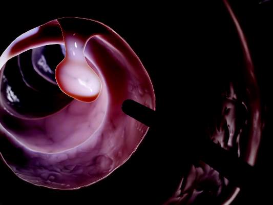
Among the 1,212 cases of DILI in the analysis, one-third were linked to antimicrobial therapies, including 41 (3.3%) in which cephalosporins were implicated, the investigators reported. Nineteen of the cases were tied to a single dose of intravenous cefazolin given before surgery. These patients developed cholestatic or mixed hepatocellular-cholestatic injury 1-3 weeks after the cefazolin infusion. They almost always had jaundice and pruritus, and usually also had fever and nausea. Signs and symptoms were self-limiting, resolving within a few days to weeks.
“Because of confusion about the specific diagnosis, patients underwent substantial diagnostic testing (including multiple computed tomography scans, magnetic resonance imaging scans, endoscopic retrograde cholangiopancreatography exams, and liver biopsies),” which in some cases led to severe complications, the investigators said.
The study also identified barriers to identifying cefazolin as a cause of DILI. Patients often did not know they had received the antibiotic and clinicians often did not know that cefazolin could cause DILI. In more than half of cases, DILI was linked to cefazolin only after careful medical record reviews. “For these reasons, we speculate that cefazolin is and has been underappreciated as a cause of DILI,” the researchers noted. “The appearance of jaundice and pruritus 1-3 weeks after minor surgery should lead to a search of surgical records and medications that might have been given during surgery. These results also imply that the merits of routine use of cefazolin at the time of uncomplicated surgery should be reconsidered carefully.”
Two patients died after receiving cephalosporins other than cefazolin, and another patient developed severe liver injury, the researchers said. “However, in each of the fatal cases, patients had a complicated clinical course, with a severe hypersensitivity reaction on top of an underlying liver disease. Therefore, we urge caution in concluding that non-cefazolin cephalosporin-induced DILI may be severe or fatal,” they said.
The study was funded by the National Institute of Diabetes and Digestive and Kidney Diseases, the National Institutes of Health, the National Cancer Institute, and by six Clinical and Translational Science Award grants. The investigators reported no conflicts of interest.
The investigators from the Drug-Induced Liver Injury Network (DILIN) report a previously unrecognized phenomenon: Patients who receive a single dose of an IV cephalosporin prior to an operative procedure may develop jaundice and biochemical cholestasis 1-3 weeks later. Remarkably, every single patient in this case series also presented with pruritus, suggesting an allergic reaction. Nearly half of the patients reported a previous “drug allergy” (although it is uncertain whether this was disclosed before their procedures), and some were in fact penicillin allergic, so likely should not have ever received cefazolin.

|
Dr. Stuart C. Gordon |
Some of the late-onset cases described in this series could have justified a misdiagnosis of “postoperative cholestasis” or have led to a fishing expedition for various “zebra” diagnoses. What is instructive in this report is that, for the most part, neither the patients nor the doctors evaluating their unexplained hepatitis ever suspected that an antibiotic had even been given. This observation highlights the fact that often medications that are used just once in the surgical suite will then disappear from a patient’s medications list and are often difficult to subsequently identify in the electronic medical record.Cefazolin is the workhorse for preoperative prophylaxis in cardiac and orthopedic surgery, and most operations involving skin, such as plastic surgery. Such antibiotic prophylaxis is generally used very appropriately and according to evidence-based clinical guidelines and it is a closely monitored and audited quality indicator at hospitals and surgical centers. The use of intravenous cefazolin as preoperative prophylaxis will likely not be diminished by these reports, but this case series again emphasizes the need to avoid cephalosporins among patients who report previous beta-lactam allergies. Early recognition of this culprit in cases of unexplained cholestatic hepatitis, especially in patients who recently underwent operative procedures, may obviate hospitalization.
Dr. Stuart Gordon is professor of medicine at Wayne State University School of Medicine and director of the Division of Hepatology at Henry Ford Health Systems, Detroit. He has no relevant conflicts of interest.
The investigators from the Drug-Induced Liver Injury Network (DILIN) report a previously unrecognized phenomenon: Patients who receive a single dose of an IV cephalosporin prior to an operative procedure may develop jaundice and biochemical cholestasis 1-3 weeks later. Remarkably, every single patient in this case series also presented with pruritus, suggesting an allergic reaction. Nearly half of the patients reported a previous “drug allergy” (although it is uncertain whether this was disclosed before their procedures), and some were in fact penicillin allergic, so likely should not have ever received cefazolin.
|
|
Dr. Stuart C. Gordon |
Some of the late-onset cases described in this series could have justified a misdiagnosis of “postoperative cholestasis” or have led to a fishing expedition for various “zebra” diagnoses. What is instructive in this report is that, for the most part, neither the patients nor the doctors evaluating their unexplained hepatitis ever suspected that an antibiotic had even been given. This observation highlights the fact that often medications that are used just once in the surgical suite will then disappear from a patient’s medications list and are often difficult to subsequently identify in the electronic medical record.Cefazolin is the workhorse for preoperative prophylaxis in cardiac and orthopedic surgery, and most operations involving skin, such as plastic surgery. Such antibiotic prophylaxis is generally used very appropriately and according to evidence-based clinical guidelines and it is a closely monitored and audited quality indicator at hospitals and surgical centers. The use of intravenous cefazolin as preoperative prophylaxis will likely not be diminished by these reports, but this case series again emphasizes the need to avoid cephalosporins among patients who report previous beta-lactam allergies. Early recognition of this culprit in cases of unexplained cholestatic hepatitis, especially in patients who recently underwent operative procedures, may obviate hospitalization.
Dr. Stuart Gordon is professor of medicine at Wayne State University School of Medicine and director of the Division of Hepatology at Henry Ford Health Systems, Detroit. He has no relevant conflicts of interest.
A single intravenous infusion of cefazolin can cause drug-induced liver injury, and the antibiotic ranked sixth among pharmacologic causes of hepatic injury in an analysis of 1,212 patients. “Cephalosporins appear to be a relatively common cause of antibiotic-associated liver injury,” said Dr. Saleh Alqahtani at the University of Texas Southwestern in Dallas and his associates in a report in the July issue of Clinical Gastroenterology and Hepatology (doi: 10.1016/j.cgh.2015.01.010).
“The latency period is typically 1-3 weeks after exposure, and patients may not become symptomatic until after the antibiotic is stopped – this is particularly true in the unique clinical syndrome in which a single infusion of cefazolin leads to drug-induced liver injury [DILI].”
Cephalosporins have been reported as rare causes of DILI, but most data come from single case reports, the researchers said. To study causes of DILI, they analyzed cases from the Drug-Induced Livery Injury Network, an ongoing prospective study at eight U.S. medical centers. Enrolled patients had strong clinical suspicion for liver injury caused by a drug or an herbal agent. Liver injury was defined based on specific criteria for liver enzymes, alkaline phosphatase, or total bilirubin levels, or as an international normalized ratio greater than 1.5 that was accompanied by elevated liver enzymes. Patients were followed for at least 6 months after their baseline visit.
Among the 1,212 cases of DILI in the analysis, one-third were linked to antimicrobial therapies, including 41 (3.3%) in which cephalosporins were implicated, the investigators reported. Nineteen of the cases were tied to a single dose of intravenous cefazolin given before surgery. These patients developed cholestatic or mixed hepatocellular-cholestatic injury 1-3 weeks after the cefazolin infusion. They almost always had jaundice and pruritus, and usually also had fever and nausea. Signs and symptoms were self-limiting, resolving within a few days to weeks.
“Because of confusion about the specific diagnosis, patients underwent substantial diagnostic testing (including multiple computed tomography scans, magnetic resonance imaging scans, endoscopic retrograde cholangiopancreatography exams, and liver biopsies),” which in some cases led to severe complications, the investigators said.
The study also identified barriers to identifying cefazolin as a cause of DILI. Patients often did not know they had received the antibiotic and clinicians often did not know that cefazolin could cause DILI. In more than half of cases, DILI was linked to cefazolin only after careful medical record reviews. “For these reasons, we speculate that cefazolin is and has been underappreciated as a cause of DILI,” the researchers noted. “The appearance of jaundice and pruritus 1-3 weeks after minor surgery should lead to a search of surgical records and medications that might have been given during surgery. These results also imply that the merits of routine use of cefazolin at the time of uncomplicated surgery should be reconsidered carefully.”
Two patients died after receiving cephalosporins other than cefazolin, and another patient developed severe liver injury, the researchers said. “However, in each of the fatal cases, patients had a complicated clinical course, with a severe hypersensitivity reaction on top of an underlying liver disease. Therefore, we urge caution in concluding that non-cefazolin cephalosporin-induced DILI may be severe or fatal,” they said.
The study was funded by the National Institute of Diabetes and Digestive and Kidney Diseases, the National Institutes of Health, the National Cancer Institute, and by six Clinical and Translational Science Award grants. The investigators reported no conflicts of interest.

Rapid antibody assay found sensitive for celiac disease
A point-of-care test for deamidated gliadin peptide antibodies was as sensitive as standard serology in detecting celiac disease, according to a single-center prospective study.
The results suggest that the new test could be used as a rapid serologic assay in outpatient care, which could save costs by reducing office visits and negating the need for duodenal biopsy in some patients, said Dr. Peter Mooney of Royal Hallamshire Hospital in Sheffield, England, and his associates. “Endoscopy is performed in both primary and secondary care in the United States, and the use of this point-of-care test before all procedures has the potential to reduce the numbers of missed cases at endoscopy significantly,” the investigators said.
Celiac disease affects about 0.2%-1.2% of the global population, and about three to four cases go undetected for every case that is diagnosed. Part of the problem is that endoscopists often do not recognize signs of celiac disease and therefore miss the chance to perform duodenal biopsy, the investigators said. A previous algorithm for when to perform duodenal biopsy detected 100% of cases of celiac disease, but relied on serology, which often is not available in clinical practice, the researchers noted (Clin. Gastroenterol. Hepatol. 2015 Jan. 2 [doi: 10.1016/j.cgh.2015.01.010]).
Therefore, they prospectively compared three point-of-care, finger prick–based assays for biomarkers of celiac disease to standard serology and duodenal biopsy. The study groups included 55 endoscopy patients who had tested positive for endomysial antibody and were considered at high risk for celiac disease, and a separate group of 508 patients who were undergoing endoscopy for any reason.
The rapid assay for deamidated gliadin peptide antibodies was the most sensitive of the point-of-case tests, detecting 63 of 68 previously undiagnosed cases of celiac disease (92.7%; 95% confidence interval, 83%-97.3%), the researchers reported. This sensitivity was very similar to that for standard serology for anti-tissue transglutaminase antibodies (91.2, 95% confidence interval, 81.1%-96.4%), they said. Among the five patients with false-negative results, one was positive for antibodies to both tissue transglutaminase and endomysial antibodies, one was positive only for endomysial antibodies, and three had seronegative celiac disease that was confirmed by a compatible human leukocyte antigen genotype. Notably, four of the five cases that the deamidated gliadin peptide antibody test failed to detect had symptoms such as weight loss, chronic diarrhea, and anemia, meaning that they would have been very likely to be detected by an algorithm that incorporated both rapid assay results and clinical findings, the investigators emphasized.
The deamidated gliadin peptide antibody assay costs about $28 per test and is not yet available in the United States, said the researchers. Using the rapid assay as a point-of-care diagnostic test for celiac disease would save about $9,500 per 1,000 endoscopies, but would miss about 7% of cases, they added. “The prevalence of celiac disease in our cohort was 13%, and represents referral bias to a tertiary celiac center,” they said. “In lower-prevalence populations, such as the 2% of 4% expected for all-comers to endoscopy, the positive predictive value inevitably will decrease and this will have an impact on cost savings.” But negative predictive value also would improve when testing lower-prevalence populations, and negative predictive value is crucial when screening in order to avoid missing true cases of the disease, they emphasized.
Tillotts Pharma makes the deamidated gliadin peptide antibody test and funded the study. One coauthor reported having received relevant research support from Tillotts Pharma, BHR Pharmaceuticals, and Coeliac UK. The other investigators reported having no conflicts of interest.
FROM CLINICAL GASTROENTEROLOGY AND HEPATOLOGY
Key clinical point: A point-of-care test for deamidated gliadin peptide antibodies could help direct duodenal biopsy for celiac disease.
Major finding: The assay detected 92.7% of cases of celiac disease, while the sensitivity of standard serology for anti-tissue transglutaminase was 91.2%.
Data source: Prospective, single-center study of 508 routine endoscopy cases and 55 additional patients who were considered high-risk for celiac disease.
Disclosures: Tillotts Pharma makes the deamidated gliadin peptide antibody test and funded the study. One coauthor reported having received relevant research support from Tillotts Pharma, BHR Pharmaceuticals, and Coeliac UK. The other investigators reported having no conflicts of interest.
Split-dosing found best for colon cleansing before colonoscopy
Split-dose regimens were more than twice as effective for colon cleansing as day-before preparations, and colonoscopy patients preferred them, a meta-analysis of 47 randomized trials showed.
Split-dose regimens might be superior for colon cleaning because of the shorter interval between the last preparation product taken and undergoing colonoscopy, said Myriam Martel at McGill University, Montreal, and her associates.
Colonoscopy is crucial to detect and prevent colon cancer, and repeating the procedure because of inadequate preparation of the colon is expensive. Colon-cleaning preparations include polyethylene glycol (PEG), sodium phosphate, picosulfate, and oral sulfate solutions, the reviewers noted.
To compare the efficacy of split-dose and other schedules and to determine the best products and volumes, they reviewed 47 randomized trials found by systematically searching MEDLINE, EMBASE, Scopus, CENTRAL, and the ISI Web of Knowledge database. The studies were published between 1980 and 2014 and did not include pediatric patients, hospitalized patients, or patients with inflammatory bowel disease, the reviewers said in the July issue of Gastroenterology (doi:10.1053/j.gastro.2015.04.004).
Split-dose preparations provided significantly better colon cleansing, compared with day-before preparations (odds ratio [OR], 2.51; 95% confidence interval [CI], 1.86-3.39), regardless of whether the day-before preparation was PEG (OR, 2.6; 95% CI, 1.46-4.63), sodium phosphate (OR, 9.34; 95% CI, 2.12-41.11), or picosulfate (OR, 3.54; 95% CI, 1.95-6.45), Ms. Martel and her associates reported.
In the intention-to-treat analysis, the highest volumes of PEG (3 L or more) led to greater bowel cleanliness than did lower split-dose volumes (OR, 1.89; 95% CI, 1.01-3.46). Patients were almost twice as likely to report being willing to repeat split-dose preparation, compared with day-before cleansing, and preferred low-volume preparations over high-volume ones, they said.
Secondary outcomes reported in the 47 trials included side effects, detection of polyps and adenomas, and resumption of daily activities, but definitions of these measures varied too much for the reviewers to reliably compare them. And since other studies are assessing the role of specific diets and adjuvants in colon preparation, they did not include those variables in their analyses. Future studies also should compare low and high-volume split-PEG dosing regimens, and regimens of PEG and those based on picosulfate and oral sulfate solutions, the investigators said.
Ms. Martel and her associates received no funding for the meta-analysis. One coauthor reported consulting relationships with Pendopharm, Boston Scientific, Olympus Canada, and Cook, and grant funding from Boston Scientific and Cook.
FROM GASTROENTEROLOGY
Key clinical point: Split-dose regimens were more effective for colon cleansing than day-before preparations, and colonoscopy patients preferred them.
Major finding: Split-dose preparations were more than twice as effective as day-before preparations (odds ratio, 2.51; 95% confidence interval, 1.86-3.39).
Data source: Meta-analysis of 47 randomized trials.
Disclosures: The investigators received no funding for the meta-analysis. One coauthor reported consulting relationships with Pendopharm, Boston Scientific, Olympus Canada, and Cook, and grant funding from Boston Scientific and Cook. The other investigators reported having no conflicts of interest.

Cefazolin ranks sixth as cause of drug-induced liver injury
A single intravenous infusion of cefazolin can cause drug-induced liver injury, and the antibiotic ranked sixth among pharmacologic causes of hepatic injury in an analysis of 1,212 patients.
“Cephalosporins appear to be a relatively common cause of antibiotic-associated liver injury,” said Dr. Saleh Alqahtani at the University of Texas Southwestern in Dallas and his associates. “The latency period is typically 1-3 weeks after exposure, and patients may not become symptomatic until after the antibiotic is stopped – this is particularly true in the unique clinical syndrome in which a single infusion of cefazolin leads to drug-induced liver injury.”
Cephalosporins have been reported as rare causes of drug-induced liver injury (DILI), but most data come from single case reports, the researchers said. To study causes of DILI, they analyzed cases from the Drug-Induced Livery Injury Network, an ongoing prospective study at eight U.S. medical centers. Enrolled patients had strong clinical suspicion for liver injury caused by a drug or an herbal agent. Liver injury was defined based on specific criteria for liver enzymes, alkaline phosphatase, or total bilirubin levels, or as an international normalized ratio greater than 1.5 that was accompanied by elevated liver enzymes or ALP. Patients were followed for at least 6 months after their baseline visit (Clin. Gastroenterol. Hepatol. 2014 Dec. 17 [doi: 10.1016/j.cgh.2015.01.010]).
Among the 1,212 cases of DILI in the analysis, one-third were linked to antimicrobial therapies, including 41 (3.3%) in which cephalosporins were implicated, the investigators reported. Nineteen of the cases were tied to a single dose of intravenous cefazolin given before surgery. These patients developed cholestatic or mixed hepatocellular-cholestatic injury 1-3 weeks after the cefazolin infusion. They almost always had jaundice and pruritus, and usually also had fever and nausea. Signs and symptoms were self-limiting, resolving within a few days to a few weeks.
“Because of confusion about the specific diagnosis, patients underwent substantial diagnostic testing (including multiple computed tomography scans, magnetic resonance imaging scans, endoscopic retrograde cholangiopancreatography exams, liver biopsies, and others), which often were unnecessary,” and in some cases led to severe complications, the investigators said.
The study also identified barriers to identifying cefazolin as a cause of DILI, they said. Patients often did not know they had received the antibiotic, and clinicians, including study investigators, often did not know that cefazolin could cause DILI. In more than half of cases, DILI was linked to cefazolin only after careful medical record reviews. “For these reasons, we speculate that cefazolin is and has been underappreciated as a cause of DILI,” the researchers noted. “The appearance of jaundice and pruritus 1-3 weeks after minor surgery should lead to a search of surgical records and medications that might have been given during surgery. These results also imply that the merits of routine use of cefazolin at the time of uncomplicated surgery should be reconsidered carefully.”
Two patients died after receiving cephalosporins other than cefazolin, and another patient developed severe liver injury, the researchers said. “However, in each of the fatal cases, patients had a complicated clinical course, with a severe hypersensitivity reaction on top of an underlying liver disease. Therefore, we urge caution in concluding that non-cefazolin cephalosporin-induced DILI may be severe or fatal,” they said. “Because cephalosporins are used commonly in clinical practice, it is likely that the overall mortality rate associated with cephalosporin use is low, but not nil, and it may be more likely in patients with underlying disorders.”
The study was funded by the National Institute of Diabetes and Digestive and Kidney Diseases, the National Institutes of Health, the National Cancer Institute, and by six Clinical and Translational Science Award grants. The investigators reported having no conflicts of interest.
FROM CLINICAL GASTROENTEROLOGY AND HEPATOLOGY
Key clinical point: A single dose of cefazolin can cause drug-induced liver injury (DILI), and the agent is implicated more often than previously thought.
Major finding: Cefazolin ranked sixth among causes of DILI, and signs and symptoms began 1-3 weeks after initial exposure.
Data source: Registry-based study of 1,212 cases of DILI.
Disclosures: The study was funded by the National Institute of Diabetes and Digestive and Kidney Diseases, the National Institutes of Health, the National Cancer Institute, and by Clinical and Translational Science Award grants. The investigators reported having no conflicts of interest.
Rapid antibody assay is sensitive for celiac disease
A point-of-care test for deamidated gliadin peptide antibodies was as sensitive as standard serology in detecting celiac disease, according to a single-center prospective study reported in the July issue of Clinical Gastroenterology and Hepatology.
The results suggest that the new test could be used as a rapid serologic assay in outpatient care, which could save costs by reducing office visits and negating the need for duodenal biopsy in some patients, said Dr. Peter Mooney of Royal Hallamshire Hospital in Sheffield, England, and his associates. “Endoscopy is performed in both primary and secondary care in the United States, and the use of this point-of-care test before all procedures has the potential to reduce the numbers of missed cases at endoscopy significantly,” the investigators said.
Celiac disease affects about 0.2%-1.2% of the global population, and about three to four cases go undetected for every case that is diagnosed. Part of the problem is that endoscopists often do not recognize signs of celiac disease and therefore miss the chance to perform duodenal biopsy, the investigators said. A previous algorithm for when to perform duodenal biopsy detected 100% of cases of celiac disease, but relied on serology, which often is not available in clinical practice, the researchers noted (Clin. Gastroenterol. Hepatol. 2015 Jan. 2 [doi: 10.1016/j.cgh.2015.01.010]).
Therefore, they prospectively compared three point-of-care, finger prick–based assays for biomarkers of celiac disease to standard serology and duodenal biopsy. The study groups included 55 endoscopy patients who had tested positive for endomysial antibody and were considered at high risk for celiac disease, and a separate group of 508 patients who were undergoing endoscopy for any reason.
The rapid assay for deamidated gliadin peptide antibodies was the most sensitive of the point-of-case tests, detecting 63 of 68 previously undiagnosed cases of celiac disease (92.7%; 95% confidence interval [CI], 83%-97.3%), the researchers reported. This sensitivity was very similar to that for standard serology for anti-tissue transglutaminase antibodies (91.2, 95% CI: 81.1%-96.4%), they said>. Among the five patients with false-negative results, one was positive for antibodies to both tissue transglutaminase and endomysial antibodies, one was positive only for endomysial antibodies, and three had seronegative celiac disease that was confirmed by a compatible human leukocyte antigen genotype. Notably, four of the five cases that the deamidated gliadin peptide antibody test failed to detect had symptoms such as weight loss, chronic diarrhea, and anemia, meaning that they would have been very likely to be detected by an algorithm that incorporated both rapid assay results and clinical findings, the investigators emphasized.
The deamidated gliadin peptide antibody assay costs about $28 per test and is not yet available in the United States, said the researchers. Using the rapid assay as a point-of-care diagnostic test for celiac disease would save about $9,500 per 1,000 endoscopies, but would miss about 7% of cases, they added. “The prevalence of celiac disease in our cohort was 13%, and represents referral bias to a tertiary celiac center,” they said. “In lower-prevalence populations, such as the 2% of 4% expected for all-comers to endoscopy, the positive predictive value inevitably will decrease and this will have an impact on cost savings.” But negative predictive value also would improve when testing lower-prevalence populations, and negative predictive value is crucial when screening in order to avoid missing true cases of the disease, they emphasized.
Tillotts Pharma makes the deamidated gliadin peptide antibody test and funded the study. One coauthor reported having received relevant research support from Tillotts Pharma, BHR Pharmaceuticals, and Coeliac UK. The other investigators reported having no conflicts of interest.
Split dose best for colon cleansing before colonoscopy Split-dose regimens were more than twice as effective for colon cleansing as day-before preparations, and colonoscopy patients preferred them, a meta-analysis of 47 randomized trials showed.
Split-dose regimens might be superior for colon cleaning because of the shorter interval between the last preparation product taken and undergoing colonoscopy, said Myriam Martel at McGill University, Montreal, and her associates in a report in the July issue of Gastroenterology (doi:10.1053/j.gastro.2015.04.004).
Repeating a colonoscopy because of inadequate preparation of the colon is expensive. Colon-cleansing preparations include polyethylene glycol (PEG), sodium phosphate, picosulfate, and oral sulfate solutions, the reviewers noted.

To compare the efficacy of split-dose and other schedules and to determine the best products and volumes, they reviewed 47 randomized trials found by systematically searching MEDLINE, EMBASE, Scopus, CENTRAL, and the ISI Web of Knowledge database. The studies were published between 1980 and 2014 and did not include pediatric, hospitalized, or inflammatory bowel disease patients, the reviewers said.
Split-dose preparations provided significantly better colon cleansing than did day-before preparations (odds ratio [OR], 2.51; 95% confidence interval [CI], 1.86-3.39), regardless of whether the day-before preparation was PEG (OR, 2.6; 95% CI, 1.46-4.63), sodium phosphate (OR, 9.34; 95% CI, 2.12-41.11), or picosulfate (OR, 3.54; 95% CI, 1.95-6.45), Ms. Martel and her associates reported.
In the intention-to-treat analysis, the highest volumes of PEG (3 L or more) led to greater bowel cleanliness than did lower split-dose volumes (OR, 1.89; 95% CI, 1.01-3.46). Patients were almost twice as likely to report being willing to repeat split-dose preparations than day-before cleansing, and preferred low-volume preparations over high-volume ones, they said.
Secondary outcomes reported in the 47 trials included side effects, detection of polyps and adenomas, and resumption of daily activities, but definitions of these measures varied too much for the reviewers to reliably compare them. And since other studies are assessing the role of specific diets and adjuvants in colon preparation, they did not include those variables in their analyses.
Ms. Martel and her associates received no funding for the study. One coauthor reported consulting relationships with Pendopharm, Boston Scientific, Olympus Canada, and Cook, and grant funding from Boston Scientific and Cook.
Split-dose regimens might be superior for colon cleaning because of the shorter interval between the last preparation product taken and undergoing colonoscopy, said Myriam Martel at McGill University, Montreal, and her associates in a report in the July issue of Gastroenterology (doi:10.1053/j.gastro.2015.04.004).
Repeating a colonoscopy because of inadequate preparation of the colon is expensive. Colon-cleansing preparations include polyethylene glycol (PEG), sodium phosphate, picosulfate, and oral sulfate solutions, the reviewers noted.
To compare the efficacy of split-dose and other schedules and to determine the best products and volumes, they reviewed 47 randomized trials found by systematically searching MEDLINE, EMBASE, Scopus, CENTRAL, and the ISI Web of Knowledge database. The studies were published between 1980 and 2014 and did not include pediatric, hospitalized, or inflammatory bowel disease patients, the reviewers said.
Split-dose preparations provided significantly better colon cleansing than did day-before preparations (odds ratio [OR], 2.51; 95% confidence interval [CI], 1.86-3.39), regardless of whether the day-before preparation was PEG (OR, 2.6; 95% CI, 1.46-4.63), sodium phosphate (OR, 9.34; 95% CI, 2.12-41.11), or picosulfate (OR, 3.54; 95% CI, 1.95-6.45), Ms. Martel and her associates reported.
In the intention-to-treat analysis, the highest volumes of PEG (3 L or more) led to greater bowel cleanliness than did lower split-dose volumes (OR, 1.89; 95% CI, 1.01-3.46). Patients were almost twice as likely to report being willing to repeat split-dose preparations than day-before cleansing, and preferred low-volume preparations over high-volume ones, they said.
Secondary outcomes reported in the 47 trials included side effects, detection of polyps and adenomas, and resumption of daily activities, but definitions of these measures varied too much for the reviewers to reliably compare them. And since other studies are assessing the role of specific diets and adjuvants in colon preparation, they did not include those variables in their analyses.
Ms. Martel and her associates received no funding for the study. One coauthor reported consulting relationships with Pendopharm, Boston Scientific, Olympus Canada, and Cook, and grant funding from Boston Scientific and Cook.

WCD: IL-23p19 inhibitors yield ‘promising’ psoriasis results
VANCOUVER, B.C. – So far, monoclonal antibodies that specifically target IL-23p19 are showing promise for treating chronic plaque psoriasis and are usually well tolerated, Dr. Elisabeth Riedl said at the World Congress of Dermatology.
The agents target the interleukin (IL)-23 cytokine, which is important in the induction of T-helper-17 cells in psoriasis and several other inflammatory disorders, said Dr. Riedl of the department of dermatology at Mount Sinai Hospital in New York. IL-23 has two subunits – p40, which it shares with IL-12, and p19, which is specific for IL-23, she said. Based on earlier research, the effectiveness of psoriasis treatments that target the p40 subunit seems to depend mostly on their ability to neutralize IL-23, not IL-12, she noted.
That observation helped spur several psoriasis trials of monoclonal antibodies that target IL-23p19, according to Dr. Riedl. Among the most promising of the monoclonal antibodies is BI 655066, which significantly outperformed placebo in a randomized, double-blind phase I trial reported earlier in 2015. At week 12, 87% of patients who received intravenous or oral BI65506 achieved a PASI (Psoriasis Area and Severity Index) 75 score, while 58% achieved a PASI 90 score and 16% achieved PASI 100, compared with none of the placebo group.
Another IL-23p19 inhibitor, guselkumab (CNTO 1959), beat placebo in a small randomized, double-blind phase I trial, Dr. Riedl noted. All patients who received a single subcutaneous injection of 300 mg of guselkumab achieved a PASI 75 score, and about 80% achieved PASI 90, compared with none of the placebo group, she noted.
The third IL-23p19 inhibitor, tildrakizumab (MK-3222), also beat placebo at all doses tested in a three-part, randomized, placebo-controlled phase I trial, Dr. Riedl said. Furthermore, the histopathology of a subgroup of patients revealed significant decreases in lesions, compared with baseline, and a significant correlation between histopathologic and clinical scores, she noted. “In phase II, there was a significant response for all dosing cohorts, with a durable response to 52 weeks,” she added.
The primary safety concern for IL-23 inhibitors appears to be increased infection risk, although patients with genetic diseases connected to the IL-23/Th17 pathway already are at increased risk for infections such as candidiasis, mycobacteriosis, salmonellosis, and staphylococcal and Klebsiella infections, Dr. Riedl noted. “Ustekinumab targets IL-12/IL-23 and so far has shown a favorable safety profile,” she said, pointing to a 5-year follow-up study of the antibody that found no increase over time in adverse events and similar rates of adverse events among dosing groups.
Infections and headaches were the most commonly reported adverse events in phase I and II trials of targeted anti–IL-23p19 antibodies, Dr. Riedl said. In the phase I trial of BI 655066, rates of adverse events were similar for treatment and placebo except for nasopharyngitis, which affected 13% of the BI 655066 group and none of the placebo group, she noted. Guselkumab also was associated with similar rates of overall adverse events, compared with placebo, in its phase I trial, although 6 of 20 patients who received guselkumab developed infections, including two upper respiratory tract infections and one event each of bronchitis, folliculitis, viral gastroenteritis, herpes simplex infection, lower respiratory tract infection, vaginal infection, and nasopharyngitis, she said. Tildrakizumab was generally well tolerated in its phase IIb trial, she added. Serious adverse events that might have been related to treatment were rare but included bacterial arthritis, lymphedema, melanoma, stroke, epiglottitis, and knee infection.
Taken together, the data for IL-23p19 inhibitors are encouraging, according to Dr. Riedl. “The results suggest that selective inhibition of IL-23p19 is a promising target for novel therapies,” she said.
Dr. Riedl reported receiving grant support or serving as a consultant, advisory board member, or speakers bureau member for Abbvie, Celgen, Merck, Pfizer, and Janssen.
EXPERT ANALYSIS FROM WCD 2015
WCD: Bad scar? Remember the four Ts
VANCOUVER, B.C. – A simple mnemonic – Talk, Time, Training, and Tools – can help dermatologists optimize outcomes when facing “bad” scars after skin surgery, Dr. Evans Bailey said at the World Congress of Dermatology.
Cosmetically challenging wounds can occur because the patient is on anticoagulants or for other reasons that the clinician cannot control, noted Dr. Bailey, a dermatologist with the Surgical Dermatology Group in Birmingham, Ala. Managing these situations begins during the informed consent process, when dermatologists should educate patients about the procedure and set reasonable expectations for cosmetic outcomes, he said.
“When it comes to scars, the battle is won or lost before you do surgery,” he said. “If you tell patients beforehand, it’s an explanation. If you tell them afterward, it’s an excuse.”
When obtaining informed consent, Dr. Bailey uses a digital tablet to take presurgical photos and draw options for surgical repair, he said. “I let them hold the tablet and show them the possibilities, because sometimes the smallest spots turn out to be the biggest tumors.”
When a patient returns after surgery with a “bad” scar, “never show surprise!” Dr. Bailey emphasized. “Don’t give up. Don’t show surprise, but be compassionate and pull hard for your patient.” Continuing to talk with patients at this point is key, he said. He shows patients the preoperative photographs and explains why he chose the surgical approach. Then he verbally commits to sticking with the patient during the entire process of wound healing and repair. “Say, ‘don’t worry; this is all under control; this will take longer than we thought. But I will make sure everything heals well.’ ”
Besides talking extensively with patients, dermatologists it is important to allow time for the scar to fully mature and involute before they intervene, said Dr. Bailey. “Allow scars to repair themselves,” he said. “Do not jump in too quickly. The body is really quite dynamic and capable – often more capable than we are.”
It’s equally important to stick to the fundamental principles of skin surgery, no matter how formidable the wound, Dr. Bailey noted. “Remember your training,” he said. “Protect the free margin and redefine the cosmetic subunits.” Tacking or suspension sutures can be used to redefine the cosmetic subunit and eliminate or minimize tension across the tissue flap, leading to better cosmetic outcomes.
For scars that still need improvement, tools such as wire brush dermabrasion can be used for raised scars, and TCA CROSS (trichloroacetic acid chemical reconstruction of skin scars) for depressed scars, Dr. Bailey noted. Both techniques can dramatically improve the final appearance of the scar, he noted.
“But the enemy of good is better,” he said. “So at some point I say, ‘you’re done.’ ”
Dr. Bailey reported having no relevant conflicts of interest.
EXPERT ANALYSIS AT WCD 2015
WCD: Expert reviews laser options for benign pigmented lesions
VANCOUVER, B.C. – Laser treatment can markedly improve the appearance of lentigines and nevi of Ota, although response is variable for most other benign pigmented lesions of the epidermis and dermis, Dr. Jason Rivers said at the World Congress of Dermatology.
“The best approach is to choose the device based on the location of pigment within the skin,” said Dr. Rivers, a dermatologist with Pacific Dermaesthetics in Vancouver. “Melanin has a fairly wide absorption spectrum, from the 400s down into the infrared spectrum. As we go further down into the infrared spectrum, we need to use higher fluencies, because we need to penetrate deeper into the skin.”
Q-switched 532-nm Nd:YAG laser is a good choice for facial lentigines, according to Dr. Rivers. “You can see marked improvement after one treatment, but you can be quite effective with various modalities,” he said. Intense pulsed light (IPL) therapy also can be effective for lentigines, including for patients with darker skin, although there is a risk of postinflammatory hyperpigmentation, he cautioned. Another option is 1,927 nm fractionated thulium fiber laser, he said. “Mopping up a lot of things at one time can be extremely effective, especially for lentigines on the face.”
For patients seeking removal of freckles, intense pulse light can clear a wide area of dyschromia in one session, Dr. Rivers continued. For mucosal melanosis, Q-switched lasers such as 532-nm frequency-doubled Nd:YAG, 694-nm ruby, or 755-nm alexandrite can be effective and are associated with minimal or no scarring, he added.
Café au lait patches typically respond well to combined treatment with 532-nm and 1,064-nm lasers, according to Dr. Rivers. A total of 10 treatment sessions spaced over 2-3 years has given him a durable response and without skin changes as a result of laser treatment, he said. However, some data indicate that café au lait patches might respond even better to Q-switched 755-nm alexandrite laser instead of conventional Q-switched lasers, he noted.
Becker’s nevus presents a much greater challenge. “Studies have looked at many kinds of lasers, and in all cases results may be suboptimal,” Dr. Rivers said. “For one of my patients with Becker’s nevus, I’ve basically thrown the kitchen sink at her, and we’re pretty much back to square one.” Q-switched 755-nm alexandrite laser can provide substantial clearing after 3 treatments in 4 weeks, but results might not be durable, he added. Laser therapy of nevus spilus also is “hit or miss,” although some studies suggest that Q-switched ruby laser is more effective than alexandrite for these lesions, he added.
Among the benign pigmented lesions of the dermis, nevus of Ota usually responds best to laser treatment, particularly with Q-switched 694-nm ruby or Q-switched 1,064-nm Nd:YAG, Dr. Rivers said. Data indicate that children younger than 10 years might have more superficially placed pigment cells that can be treated with lower fluence, such as 2.2 instead of 2.8 J, he noted.
Both congenital and acquired melanocytic nevi respond variably to laser therapy, Dr. Rivers said. For acquired lesions, options include Q-switched 532-nm, fully ablative carbon dioxide resurfacing, and continuous wave laser, he said. Long-pulsed alexandrite laser therapy can partiallyly clear congenital lesions after just one treatment session, and patients can then be given the option of fully ablative carbon dioxide resurfacing, he said.
Dermatologic laser therapy can, of course, cause adverse effects. The best treatment for laser-induced chrysiasis is prevention, but lesions can clear with Q-switched 1,064-nm laser treatment, Dr. Rivers said. Pigmentation might occur at the lesion margin, but fades with time, he noted.
Melanoma also has been reported as a consequence of dermatologic laser therapy. While sublethal laser damage can increase DNA damage in vitro, Q-switched ruby laser does not seem to induce genetic changes, Dr. Rivers said. In addition, some lesions were probably melanoma to begin with, he said, noting that failure to diagnose malignant melanoma is among the most common allegations in medical liability claims against dermatologists.
Dr. Rivers disclosed no relevant conflicts of interest.
AT WCD 2015
WCD: Methotrexate found safer but less effective than cyclosporine in atopic dermatitis
VANCOUVER, B.C. – In the first head-to-head trial in atopic dermatitis, cyclosporine was about five times more effective in terms of SCORAD 50 but was associated with more adverse events, Dr. Catherine Goujon-Henry reported at the World Congress of Dermatology.
Based on the findings, dermatologists should consider treating AD patients with a relatively high dose of methotrexate, such as 15 mg weekly, or first stabilize flaring patients with cyclosporine and then switch them to methotrexate for long-term disease control, said Dr. Goujon-Henry, who is with the Clinical Research Unit in Immunology at Lyon-Sud Hospital in Lyon, France.
Methotrexate is recommended in atopic dermatitis but has never been directly compared with cyclosporine, Dr. Goujon-Henry noted. For the study, she and her colleagues randomized 97 adults with moderate to severe AD to 8 weeks of treatment with either methotrexate at 15 mg per week or cyclosporine at 2.5 mg per kg per day. Patients only were allowed to use topical glucocorticoids or tacrolimus during the first month of the trial.
Only 8% of the methotrexate group achieved SCORAD 50 by week 8, compared with 42% of cyclosporine patients, Dr. Goujon-Henry reported. Furthermore, only 35% of the methotrexate group achieved EASI 50 compared with 68% of the cyclosporine group, she said. In addition, Dermatology Life Quality Index (DLQI) scores were 5 or less for only 37% of methotrexate patients, compared with 56% of cyclosporine patients, she reported.
The protocol called for increasing the methotrexate dose to 25 mg per week and the cyclosporine dose to 5 mg per kg per day for patients who did not achieve SCORAD 50 by week 8. Dose increases were performed for 74% of the methotrexate group and for 58% of the cyclosporine group, Dr. Goujon-Henry said. At week 12, 15 of the 50 methotrexate patients were considered non-responders and went off study, as did three cyclosporine patients. At this point, 43% of the methotrexate patients who remained on study had achieved SCORAD 50, as had 62% of cyclosporine patients.
Also at week 16, two-thirds of methotrexate patients and 79% of cyclosporine patients had achieved EASI 50, and 61% of methotrexate patients and 77% of cyclosporine patients scored 5 or less on the DLQI, Dr. Goujon-Henry reported. However, about 53% of cyclosporine patients experienced drug-related adverse events, compared with 40% of the methotrexate group, she said. Serious adverse events that led to treatment discontinuation included arterial hypertension and hypertrichosis for the cyclosporine group, and lymphopenia and hepatitis for the methotrexate group, she reported.
Taken together, the findings support using methotrexate as first-line therapy for moderate to severe atopic dermatitis, but patients need to expect a slower treatment response in exchange for the better safety profile, Dr. Goujon-Henry said.
She disclosed no relevant conflicts of interest.
AT WDC 2015
Key clinical point: Methotrexate has a better safety profile but is less effective than cyclosporine in clearing moderate to severe atopic dermatitis.
Major finding: Only 8% of the methotrexate group achieved SCORAD 50 at week 8, compared with 42% of those in the cyclosporine group.
Data source: Randomized, controlled, single-blind, non-inferiority trial of methotrexate and cyclosporine in adults with atopic dermatitis.
Disclosures: Dr. Goujon-Henry disclosed no relevant conflicts of interest.