User login
Official Newspaper of the American College of Surgeons
CMS extends 2014 Medicare meaningful use attestation deadline
Physicians and other eligible professionals will now have until March 20 to attest to meaningful use of electronic health records for the 2014 reporting year and avoid a Medicare penalty for 2016.

The Centers for Medicare & Medicaid Services announced it was extending the deadline from Feb. 28. Eligible providers must attest to meaningful use every year to receive bonus payments under the EHR Incentive Program. Those who fail to attest by March 20 for the 2014 reporting year will see a 2% reduction in Medicare payments in 2016.
During this extension period, providers can make a one-time switch between the Medicare and Medicaid EHR Incentive Programs.
Physicians and other eligible professionals will now have until March 20 to attest to meaningful use of electronic health records for the 2014 reporting year and avoid a Medicare penalty for 2016.
The Centers for Medicare & Medicaid Services announced it was extending the deadline from Feb. 28. Eligible providers must attest to meaningful use every year to receive bonus payments under the EHR Incentive Program. Those who fail to attest by March 20 for the 2014 reporting year will see a 2% reduction in Medicare payments in 2016.
During this extension period, providers can make a one-time switch between the Medicare and Medicaid EHR Incentive Programs.

VIDEO: Challenges abound in rolling out stroke embolectomy
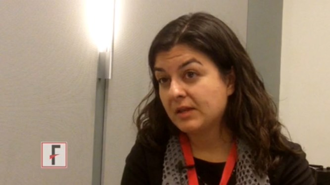
NASHVILLE, TENN. – U.S. stroke specialists now face the challenge of making endovascular embolectomy a routinely available option for selected patients with acute ischemic stroke, Dr. Pooja Khatri said in an interview at the International Stroke Conference.
During the conference, which was sponsored by the American Heart Association, new reports from three independent, randomized controlled trials, as well as data from a fourth study published in January, collectively established endovascular embolectomy as the new standard-of-care treatment for acute ischemic stroke patients with a proximal occlusion of a large, intracerebral artery. The stroke community, however, now faces the responsibility of figuring out how to make this a reality.
Among the hurdles they face are using CT imaging or other methods to identify in daily practice the specific patients who will get the biggest benefit from endovascular treatment and finding a consensus within each region on how to triage acute stroke patients to centers that can perform embolectomy, said Dr. Khatri, professor of neurology and director of acute stroke at the University of Cincinnati. In Cincinnati, Dr. Khatri and her colleagues are planning to soon hold a retreat with representatives from other area hospitals to try to work out the logistics.Dr. Khatri has received research support from Penumbra and Genentech.
The video associated with this article is no longer available on this site. Please view all of our videos on the MDedge YouTube channel
[email protected]
On Twitter @mitchelzoler
EXPERT ANALYSIS FROM THE INTERNATIONAL STROKE CONFERENCE
Study: Osteoarthritis develops sooner than thought after ACL injury, repair
People who have had a knee reconstruction following trauma may be susceptible to osteoarthritis sooner than currently thought, according to new MRI findings at 1 year after anterior cruciate ligament reconstruction.
Almost a third of people studied had some evidence of early osteoarthritis (OA) at that early time point, challenging “existing dogma that degenerative joint disease does not become apparent for years post-ACLR,” reported Dr. Kay Crossley of the University of Queensland in Brisbane, Australia (Arthritis Rheumatol. 2015 Feb. 18 [doi:10.1002/art.39005]).
However, as they did not have access to preoperative images, they could not rule out that some OA features may have been preexisting and not related to knee trauma, they said.
“This is a sample that was taken after the injury and after the reconstruction, so they truly don’t know that what they’re finding is as a result of even the injury, surgery, or the meniscal damage or meniscal resection they had done at the time,” Dr. David J. Hunter, a leading OA expert from Sydney (Australia) University, said when asked to comment on the study’s findings.
“It may well be that these were people that had some underlying structural damage,” he added.
The researchers noted that radiographic knee OA was thought to be as high as 50%-90% a decade after anterior cruciate ligament reconstruction (ACLR). The issue is particularly important because ACL injuries typically occur in younger adults who are then prone to developing knee OA before they reach 40 years, they said.
“Early detection of knee OA after ACLR may permit early intervention such as load management, which is likely to be more effective prior to the development of advanced disease,” they wrote.
Their study included 111 patients aged 18-50 years who had undergone single-bundle hamstring-tendon autograft ACLR 1 year earlier.
MRI scans of their knees were compared with 20 uninjured asymptomatic matched controls. The researchers used the MRI Osteoarthritis Knee Score (MOAKS) to score specific OA features because the more recent Anterior Cruciate Ligament Osteoarthritis Score (ACLOAS) had not been published at the time of their study.
Results showed that 34 (31%) patients had MRI-defined knee OA following an ACLR a year earlier.
MRI-OA features were most frequently found in the patellofemoral compartment, particularly the medial femoral trochlea, a potentially underrecognized site of knee pathology following reconstruction, the researchers said.
Pathology in the patellofemoral joint included not only “early” features of OA, such as bone marrow lesions and partial-thickness cartilage loss, but also frank osteophytes on MRI, they noted.
None of the uninjured control knees had MRI-defined patellofemoral or tibiofemoral OA.
The authors acknowledged that a lack of access to preoperative knee images limited the conclusions they could reach in their study, but they noted that MRI-OA features were rarely seen in the small sample of uninjured matched control knees.
“Combined with the observation that six times as many reconstructed knees had radiographic osteophytes than uninjured contralateral knees, these findings suggest that knee trauma and/or reconstruction was strongly implicated in the development of OA features,” they wrote.
Another limitation that the authors acknowledged was that the MRI definition of OA was relatively new and was likely to be refined as the understanding of OA pathology evolved.
Dr. Hunter, who was the lead investigator involved in developing the MOAKS, agreed that the definition needed more validity and testing.
“This is the third study that uses that definition, and I do think that long-term clinical implications of what MRI definition means is unknown,” he said. “The challenge that we have is that we do kick up a lot of abnormalities, and we don’t truly know what the long-term clinical implications of those abnormalities are at this point.”
“There are a lot of problems with the way this study has been done, but I do think it is really helpful that it highlights how important injury is with regards to predisposing to early OA.”
“It’s something that a lot of people don’t really highlight or pay attention to,” he said.
The study was partly funded by the Queensland Orthopaedic Physiotherapy Network, a University of Melbourne Research Collaboration grant, and University of British Columbia’s Centre for Hip Health and Mobility via the Society for Mobility and Health. One study author is president of Boston Imaging Core Lab, LLC, and is a consultant to Merck Serono, Sanofi-Aventis, Genzyme and TissueGene. No other authors declared conflicts of interest.
FROM ARTHRITIS & RHEUMATOLOGY
Key clinical point: People who have undergone anterior cruciate ligament reconstruction following trauma may be susceptible to early OA sooner than previously thought, but the study authors did not have access to baseline images to rule out existing pathology.
Major finding: A third of the 111 patients studied had evidence of MRI-defined OA a year after their surgery.
Data source: MRI study of 111 patients who had undergone ACL surgery matched with 20 uninjured asymptomatic controls.
Disclosures: The study was partly funded by the Queensland Orthopaedic Physiotherapy Network, a University of Melbourne Research Collaboration grant, and University of British Columbia’s Centre for Hip Health and Mobility via the Society for Mobility and Health. One study author is president of Boston Imaging Core Lab, LLC, and is a consultant to Merck Serono, Sanofi-Aventis, Genzyme and TissueGene. No other authors declared conflicts of interest.

Report: Newly insured patients won’t strain health system
Even though millions of Americans have gained health insurance through the Affordable Care Act over the last year, researchers at the Commonwealth Fund say the health care system isn’t likely to be overwhelmed caring for them.
In an analysis released on Feb. 25, researchers at the think tank estimated that on average there would be only about 1.34 additional visits to primary care offices per week, an increase of 3.8% nationally. Hospital outpatient departments will see between 1.2 and 11 additional visits weekly, on average, an uptick of about 2.6% nationally.

“Although analysts have expressed concern that greater access to care will strain the service delivery system, our projections suggest that increased use of health services by the newly insured will be relatively modest for most services,” the researchers wrote in the report.
The modest increase in the use of health care services means that the existing health care workforce will be able to meet the demands, according to the Commonwealth Fund. And structural changes – some of which are already underway – will help, including physician pooling and the greater use of nurses and physician assistants as part of a care-delivery team. Technological advances, such as telemedicine and the electronic exchange of health information, can also help the existing physician workforce meet the increased demand for services, according to the report.
“The U.S. health system is likely to be able to absorb these increases.” Sherry Glied, Ph. D., dean of the Robert Wagner Graduate School of Public Service at New York University and lead author of the report, wrote.
Researchers used the Medical Expenditure Panel Survey to estimate current utilization rates and then calculated additional use based on coverage gains under the Affordable Care Act.
Even though millions of Americans have gained health insurance through the Affordable Care Act over the last year, researchers at the Commonwealth Fund say the health care system isn’t likely to be overwhelmed caring for them.
In an analysis released on Feb. 25, researchers at the think tank estimated that on average there would be only about 1.34 additional visits to primary care offices per week, an increase of 3.8% nationally. Hospital outpatient departments will see between 1.2 and 11 additional visits weekly, on average, an uptick of about 2.6% nationally.
“Although analysts have expressed concern that greater access to care will strain the service delivery system, our projections suggest that increased use of health services by the newly insured will be relatively modest for most services,” the researchers wrote in the report.
The modest increase in the use of health care services means that the existing health care workforce will be able to meet the demands, according to the Commonwealth Fund. And structural changes – some of which are already underway – will help, including physician pooling and the greater use of nurses and physician assistants as part of a care-delivery team. Technological advances, such as telemedicine and the electronic exchange of health information, can also help the existing physician workforce meet the increased demand for services, according to the report.
“The U.S. health system is likely to be able to absorb these increases.” Sherry Glied, Ph. D., dean of the Robert Wagner Graduate School of Public Service at New York University and lead author of the report, wrote.
Researchers used the Medical Expenditure Panel Survey to estimate current utilization rates and then calculated additional use based on coverage gains under the Affordable Care Act.

Giant intracranial aneurysm treatment confers some long-term survival benefit
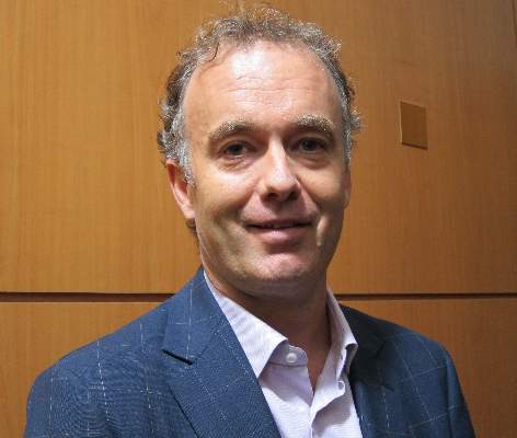
NASHVILLE, TENN. – The 10-year outcome for most giant intracranial aneurysms is rather dismal even if the lesions are initially successfully treated, according to findings from the International Study of Unruptured Intracranial Aneurysms.
Most patients lived through the first 2 years after diagnosis, but by 10 years, 37% of treated patients had died. Untreated patients had significantly higher mortality at 57%, Dr. James C. Torner said at the International Stroke Conference, which was sponsored by the American Heart Association.
“Compared to small aneurysms, the risk of all-cause mortality for these giant aneurysms is seven times greater in the first year, and the risk of hemorrhage or procedure-related death is 15 times higher. The risk does decrease over time, but it’s still elevated. It’s twice as high for death from hemorrhage and six times more likely from death due to rupture or a procedure by 10 years.”
Dr. Torner of the University of Iowa, Iowa City, presented 10-year outcomes from the International Study of Unruptured Intracranial Aneurysms (ISUIA), which followed 4,059 patients, of whom 187 had a giant unruptured intracranial aneurysm.
The mean lesion size was about 30 mm, but ranged from 25 to 63 mm. A third involved the internal carotid, and another third were cavernous. The remainder were either vestibular, posterior communicating, anterior cerebral, or middle cerebral.
Most were irregular; about 40% were a single sac. Daughter sacs were present in the rest. The volume ranged from 1,100 to 38,000 mm3, with about 10% having a volume of greater than 20,000 mm3.
Most of the patients were women (91%). Age was not related to occurrence; patients ranged from 25 to 82 years. There were some common risk factors, including smoking (in 70%), a family history of aneurysm (in 10%), a family history of coronary artery disease (in up to 45%, depending on patient age), hypertension (in up to 40%), and vascular headache. Patients aged 50 years or older were most likely to present with vascular headache (75%).
The baseline score on the modified Rankin Scale (mRS) was 1 in about 93% of the group. But 80% presented with some symptoms, including cranial nerve deficit (47%; commonly in cranial nerves III and IV), mass effect (16%), headache (44%), orbital pain (21%), and partial vision loss (25%).
Surgery was performed in 39% and endovascular treatment in 27%; the rest of the patients were initially untreated, although 3% later received endovascular treatment and 5% underwent surgical treatment during the follow-up period.
By 10 years, many of the lesions had ruptured. Posterior aneurysms were most likely to rupture (53%), while cavernous aneurysms were least likely to rupture (5%). About a third of the posterior communicating and 36% of the anterior lesions were unruptured.
The risk of rupture increased over the first 5 years, rising from almost 0% in year 1 to 20% by year 5. “Hemorrhage risk is huge over the first 5 years, even with the cavernous aneurysms – even they can rupture,” Dr. Torner said. “In those with smaller aneurysms, the risk of rupture may be high in the first 2 years, but then it subsides somewhat.”
Among the 59% of the overall group who survived, the mRS score remained excellent (1 or 2) by 10 years. Of the remainder who died, 23% died of a cranial or subarachnoid hemorrhage, and 11% of a cerebral infarct. Coronary disease, respiratory disease, and cancer were the other causes.
Treated patients generally fared better than untreated, although mortality varied significantly by lesion location. For anterior lesions, the death rate was 25% in untreated patients , 32% for surgical patients, and 19% in endovascular patients. Death rates for those with posterior communicating lesions were 90% for untreated and 50% for surgically treated patients; all of those with endovascular treatment died. For those with posterior lesions, death rates approached 100% for all groups.
Dr. Torner cautioned that because the study outcomes were collected during the 1990s, most treated patients underwent only coiling or clipping; better outcomes may be seen with more advanced therapies.
He said he had no relevant financial disclosures.
[email protected]
On Twitter @alz_gal
AT THE INTERNATIONAL STROKE CONFERENCE
Key clinical point: Giant intracranial aneurysms have a generally poor 10-year outcome, although treated patients fare better than untreated.
Major finding: By 10 years, the death rate was 37% among the treated patients and 57% among the untreated.
Data source: The retrospective analysis comprised 187 patients with aneurysms greater than 25 mm.
Disclosures: Dr. Torner said he had no relevant financial disclosures.
Medical societies again call for gun law changes
Calling gun violence and related deaths a public health issue, the leaders of seven professional medical societies have joined together with the American Bar Association to push for changes in the nation’s gun laws and policies.
“Because many of the efforts in the past have not been successful to counteract the very heavy lobbying from the gun lobby and the National Rifle Association, our hope is that this unique type of collaboration across health care professional organizations as well as the legal profession – we’re hoping that that type of alliance is something that is so powerful and addresses the concerns ... raised by the NRA and the gun lobby,” Dr. Steven E. Weinberger, chief executive officer of the American College of Physicians, said in an interview.

The policy recommendations were published online Feb. 23 in Annals of Internal Medicine.
The organizations include: American Academy of Family Physicians, American Academy of Pediatricians, American College of Emergency Physicians, American Congress of Obstetricians and Gynecologists, American College of Physicians, American College of Surgeons, and American Psychiatric Association. Also joining in on the recommendations are the American Public Health Association and the American Bar Association.
“Our organizations support a public health approach to firearm-related violence and prevention of firearm injuries and deaths,” Dr. Weinberger and colleagues wrote. “Similar approaches have produced major achievements in the reduction of tobacco use, motor vehicle deaths (seat belts), and unintentional poisoning and can serve as models going forward.”
• The policy recommendations include:
• Supporting criminal background checks for all firearm purchases, including sales by gun dealers, sales at gun shows, and private sales.
• Opposing state and federal mandates that interfere with physician free speech and patient-physician relationship, including physician “gag laws” that forbid physicians to discuss gun ownership and guns in the home.
• Opposing the sale or ownership of assault weapons and large-capacity magazines for private citizens.
• Advocating for research into the causes and consequences of firearm violence and unintentional injuries so that evidence-based policies may be developed.
• Supporting improved access to mental health care, with caution against broadly including all persons with any mental or substance abuse disorder in a category of persons prohibited from purchasing firearms.
• Opposing blanket reporting laws that require physicians to report patients with mental or substance use disorders, as these laws may stigmatize the patients and inhibit them from seeking treatment.
The authors note that these recommendations have been confirmed by the American Bar Association as being “constitutionally sound” and do not interfere with the Second Amendment.
“We’re hoping that ... this will create a groundswell of support from a number of large, prestigious, and influential organizations,” Dr. Weinberger said, noting that the group will be targeting “most of the medical societies for starters” as well as “some of the prominent patient and consumer organizations and obviously organizations that have been involved in firearm violence and advocating for more appropriate firearms control.”
Calling gun violence and related deaths a public health issue, the leaders of seven professional medical societies have joined together with the American Bar Association to push for changes in the nation’s gun laws and policies.
“Because many of the efforts in the past have not been successful to counteract the very heavy lobbying from the gun lobby and the National Rifle Association, our hope is that this unique type of collaboration across health care professional organizations as well as the legal profession – we’re hoping that that type of alliance is something that is so powerful and addresses the concerns ... raised by the NRA and the gun lobby,” Dr. Steven E. Weinberger, chief executive officer of the American College of Physicians, said in an interview.
The policy recommendations were published online Feb. 23 in Annals of Internal Medicine.
The organizations include: American Academy of Family Physicians, American Academy of Pediatricians, American College of Emergency Physicians, American Congress of Obstetricians and Gynecologists, American College of Physicians, American College of Surgeons, and American Psychiatric Association. Also joining in on the recommendations are the American Public Health Association and the American Bar Association.
“Our organizations support a public health approach to firearm-related violence and prevention of firearm injuries and deaths,” Dr. Weinberger and colleagues wrote. “Similar approaches have produced major achievements in the reduction of tobacco use, motor vehicle deaths (seat belts), and unintentional poisoning and can serve as models going forward.”
• The policy recommendations include:
• Supporting criminal background checks for all firearm purchases, including sales by gun dealers, sales at gun shows, and private sales.
• Opposing state and federal mandates that interfere with physician free speech and patient-physician relationship, including physician “gag laws” that forbid physicians to discuss gun ownership and guns in the home.
• Opposing the sale or ownership of assault weapons and large-capacity magazines for private citizens.
• Advocating for research into the causes and consequences of firearm violence and unintentional injuries so that evidence-based policies may be developed.
• Supporting improved access to mental health care, with caution against broadly including all persons with any mental or substance abuse disorder in a category of persons prohibited from purchasing firearms.
• Opposing blanket reporting laws that require physicians to report patients with mental or substance use disorders, as these laws may stigmatize the patients and inhibit them from seeking treatment.
The authors note that these recommendations have been confirmed by the American Bar Association as being “constitutionally sound” and do not interfere with the Second Amendment.
“We’re hoping that ... this will create a groundswell of support from a number of large, prestigious, and influential organizations,” Dr. Weinberger said, noting that the group will be targeting “most of the medical societies for starters” as well as “some of the prominent patient and consumer organizations and obviously organizations that have been involved in firearm violence and advocating for more appropriate firearms control.”
FROM ANNALS OF INTERNAL MEDICINE
FDA approves adhesive treatment for superficial varicose veins
The VenaSeal closure system, which uses an adhesive directly injected into the vein, has been approved as a permanent treatment for symptomatic, superficial varicose veins, the Food and Drug Administration announced on Feb. 20.
“This new system is the first to permanently treat varicose veins by sealing them with an adhesive,” Dr. William Maisel, acting director of the Office of Device Evaluation in the FDA’s Center for Devices and Radiological Health, said in the FDA’s statement. Because the system “does not incorporate heat application or cutting, the in-office procedure can allow patients to quickly return to their normal activities, with less bruising,” he added.
The VenaSeal system differs from other procedures used to treat varicose veins, which use drugs, lasers, radiofrequency, or incisions, the FDA statement points out. The complete sterile kit includes the adhesive (n-butyl-2-cyanoacrylate), which solidifies when injected directly into the target vein via a catheter, under ultrasound guidance. The additional system components include the catheter, the adhesive, a guidewire, dispenser gun, dispenser tips, and syringes.
Approval was based on data from three clinical trials sponsored by the manufacturer. In the U.S. study that compared results in 108 patients treated with the VenaSeal system and 114 patients treated with radiofrequency ablation therapy, the device was shown “to be safe and effective for vein closure for the treatment of symptomatic superficial varicose veins of the legs,” according to the FDA. In the study, adverse events associated with the VenaSeal treatment included phlebitis and paresthesias in the treated areas, which are “generally associated with treatments of this condition,” the FDA statement noted.
The agency reviewed the VenaSeal System as a class III medical device, considered the highest risk type of medical devices that are subjected to the highest level of regulatory control, and which must be approved before marketing.
VenaSeal is manufactured by Covidien, which acquired Sapheon, the company that developed VenaSeal, in 2014. The system has also been approved in Canada, Europe, and Hong Kong, according to a Covidien statement issued last year.
Baseline QOL measures not associated with outcomes in high-risk operable lung cancer patients
Poor baseline quality-of-life scores were not predictive of worse overall or recurrence-free survival, or of higher risk for adverse events following sublobar resection in high-risk surgical patients with lung cancer.
In addition, quality of life (QOL) and dyspnea scores did not deteriorate significantly overall, based upon the results of a prospective, multicenter study. Low dyspnea scores at baseline, however, did predict subsequent poor overall survival, according to Dr. Hiran C. Fernando of the Boston Medical Center and his colleagues.

The researchers assessed QOL using the 36-item Short-Form Health Survey (SF36) and the dyspnea score from the University of California, San Diego, Shortness of Breath Questionnaire (SOBQ). Both were measured at baseline, 3, 12, and 24 months. The SF36 scores were further broken down into the physical component summary (PCS) and the mental component summary (MCS), according to their report published online and in the March issue of the Journal of Thoracic and Cardiovascular Surgery (2014 Nov. 13 [doi:10.1016/j.jtcvs.2014.11.003]) .
A total of 212 eligible patients in the American College of Surgeons Oncology Group Z4032 trial were randomized to sublobar resection (108 patients) or sublobar resection with brachytherapy (104). The mean age was about 70.5 years, and 56% were women. There were no significant differences in baseline QOL scores between arms. Baseline PCS and MCS scores were at least 1 standard deviation below those of the U.S. general population in 65% and 46.5% of the patients, respectively.
Overall, there were no significant differences in grade 3+ adverse events, overall survival, or recurrence-free survival seen in patients with baseline scores greater than or equal to median QOL scores or less than median scores. There was, however, significantly worse overall survival for patients with baseline SOBQ scores less than or equal to median. In addition, a 10-point drop in SOBQ score at 12 months also predicted poor overall survival, according to Dr. Fernando and his associates.
In terms of results for operative procedures and tumor types, there was a significantly higher percentage of patients with a decline of 10 points or more in SOBQ scores with segmentectomy, compared with wedge resection (40.5% vs. 21.9%) at 12 months, with thoracotomy vs. video-assisted thoracic surgery (VATS) (38.8% vs. 20.4%, P = .03) at 12 months, and for T1b vs. T1a tumors (46.9% vs. 23.5%) at 24 months. In addition, there was a significantly greater than or equal to 10-point improvement in PCS scores at 3 months with VATS vs. thoracotomy (16.5% vs. 3.6%).
The researchers pointed out that, although QOL measurements can be useful to help decide the optimal surgery procedure, it has even more relevance when considering surgical versus nonsurgical therapies, such as using stereotactic body radiation therapy, for high-risk patients with early-stage lung cancer.
“Some advantages relating to minimizing postoperative dyspnea, as measured by the SOBQ, were gained by using VATS (rather than thoracotomy) or wedge resection (rather than segmentectomy). In addition, VATS, as opposed to thoracotomy, patients had improved PCS scores at 3 months, lending support to the preferential use of VATS when SR is performed,” the researchers concluded.
The study was supported by the National Cancer Institute. Dr. Fernando reported receiving consulting fees from Galil and CSA Medical.
This research is a noteworthy contribution because there source of prospectively acquired QOL data for such a high-risk group facing lung surgery, Dr. Michael T. Jaklitsch said in his invited editorial commentary (J. Thorac. Cardiovasc. Surg. 2015 Dec. 2 [doi:10.1016/j.jtcvs.2014.11.068]).

|
Dr. Michael T. Jaklitsch |
Although there was no evidence of predictive ability of QOL data for this population, “the predictive value of self-assessment may be more powerful in a broader population, however, than in a selected high-risk population such as the Alliance Z4032 trial,” he said. “The amount of pulmonary impairment required to enter this trial was likely the prime determinant of morbidity.” Thus QOL tools may be predictive with certain populations, but not in others. Overall, however, one benefit of self-assessment tools is that they allow patients to be seen more as people than as disease cases by the surgeons.
“Self-assessment tools allow our patients to tell us more completely about themselves and frequently become a springboard to discuss fears of the near future after surgery and what that might look like,” he added. “They allow us to ask our patients more completely, ‘How are you doing?’ ”
Dr. Jaklitsch is a thoracic surgeon at Brigham and Woman’s Hospital, Harvard Medical School, Boston.
This research is a noteworthy contribution because there source of prospectively acquired QOL data for such a high-risk group facing lung surgery, Dr. Michael T. Jaklitsch said in his invited editorial commentary (J. Thorac. Cardiovasc. Surg. 2015 Dec. 2 [doi:10.1016/j.jtcvs.2014.11.068]).
|
|
Dr. Michael T. Jaklitsch |
Although there was no evidence of predictive ability of QOL data for this population, “the predictive value of self-assessment may be more powerful in a broader population, however, than in a selected high-risk population such as the Alliance Z4032 trial,” he said. “The amount of pulmonary impairment required to enter this trial was likely the prime determinant of morbidity.” Thus QOL tools may be predictive with certain populations, but not in others. Overall, however, one benefit of self-assessment tools is that they allow patients to be seen more as people than as disease cases by the surgeons.
“Self-assessment tools allow our patients to tell us more completely about themselves and frequently become a springboard to discuss fears of the near future after surgery and what that might look like,” he added. “They allow us to ask our patients more completely, ‘How are you doing?’ ”
Dr. Jaklitsch is a thoracic surgeon at Brigham and Woman’s Hospital, Harvard Medical School, Boston.
Poor baseline quality-of-life scores were not predictive of worse overall or recurrence-free survival, or of higher risk for adverse events following sublobar resection in high-risk surgical patients with lung cancer.
In addition, quality of life (QOL) and dyspnea scores did not deteriorate significantly overall, based upon the results of a prospective, multicenter study. Low dyspnea scores at baseline, however, did predict subsequent poor overall survival, according to Dr. Hiran C. Fernando of the Boston Medical Center and his colleagues.
The researchers assessed QOL using the 36-item Short-Form Health Survey (SF36) and the dyspnea score from the University of California, San Diego, Shortness of Breath Questionnaire (SOBQ). Both were measured at baseline, 3, 12, and 24 months. The SF36 scores were further broken down into the physical component summary (PCS) and the mental component summary (MCS), according to their report published online and in the March issue of the Journal of Thoracic and Cardiovascular Surgery (2014 Nov. 13 [doi:10.1016/j.jtcvs.2014.11.003]) .
A total of 212 eligible patients in the American College of Surgeons Oncology Group Z4032 trial were randomized to sublobar resection (108 patients) or sublobar resection with brachytherapy (104). The mean age was about 70.5 years, and 56% were women. There were no significant differences in baseline QOL scores between arms. Baseline PCS and MCS scores were at least 1 standard deviation below those of the U.S. general population in 65% and 46.5% of the patients, respectively.
Overall, there were no significant differences in grade 3+ adverse events, overall survival, or recurrence-free survival seen in patients with baseline scores greater than or equal to median QOL scores or less than median scores. There was, however, significantly worse overall survival for patients with baseline SOBQ scores less than or equal to median. In addition, a 10-point drop in SOBQ score at 12 months also predicted poor overall survival, according to Dr. Fernando and his associates.
In terms of results for operative procedures and tumor types, there was a significantly higher percentage of patients with a decline of 10 points or more in SOBQ scores with segmentectomy, compared with wedge resection (40.5% vs. 21.9%) at 12 months, with thoracotomy vs. video-assisted thoracic surgery (VATS) (38.8% vs. 20.4%, P = .03) at 12 months, and for T1b vs. T1a tumors (46.9% vs. 23.5%) at 24 months. In addition, there was a significantly greater than or equal to 10-point improvement in PCS scores at 3 months with VATS vs. thoracotomy (16.5% vs. 3.6%).
The researchers pointed out that, although QOL measurements can be useful to help decide the optimal surgery procedure, it has even more relevance when considering surgical versus nonsurgical therapies, such as using stereotactic body radiation therapy, for high-risk patients with early-stage lung cancer.
“Some advantages relating to minimizing postoperative dyspnea, as measured by the SOBQ, were gained by using VATS (rather than thoracotomy) or wedge resection (rather than segmentectomy). In addition, VATS, as opposed to thoracotomy, patients had improved PCS scores at 3 months, lending support to the preferential use of VATS when SR is performed,” the researchers concluded.
The study was supported by the National Cancer Institute. Dr. Fernando reported receiving consulting fees from Galil and CSA Medical.
FROM THE JOURNAL OF THORACIC AND CARDIOVASCULAR SURGERY
Key clinical point: Baseline quality-of-life measures were not predictive of outcomes after lung cancer surgery in high-risk operable patients.
Major finding: A significantly greater improvement in the physical component of quality of life at 3 months and in dyspnea at 1 year was seen from using VATS, compared with thoracotomy.
Data source: Researchers reviewed self-assessment QOL data from 212 eligible high-risk operable patients from the ACSOG Z4032 trial who had sublobar resections with or without brachytherapy using VATS or thoracotomy.
Disclosures: The study was supported by the National Cancer Institute. Dr. Fernando reported receiving consulting fees from Galil and CSA Medical.
A percutaneous modification of RVAD placement shows promise
The use of a modified percutaneous placement technique for right ventricular assist device (RVAD) implantation showed the potential to lessen complications and improve outcomes, according to the results of a retrospective review of 21 patients with right ventricular failure (RVF) implanted with RVADs using this technique.
“This study shows the feasibility and safety of the proposed RVAD implantation technique for various forms of perioperative RVF. A satisfactory outcome can be achieved with a minimal rate of complications,” according to Dr. Diyar Saeed and colleagues at the Clinic for Cardiovascular Surgery, Heinrich-Heine University of Dusseldorf (Germany). Their report appears in the March issue of The Journal of Thoracic and Cardiovascular Surgery [doi:10.1016/j/jtcvs.2014.10.104].
Although this technique has been previously described anecdotally in a few case reports, this study is the largest series reported to date, according to the authors.
A total of 63 left ventricular assist devices (LVADs) were placed during the study period (January 2010 to February 2014); 17 (27%) of these patients subsequently received RVADs. An additional four patients were included who received RVADs after developing postcardiotomy RVF. The mean patient age of the RVAD patients 58 years, and 71% were men, The primary diagnosis was ischemic cardiomyopathy in 57% of the patients, dilative cardiomyopathy in 24%, and acute myocardial infarction in 19% .
The initial operation for RVAD implantation required a median sternotomy and was performed via cardiopulmonary bypass in 14 patients, LVAD support in 3 patients, ECMO support in 3 patients, and off-pump in 1 patient. A dacron graft was attached to the pulmonary artery and passed through a subxiphoid exit, where the RVAD outflow cannula was inserted. The inflow cannula was percutaneously cannulated in the femoral vein, and the sternum was primarily closed.
The median duration of RVAD support was 9 days (range 2-88 days). Explantation of the RVAD was performed by pulling and ligating the outflow graft followed by closure of the insertion site. The RVAD inflow cannula was removed and direct pressure applied.
The overall outcomes were that 52% of patients were successfully weaned from the RVAD; 38% of patients died; and 10% of patients received cardiac transplants, giving a survival rate to discharge or heart transplantation of 62%. The overall 1-year survival rate was 52%, but this comprised an improved survival rate of the 4 postcardiotomy patients (75%) compared with the 17 LVAD patients who received an RVAD (47%). Because of the small population size, these latter differences were not significant.
The main drawback of the technique is the limited mobility of the patients owing to the presence of the inflow cannula in the groin, according to the authors, with the majority of the patients remaining bed or chair bound.
“Early extubation, extended support duration, and reduction of resternotomy risks may be the main advantage of this technique. The RVAD removal can be reproducibly performed under minimal anesthesia and without the need for resternotomy. [However], the survival rate remains limited in patients requiring RVAD support after LVAD implantation,” the researchers concluded.
The authors reported having no financial disclosures.
What makes this series unique is the fact that Dr. Saeed and his colleagues placed the RVAD inflow and outflow cannulae percutaneously to avoid the need for resternotomy for removal, according to the invited editorial commentary by Dr. Robert L. Kormos [doi:10.1016/j/jtcvs.2014.11.031].
“Although this is an expanded observational study, it should make us consider further trials to define the real benefits of this approach,” he stated. “The lingering question for all of us, however, is: How does the clinical community define the need for these systems? The balance, of course, will be between utilizing a lower clinical threshold for implantation of LVADs, thus reducing the inherent RV dysfunction, versus ease of insertion and management of percutaneous RVADs, which could lead to earlier RV support decisions, obviating the syndrome of multiorgan failure that accompanies RV failure, and promoting the potential for RV recovery.”
Dr. Kormos is is a professor of surgery at the Presbyterian University Hospital, University of Pittsburgh.
The use of a modified percutaneous placement technique for right ventricular assist device (RVAD) implantation showed the potential to lessen complications and improve outcomes, according to the results of a retrospective review of 21 patients with right ventricular failure (RVF) implanted with RVADs using this technique.
“This study shows the feasibility and safety of the proposed RVAD implantation technique for various forms of perioperative RVF. A satisfactory outcome can be achieved with a minimal rate of complications,” according to Dr. Diyar Saeed and colleagues at the Clinic for Cardiovascular Surgery, Heinrich-Heine University of Dusseldorf (Germany). Their report appears in the March issue of The Journal of Thoracic and Cardiovascular Surgery [doi:10.1016/j/jtcvs.2014.10.104].
Although this technique has been previously described anecdotally in a few case reports, this study is the largest series reported to date, according to the authors.
A total of 63 left ventricular assist devices (LVADs) were placed during the study period (January 2010 to February 2014); 17 (27%) of these patients subsequently received RVADs. An additional four patients were included who received RVADs after developing postcardiotomy RVF. The mean patient age of the RVAD patients 58 years, and 71% were men, The primary diagnosis was ischemic cardiomyopathy in 57% of the patients, dilative cardiomyopathy in 24%, and acute myocardial infarction in 19% .
The initial operation for RVAD implantation required a median sternotomy and was performed via cardiopulmonary bypass in 14 patients, LVAD support in 3 patients, ECMO support in 3 patients, and off-pump in 1 patient. A dacron graft was attached to the pulmonary artery and passed through a subxiphoid exit, where the RVAD outflow cannula was inserted. The inflow cannula was percutaneously cannulated in the femoral vein, and the sternum was primarily closed.
The median duration of RVAD support was 9 days (range 2-88 days). Explantation of the RVAD was performed by pulling and ligating the outflow graft followed by closure of the insertion site. The RVAD inflow cannula was removed and direct pressure applied.
The overall outcomes were that 52% of patients were successfully weaned from the RVAD; 38% of patients died; and 10% of patients received cardiac transplants, giving a survival rate to discharge or heart transplantation of 62%. The overall 1-year survival rate was 52%, but this comprised an improved survival rate of the 4 postcardiotomy patients (75%) compared with the 17 LVAD patients who received an RVAD (47%). Because of the small population size, these latter differences were not significant.
The main drawback of the technique is the limited mobility of the patients owing to the presence of the inflow cannula in the groin, according to the authors, with the majority of the patients remaining bed or chair bound.
“Early extubation, extended support duration, and reduction of resternotomy risks may be the main advantage of this technique. The RVAD removal can be reproducibly performed under minimal anesthesia and without the need for resternotomy. [However], the survival rate remains limited in patients requiring RVAD support after LVAD implantation,” the researchers concluded.
The authors reported having no financial disclosures.
FROM THE JOURNAL OF THORACIC AND CARDIOVASCULAR SURGERY
Key clinical point: Use of a percutaneous modification of right ventricular assist device (RVAD) implantation appears to provide extended support duration and reduces the resternotomy risks, compared with conventional RVAD implantation.
Major finding: The survival rates to discharge or heart transplantation, and to 1 year, were 62% and 52%, respectively.
Data source: A retrospective review of 21 patients with RVF implanted with RVADs via the modified percutaneous placement technique.
Disclosures: The authors reported having no financial disclosures.
Black surgeons transcend artificial barriers
The emergence of African Americans as acclaimed leaders in the field of surgery over the last century is a triumph of personal struggle, brilliant minds, and sheer determination.
Disparities in educational and professional opportunities related to racial/ethnic identity persist, but LaSalle D. Leffall, Jr., MD, FACS, fondly recalls a favorite quote from pioneering African American surgeon Charles Drew: “Excellence of performance will transcend artificial barriers created by man.” The goals and abundant talent shared by the membership of the American College of Surgeons (ACS) and Society of Black Academic Surgeons (SBAS) are testimony to the ongoing dissolution of these artificial barriers.
Parallels are evident between the history of organized surgery in America and African American efforts to achieve health care equity. While the spectrum of surgical procedures mushroomed in the late 19th century, surgical training was characterized by inconsistency and instability. The ACS was established in 1913 with the mission of “improving the care of the surgical patient and to safeguarding standards of care in an optimal and ethical practice environment.”
Significant disparities in medical and surgical care existed for African Americans in this era. During the decades following the Civil War, black citizens were routinely denied care or they received substandard care delivered in segregated hospitals. Medical education opportunities for African Americans were nearly nonexistent.
Nonetheless, African Americans mobilized their talent and energy to address the same threats to quality medical care as those faced by the ACS founders, but having to overcome even greater obstacles in the form of systematic racism and exclusion.
Chicago’s Provident Hospital and Training School (the first African American-owned and -operated hospital) was established in 1891. Howard University College of Medicine in Washington, and Meharry Medical College in Nashville, Tenn., founded in 1868 and 1876, respectively, remained the predominent options for prospective African American medical students for several decades. Because African American medical professionals were denied membership in the American Medical Association, they formed their own professional society, the National Medical Association, in 1895.
In contrast, Dr. Daniel Hale Williams, founder of the Provident Hospital and founding member of the National Medical Association, was a charter member of ACS in 1913. However, more than 20 years passed before another African American surgeon (Louis Tompkins Wright, MD, FACS) became a Fellow of the College.
The number of African American surgeons in the College grew steadily after World War II, and in the past 3 years, approximately 6% of ACS inductees have been African Americans.
The value of race/ethnic diversity in optimizing quality of care is summarized by Dr. Otis Brawley, Chief Medical Officer for the American Cancer Society and past Director of the Office for Special Populations Research at the National Institutes of Health: “The practice of surgery is both a science and an art. Part of the art is understanding the patient’s needs and communicating well. Modern medicine has used the phrase ‘cultural competence’ to describe this. While a physician of one race certainly can and often does provide excellent service to a patient of another, diverse membership in the surgical community is essential for cultural competence of that community.”
Pioneering black surgeons
The following lists a few of the many African American surgical luminaries who fought incredible obstacles in order to improve health outcomes for all:
• Daniel Hale Williams, MD, FACS (1856-1931): Founder of Provident Hospital; performed one of the first successful open heart surgeries; charter member of the ACS.
• Louis Tompkins Wright, MD, FACS (1891-1952): Second African American admitted to ACS Fellowship (in 1934) amid much debate and controversy, despite graduating cum laude from Harvard Medical School and having an illustrious career as a decorated Army surgeon. Dr. Wright was the son of a slave (Ceah Ketcham Wright, MD) who pursued medical education at Meharry after obtaining his freedom.
• Charles Richard Drew, MD, FACS (1904-1950): Pioneer transfusion researcher; first American Red Cross Blood Bank director. Dr. Leffall, a Drew trainee, recalls, “After several applications for fellowship in ACS, Drew was approved for admission at the annual convocation October 1950. He was killed in an automobile accident April 1, 1950, en route to a medical meeting in Tuskegee, Ala. In a highly unusual action, the College’s Board of Regents approved him for posthumous fellowship October 1951.”

• Vivien Thomas (1910-1985): surgical technical assistant to Dr. Alfred Blalock at Vanderbilt, and supervisor of surgical laboratories at Johns Hopkins for 35 years; became pioneer in cardiac surgery despite having no formal education beyond high school and awarded honorary doctorate at Hopkins in 1976.
• Samuel Kountz, MD, FACS (1930-1981): Pioneer transplant surgeon; performed first successful human non-identical twin kidney transplant; developed Belzer kidney perfusion prototype.
• Claude Organ, MD, FACS (1926-2005): Professor of surgery at the University of California, Davis, and University of California, San Francisco East; editor of JAMA Archives of Surgery; founding member and President of SBAS (1995-1997) and second African American President of ACS (2003-2004).
• LaSalle D. Leffall, Jr., MD, FACS: Charles R. Drew Professor of Surgery and former chairman of surgery at Howard University; former chairman of the President’s Cancer Panel; SBAS president (1997-1998); first African American president of the American Cancer Society; first African American president of the ACS (1995-1996).
• Levi Watkins, MD: Professor of cardiac surgery, Johns Hopkins University; performed first human implantation of automatic implantable defibrillator; carried out landmark efforts to strengthen diversity among health care professionals. Dr. Watkins commented: “On the occasion of the national holiday of my former pastor, Dr. Martin Luther King, Jr., I am happy to say that given the opportunity the African American surgeon has shown very well that he or she is capable and worthy to serve and lead this nation in health equity and well-being. We must, however, not allow this progress and opportunity to slip from us using slogans to replace direct action on our part. I must compliment the ACS for its role in helping to bring about change in this area.”
• Harold P. Freeman, MD, FACS: Past president, American Cancer Society; past chairman, President’s Cancer Panel; pioneer architect of patient navigation programs; founder and president/CEO, Harold P. Freeman Patient Navigation Institute.
• Haile Debas, MD, FACS: Past chairman, University of California, San Francisco (UCSF) department of surgery; past dean, School of Medicine; founding executive director, UCSF Global Health Sciences; president, the American Surgical Association (2002-2003).
• Benjamin S. Carson, Sr., MD: Emeritus professor of neurosurgery, Johns Hopkins School of Medicine; president/CEO American Business Collaborative, LLC; awarded Presidential Medal of Freedom; renowned for historic 1987 surgical procedure separating craniopagus conjoined twins.
He recalled: “Twins joined at the back of the head had never before been separated with both surviving. The 22-hour operation which combined advanced neurosurgical techniques with hypothermic cardiac arrest proved successful. I intentionally remained in the background and did not reveal my role as the primary neurosurgeon until the press conference several hours after the completion of the operation. Historically, the accomplishments of Blacks in scientific endeavors have not been heavily covered by the news media. In order to inspire millions of Black youngsters who lacked scientific role models, I waited until the story was so big that the media could not back off. Thankfully our society has advanced to the point that this kind of thing is no longer necessary.”

• Alexa Canady, MD, FACS: First African American female neurosurgery resident (University of Minnesota); became chief of neurosurgery at the Children’s Hospital of Detroit at age 36.
• Henri R. Ford, MD, FACS: Past surgeon-in-chief, Children’s Hospital of Pittsburgh; past vice-president, chief of surgery, University of California, Los Angeles (UCLA); vice-dean for Medical Education, Keck School/UCLA; SBAS president (2010-2011).
• L.D. Britt, MD, MPH, D.Sc (Hon), FACS, FCCM, FRCSEng (Hon), FRCSEd (Hon), FWACS (Hon), FRCSI (Hon), FCS(SA) (Hon), FRCS(Glasg) (Hon):
Chairman, department of surgery at Eastern Virginia Medical School; past president of numerous academic societies (American College of Surgeons, 2010-2011; Southern Surgical Congress; American Association of Trauma; Halsted Society, SBAS 1999-2001; American Surgical Association; Society of Surgical Chairs); current SBAS executive director; Robert J. Glaser Distinguished Educator Award recipient (highest teaching award granted by Association of American Medical Colleges).
Professional and personal challenges
Many of these African American professionals tackle the same barriers to productive longevity as the African American general population – disparities in professional promotion opportunities, socioeconomic disadvantages, and reduced life expectancy (National Vital Statistics Reports; vol. 61 no. 4., National Center for Health Statistics 2013).
Black physicians are less likely to receive academic promotion in university programs and are underrepresented within the full professorship rank (JAMA 2000; 284:1085-92); African American researchers are less likely to receive NIH research funding (Science 2011;333:1015-19); and society has lost many academically accomplished African American surgeons to premature death from cancer and other illnesses as well as trauma. Icons Dr. Charles Drew and Dr. Sam Kountz both died in the prime of their professional careers. More recently, the surgical oncology community mourned the losses of Keith Amos, MD, FACS, assistant professor of surgery, University of North Carolina and Yvedt Matory, MD, FACS, assistant professor of surgery, Harvard Medical School. Both of these talented surgeons died before reaching age 50.
Socioeconomic disparities and downstream effects on the pipeline of African American students and trainees remain apparent in the pool of surgeons available to pursue academic careers. These inequities are poignantly described by Sha’shonda Revels, MD, chief resident in general surgery at the University of Michigan, Ann Arbor, with plans to pursue an academic career in cardiothoracic surgery: “I consider myself truly blessed to have the opportunity to learn a tangible skill that I can use to effect change in the lives of others. … I appreciate the struggles that others have made for me to have this opportunity. Those not so well known ‘others’ include my grandmother who cleaned houses, and took care of me so that my mother could finish high school and attend college. They also include my parents who were tenacious about academics and would not accept a B average.”
The SBAS is born
Despite post-WWII gains, opportunities remained limited for black surgeons to achieve prominence and recognition in academic surgical societies. The Society for Black Academic Surgeons was therefore established as a network that would promote the careers of African American surgeons in academia and accelerate their upward professional trajectory.
SBAS founding member and President (1993-1995) Eddie Hoover, MD, FACS, professor of surgery at the State University of New York Buffalo, and Editor-in-Chief for the Journal of the National Medical Association, provides this passionate account of its history and accomplishments: “SBAS was created in a hotel room at the Marriott in New Orleans in 1987 to address the paucity of academic African American surgeons; their poor retention, promotion, and research funding; and lack of a leadership role in American surgery. The hero of academic African American surgeons, Dr. Charles Drew, adorns the shield of SBAS as much for his defiance of ACS for refusing to accept other well-qualified African American surgeons in the 1940’s as for his scientific contributions …With strong ACS support, SBAS has been stunningly successful over the past 27 years with a dozen SBAS members serving as surgical chairs and four as deans of majority schools.”

Dr. Britt, first African American chair of the ACS Board of Regents and later SBAS president, emphasizes that these two organizations have shared goals. “The evolution of the American College of Surgeons, the world’s largest organization for surgeons, and its growing partnership with the Society of Black Academic Surgeons (demonstrated formally, informally, and sometimes tacitly) needs to be recognized, underscored, and continually enhanced. Many of the ideals of SBAS now mirror the ideals and achievements of the ACS, including an ever growing diverse membership, improved diversity at all levels of leadership, meaningful mentorships for underrepresented minorities, and the establishment of several initiatives to address severe health care disparities in the world’s wealthiest nation.”
The 25th Annual Scientific Meeting of the SBAS will be hosted by the University of North Carolina, Chapel Hill, April 9-11, 2015.
Frederick Cason, MD, FACS, professor of surgery and chief, Division of Surgical Education at Morehouse School of Medicine and the SBAS historian and archivist, stated, “With the strong academic activities of SBAS, the networking it fosters, and the partnerships with numerous academic institutions and members of the College… there developed some 10 academic chairmen and at least 4 deans leading our major medical centers in America.”

Andrea Hayes-Jordan, MD, SBAS 2015 Annual Meeting program chair and associate professor of surgery and Pediatrics and Director of Pediatric Surgical Oncology at the University of Texas M.D. Anderson Cancer Center, notes, “At the annual meeting we are not only able to see the results of excellent research efforts from minority surgeons from around the country, but one has the unique opportunity to interact with them on a personal level, at an intimate meeting. This personal interaction with successful chair persons of color is invaluable in receiving pearls of advice, and understanding the nuances of successful academic practice. We hope in the future to increase the membership of SBAS and continue to promote the timely advancement of our members.”

Robert Higgins, MD, FACS, Professor and Chairman of the Department of Surgery, Ohio State University and past SBAS president (2008-2009), commented, “As someone who has benefitted from the progress that the SBAS/ACS relationship has fostered, I think the critical strength of this effort in the future is based upon its ability to create foundations for the development of underrepresented men and women of diverse backgrounds to reach new heights in surgery.”
Gender diversity addressed
Health equity efforts must also address gender imbalance. Edward E. Cornwell III, MD, FACS, LaSalle D. Leffall, Jr. Professor and Chairman of Surgery, Howard University, and SBAS president (2003-2004), reminisced about the first national meeting photo (above) of SBAS leadership and notes the “most obvious sign of the times in April 1989 … no women surgeons in that photo (the woman in the top row wasn’t a physician). Today, at a time when 14 of my 25 categorical surgical residents are women … we stand on the precipice of the next 25 years of career milestones trumpeting gender diversity – that will surpass the explosion we saw over the last 25 years with male surgeons of color.”
Dr. Brawley also commented on the meteoric rise of African American women surgeons: “I take particular pride in the contribution today of black women who have come on strong in the past half-century. I appreciate and celebrate their achievement.”
Patricia Turner, MD, FACS, Director, ACS Division of Member Services, and Associate Professor of Surgery at the University of Chicago, summarized the African American female experience in surgery: “It is indeed a pleasure to note that in the years since the first SBAS photograph in 1989 … the number of women in medicine and surgery continues to surge. This recent growth is as much a reflection of an increased interest in surgery among women who may have been discouraged previously, as it is of a profession that has realized that previously accepted constraints around gender, race, and other factors limited access to colleagues exemplifying excellence in surgery. ACS and SBAS have consistently placed excellence, integrity, and outcomes at the forefront of their mission. Diversity brings quality, and the house of surgery is no different.”

Mentorship and Giving Back
Despite facing myriad obstacles, successful contemporary African American surgeons that are testimony to the mentorship and outreach efforts of SBAS as well as the ACS leadership have been acknowledged by an exhibit developed by the National Library of Medicine titled “Opening Doors: Contemporary African American Academic Surgeons,” and by a chapter contributed by Dr. Britt for the ACS Centennial textbook, “Setting the Course and Establishing Alliances: Fellows of the American College of Surgeons with African American Heritage.” The NLM exhibit specifically highlights the achievements of contemporary surgeons Drs. Alexa Canady; LaSalle Lefall; Claude Organ (1926-2005); Rosalyn Scott; L.D. Britt; Malcolm V. Brock; Karyn L.Butler; Benjamin Carson; Edward Cornwell; Kenneth Davis; Sharon Henry; Carla Pugh; Velma Scantlebury; Claudia Thomas; Errington Thompson; Levi Watkins; Patricia Bath; and Richard Scott. Dr. Britt (a legend in his own right as well as a contemporary icon) also acknowledges the accomplishments of the recent generations of academic surgeons (many of which overlap with the notables listed by the NLM), including Drs. Levi Watkins; Kenneth Forde; Alexa Canady; Clive Callender; Arthur Fleming; Lenworth Jacobs; Velma Scantlebury; Sharon Henry; Patricia Turner; Cato Laurencin; Butch Rossner; Kimberly Joseph; Debra Ford; Robert Higgins; Lisa Newman; Carla Pugh; Electron Kebenew; Terrence Fullum; David Jacobs; Andre Campbell; Kenneth Davis; Rhonda Henry-Tillman; Sherilyn Gordon-Burroughs; Hobart Harris; Michael Watkins; Raphael Lee; Karyn Butler; Edward Barksdale; Orlando Kirton; Jeffrey Upperman; Frederick Cason; Malcolm Brock; and Raymond Bynoe.

African Americans in medicine and surgery remain committed to improving the landscape of health care for underrepresented minority patients, students, and trainees today as in the past. This dedication was expressed by Dr. Daniel Hale Williams many decades ago: “My greatest reward is knowing that I can help my fellow man, especially those of my own race, who so deserve a better way of life.”
Today, trainee Dr. Revels echoes a similar allegiance to eradicating health care disparities by strengthening gender balance as well as racial/ethnic diversity in the health care profession: “I have a responsibility to stand for the next generation of young women of color who may be told that they didn’t need to take that algebra class or that AP classes would be too hard for them.”
Data continue to document that African American physicians are substantially more likely to establish practices that provide care to minority and impoverished patient populations (The Rationale for Diversity in the Health Professions: A Review of the Evidence. Washington, DC: U.S. Department of Health and Human Services Health Resources and Services Administration Bureau of Health Professions, October 2006).
Transcending artificial barriers
The ACS and the SBAS are in many ways bound together by their shared history, goals, and membership, and both have a critical role to play in the continuing struggle for equity and diversity in the surgical profession. Dr. Britt eloquently stated: “The month of February should not be the only time to highlight these milestones. On the contrary, there should be around-the-clock recognition, along with continual emphasis on even more advances. Let me be one of the first to toast this special partnership.”

Lisa Newman MD, MPH, FACS, FASCO, is Professor of Surgery and Director of the Breast Care Center at the University of Michigan in Ann Arbor.
Her research focus includes studies of high-risk/triple negative breast cancer and breast cancer disparities related to African ancestry. This research involves an international breast cancer registry. Dr. Newman also serves on the Program Committee for the Society of Black Academic Surgeons.
Several resources are available to those interested in learning more about the history of African Americans in medicine and surgery in particular:
“Opening Doors: Contemporary African American Academic Surgeons”; exhibit developed by the National Library of Medicine and the Reginald F. Lewis Museum of Maryland African American History and Culture” (http://www.nlm.nih.gov/exhibition/aframsurgeons/)
Organ, Claude, A Century of Black Surgeons: The USA Experience. Norman, OK: Transcript Press, 1987.
Leffall, LaSalle D., No Boundaries: A Cancer Surgeon’s Odyssey. Washington DC: Howard University Press, 2005.
O’Shea, JS., “Louis T. Wright and Henry W. Cave: How they paved the way for fellowships for black surgeons.” Bulletin of the American College of Surgeons 2005; 90(10):22-29.
Stain, SC, “Presidential Address: Dr. Organ, how are we doing”, American Journal of Surgery 2009; 197:137-41.
ACS Centennial textbook, pages 34-41, by Dr. L.D. Britt “Setting the Course and Establishing Alliances: Fellows of the American College of Surgeons with an African American Heritage”
The emergence of African Americans as acclaimed leaders in the field of surgery over the last century is a triumph of personal struggle, brilliant minds, and sheer determination.
Disparities in educational and professional opportunities related to racial/ethnic identity persist, but LaSalle D. Leffall, Jr., MD, FACS, fondly recalls a favorite quote from pioneering African American surgeon Charles Drew: “Excellence of performance will transcend artificial barriers created by man.” The goals and abundant talent shared by the membership of the American College of Surgeons (ACS) and Society of Black Academic Surgeons (SBAS) are testimony to the ongoing dissolution of these artificial barriers.
Parallels are evident between the history of organized surgery in America and African American efforts to achieve health care equity. While the spectrum of surgical procedures mushroomed in the late 19th century, surgical training was characterized by inconsistency and instability. The ACS was established in 1913 with the mission of “improving the care of the surgical patient and to safeguarding standards of care in an optimal and ethical practice environment.”
Significant disparities in medical and surgical care existed for African Americans in this era. During the decades following the Civil War, black citizens were routinely denied care or they received substandard care delivered in segregated hospitals. Medical education opportunities for African Americans were nearly nonexistent.
Nonetheless, African Americans mobilized their talent and energy to address the same threats to quality medical care as those faced by the ACS founders, but having to overcome even greater obstacles in the form of systematic racism and exclusion.
Chicago’s Provident Hospital and Training School (the first African American-owned and -operated hospital) was established in 1891. Howard University College of Medicine in Washington, and Meharry Medical College in Nashville, Tenn., founded in 1868 and 1876, respectively, remained the predominent options for prospective African American medical students for several decades. Because African American medical professionals were denied membership in the American Medical Association, they formed their own professional society, the National Medical Association, in 1895.
In contrast, Dr. Daniel Hale Williams, founder of the Provident Hospital and founding member of the National Medical Association, was a charter member of ACS in 1913. However, more than 20 years passed before another African American surgeon (Louis Tompkins Wright, MD, FACS) became a Fellow of the College.
The number of African American surgeons in the College grew steadily after World War II, and in the past 3 years, approximately 6% of ACS inductees have been African Americans.
The value of race/ethnic diversity in optimizing quality of care is summarized by Dr. Otis Brawley, Chief Medical Officer for the American Cancer Society and past Director of the Office for Special Populations Research at the National Institutes of Health: “The practice of surgery is both a science and an art. Part of the art is understanding the patient’s needs and communicating well. Modern medicine has used the phrase ‘cultural competence’ to describe this. While a physician of one race certainly can and often does provide excellent service to a patient of another, diverse membership in the surgical community is essential for cultural competence of that community.”
Pioneering black surgeons
The following lists a few of the many African American surgical luminaries who fought incredible obstacles in order to improve health outcomes for all:
• Daniel Hale Williams, MD, FACS (1856-1931): Founder of Provident Hospital; performed one of the first successful open heart surgeries; charter member of the ACS.
• Louis Tompkins Wright, MD, FACS (1891-1952): Second African American admitted to ACS Fellowship (in 1934) amid much debate and controversy, despite graduating cum laude from Harvard Medical School and having an illustrious career as a decorated Army surgeon. Dr. Wright was the son of a slave (Ceah Ketcham Wright, MD) who pursued medical education at Meharry after obtaining his freedom.
• Charles Richard Drew, MD, FACS (1904-1950): Pioneer transfusion researcher; first American Red Cross Blood Bank director. Dr. Leffall, a Drew trainee, recalls, “After several applications for fellowship in ACS, Drew was approved for admission at the annual convocation October 1950. He was killed in an automobile accident April 1, 1950, en route to a medical meeting in Tuskegee, Ala. In a highly unusual action, the College’s Board of Regents approved him for posthumous fellowship October 1951.”
• Vivien Thomas (1910-1985): surgical technical assistant to Dr. Alfred Blalock at Vanderbilt, and supervisor of surgical laboratories at Johns Hopkins for 35 years; became pioneer in cardiac surgery despite having no formal education beyond high school and awarded honorary doctorate at Hopkins in 1976.
• Samuel Kountz, MD, FACS (1930-1981): Pioneer transplant surgeon; performed first successful human non-identical twin kidney transplant; developed Belzer kidney perfusion prototype.
• Claude Organ, MD, FACS (1926-2005): Professor of surgery at the University of California, Davis, and University of California, San Francisco East; editor of JAMA Archives of Surgery; founding member and President of SBAS (1995-1997) and second African American President of ACS (2003-2004).
• LaSalle D. Leffall, Jr., MD, FACS: Charles R. Drew Professor of Surgery and former chairman of surgery at Howard University; former chairman of the President’s Cancer Panel; SBAS president (1997-1998); first African American president of the American Cancer Society; first African American president of the ACS (1995-1996).
• Levi Watkins, MD: Professor of cardiac surgery, Johns Hopkins University; performed first human implantation of automatic implantable defibrillator; carried out landmark efforts to strengthen diversity among health care professionals. Dr. Watkins commented: “On the occasion of the national holiday of my former pastor, Dr. Martin Luther King, Jr., I am happy to say that given the opportunity the African American surgeon has shown very well that he or she is capable and worthy to serve and lead this nation in health equity and well-being. We must, however, not allow this progress and opportunity to slip from us using slogans to replace direct action on our part. I must compliment the ACS for its role in helping to bring about change in this area.”
• Harold P. Freeman, MD, FACS: Past president, American Cancer Society; past chairman, President’s Cancer Panel; pioneer architect of patient navigation programs; founder and president/CEO, Harold P. Freeman Patient Navigation Institute.
• Haile Debas, MD, FACS: Past chairman, University of California, San Francisco (UCSF) department of surgery; past dean, School of Medicine; founding executive director, UCSF Global Health Sciences; president, the American Surgical Association (2002-2003).
• Benjamin S. Carson, Sr., MD: Emeritus professor of neurosurgery, Johns Hopkins School of Medicine; president/CEO American Business Collaborative, LLC; awarded Presidential Medal of Freedom; renowned for historic 1987 surgical procedure separating craniopagus conjoined twins.
He recalled: “Twins joined at the back of the head had never before been separated with both surviving. The 22-hour operation which combined advanced neurosurgical techniques with hypothermic cardiac arrest proved successful. I intentionally remained in the background and did not reveal my role as the primary neurosurgeon until the press conference several hours after the completion of the operation. Historically, the accomplishments of Blacks in scientific endeavors have not been heavily covered by the news media. In order to inspire millions of Black youngsters who lacked scientific role models, I waited until the story was so big that the media could not back off. Thankfully our society has advanced to the point that this kind of thing is no longer necessary.”
• Alexa Canady, MD, FACS: First African American female neurosurgery resident (University of Minnesota); became chief of neurosurgery at the Children’s Hospital of Detroit at age 36.
• Henri R. Ford, MD, FACS: Past surgeon-in-chief, Children’s Hospital of Pittsburgh; past vice-president, chief of surgery, University of California, Los Angeles (UCLA); vice-dean for Medical Education, Keck School/UCLA; SBAS president (2010-2011).
• L.D. Britt, MD, MPH, D.Sc (Hon), FACS, FCCM, FRCSEng (Hon), FRCSEd (Hon), FWACS (Hon), FRCSI (Hon), FCS(SA) (Hon), FRCS(Glasg) (Hon):
Chairman, department of surgery at Eastern Virginia Medical School; past president of numerous academic societies (American College of Surgeons, 2010-2011; Southern Surgical Congress; American Association of Trauma; Halsted Society, SBAS 1999-2001; American Surgical Association; Society of Surgical Chairs); current SBAS executive director; Robert J. Glaser Distinguished Educator Award recipient (highest teaching award granted by Association of American Medical Colleges).
Professional and personal challenges
Many of these African American professionals tackle the same barriers to productive longevity as the African American general population – disparities in professional promotion opportunities, socioeconomic disadvantages, and reduced life expectancy (National Vital Statistics Reports; vol. 61 no. 4., National Center for Health Statistics 2013).
Black physicians are less likely to receive academic promotion in university programs and are underrepresented within the full professorship rank (JAMA 2000; 284:1085-92); African American researchers are less likely to receive NIH research funding (Science 2011;333:1015-19); and society has lost many academically accomplished African American surgeons to premature death from cancer and other illnesses as well as trauma. Icons Dr. Charles Drew and Dr. Sam Kountz both died in the prime of their professional careers. More recently, the surgical oncology community mourned the losses of Keith Amos, MD, FACS, assistant professor of surgery, University of North Carolina and Yvedt Matory, MD, FACS, assistant professor of surgery, Harvard Medical School. Both of these talented surgeons died before reaching age 50.
Socioeconomic disparities and downstream effects on the pipeline of African American students and trainees remain apparent in the pool of surgeons available to pursue academic careers. These inequities are poignantly described by Sha’shonda Revels, MD, chief resident in general surgery at the University of Michigan, Ann Arbor, with plans to pursue an academic career in cardiothoracic surgery: “I consider myself truly blessed to have the opportunity to learn a tangible skill that I can use to effect change in the lives of others. … I appreciate the struggles that others have made for me to have this opportunity. Those not so well known ‘others’ include my grandmother who cleaned houses, and took care of me so that my mother could finish high school and attend college. They also include my parents who were tenacious about academics and would not accept a B average.”
The SBAS is born
Despite post-WWII gains, opportunities remained limited for black surgeons to achieve prominence and recognition in academic surgical societies. The Society for Black Academic Surgeons was therefore established as a network that would promote the careers of African American surgeons in academia and accelerate their upward professional trajectory.
SBAS founding member and President (1993-1995) Eddie Hoover, MD, FACS, professor of surgery at the State University of New York Buffalo, and Editor-in-Chief for the Journal of the National Medical Association, provides this passionate account of its history and accomplishments: “SBAS was created in a hotel room at the Marriott in New Orleans in 1987 to address the paucity of academic African American surgeons; their poor retention, promotion, and research funding; and lack of a leadership role in American surgery. The hero of academic African American surgeons, Dr. Charles Drew, adorns the shield of SBAS as much for his defiance of ACS for refusing to accept other well-qualified African American surgeons in the 1940’s as for his scientific contributions …With strong ACS support, SBAS has been stunningly successful over the past 27 years with a dozen SBAS members serving as surgical chairs and four as deans of majority schools.”
Dr. Britt, first African American chair of the ACS Board of Regents and later SBAS president, emphasizes that these two organizations have shared goals. “The evolution of the American College of Surgeons, the world’s largest organization for surgeons, and its growing partnership with the Society of Black Academic Surgeons (demonstrated formally, informally, and sometimes tacitly) needs to be recognized, underscored, and continually enhanced. Many of the ideals of SBAS now mirror the ideals and achievements of the ACS, including an ever growing diverse membership, improved diversity at all levels of leadership, meaningful mentorships for underrepresented minorities, and the establishment of several initiatives to address severe health care disparities in the world’s wealthiest nation.”
The 25th Annual Scientific Meeting of the SBAS will be hosted by the University of North Carolina, Chapel Hill, April 9-11, 2015.
Frederick Cason, MD, FACS, professor of surgery and chief, Division of Surgical Education at Morehouse School of Medicine and the SBAS historian and archivist, stated, “With the strong academic activities of SBAS, the networking it fosters, and the partnerships with numerous academic institutions and members of the College… there developed some 10 academic chairmen and at least 4 deans leading our major medical centers in America.”
Andrea Hayes-Jordan, MD, SBAS 2015 Annual Meeting program chair and associate professor of surgery and Pediatrics and Director of Pediatric Surgical Oncology at the University of Texas M.D. Anderson Cancer Center, notes, “At the annual meeting we are not only able to see the results of excellent research efforts from minority surgeons from around the country, but one has the unique opportunity to interact with them on a personal level, at an intimate meeting. This personal interaction with successful chair persons of color is invaluable in receiving pearls of advice, and understanding the nuances of successful academic practice. We hope in the future to increase the membership of SBAS and continue to promote the timely advancement of our members.”
Robert Higgins, MD, FACS, Professor and Chairman of the Department of Surgery, Ohio State University and past SBAS president (2008-2009), commented, “As someone who has benefitted from the progress that the SBAS/ACS relationship has fostered, I think the critical strength of this effort in the future is based upon its ability to create foundations for the development of underrepresented men and women of diverse backgrounds to reach new heights in surgery.”
Gender diversity addressed
Health equity efforts must also address gender imbalance. Edward E. Cornwell III, MD, FACS, LaSalle D. Leffall, Jr. Professor and Chairman of Surgery, Howard University, and SBAS president (2003-2004), reminisced about the first national meeting photo (above) of SBAS leadership and notes the “most obvious sign of the times in April 1989 … no women surgeons in that photo (the woman in the top row wasn’t a physician). Today, at a time when 14 of my 25 categorical surgical residents are women … we stand on the precipice of the next 25 years of career milestones trumpeting gender diversity – that will surpass the explosion we saw over the last 25 years with male surgeons of color.”
Dr. Brawley also commented on the meteoric rise of African American women surgeons: “I take particular pride in the contribution today of black women who have come on strong in the past half-century. I appreciate and celebrate their achievement.”
Patricia Turner, MD, FACS, Director, ACS Division of Member Services, and Associate Professor of Surgery at the University of Chicago, summarized the African American female experience in surgery: “It is indeed a pleasure to note that in the years since the first SBAS photograph in 1989 … the number of women in medicine and surgery continues to surge. This recent growth is as much a reflection of an increased interest in surgery among women who may have been discouraged previously, as it is of a profession that has realized that previously accepted constraints around gender, race, and other factors limited access to colleagues exemplifying excellence in surgery. ACS and SBAS have consistently placed excellence, integrity, and outcomes at the forefront of their mission. Diversity brings quality, and the house of surgery is no different.”
Mentorship and Giving Back
Despite facing myriad obstacles, successful contemporary African American surgeons that are testimony to the mentorship and outreach efforts of SBAS as well as the ACS leadership have been acknowledged by an exhibit developed by the National Library of Medicine titled “Opening Doors: Contemporary African American Academic Surgeons,” and by a chapter contributed by Dr. Britt for the ACS Centennial textbook, “Setting the Course and Establishing Alliances: Fellows of the American College of Surgeons with African American Heritage.” The NLM exhibit specifically highlights the achievements of contemporary surgeons Drs. Alexa Canady; LaSalle Lefall; Claude Organ (1926-2005); Rosalyn Scott; L.D. Britt; Malcolm V. Brock; Karyn L.Butler; Benjamin Carson; Edward Cornwell; Kenneth Davis; Sharon Henry; Carla Pugh; Velma Scantlebury; Claudia Thomas; Errington Thompson; Levi Watkins; Patricia Bath; and Richard Scott. Dr. Britt (a legend in his own right as well as a contemporary icon) also acknowledges the accomplishments of the recent generations of academic surgeons (many of which overlap with the notables listed by the NLM), including Drs. Levi Watkins; Kenneth Forde; Alexa Canady; Clive Callender; Arthur Fleming; Lenworth Jacobs; Velma Scantlebury; Sharon Henry; Patricia Turner; Cato Laurencin; Butch Rossner; Kimberly Joseph; Debra Ford; Robert Higgins; Lisa Newman; Carla Pugh; Electron Kebenew; Terrence Fullum; David Jacobs; Andre Campbell; Kenneth Davis; Rhonda Henry-Tillman; Sherilyn Gordon-Burroughs; Hobart Harris; Michael Watkins; Raphael Lee; Karyn Butler; Edward Barksdale; Orlando Kirton; Jeffrey Upperman; Frederick Cason; Malcolm Brock; and Raymond Bynoe.
African Americans in medicine and surgery remain committed to improving the landscape of health care for underrepresented minority patients, students, and trainees today as in the past. This dedication was expressed by Dr. Daniel Hale Williams many decades ago: “My greatest reward is knowing that I can help my fellow man, especially those of my own race, who so deserve a better way of life.”
Today, trainee Dr. Revels echoes a similar allegiance to eradicating health care disparities by strengthening gender balance as well as racial/ethnic diversity in the health care profession: “I have a responsibility to stand for the next generation of young women of color who may be told that they didn’t need to take that algebra class or that AP classes would be too hard for them.”
Data continue to document that African American physicians are substantially more likely to establish practices that provide care to minority and impoverished patient populations (The Rationale for Diversity in the Health Professions: A Review of the Evidence. Washington, DC: U.S. Department of Health and Human Services Health Resources and Services Administration Bureau of Health Professions, October 2006).
Transcending artificial barriers
The ACS and the SBAS are in many ways bound together by their shared history, goals, and membership, and both have a critical role to play in the continuing struggle for equity and diversity in the surgical profession. Dr. Britt eloquently stated: “The month of February should not be the only time to highlight these milestones. On the contrary, there should be around-the-clock recognition, along with continual emphasis on even more advances. Let me be one of the first to toast this special partnership.”
Lisa Newman MD, MPH, FACS, FASCO, is Professor of Surgery and Director of the Breast Care Center at the University of Michigan in Ann Arbor.
Her research focus includes studies of high-risk/triple negative breast cancer and breast cancer disparities related to African ancestry. This research involves an international breast cancer registry. Dr. Newman also serves on the Program Committee for the Society of Black Academic Surgeons.
