User login
Official Newspaper of the American College of Surgeons
Women with ovarian cancer wait over a month to start treatment
SAN DIEGO – Women diagnosed with ovarian cancer waited about 38 days, on average, from the initial concerning imaging test to initiation of treatment, according to a study analyzing health care transit times at a single community hospital.
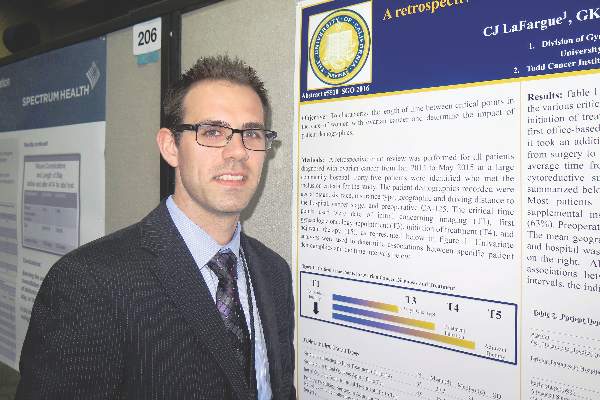
“Ovarian cancer is such a hard disease to diagnose early,” Christopher J. LaFargue, MD, lead study author, said in an interview at the annual meeting of the Society of Gynecologic Oncology. “Once it’s diagnosed, it’s imperative that women get in to see the gynecologic oncologist [and] make sure their treatment care isn’t lagged for any reason.”

In an effort to characterize the length of time between critical points in the care of women with ovarian cancer and determine the impact of patient demographics, Dr. LaFargue and his associates retrospectively evaluated the medical records of 45 women who were diagnosed with ovarian cancer at Long Beach (Calif.) Memorial Medical Center between January 2012 and May 2015.
They examined patient demographics, including preoperative CA-125. Time points of interest were first concerning imaging test, first gynecologic oncology appointment, initiation of treatment, and adjuvant therapy. They used univariate analyses to determine associations between specific patient demographics and the time intervals of interest.
Dr. LaFargue, a resident in the department of obstetrics and gynecology at the University of California, Irvine, reported that the mean age of patients was 61 years, and the mean driving distance to the hospital from the patients’ home was 11 miles. More than half of the patients were white (58%) and 62% of patients were diagnosed with stage III or IV disease. Preoperative CA-125 exceeded 200 U/mL in 62% of patients. Medicare enrollees with supplemental insurance made up less than half of the group (44%).
The researchers found that the average time from initial concerning imaging to start of treatment was about 38 days. The average time from initial imaging to the first office visit with a gynecologic oncologist was about 18 days. The time from that appointment to initial treatment was 19 days, on average. The time from the start of neoadjuvant chemotherapy to interval cytoreductive surgery was 103 days, on average.
The only factor that significantly impacted transit time was a patient’s CA-125 level. Those who had a level of 200 U/mL or greater were more likely to receive surgery or treatment quicker, compared with those who had a CA-125 level less than 200 U/mL. No other statistically significant associations between patient demographics and length of time intervals were observed.
“It would have been nice to have seen a correlation with insurance status,” Dr. LaFargue said. “That’s kind of what we were hoping for, because then you can make an argument with insurance payers that patients who have Medicare aren’t getting treated as quickly as those who have a PPO, for example.”
He acknowledged certain limitations of the study, including its small sample size, lack of outcomes data, and the fact that it was conducted in a community hospital setting.
Dr. LaFargue reported having no financial disclosures.
SAN DIEGO – Women diagnosed with ovarian cancer waited about 38 days, on average, from the initial concerning imaging test to initiation of treatment, according to a study analyzing health care transit times at a single community hospital.
“Ovarian cancer is such a hard disease to diagnose early,” Christopher J. LaFargue, MD, lead study author, said in an interview at the annual meeting of the Society of Gynecologic Oncology. “Once it’s diagnosed, it’s imperative that women get in to see the gynecologic oncologist [and] make sure their treatment care isn’t lagged for any reason.”
In an effort to characterize the length of time between critical points in the care of women with ovarian cancer and determine the impact of patient demographics, Dr. LaFargue and his associates retrospectively evaluated the medical records of 45 women who were diagnosed with ovarian cancer at Long Beach (Calif.) Memorial Medical Center between January 2012 and May 2015.
They examined patient demographics, including preoperative CA-125. Time points of interest were first concerning imaging test, first gynecologic oncology appointment, initiation of treatment, and adjuvant therapy. They used univariate analyses to determine associations between specific patient demographics and the time intervals of interest.
Dr. LaFargue, a resident in the department of obstetrics and gynecology at the University of California, Irvine, reported that the mean age of patients was 61 years, and the mean driving distance to the hospital from the patients’ home was 11 miles. More than half of the patients were white (58%) and 62% of patients were diagnosed with stage III or IV disease. Preoperative CA-125 exceeded 200 U/mL in 62% of patients. Medicare enrollees with supplemental insurance made up less than half of the group (44%).
The researchers found that the average time from initial concerning imaging to start of treatment was about 38 days. The average time from initial imaging to the first office visit with a gynecologic oncologist was about 18 days. The time from that appointment to initial treatment was 19 days, on average. The time from the start of neoadjuvant chemotherapy to interval cytoreductive surgery was 103 days, on average.
The only factor that significantly impacted transit time was a patient’s CA-125 level. Those who had a level of 200 U/mL or greater were more likely to receive surgery or treatment quicker, compared with those who had a CA-125 level less than 200 U/mL. No other statistically significant associations between patient demographics and length of time intervals were observed.
“It would have been nice to have seen a correlation with insurance status,” Dr. LaFargue said. “That’s kind of what we were hoping for, because then you can make an argument with insurance payers that patients who have Medicare aren’t getting treated as quickly as those who have a PPO, for example.”
He acknowledged certain limitations of the study, including its small sample size, lack of outcomes data, and the fact that it was conducted in a community hospital setting.
Dr. LaFargue reported having no financial disclosures.
AT THE ANNUAL MEETING ON WOMEN’S CANCER
Key clinical point: Initiation of treatment for ovarian cancer following the first concerning imaging test took an average of about 38 days.
Major finding: The average time from the initial imaging test to the first visit with a gynecologic oncologist was about 18 days.
Data source: A retrospective evaluation of 45 women who were diagnosed with ovarian cancer between January 2012 and May 2015 at a community hospital.
Disclosures: Dr. LaFargue reported having no financial disclosures.
Subtotal fenestrating cholecystectomy: Optimal ‘bailout’ for difficult cases
Subtotal fenestrating cholecystectomy with drain placement appears optimal, compared with the reconstituting procedure, for experienced surgeons seeking a “bailout” operation in both open and minimally invasive cholecystectomy where the critical view of safety (CVS) is not easily attainable, according to a report written on behalf of the SAGES Safe Cholecystectomy Task Force 2015.
The rise in laparoscopic cholecystectomy has been associated with an increase in the rate of bile duct injury, most commonly when secure ductal identification using CVS is not possible because of an inflamed hepatocystic triangle occluding the cystic duct, cystic artery, and cystic plate. In such cases, a safe and effective bailout technique (one not requiring a second operation) must be decided upon in preference to simply closing and proceeding to a later open procedure, according to Steven M. Strasberg, MD, of Washington University in Saint Louis and his colleagues (J Am Coll Surg. 2016;222:89-96).

In order to clarify the two most common and effective “partial cholecystectomy” procedures being performed, Dr. Strasburg and his colleagues have suggested the use of the term “subtotal” in place of “partial” and the terms “fenestrating” vs. “reconstituting,” to define whether there is an open or closed gallbladder remnant, respectively, after the procedure.
In subtotal fenestrating cholecystectomy, the free peritonealized portion of the gallbladder is excised, except for a tip at the lowest portion that acts as a shield to protect against inadvertently entering the hepatocystic triangle, according to the authors. There is no sealed lumen remaining, thus the cystic duct requires closure. The cystic duct may be closed from the inside with a purse-string suture. Attempts to ligate the cystic duct outside the gallbladder may injure the common bile duct and can potentially result in fistulas.
In subtotal reconstituting cholecystectomy, the free peritonealized portion of the gallbladder is excised, but the lowest portion of the gallbladder is closed with sutures or staples and reconstitutes an intact lumen in which stones may be re-formed, which can in turn require reoperation.
“Whether the subtotal cholecystectomy is ‘fenestrating’ or ‘reconstituting’ depends on whether the lowest part of the gallbladder is left open (fenestrating) or closed (reconstituting) and not on the amount of gallbladder that is left attached to the liver,” according to the authors.
Subtotal fenestrating cholecystectomy is most likely done when an open approach is used, whereas subtotal reconstituting cholecystectomies are probably easier to do laparoscopically and are preferred by surgeons doing minimally invasive procedures, they said.
Despite the fact that there have been no head-to-head comparisons of fenestrating vs. reconstituting techniques, the authors said they prefer the fenestrating method, although the technique chosen may be based on the experience of the surgeon, they noted.
“The principle is that a subtotal fenestrating cholecystectomy is a standard operation that should be used liberally when surgeons encounter difficulty getting to the CVS,” the authors wrote. “We believe that clarification of the procedures and what they are called will help to choose which type of procedure to select, and it will also facilitate the performance of clinical studies in this area,“ they concluded.
The authors reported having no relevant financial disclosures.
A transcript of an interactive discussion of this paper and topic is available online (www.journalacs.org/RAS-ACS-discussion-2016).
Subtotal fenestrating cholecystectomy with drain placement appears optimal, compared with the reconstituting procedure, for experienced surgeons seeking a “bailout” operation in both open and minimally invasive cholecystectomy where the critical view of safety (CVS) is not easily attainable, according to a report written on behalf of the SAGES Safe Cholecystectomy Task Force 2015.
The rise in laparoscopic cholecystectomy has been associated with an increase in the rate of bile duct injury, most commonly when secure ductal identification using CVS is not possible because of an inflamed hepatocystic triangle occluding the cystic duct, cystic artery, and cystic plate. In such cases, a safe and effective bailout technique (one not requiring a second operation) must be decided upon in preference to simply closing and proceeding to a later open procedure, according to Steven M. Strasberg, MD, of Washington University in Saint Louis and his colleagues (J Am Coll Surg. 2016;222:89-96).
In order to clarify the two most common and effective “partial cholecystectomy” procedures being performed, Dr. Strasburg and his colleagues have suggested the use of the term “subtotal” in place of “partial” and the terms “fenestrating” vs. “reconstituting,” to define whether there is an open or closed gallbladder remnant, respectively, after the procedure.
In subtotal fenestrating cholecystectomy, the free peritonealized portion of the gallbladder is excised, except for a tip at the lowest portion that acts as a shield to protect against inadvertently entering the hepatocystic triangle, according to the authors. There is no sealed lumen remaining, thus the cystic duct requires closure. The cystic duct may be closed from the inside with a purse-string suture. Attempts to ligate the cystic duct outside the gallbladder may injure the common bile duct and can potentially result in fistulas.
In subtotal reconstituting cholecystectomy, the free peritonealized portion of the gallbladder is excised, but the lowest portion of the gallbladder is closed with sutures or staples and reconstitutes an intact lumen in which stones may be re-formed, which can in turn require reoperation.
“Whether the subtotal cholecystectomy is ‘fenestrating’ or ‘reconstituting’ depends on whether the lowest part of the gallbladder is left open (fenestrating) or closed (reconstituting) and not on the amount of gallbladder that is left attached to the liver,” according to the authors.
Subtotal fenestrating cholecystectomy is most likely done when an open approach is used, whereas subtotal reconstituting cholecystectomies are probably easier to do laparoscopically and are preferred by surgeons doing minimally invasive procedures, they said.
Despite the fact that there have been no head-to-head comparisons of fenestrating vs. reconstituting techniques, the authors said they prefer the fenestrating method, although the technique chosen may be based on the experience of the surgeon, they noted.
“The principle is that a subtotal fenestrating cholecystectomy is a standard operation that should be used liberally when surgeons encounter difficulty getting to the CVS,” the authors wrote. “We believe that clarification of the procedures and what they are called will help to choose which type of procedure to select, and it will also facilitate the performance of clinical studies in this area,“ they concluded.
The authors reported having no relevant financial disclosures.
A transcript of an interactive discussion of this paper and topic is available online (www.journalacs.org/RAS-ACS-discussion-2016).
FROM THE JOURNAL OF THE AMERICAN COLLEGE OF SURGEONS
Key clinical point: Subtotal fenestrating cholecystectomy should be used liberally when surgeons have difficulty getting to the critical view of safety (CVS).
Major finding: Subtotal fenestrating cholecystectomy with drain placement, despite its difficulty in laparoscopic cases, should be the procedure of choice for experienced surgeons in both open and minimally invasive procedures where the CVS is not safely attainable.
Data source: An expert analysis of historical data and the literature to determine optimal surgical technique, on behalf of the SAGES Safe Cholecystectomy Task Force 2015.
Disclosures: The authors reported having no relevant financial disclosures.

CMS seeks input on future of Open Payments program
The Centers for Medicare & Medicaid Services is seeking physician input on the Open Payments program.
The agency signaled its intent to gather information in its proposed Medicare Physician Fee Schedule for 2017 and will host a conference call for that purpose on August 2.

The agency already has released a slide presentation to be used during the call that highlights the information being requested, including whether the payment categories are inclusive enough; how many years of payment data is relevant; whether reporting entities should pre-vet data before reporting to the Open Payment system; the adequacy of the definition of a covered recipient teaching hospital; whether reporting entities should be able to submit data continuously throughout the calendar year; how mergers affect reporting; clarity on reporting of ownership and investment interests; clarity on the definition of physician-owned distributors; and clarity on ways to streamline the reporting process.
Details for participating in the conference call can be found here.
The Centers for Medicare & Medicaid Services is seeking physician input on the Open Payments program.
The agency signaled its intent to gather information in its proposed Medicare Physician Fee Schedule for 2017 and will host a conference call for that purpose on August 2.
The agency already has released a slide presentation to be used during the call that highlights the information being requested, including whether the payment categories are inclusive enough; how many years of payment data is relevant; whether reporting entities should pre-vet data before reporting to the Open Payment system; the adequacy of the definition of a covered recipient teaching hospital; whether reporting entities should be able to submit data continuously throughout the calendar year; how mergers affect reporting; clarity on reporting of ownership and investment interests; clarity on the definition of physician-owned distributors; and clarity on ways to streamline the reporting process.
Details for participating in the conference call can be found here.

Feds sue to block mega-mergers by health insurers
The U.S. Department of Justice (DOJ) is suing to block two mega-mergers between four of the largest health insurers in the nation, claiming the alignments will harm competition and reduce patient choice.
The DOJ and a number of state attorneys general filed legal challenges July 21 in the U.S. District Court for the District of Columbia seeking to ban Anthem’s proposed acquisition of Cigna and Aetna’s proposed acquisition of Humana. The lawsuits allege the two mergers – valued at $54 billion and $37 billion respectively – would negatively affect doctors, patients, and employers, by limiting price competition, reducing benefits, decreasing incentives to provide innovative wellness programs, and lowering quality of care.

“These mergers would restrict competition for health insurance products sold in markets across the country and would give tremendous power over the nation’s health insurance industry to just three large companies,” U.S. Attorney General Loretta E. Lynch said in a statement. “Our actions seek to preserve competition that keeps premiums down and drives insurers to collaborate with doctors and hospitals to provide better health care for all Americans.”
Anthem called the lawsuit “an unfortunate and misguided step backward for access to affordable health care.
“The DOJ’s action is based on a flawed analysis and misunderstanding of the dynamic, competitive, and highly regulated health care landscape and is inconsistent with the way that the DOJ has reviewed past health care transactions,” Anthem officials said in a statement. “Anthem has an unwavering commitment to enhancing access to affordable health care, and the benefits and efficiencies from its merger with Cigna is one way that Anthem will continue its mission of improving consumer choice, quality, and affordability.”
In a statement, Cigna officials said that company is evaluating its options given the nature of the concerns raised by the DOJ. Aetna and Humana meanwhile, vowed to vigorously defend their pending merger.
“A combined company will result in a broader choice of products, access to higher quality, and more affordable care, and a better overall experience for consumers,” according to a joint statement. “Aetna and Humana look forward to making this clear in court, where a judge will review the transaction based on its merits.”
Anthem’s proposed acquisition of Cigna would be the largest merger in the history of the health insurance industry, according to the DOJ. The companies began talks of a possible merger in early 2014 and Anthem agreed to acquire Cigna for $54 billion in 2015, according to court documents.
Meanwhile, Aetna began inquiring about a deal with Humana in March 2015, entering into a definitive agreement to acquire Humana for $37 billion later that year. The two proposed mergers have been closely watched by the DOJ and other regulatory agencies from the start.
The DOJ’s suit against Anthem and Cigna alleges the merger would substantially reduce competition for millions of patients who receive commercial health insurance coverage, from large-group employers in at least 35 metropolitan areas and from public exchanges created by the Affordable Care Act. The elimination of Cigna also threatens competition among commercial insurers for the purchase of health care services from hospitals, physicians, and other providers, the suit alleges. Eleven states and the District of Columbia joined the department’s challenge of the Cigna acquisition.
The government’s challenge against Aetna and Humana alleges the merger would greatly reduce Medicare Advantage competition in more than 350 counties in 21 states, affecting more than 1.5 million Medicare Advantage patients. The lawsuit also claims that Aetna’s purchase of Humana would substantially reduce competition to sell commercial health insurance to individuals and families on the public exchanges in 17 counties in Florida, Georgia, and Missouri. Eight states and the District of Columbia joined the department’s challenge of the Humana acquisition.
The American Medical Association voiced support for the lawsuit, condemning the proposed transactions as moves that will lessen competition and choice. In 2015, AMA issued special analyses showing that the combined impact of the proposed Anthem/Cigna and Aetna/Humana mergers and urged the federal government to block the transactions.

“The prospect of reducing five national health insurance carriers to just three is unacceptable,” AMA President Andrew W. Gurman, MD, said in a statement. “[The] action by the DOJ acknowledges the AMA’s concern that patients’ interests can be harmed when big insurers acquire rivals and develop strangleholds on local markets. Allowing commercial health insurers to become too big and exert control over the delivery of health care would be bad for patients and vitality of the nation’s health care system.”
On Twitter @legal_med
The U.S. Department of Justice (DOJ) is suing to block two mega-mergers between four of the largest health insurers in the nation, claiming the alignments will harm competition and reduce patient choice.
The DOJ and a number of state attorneys general filed legal challenges July 21 in the U.S. District Court for the District of Columbia seeking to ban Anthem’s proposed acquisition of Cigna and Aetna’s proposed acquisition of Humana. The lawsuits allege the two mergers – valued at $54 billion and $37 billion respectively – would negatively affect doctors, patients, and employers, by limiting price competition, reducing benefits, decreasing incentives to provide innovative wellness programs, and lowering quality of care.
“These mergers would restrict competition for health insurance products sold in markets across the country and would give tremendous power over the nation’s health insurance industry to just three large companies,” U.S. Attorney General Loretta E. Lynch said in a statement. “Our actions seek to preserve competition that keeps premiums down and drives insurers to collaborate with doctors and hospitals to provide better health care for all Americans.”
Anthem called the lawsuit “an unfortunate and misguided step backward for access to affordable health care.
“The DOJ’s action is based on a flawed analysis and misunderstanding of the dynamic, competitive, and highly regulated health care landscape and is inconsistent with the way that the DOJ has reviewed past health care transactions,” Anthem officials said in a statement. “Anthem has an unwavering commitment to enhancing access to affordable health care, and the benefits and efficiencies from its merger with Cigna is one way that Anthem will continue its mission of improving consumer choice, quality, and affordability.”
In a statement, Cigna officials said that company is evaluating its options given the nature of the concerns raised by the DOJ. Aetna and Humana meanwhile, vowed to vigorously defend their pending merger.
“A combined company will result in a broader choice of products, access to higher quality, and more affordable care, and a better overall experience for consumers,” according to a joint statement. “Aetna and Humana look forward to making this clear in court, where a judge will review the transaction based on its merits.”
Anthem’s proposed acquisition of Cigna would be the largest merger in the history of the health insurance industry, according to the DOJ. The companies began talks of a possible merger in early 2014 and Anthem agreed to acquire Cigna for $54 billion in 2015, according to court documents.
Meanwhile, Aetna began inquiring about a deal with Humana in March 2015, entering into a definitive agreement to acquire Humana for $37 billion later that year. The two proposed mergers have been closely watched by the DOJ and other regulatory agencies from the start.
The DOJ’s suit against Anthem and Cigna alleges the merger would substantially reduce competition for millions of patients who receive commercial health insurance coverage, from large-group employers in at least 35 metropolitan areas and from public exchanges created by the Affordable Care Act. The elimination of Cigna also threatens competition among commercial insurers for the purchase of health care services from hospitals, physicians, and other providers, the suit alleges. Eleven states and the District of Columbia joined the department’s challenge of the Cigna acquisition.
The government’s challenge against Aetna and Humana alleges the merger would greatly reduce Medicare Advantage competition in more than 350 counties in 21 states, affecting more than 1.5 million Medicare Advantage patients. The lawsuit also claims that Aetna’s purchase of Humana would substantially reduce competition to sell commercial health insurance to individuals and families on the public exchanges in 17 counties in Florida, Georgia, and Missouri. Eight states and the District of Columbia joined the department’s challenge of the Humana acquisition.
The American Medical Association voiced support for the lawsuit, condemning the proposed transactions as moves that will lessen competition and choice. In 2015, AMA issued special analyses showing that the combined impact of the proposed Anthem/Cigna and Aetna/Humana mergers and urged the federal government to block the transactions.
“The prospect of reducing five national health insurance carriers to just three is unacceptable,” AMA President Andrew W. Gurman, MD, said in a statement. “[The] action by the DOJ acknowledges the AMA’s concern that patients’ interests can be harmed when big insurers acquire rivals and develop strangleholds on local markets. Allowing commercial health insurers to become too big and exert control over the delivery of health care would be bad for patients and vitality of the nation’s health care system.”
On Twitter @legal_med

Fresh Press: ACS Surgery News digital July issue is available on the website
The July issue of ACS Surgery News in digital format is available online. Use the mobile app to download or view as a pdf.
This month’s issue features coverage of the report, “A National Trauma Care System: Integrating Civilian and Military Trauma Systems to Achieve Zero Preventable Deaths After Injury,” released by the National Academies of Sciences, Engineering, and Medicine. The report, supported in part by the American College of Surgeons, gives a detailed list of policy recommendations that would create a unified trauma response system incorporating lessons learned from combat mission with the aim of eliminating unnecessary deaths in civilian trauma situations (p. 1).

Looking for some summer reading? Dr. Carol Scott-Conner, moderator of the ACS Communities Surgeon Writers discussion group, reviews three books by surgeons that should be of special interest to the readers of ACS Surgery News. All three works concern personal experience and reflections on the profession (p. 4).
Don’t miss the story (p. 7) on peer review hearings and how best to approach this situation. These hearings should be informative and educational, but can also play out in unexpected ways. Be prepared!
The July issue of ACS Surgery News in digital format is available online. Use the mobile app to download or view as a pdf.
This month’s issue features coverage of the report, “A National Trauma Care System: Integrating Civilian and Military Trauma Systems to Achieve Zero Preventable Deaths After Injury,” released by the National Academies of Sciences, Engineering, and Medicine. The report, supported in part by the American College of Surgeons, gives a detailed list of policy recommendations that would create a unified trauma response system incorporating lessons learned from combat mission with the aim of eliminating unnecessary deaths in civilian trauma situations (p. 1).
Looking for some summer reading? Dr. Carol Scott-Conner, moderator of the ACS Communities Surgeon Writers discussion group, reviews three books by surgeons that should be of special interest to the readers of ACS Surgery News. All three works concern personal experience and reflections on the profession (p. 4).
Don’t miss the story (p. 7) on peer review hearings and how best to approach this situation. These hearings should be informative and educational, but can also play out in unexpected ways. Be prepared!
Lessons learned from merging EHR systems
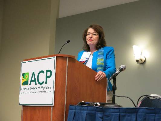
WASHINGTON – As practices merge, how hard is it to merge EHRs?
Even in what might seem to be the best circumstances, it can be a huge challenge, according to Jacqueline Fincher, MD, of McDuffie Medical Associates, Thompson, Ga.
One appealing aspect of the merger of Dr. Fincher’s practice with another was that each practice used an EHR from the same vendor and each was operating with the same updates.

“This is the perfect setup,” she said at the annual meeting of the American College of Physicians. “We don’t have to go to Epic. We can stay on the same EHR. In fact, this group had gone on the same EHR back in 2006 within a month of the time that we went on; we were on the exact same version and the same everything. We were totally even. The thought among the corporate and IT staff of the new entity was that this is going to be seamless. We’re just going to renumber the accounts and everything will be just fine.”
A call to the EHR vendor, whom she did not name, revealed that the process would be anything but seamless.
“Our IT staff contacted our common EHR vendor and said we want to merge this practice with our bigger practice and the EHR company said, ‘Wow, we’ve never done that before.’ What? In this day of consolidation and integration, they had never done it before? Nor did they have a business model to do so, much less a digital plan to do so. That was pretty shocking,” she said.
Dr. Fincher noted that the EHR vendor recommended a third party vendor to handle creating an interface between the two EHRs. “Most EHR companies do not, I say, do not have a dedicated service to migrate data. It’s almost always going out to a third-party conversion service that doesn’t know you, doesn’t know your work flow, and makes everything even more difficult.”
Two and a half months – and 10 interfaces – later, the launch of the combined EHR was a disaster, Dr. Fincher said.
Even though both practices were using the exact same version of the EHR, each had very different work flows, defaults, and other nuances that meant data didn’t transfer smoothly – cheaply.
Among the surprise expenses: about $55,000 for additional hardware, network cabling and interfaces; $35,000 for additional servers; and at least $100,000 for personnel expenses related to the data migration.
Given her experience, Dr. Fincher advised her peers “to start at least 6 months in advance to map and convert the data.”
And it is vital to get input and participation from all office stakeholders – both clinical and administrative staff – regarding how the data is migrated, she said. Be sure to completely understand all work flows from both practices so that you know how the data is going to migrate.
“Understanding work flow, that is absolutely critical. Every single office has a different work flow for every type of encounter by any method. How these work flows are the same or different between your office and the new practice or your EHR and the new EHR that you’re going to, they are different. You have to understand the differences,” she added.
“We didn’t perceive that there was that much difference but when everything crashed, we discovered there were because they had different work flows, they had different defaults in place, those types of things.”
Other key questions: Which data needs to be migrated? How long should the old system remain in place? Should the data be migrated manually or digitally? How much time will the EHR merger take?
“You want to establish that structured planning time,” Dr. Fincher said. “You’ve got to carve out scheduled time with the group in order to do this. Establish the tasks that need to be accomplished, so just making a to-do list every week and who’s going to be accountable to accomplish those parts of the list” is important.
WASHINGTON – As practices merge, how hard is it to merge EHRs?
Even in what might seem to be the best circumstances, it can be a huge challenge, according to Jacqueline Fincher, MD, of McDuffie Medical Associates, Thompson, Ga.
One appealing aspect of the merger of Dr. Fincher’s practice with another was that each practice used an EHR from the same vendor and each was operating with the same updates.
“This is the perfect setup,” she said at the annual meeting of the American College of Physicians. “We don’t have to go to Epic. We can stay on the same EHR. In fact, this group had gone on the same EHR back in 2006 within a month of the time that we went on; we were on the exact same version and the same everything. We were totally even. The thought among the corporate and IT staff of the new entity was that this is going to be seamless. We’re just going to renumber the accounts and everything will be just fine.”
A call to the EHR vendor, whom she did not name, revealed that the process would be anything but seamless.
“Our IT staff contacted our common EHR vendor and said we want to merge this practice with our bigger practice and the EHR company said, ‘Wow, we’ve never done that before.’ What? In this day of consolidation and integration, they had never done it before? Nor did they have a business model to do so, much less a digital plan to do so. That was pretty shocking,” she said.
Dr. Fincher noted that the EHR vendor recommended a third party vendor to handle creating an interface between the two EHRs. “Most EHR companies do not, I say, do not have a dedicated service to migrate data. It’s almost always going out to a third-party conversion service that doesn’t know you, doesn’t know your work flow, and makes everything even more difficult.”
Two and a half months – and 10 interfaces – later, the launch of the combined EHR was a disaster, Dr. Fincher said.
Even though both practices were using the exact same version of the EHR, each had very different work flows, defaults, and other nuances that meant data didn’t transfer smoothly – cheaply.
Among the surprise expenses: about $55,000 for additional hardware, network cabling and interfaces; $35,000 for additional servers; and at least $100,000 for personnel expenses related to the data migration.
Given her experience, Dr. Fincher advised her peers “to start at least 6 months in advance to map and convert the data.”
And it is vital to get input and participation from all office stakeholders – both clinical and administrative staff – regarding how the data is migrated, she said. Be sure to completely understand all work flows from both practices so that you know how the data is going to migrate.
“Understanding work flow, that is absolutely critical. Every single office has a different work flow for every type of encounter by any method. How these work flows are the same or different between your office and the new practice or your EHR and the new EHR that you’re going to, they are different. You have to understand the differences,” she added.
“We didn’t perceive that there was that much difference but when everything crashed, we discovered there were because they had different work flows, they had different defaults in place, those types of things.”
Other key questions: Which data needs to be migrated? How long should the old system remain in place? Should the data be migrated manually or digitally? How much time will the EHR merger take?
“You want to establish that structured planning time,” Dr. Fincher said. “You’ve got to carve out scheduled time with the group in order to do this. Establish the tasks that need to be accomplished, so just making a to-do list every week and who’s going to be accountable to accomplish those parts of the list” is important.
EXPERT ANALYSIS FROM ACP INTERNAL MEDICINE 2016
New data shed light on impact of resecting the primary tumor in stage IV breast cancer
CHICAGO – The survival impact of resecting the primary tumor in women with de novo stage IV breast cancer depends on receipt of and response to prior systemic therapy, suggested a pair of studies reported at the annual meeting of the American Society of Clinical Oncology.
A randomized trial conducted in Turkey found that, relative to peers who received initial systemic therapy, women who underwent initial resection of the primary tumor had a one-third lower risk of death at 5 years. But a prospective registry study conducted in the United States found that elective resection after a response to first-line therapy did not significantly improve overall survival, with patients living roughly 6 years regardless of whether they had the surgery or not.
Findings in context

“I think these studies have just confirmed what we know, and that is that tumor biology is critical,” said invited discussant Elizabeth A. Mittendorf, MD, PhD, of University of Texas MD Anderson Cancer Center, Houston. “Patients who do not respond to systemic therapy will do poorly, so I don’t think it’s unwise to consider a biologic ‘stress test’ with initiation of first-line therapy, knowing that patients who do not respond will not benefit from surgery.”
Those with hormone receptor–positive or HER2-positive disease are most likely to benefit from targeted therapy and may see even higher response rates as novel targeted agents are introduced. “But despite the increase in response to therapy, we really have no data at this time to suggest any benefit from surgery,” she added. “There may be some utility in continuing to enroll these patients in a clinical trial. I would suggest that it would need to be a subtype-specific trial and would question whether we have the appetite to conduct such a study.”
More information on managing de novo stage IV breast cancer is expected from ongoing trials such as the Eastern Cooperative Oncology Group’s 2108 trial, which is randomizing patients having a response or stable disease with first-line therapy to either early local therapy or delayed local therapy only at the time of progression, according to Dr. Mittendorf.
Poor accrual necessitated redesign of the trial. “As part of that redesign, there was a decrease in the target enrollment, which causes me concern that the trial will not be powered to inform its primary endpoint of overall survival,” she commented. However, “it’s interesting to note that in early 2014, shortly after the report of the trials from India and Turkey at the San Antonio Breast Cancer Symposium, there was a significant increase in enrollment, suggesting that this is a clinically important question.”
Turkish study: MF07-01
The first study – trial MF07-01 of the Turkish Federation of Breast Diseases Societies – was presented by Atilla Soran, MD, of Magee-Womens Hospital of University of Pittsburgh Medical Center.
He and his colleagues enrolled in the trial women with de novo stage IV breast cancer whose primary tumor was amenable to complete surgical resection and who were healthy enough to be treated.
The women were randomized evenly either to initial systemic therapy followed by local therapy only if local progression occurred, or to initial local therapy, consisting of surgery with or without radiation therapy of the breast and axilla, followed by systemic therapy.
Among the 274 evaluable women having a median follow-up of about 40 months, the 3-year rate of overall survival did not differ significantly between the two groups, Dr. Soran reported.
However, the 5-year rate of overall survival was 41.6% in the initial surgery group and 24.4% in the initial systemic therapy group, a difference translating to a significant reduction in the risk of death (hazard ratio, 0.66; P = .005). Median survival was 46 months and 37 months, respectively.
The benefit was similar in women whose tumors had hormone receptors, whose tumors were negative for HER2, and who were younger than age 55. There was no significant benefit of up-front surgery for women with bone-only metastases, “but we believe that when we follow these patients longer, the difference is going to be statistically significant,” he said.
On the other hand, there was a trend among women who had multiple pulmonary and/or liver metastases whereby they were more likely to die if they initially had surgery instead of systemic therapy.
Locoregional progression/relapse occurred in 1% of the initial surgery group but 11% of the initial systemic therapy group. Among women who did not have locoregional progression/relapse, surgery still had a survival benefit (HR, 0.61; P = .001).
“We know that with systemic therapy, immunotherapy, radiation therapy, and imaging as developments, patients are living longer when you compare to a decade ago or 20 years ago,” said Dr. Soran. “But we also believe that there is a role for surgery of the primary tumor in those patients.”
“Performance status, age, and comorbidities must be taken into account, and the burden of metastatic disease needs to be considered,” he maintained. “The benefit of surgery at presentation is dependent on the completeness of resection, and axillary surgery and locoregional radiation therapy should be considered regardless of the metastasis.”
U.S. study: TBCRC 013
The second study – the Translational Breast Cancer Research Consortium’s study 013 – was presented by Tari A. King, MD, chief of breast surgery at the Dana-Farber Cancer Institute, associate division chief for breast surgery at Brigham and Women’s Hospital, and associate professor of surgery at Harvard Medical School, all in Boston.
The investigators analyzed data from the study’s cohort A, consisting of 112 patients with de novo stage IV breast cancer who had an intact primary tumor. All patients were given first-line systemic therapy; those who had a response were additionally offered elective resection of their primary tumor.
The median duration of follow-up was 54 months. Overall, 85% of the women had a response to their first-line therapy, Dr. King reported.
Some 43% of responders opted to undergo elective surgery to resect their primary tumor, defined as surgery performed in the absence of local symptoms or the need for local control, with specific type and extent left up to the treating physician.
In a multivariate analysis among responders surviving at least 6 months, median survival was 71 months with elective surgery and 65 months without it, a nonsignificant difference.
Findings were similar among subsets of women having estrogen receptor–positive tumors or HER2-positive tumors, and various combinations of these features.
In recursive partitioning analysis, response to first-line therapy, HER2 status, and age were the major determinants of survival.
“Importantly, although we were not able to demonstrate a survival benefit with the use of surgery, surgery also did not impact progression-free survival,” noted Dr. King.
Ultimately, 4% of responders who did not have elective surgery and 18% of nonresponders went on to have palliative resection of their primary.
“As this was a registry study, patients selected for surgery were more likely to have single-organ metastatic disease and to have received first-line chemotherapy, yet despite this selection bias, surgery did not impact survival in any tumor subtype,” Dr. King summarized. “Among patients who responded to therapy, HER2 status and patient age remained strong prognostic factors. Further investigation is needed to determine if subsets of patients will ultimately benefit from surgery.”
“In the absence of additional prospective data, our findings do not support surgery for the primary tumor outside of a clinical trial,” she concluded.
CHICAGO – The survival impact of resecting the primary tumor in women with de novo stage IV breast cancer depends on receipt of and response to prior systemic therapy, suggested a pair of studies reported at the annual meeting of the American Society of Clinical Oncology.
A randomized trial conducted in Turkey found that, relative to peers who received initial systemic therapy, women who underwent initial resection of the primary tumor had a one-third lower risk of death at 5 years. But a prospective registry study conducted in the United States found that elective resection after a response to first-line therapy did not significantly improve overall survival, with patients living roughly 6 years regardless of whether they had the surgery or not.
Findings in context
“I think these studies have just confirmed what we know, and that is that tumor biology is critical,” said invited discussant Elizabeth A. Mittendorf, MD, PhD, of University of Texas MD Anderson Cancer Center, Houston. “Patients who do not respond to systemic therapy will do poorly, so I don’t think it’s unwise to consider a biologic ‘stress test’ with initiation of first-line therapy, knowing that patients who do not respond will not benefit from surgery.”
Those with hormone receptor–positive or HER2-positive disease are most likely to benefit from targeted therapy and may see even higher response rates as novel targeted agents are introduced. “But despite the increase in response to therapy, we really have no data at this time to suggest any benefit from surgery,” she added. “There may be some utility in continuing to enroll these patients in a clinical trial. I would suggest that it would need to be a subtype-specific trial and would question whether we have the appetite to conduct such a study.”
More information on managing de novo stage IV breast cancer is expected from ongoing trials such as the Eastern Cooperative Oncology Group’s 2108 trial, which is randomizing patients having a response or stable disease with first-line therapy to either early local therapy or delayed local therapy only at the time of progression, according to Dr. Mittendorf.
Poor accrual necessitated redesign of the trial. “As part of that redesign, there was a decrease in the target enrollment, which causes me concern that the trial will not be powered to inform its primary endpoint of overall survival,” she commented. However, “it’s interesting to note that in early 2014, shortly after the report of the trials from India and Turkey at the San Antonio Breast Cancer Symposium, there was a significant increase in enrollment, suggesting that this is a clinically important question.”
Turkish study: MF07-01
The first study – trial MF07-01 of the Turkish Federation of Breast Diseases Societies – was presented by Atilla Soran, MD, of Magee-Womens Hospital of University of Pittsburgh Medical Center.
He and his colleagues enrolled in the trial women with de novo stage IV breast cancer whose primary tumor was amenable to complete surgical resection and who were healthy enough to be treated.
The women were randomized evenly either to initial systemic therapy followed by local therapy only if local progression occurred, or to initial local therapy, consisting of surgery with or without radiation therapy of the breast and axilla, followed by systemic therapy.
Among the 274 evaluable women having a median follow-up of about 40 months, the 3-year rate of overall survival did not differ significantly between the two groups, Dr. Soran reported.
However, the 5-year rate of overall survival was 41.6% in the initial surgery group and 24.4% in the initial systemic therapy group, a difference translating to a significant reduction in the risk of death (hazard ratio, 0.66; P = .005). Median survival was 46 months and 37 months, respectively.
The benefit was similar in women whose tumors had hormone receptors, whose tumors were negative for HER2, and who were younger than age 55. There was no significant benefit of up-front surgery for women with bone-only metastases, “but we believe that when we follow these patients longer, the difference is going to be statistically significant,” he said.
On the other hand, there was a trend among women who had multiple pulmonary and/or liver metastases whereby they were more likely to die if they initially had surgery instead of systemic therapy.
Locoregional progression/relapse occurred in 1% of the initial surgery group but 11% of the initial systemic therapy group. Among women who did not have locoregional progression/relapse, surgery still had a survival benefit (HR, 0.61; P = .001).
“We know that with systemic therapy, immunotherapy, radiation therapy, and imaging as developments, patients are living longer when you compare to a decade ago or 20 years ago,” said Dr. Soran. “But we also believe that there is a role for surgery of the primary tumor in those patients.”
“Performance status, age, and comorbidities must be taken into account, and the burden of metastatic disease needs to be considered,” he maintained. “The benefit of surgery at presentation is dependent on the completeness of resection, and axillary surgery and locoregional radiation therapy should be considered regardless of the metastasis.”
U.S. study: TBCRC 013
The second study – the Translational Breast Cancer Research Consortium’s study 013 – was presented by Tari A. King, MD, chief of breast surgery at the Dana-Farber Cancer Institute, associate division chief for breast surgery at Brigham and Women’s Hospital, and associate professor of surgery at Harvard Medical School, all in Boston.
The investigators analyzed data from the study’s cohort A, consisting of 112 patients with de novo stage IV breast cancer who had an intact primary tumor. All patients were given first-line systemic therapy; those who had a response were additionally offered elective resection of their primary tumor.
The median duration of follow-up was 54 months. Overall, 85% of the women had a response to their first-line therapy, Dr. King reported.
Some 43% of responders opted to undergo elective surgery to resect their primary tumor, defined as surgery performed in the absence of local symptoms or the need for local control, with specific type and extent left up to the treating physician.
In a multivariate analysis among responders surviving at least 6 months, median survival was 71 months with elective surgery and 65 months without it, a nonsignificant difference.
Findings were similar among subsets of women having estrogen receptor–positive tumors or HER2-positive tumors, and various combinations of these features.
In recursive partitioning analysis, response to first-line therapy, HER2 status, and age were the major determinants of survival.
“Importantly, although we were not able to demonstrate a survival benefit with the use of surgery, surgery also did not impact progression-free survival,” noted Dr. King.
Ultimately, 4% of responders who did not have elective surgery and 18% of nonresponders went on to have palliative resection of their primary.
“As this was a registry study, patients selected for surgery were more likely to have single-organ metastatic disease and to have received first-line chemotherapy, yet despite this selection bias, surgery did not impact survival in any tumor subtype,” Dr. King summarized. “Among patients who responded to therapy, HER2 status and patient age remained strong prognostic factors. Further investigation is needed to determine if subsets of patients will ultimately benefit from surgery.”
“In the absence of additional prospective data, our findings do not support surgery for the primary tumor outside of a clinical trial,” she concluded.
AT THE 2016 ASCO ANNUAL MEETING
Key clinical point: Resecting the primary tumor up front had a survival benefit, whereas resecting it after a response to systemic therapy did not.
Major finding: Compared with initial systemic therapy, initial resection of the primary tumor reduced the risk of death (HR, 0.66). But after a response to first-line therapy, median survival with elective resection was not significantly superior to that without it (71 vs. 65 months).
Data source: A randomized, controlled trial among 274 women with de novo stage IV breast cancer (MF07-01 trial) and a prospective registry study among 112 women with de novo stage IV breast cancer (TBCRC 013 study).
Disclosures: Dr. Soran disclosed that he has a consulting or advisory role with NanoVision. Dr. King disclosed that she had no relevant conflicts of interest.

Anatomic repair of ccTGA did not yield superior survival
BALTIMORE – Anatomic repair did not outperform physiologic repair in patients with congenitally corrected transposition of the great arteries (ccTGA), according to a study presented by Maryam Al-Omair, M.D., of the University of Toronto at the annual meeting of the American Association for Thoracic Surgery.
Dr. Al-Omair and her colleagues hypothesized that patients undergoing anatomic repair for ccTGA would have superior systemic ventricular function and survival. However, their results showed that anatomic repair of ccTGA did not yield superior survival, compared with physiologic repair, and the long-term impact on systemic ventricular function was not certain.
Because of early evidence showing better outcomes of anatomic over physiologic repair for ccTGA, the surgical trend over time greatly favored the use of anatomic repair: At her team’s institution, anatomic repair went from 2.3% in the 1982-1989 period to 92.3% in the 2010-2015 period, Dr. Al-Omair said.
Their study assessed 200 patients (165 with biventricular ccTGA and 35 Fontan patients) who were managed from 1982 to 2015 at the Hospital for Sick Children, Toronto. The patient treatment groups were anatomic repair (38 patients), physiologic repair (89), single-ventricle (Fontan) repair (35), and palliated (no intracardiac repair) patients (38). The median follow-up was 3.4 years for anatomic repair, 13.5 years for physiologic repair, 7.5 years for single-ventricle repair, and 11.8 years with no repair (11.8 years), reflecting their change in practice.
The investigators followed the primary outcome of transplant-free survival and secondary outcomes of late systemic ventricular function and systemic atrioventricular valve function.

They found no significant difference in transplant-free survival at 20 years in the three repair groups assessed from 1892 to 2105: anatomic repair (58%), physiologic repair (71%), and single-ventricle (Fontan) repair (78%). Looking at the latter period of 2000-2015 for 10-year transplant-free survival, they found similar results: anatomic repair (77%), physiologic repair (85%), and single-ventricle (Fontan) repair (100%).
They also found that transplant-free survival in patients who required no intracadiac repair and had no associated lesions such as ventral septal defect or ventral septal defect with pulmonary stenosis was nearly 95% at 25 years.
A multivariate analysis showed no independent predictors of mortality among the three treatments, patient age at index operation, or period of treatment, as well as the need for a permanent pacemaker, or moderately to severely reduced ventricular function or moderate to severe valve regurgitation after the index operation, according to Dr. Al-Omair.
For the secondary outcome of late systemic ventricular function, a multivariate analysis showed that two of the variables were independent predictors: Index operation at or after 2000 was shown to be protective (hazard ratio, 0.152), while a negative association was seen with moderately to severely reduced ventricular function after the index operation (HR, 12.4).
For the secondary outcome of late systemic valve function, a multivariate analysis showed that three of the variables were independent predictors: Fontan operation (HR, 0.124) and index operation at or after 2000 (HR, 0.258) were shown to be protective, while a negative association was seen with moderately to severely reduced valve regurgitation after the index operation (HR, 9.00).
The researchers concluded that midterm Fontan survival was relatively favorable, pushing borderline repair may not be necessary, and “prophylactic banding” and the double-switch procedure should be looked on with caution for lower-risk patients.
“Our study also showed that survival was best in those having no associated lesions requiring operation, indicating that performing an anatomic repair for those not having associated lesions could be counterproductive,” Dr. Al-Omair concluded.
The webcast of the annual meeting presentation is available at www.aats.org.
Dr. Al-Omair reported that she and her colleagues had no relevant financial disclosures.
The choice of anatomic vs. physiologic repair of congenitally corrected transposition of the great arteries is a controversial area, with many well-known surgeons and centers advocating for anatomic repair (a much tougher and more challenging operation) as opposed to physiologic repair. The Toronto group is to be applauded for this honest conclusion, which goes a bit against the currently fashionable “more is better” approach.
Robert Jaquiss, M.D., of Duke University, Durham, N.C., is the congenital heart disease associate medical editor for Thoracic Surgery News.
BALTIMORE – Anatomic repair did not outperform physiologic repair in patients with congenitally corrected transposition of the great arteries (ccTGA), according to a study presented by Maryam Al-Omair, M.D., of the University of Toronto at the annual meeting of the American Association for Thoracic Surgery.
Dr. Al-Omair and her colleagues hypothesized that patients undergoing anatomic repair for ccTGA would have superior systemic ventricular function and survival. However, their results showed that anatomic repair of ccTGA did not yield superior survival, compared with physiologic repair, and the long-term impact on systemic ventricular function was not certain.
Because of early evidence showing better outcomes of anatomic over physiologic repair for ccTGA, the surgical trend over time greatly favored the use of anatomic repair: At her team’s institution, anatomic repair went from 2.3% in the 1982-1989 period to 92.3% in the 2010-2015 period, Dr. Al-Omair said.
Their study assessed 200 patients (165 with biventricular ccTGA and 35 Fontan patients) who were managed from 1982 to 2015 at the Hospital for Sick Children, Toronto. The patient treatment groups were anatomic repair (38 patients), physiologic repair (89), single-ventricle (Fontan) repair (35), and palliated (no intracardiac repair) patients (38). The median follow-up was 3.4 years for anatomic repair, 13.5 years for physiologic repair, 7.5 years for single-ventricle repair, and 11.8 years with no repair (11.8 years), reflecting their change in practice.
The investigators followed the primary outcome of transplant-free survival and secondary outcomes of late systemic ventricular function and systemic atrioventricular valve function.
They found no significant difference in transplant-free survival at 20 years in the three repair groups assessed from 1892 to 2105: anatomic repair (58%), physiologic repair (71%), and single-ventricle (Fontan) repair (78%). Looking at the latter period of 2000-2015 for 10-year transplant-free survival, they found similar results: anatomic repair (77%), physiologic repair (85%), and single-ventricle (Fontan) repair (100%).
They also found that transplant-free survival in patients who required no intracadiac repair and had no associated lesions such as ventral septal defect or ventral septal defect with pulmonary stenosis was nearly 95% at 25 years.
A multivariate analysis showed no independent predictors of mortality among the three treatments, patient age at index operation, or period of treatment, as well as the need for a permanent pacemaker, or moderately to severely reduced ventricular function or moderate to severe valve regurgitation after the index operation, according to Dr. Al-Omair.
For the secondary outcome of late systemic ventricular function, a multivariate analysis showed that two of the variables were independent predictors: Index operation at or after 2000 was shown to be protective (hazard ratio, 0.152), while a negative association was seen with moderately to severely reduced ventricular function after the index operation (HR, 12.4).
For the secondary outcome of late systemic valve function, a multivariate analysis showed that three of the variables were independent predictors: Fontan operation (HR, 0.124) and index operation at or after 2000 (HR, 0.258) were shown to be protective, while a negative association was seen with moderately to severely reduced valve regurgitation after the index operation (HR, 9.00).
The researchers concluded that midterm Fontan survival was relatively favorable, pushing borderline repair may not be necessary, and “prophylactic banding” and the double-switch procedure should be looked on with caution for lower-risk patients.
“Our study also showed that survival was best in those having no associated lesions requiring operation, indicating that performing an anatomic repair for those not having associated lesions could be counterproductive,” Dr. Al-Omair concluded.
The webcast of the annual meeting presentation is available at www.aats.org.
Dr. Al-Omair reported that she and her colleagues had no relevant financial disclosures.
AT THE AATS ANNUAL MEETING
Key clinical point: Performing an anatomic repair for ccTGA in patients without associated lesions could be counterproductive.
Major finding: There was no significant difference in transplant-free survival at 20 years among anatomic repair (58%), physiologic repair (71%), and single-ventricle repair (78%).
Data source: A single-institution study assessing 200 patients with ccGTA/Fontan who were managed from 1982 to 2015.
Disclosures: Dr. Al-Omair reported that she and her colleagues had no relevant financial disclosures.

MRI-VA improves view of anomalous coronary arteries
Failure to achieve a rounded and unobstructed ostia in children who have surgery to repair anomalous coronary arteries can put these children at continued risk for sudden death, but cardiac MRI with virtual angioscopy (VA) before and after the operation can give cardiologists a clear picture of a patient’s risk for sudden death and help direct ongoing management, according to a study in the July issue of the Journal of Thoracic and Cardiovascular Surgery (2016;152:205-10).
“Cardiac MRI with virtual angioscopy is an important tool for evaluating anomalous coronary anatomy, myocardial function, and ischemia and should be considered for initial and postoperative assessment of children with anomalous coronary arteries,” lead author Julie A. Brothers, MD, and her coauthors said in reporting their findings.
Anomalous coronary artery is a rare congenital condition in which the left coronary artery (LCA) originates from the right sinus or the right coronary artery (RCA) originates from the left coronary sinus. Dr. Brothers, a pediatric cardiologist, and her colleagues from the Children’s Hospital of Philadelphia and the University of Pennsylvania, also in Philadelphia, studied nine male patients who had operations for anomalous coronary arteries during Feb. 2009-May 2015 in what they said is the first study to document anomalous coronary artery anatomy both before and after surgery. The patients’ average age was 14.1 years; seven had right anomalous coronary arteries and two had left anomalous arteries. After the operations, MRI-VA revealed that two patients still had narrowing in the neo-orifices.
Previous reports recommend surgical repair for all patients with anomalous LCA and for symptomatic patients with anomalous RCA anatomy (Ann Thorac Surg. 2011;92:691-7; Ann Thorac Surg. 2014;98:941-5). MRI-VA allows the surgical team to survey the ostial stenosis before the operation “as if standing within the vessel itself,” Dr. Brothers and her coauthors wrote. Afterward, MRI-VA lets the surgeon and team see if the operation succeeded in repairing the orifices.
In the study population, VA before surgery confirmed elliptical, slit-like orifices in all patients. The operations involved unroofing procedures; two patients also had detachment and resuspension procedures during surgery. After surgery, VA showed that seven patients had round, patent, unobstructed repaired orifices; but two had orifices that were still narrow and somewhat stenotic, Dr. Brothers and her coauthors said. The study group had postoperative MRI-VA an average of 8.6 months after surgery.
“The significance of these findings is unknown; however, if the proposed mechanism of ischemia is due to a slit-like orifice, a continued stenotic orifice may place subjects at risk for sudden death,” the researchers said. The two study patients with the narrowed, stenotic orifices have remained symptom free, with no evidence of ischemia on exercise stress test or cardiac MRI. “These subjects will need to be followed up in the future to monitor for progression or resolution,” the study authors wrote.
Sudden cardiac death (SCD) is more common in anomalous aortic origin of the LCA than the RCA, Dr. Brothers and her colleagues said. Thus, an elliptical, slit-like neo-orifice is a concern because it can become blocked during exercise, possibly leading to lethal ventricular arrhythmia, they said. Ischemia in patients with anomalous coronary artery seems to result from a cumulative effect of exercise.
Patients who undergo the modified unroofing procedure typically have electrocardiography and echocardiography afterward and then get cleared to return to competitive sports in about 3 months if their stress test indicates it. Dr. Brothers and her colleagues said this activity recommendation may need alteration for those patients who have had a heart attack or sudden cardiac arrest, because they may remain at increased risk of SCD after surgery. “At the very least, additional imaging, such as with MRI-VA, should be used in this population,” the study authors said.
While Dr. Brothers and her colleagues acknowledged the small sample size is a limitation of the study, they also pointed out that anomalous coronary artery is a rare disease. They also noted that high-quality VA images can be difficult to obtain in noncompliant patients or those have arrhythmia or irregular breathing. “The images obtained in this study were acquired at an institution very familiar with pediatric cardiac coronary MRI and would be appropriate for assessing the coronary ostia with VA,” they said.
Dr. Brothers and her coauthors had no financial disclosures.
The MRI technique that Dr. Brothers and her colleagues reported on can provide important details of the anomalous coronary anatomy and about myocardial function, Philip S. Naimo, MD, Edward Buratto, MBBS, and Igor Konstantinov, MD, PhD, FRACS, of the Royal Children’s Hospital, University of Melbourne, wrote in their invited commentary. But, the ability to evaluate the neo-ostium after surgery had “particular value,” the commentators said (J. Thorac. Cardiovasc. Surg. 2016 Jul;152:211-12).
MRI with virtual angioscopy can fill help fill in the gaps where the significance of a narrowed neo-ostium is unknown, the commentators said. “The combination of anatomic information on the ostium size, shape, and location, as well as functional information on wall motion and myocardial perfusion, which can be provided by MRI-VA, would be particularly valuable in these patients,” they said.
They also pointed out that MRI-VA could be used in patients who have ongoing but otherwise undetected narrowing of the ostia after the unroofing procedure. At the same time, the technique will also require sufficient caseloads to maintain expertise. “It is safe to say that MRI-VA is here to stay,” Dr. Naimo, Dr. Buratto, and Dr. Konstantinov wrote. “The actual application of this virtual modality will need further refinement to be used routinely.”
The commentary authors had no financial relationships to disclose.
Failure to achieve a rounded and unobstructed ostia in children who have surgery to repair anomalous coronary arteries can put these children at continued risk for sudden death, but cardiac MRI with virtual angioscopy (VA) before and after the operation can give cardiologists a clear picture of a patient’s risk for sudden death and help direct ongoing management, according to a study in the July issue of the Journal of Thoracic and Cardiovascular Surgery (2016;152:205-10).
“Cardiac MRI with virtual angioscopy is an important tool for evaluating anomalous coronary anatomy, myocardial function, and ischemia and should be considered for initial and postoperative assessment of children with anomalous coronary arteries,” lead author Julie A. Brothers, MD, and her coauthors said in reporting their findings.
Anomalous coronary artery is a rare congenital condition in which the left coronary artery (LCA) originates from the right sinus or the right coronary artery (RCA) originates from the left coronary sinus. Dr. Brothers, a pediatric cardiologist, and her colleagues from the Children’s Hospital of Philadelphia and the University of Pennsylvania, also in Philadelphia, studied nine male patients who had operations for anomalous coronary arteries during Feb. 2009-May 2015 in what they said is the first study to document anomalous coronary artery anatomy both before and after surgery. The patients’ average age was 14.1 years; seven had right anomalous coronary arteries and two had left anomalous arteries. After the operations, MRI-VA revealed that two patients still had narrowing in the neo-orifices.
Previous reports recommend surgical repair for all patients with anomalous LCA and for symptomatic patients with anomalous RCA anatomy (Ann Thorac Surg. 2011;92:691-7; Ann Thorac Surg. 2014;98:941-5). MRI-VA allows the surgical team to survey the ostial stenosis before the operation “as if standing within the vessel itself,” Dr. Brothers and her coauthors wrote. Afterward, MRI-VA lets the surgeon and team see if the operation succeeded in repairing the orifices.
In the study population, VA before surgery confirmed elliptical, slit-like orifices in all patients. The operations involved unroofing procedures; two patients also had detachment and resuspension procedures during surgery. After surgery, VA showed that seven patients had round, patent, unobstructed repaired orifices; but two had orifices that were still narrow and somewhat stenotic, Dr. Brothers and her coauthors said. The study group had postoperative MRI-VA an average of 8.6 months after surgery.
“The significance of these findings is unknown; however, if the proposed mechanism of ischemia is due to a slit-like orifice, a continued stenotic orifice may place subjects at risk for sudden death,” the researchers said. The two study patients with the narrowed, stenotic orifices have remained symptom free, with no evidence of ischemia on exercise stress test or cardiac MRI. “These subjects will need to be followed up in the future to monitor for progression or resolution,” the study authors wrote.
Sudden cardiac death (SCD) is more common in anomalous aortic origin of the LCA than the RCA, Dr. Brothers and her colleagues said. Thus, an elliptical, slit-like neo-orifice is a concern because it can become blocked during exercise, possibly leading to lethal ventricular arrhythmia, they said. Ischemia in patients with anomalous coronary artery seems to result from a cumulative effect of exercise.
Patients who undergo the modified unroofing procedure typically have electrocardiography and echocardiography afterward and then get cleared to return to competitive sports in about 3 months if their stress test indicates it. Dr. Brothers and her colleagues said this activity recommendation may need alteration for those patients who have had a heart attack or sudden cardiac arrest, because they may remain at increased risk of SCD after surgery. “At the very least, additional imaging, such as with MRI-VA, should be used in this population,” the study authors said.
While Dr. Brothers and her colleagues acknowledged the small sample size is a limitation of the study, they also pointed out that anomalous coronary artery is a rare disease. They also noted that high-quality VA images can be difficult to obtain in noncompliant patients or those have arrhythmia or irregular breathing. “The images obtained in this study were acquired at an institution very familiar with pediatric cardiac coronary MRI and would be appropriate for assessing the coronary ostia with VA,” they said.
Dr. Brothers and her coauthors had no financial disclosures.
FROM THE JOURNAL OF THORACIC AND CARDIOVASCULAR SURGERY
Key clinical point: Cardiac MRI with virtual angioscopy (VA) can perform pre- and postoperative assessment in pediatric patients with anomalous coronary arteries.
Major finding: MRI-VA showed that neo-ostium in seven patients were round and unobstructed after surgery, but remained elliptical and somewhat stenotic in two patients.
Data source: Nine male patients aged 5-19 years who had modified unroofing procedure for anomalous coronary artery anatomy at a single institution between February 2009 and May 2015.
Disclosures: Dr. Brothers and coauthors had no financial relationships to disclose.
Supreme Court offers mixed take on false claim liability
A U.S. Supreme Court ruling that expands liability under the federal False Claims Act (FCA) could have both positive and negative implications for physicians accused of submitting false claims to the government.
Justices ruled June 16 that health care providers can be held liable under the FCA if they bill for a service, but fail to comply with underlying regulations, even if the violation is not explicit in the claim. The decision upholds use of the “implied false certification theory” in FCA cases, which provides that any submission for government payment include an implicit certification of compliance with all applicable contract requirements, laws, and regulations.
The ruling allows a lawsuit by a patient’s family to continue against Universal Health Services, a national hospital management company. The plaintiff, Julio Escobar, claims that Universal presented false claims to Medicaid by seeking payments for services provided by unlicensed, unsupervised health care providers. Although the reimbursement claims submitted to the government accurately described the services provided and cited the correct charges, the plaintiffs alleged that because the clinic’s operations violated state requirements to participate in Medicaid, Universal had also violated the FCA. Universal argued the FCA suit was invalid because a reimbursement claim cannot be false unless its details are untrue or inaccurate.
The Supreme Court ruled in favor of the plaintiffs. By using National Provider Identification numbers that corresponded to specific job titles without disclosing many violations of staff and licensing requirements, Universal’s claims constituted misrepresentations, the justices said.

The opinion includes language that is both helpful and harmful to physicians, according to health law attorneys. On the one hand, the justices supported the implied certification theory, thus expanding the scope of potential liability under the FCA in certain circumstances, said George B. Breen, a Washington-based health law attorney.
“The court’s decision is significant for health care providers and suppliers that submit claims to federally funded health care programs, including Medicare and Medicaid, because the FCA remains one of the federal government’s primary enforcement tools,” Mr. Breen said in an interview. “In the court’s view, half-truths in a claim for reimbursement from a government program … [are] just as actionable as an outright lie if the failure to disclose noncompliance with a statute, regulation, or contract term makes those representations misleading half-truths.”

However, the Supreme Court rejected the government’s notion that every omission, regulatory violation, or contract breach can result in fines and damages under the FCA. The compliance violation must be material to the government’s payment decision, the justices said. This means an alleged regulatory or contractual violation must have mattered to the agency’s payment decision to be actionable under the FCA, said David L. Douglass, a Washington-based health law attorney.
In their written opinion, the justices offered this example: If the government adds a requirement that health providers who participate in Medicaid must buy American-made staplers, and a health provider submits a claim but fails to disclose the use of foreign staplers, that provider should not be held liable under the FCA.
“An undisclosed fact is material if, for instance, no one can say with reason that the plaintiff would have signed this contract if informed of the likelihood of the undisclosed fact,” the justices wrote. “A misrepresentation cannot be deemed material merely because the government designates compliance with a particular requirement as a condition of payment. … The False Claims Act does not adopt such an extraordinarily expansive view of liability.”
This aspect of the ruling is good news for defendants and potential FCA targets, Mr. Douglass said in an interview.
“The court has restored the role of the materiality element,” he said. “The materiality element is a stringent protection against the risk that technical instances of noncompliance can become the basis for punitive liability.”

The high court also noted that if the underlying violation is known to widely occur and, nonetheless, Medicare regularly pays such claims, the violation is likely not material to payment, added William W. Horton, a Birmingham, Ala.–based health law attorney and chair of the American Bar Association Health Law Section.
“What I think this does, from the standpoint of defense of these claims, is open up new possibilities for arguing about whether a technical violation of a legal requirement, in fact, satisfies this test,” Mr. Horton said in an interview. “Whether, if CMS had known the violation occurred, would that have been material to CMS’s decision to pay the claim or not?”
But attorneys for plaintiffs and whistle-blowers are also hailing the Supreme Court opinion as positive for their clients.
Stephen M. Kohn, executive director of the National Whistleblower Center, said the Supreme Court correctly ruled on the issue.
“The legal position taken by the Chamber of Commerce and health care industry in this case was dumbfounding,” Mr. Kohn said in a statement. “Had the court agreed with the chamber and its allies – that you can bill the taxpayers for the services of a so-called doctor who was in fact unlicensed, and whose degree came from an unaccredited Internet college – then the floodgates would be opened for fraud in government contracting.”

Houston-based plaintiffs’ attorney Joel Androphy, who represents whistle-blowers, called the opinion “very favorable” for implied certification claims.
“Eliminating barriers such as the mandated condition of payment language in statutes or regulations was a proper change in direction for evaluating pleadings,” Mr. Androphy said in an interview. “If the defendant cheats, there should not be a free pass because Congress did not include magic wording in statute or regulation to support a false claims case. Materiality may be a lingering area of dispute; however the court made clear that a commonsense approach should be applied.”
On Twitter @legal_med
A U.S. Supreme Court ruling that expands liability under the federal False Claims Act (FCA) could have both positive and negative implications for physicians accused of submitting false claims to the government.
Justices ruled June 16 that health care providers can be held liable under the FCA if they bill for a service, but fail to comply with underlying regulations, even if the violation is not explicit in the claim. The decision upholds use of the “implied false certification theory” in FCA cases, which provides that any submission for government payment include an implicit certification of compliance with all applicable contract requirements, laws, and regulations.
The ruling allows a lawsuit by a patient’s family to continue against Universal Health Services, a national hospital management company. The plaintiff, Julio Escobar, claims that Universal presented false claims to Medicaid by seeking payments for services provided by unlicensed, unsupervised health care providers. Although the reimbursement claims submitted to the government accurately described the services provided and cited the correct charges, the plaintiffs alleged that because the clinic’s operations violated state requirements to participate in Medicaid, Universal had also violated the FCA. Universal argued the FCA suit was invalid because a reimbursement claim cannot be false unless its details are untrue or inaccurate.
The Supreme Court ruled in favor of the plaintiffs. By using National Provider Identification numbers that corresponded to specific job titles without disclosing many violations of staff and licensing requirements, Universal’s claims constituted misrepresentations, the justices said.
The opinion includes language that is both helpful and harmful to physicians, according to health law attorneys. On the one hand, the justices supported the implied certification theory, thus expanding the scope of potential liability under the FCA in certain circumstances, said George B. Breen, a Washington-based health law attorney.
“The court’s decision is significant for health care providers and suppliers that submit claims to federally funded health care programs, including Medicare and Medicaid, because the FCA remains one of the federal government’s primary enforcement tools,” Mr. Breen said in an interview. “In the court’s view, half-truths in a claim for reimbursement from a government program … [are] just as actionable as an outright lie if the failure to disclose noncompliance with a statute, regulation, or contract term makes those representations misleading half-truths.”
However, the Supreme Court rejected the government’s notion that every omission, regulatory violation, or contract breach can result in fines and damages under the FCA. The compliance violation must be material to the government’s payment decision, the justices said. This means an alleged regulatory or contractual violation must have mattered to the agency’s payment decision to be actionable under the FCA, said David L. Douglass, a Washington-based health law attorney.
In their written opinion, the justices offered this example: If the government adds a requirement that health providers who participate in Medicaid must buy American-made staplers, and a health provider submits a claim but fails to disclose the use of foreign staplers, that provider should not be held liable under the FCA.
“An undisclosed fact is material if, for instance, no one can say with reason that the plaintiff would have signed this contract if informed of the likelihood of the undisclosed fact,” the justices wrote. “A misrepresentation cannot be deemed material merely because the government designates compliance with a particular requirement as a condition of payment. … The False Claims Act does not adopt such an extraordinarily expansive view of liability.”
This aspect of the ruling is good news for defendants and potential FCA targets, Mr. Douglass said in an interview.
“The court has restored the role of the materiality element,” he said. “The materiality element is a stringent protection against the risk that technical instances of noncompliance can become the basis for punitive liability.”
The high court also noted that if the underlying violation is known to widely occur and, nonetheless, Medicare regularly pays such claims, the violation is likely not material to payment, added William W. Horton, a Birmingham, Ala.–based health law attorney and chair of the American Bar Association Health Law Section.
“What I think this does, from the standpoint of defense of these claims, is open up new possibilities for arguing about whether a technical violation of a legal requirement, in fact, satisfies this test,” Mr. Horton said in an interview. “Whether, if CMS had known the violation occurred, would that have been material to CMS’s decision to pay the claim or not?”
But attorneys for plaintiffs and whistle-blowers are also hailing the Supreme Court opinion as positive for their clients.
Stephen M. Kohn, executive director of the National Whistleblower Center, said the Supreme Court correctly ruled on the issue.
“The legal position taken by the Chamber of Commerce and health care industry in this case was dumbfounding,” Mr. Kohn said in a statement. “Had the court agreed with the chamber and its allies – that you can bill the taxpayers for the services of a so-called doctor who was in fact unlicensed, and whose degree came from an unaccredited Internet college – then the floodgates would be opened for fraud in government contracting.”
Houston-based plaintiffs’ attorney Joel Androphy, who represents whistle-blowers, called the opinion “very favorable” for implied certification claims.
“Eliminating barriers such as the mandated condition of payment language in statutes or regulations was a proper change in direction for evaluating pleadings,” Mr. Androphy said in an interview. “If the defendant cheats, there should not be a free pass because Congress did not include magic wording in statute or regulation to support a false claims case. Materiality may be a lingering area of dispute; however the court made clear that a commonsense approach should be applied.”
On Twitter @legal_med
