User login
Barrett’s Esophagus: No Survival Difference Between Regular and At-Need Surveillance
SAN DIEGO—Gastroenterologists have debated the best course of action for patients with Barrett’s esophagus for decades. Which is better for detecting early malignancy and preventing progression to esophageal adenocarcinoma (EAC) — surveillance endoscopy at regular intervals or only when symptoms occur? Does one offer a better chance of survival than the other?
Now, researchers who conducted what they believe is the first randomized clinical trial comparing the two approaches say they have the answer.
, said Oliver Old, MD, a consultant upper-GI surgeon at Gloucestershire Royal Hospital, England, who presented the findings at Digestive Disease Week® (DDW) 2025.
At-need endoscopy may be a safe alternative for low-risk patients, the research team concluded.
The BOSS Trial
The Barrett’s Oesophagus Surveillance Versus Endoscopy At Need Study (BOSS) ran from 2009 to 2024 at 109 centers in the UK, and 3452 patients with Barrett’s esophagus of 1 cm circumferential or a 2 cm noncircumferential tongue or island were followed for a minimum of 10 years.
Researchers randomly assigned patients to undergo upper gastrointestinal endoscopy with biopsy every 2 years (the standard of care when the trial was set up) or endoscopy “at-need” when symptoms developed. Patients in the latter group were counseled about risk and were offered endoscopy for a range of alarm symptoms.
The study found no statistically significant difference in all-cause mortality risk between the two groups. Over the study period, 333 of 1733 patients (19.2%) in the surveillance group died, as did 356 of 1719 patients (20.7%) in the at-need group.
Similarly, no statistically significant between-group difference was found in the risk for cancer-specific mortality. About 6.2% of patients died from cancer in both groups — 108 in the regular surveillance group and 106 in the at-need group.
Nor was there a statistically significant difference in diagnosis of EAC, with 40 regular surveillance patients (2.3%) and 31 at-need patients (1.8%) receiving the diagnosis over median follow-up of 12.8 years. Cancer stage at diagnosis did not differ significantly between groups.
“The really low rate of progression to esophageal adenocarcinoma” was a key finding, Old said. The rate of progression to EAC was 0.23% per patient per year, he said.
Low- or high-grade dysplasia was detected in 10% of patients in the regular surveillance group, compared with 4% in the at-need group.
The mean interval between endoscopies was 22.9 months for the regular surveillance group and 31.5 months for the at-need group, and the median interval was 24.8 months and 25.7 months, respectively. The mean number of endoscopies was 3.5 in the regular surveillance group and 1.4 in the at-need group.
Eight patients in the regular surveillance group (0.46%) and seven in the at-need group (0.41%) reported serious adverse events.
Will BOSS Change Minds?
Current surveillance practices “are based on pure observational data, and the question of whether surveillance EGD [esophagogastroduodenoscopy] impacts EAC diagnosis and mortality has been ongoing,” said Margaret Zhou, MD, MS, clinical assistant professor at Stanford University School of Medicine, Stanford, California. A randomized clinical trial on the subject has been needed for years, she added.

However, Zhou said, “In my opinion, this study does not end the debate and will not change my practice of doing surveillance endoscopy on NDBE [nondysplastic Barrett’s esophagus], which I typically perform every 3-5 years, based on current guidelines.”
The American Gastroenterological Association clinical practice guideline, issued in June 2024, addresses surveillance and focuses on a patient-centered approach when deciding on treatment or surveillance.
Patients in the at-need endoscopy arm underwent endoscopy almost as frequently as the patients randomly assigned to regular surveillance, at a median interval of about 2 years, Zhou noted. Therefore, she said, “It’s difficult to conclude from this study that surveillance endoscopy has no impact.”
Additionally, the study was underpowered to detect a difference in all-cause mortality and assumed a progression rate for nondysplastic Barrett’s esophagus that is higher than the current understanding, Zhou said. “It also did not address the important question of EAC-related mortality, which would be an important outcome to be able to assess whether surveillance EGD has an impact,” she said.
Joel H. Rubenstein, MD, MSc, AGAF, director of the Barrett’s Esophagus Program and professor in the Division of Gastroenterology at the University of Michigan Medical School, Ann Arbor, agreed that the study doesn’t answer the pressing question of whether surveillance works.

While Rubenstein said he would not tell colleagues or patients to stop routine surveillance in patients with Barrett’s esophagus on the basis of these results, “it is a reminder that we should be circumspect in who we label as having Barrett’s esophagus, and we should be more proactive in discussing discontinuation of surveillance in patients based on advancing age and comorbidities.”
The study was funded by the UK’s National Institute for Health and Care Research. Zhou is a consultant for CapsoVision and Neptune Medical. Rubenstein has received research funding from Lucid Diagnostics. Old reported no disclosures.
A version of this article appeared on Medscape.com.
SAN DIEGO—Gastroenterologists have debated the best course of action for patients with Barrett’s esophagus for decades. Which is better for detecting early malignancy and preventing progression to esophageal adenocarcinoma (EAC) — surveillance endoscopy at regular intervals or only when symptoms occur? Does one offer a better chance of survival than the other?
Now, researchers who conducted what they believe is the first randomized clinical trial comparing the two approaches say they have the answer.
, said Oliver Old, MD, a consultant upper-GI surgeon at Gloucestershire Royal Hospital, England, who presented the findings at Digestive Disease Week® (DDW) 2025.
At-need endoscopy may be a safe alternative for low-risk patients, the research team concluded.
The BOSS Trial
The Barrett’s Oesophagus Surveillance Versus Endoscopy At Need Study (BOSS) ran from 2009 to 2024 at 109 centers in the UK, and 3452 patients with Barrett’s esophagus of 1 cm circumferential or a 2 cm noncircumferential tongue or island were followed for a minimum of 10 years.
Researchers randomly assigned patients to undergo upper gastrointestinal endoscopy with biopsy every 2 years (the standard of care when the trial was set up) or endoscopy “at-need” when symptoms developed. Patients in the latter group were counseled about risk and were offered endoscopy for a range of alarm symptoms.
The study found no statistically significant difference in all-cause mortality risk between the two groups. Over the study period, 333 of 1733 patients (19.2%) in the surveillance group died, as did 356 of 1719 patients (20.7%) in the at-need group.
Similarly, no statistically significant between-group difference was found in the risk for cancer-specific mortality. About 6.2% of patients died from cancer in both groups — 108 in the regular surveillance group and 106 in the at-need group.
Nor was there a statistically significant difference in diagnosis of EAC, with 40 regular surveillance patients (2.3%) and 31 at-need patients (1.8%) receiving the diagnosis over median follow-up of 12.8 years. Cancer stage at diagnosis did not differ significantly between groups.
“The really low rate of progression to esophageal adenocarcinoma” was a key finding, Old said. The rate of progression to EAC was 0.23% per patient per year, he said.
Low- or high-grade dysplasia was detected in 10% of patients in the regular surveillance group, compared with 4% in the at-need group.
The mean interval between endoscopies was 22.9 months for the regular surveillance group and 31.5 months for the at-need group, and the median interval was 24.8 months and 25.7 months, respectively. The mean number of endoscopies was 3.5 in the regular surveillance group and 1.4 in the at-need group.
Eight patients in the regular surveillance group (0.46%) and seven in the at-need group (0.41%) reported serious adverse events.
Will BOSS Change Minds?
Current surveillance practices “are based on pure observational data, and the question of whether surveillance EGD [esophagogastroduodenoscopy] impacts EAC diagnosis and mortality has been ongoing,” said Margaret Zhou, MD, MS, clinical assistant professor at Stanford University School of Medicine, Stanford, California. A randomized clinical trial on the subject has been needed for years, she added.
However, Zhou said, “In my opinion, this study does not end the debate and will not change my practice of doing surveillance endoscopy on NDBE [nondysplastic Barrett’s esophagus], which I typically perform every 3-5 years, based on current guidelines.”
The American Gastroenterological Association clinical practice guideline, issued in June 2024, addresses surveillance and focuses on a patient-centered approach when deciding on treatment or surveillance.
Patients in the at-need endoscopy arm underwent endoscopy almost as frequently as the patients randomly assigned to regular surveillance, at a median interval of about 2 years, Zhou noted. Therefore, she said, “It’s difficult to conclude from this study that surveillance endoscopy has no impact.”
Additionally, the study was underpowered to detect a difference in all-cause mortality and assumed a progression rate for nondysplastic Barrett’s esophagus that is higher than the current understanding, Zhou said. “It also did not address the important question of EAC-related mortality, which would be an important outcome to be able to assess whether surveillance EGD has an impact,” she said.
Joel H. Rubenstein, MD, MSc, AGAF, director of the Barrett’s Esophagus Program and professor in the Division of Gastroenterology at the University of Michigan Medical School, Ann Arbor, agreed that the study doesn’t answer the pressing question of whether surveillance works.
While Rubenstein said he would not tell colleagues or patients to stop routine surveillance in patients with Barrett’s esophagus on the basis of these results, “it is a reminder that we should be circumspect in who we label as having Barrett’s esophagus, and we should be more proactive in discussing discontinuation of surveillance in patients based on advancing age and comorbidities.”
The study was funded by the UK’s National Institute for Health and Care Research. Zhou is a consultant for CapsoVision and Neptune Medical. Rubenstein has received research funding from Lucid Diagnostics. Old reported no disclosures.
A version of this article appeared on Medscape.com.
FROM DDW 2025
Train Advanced Practice Providers in Transnasal Endoscopy?
SAN DIEGO – , a pilot study showed.
“Our study showed that TNE can be performed safely by APPs, is well tolerated by patients, and significantly impacted patient management,” Whitney Kucher, PA-C, with Northwestern Medicine, Chicago, told GI & Hepatology News.
“The chief benefit of having APPs perform TNEs is increasing patient access and expediting management of upper GI [gastrointestinal] symptoms in patients,” said Kucher who presented the study at Digestive Disease Week® (DDW) 2025.
The EvoEndo single-use endoscopy system received 510(k) clearance from the US Food and Drug Administration in early 2022.
The system includes a sterile, single-use, flexible gastroscope designed for unsedated transnasal upper endoscopy and a small portable video controller.
Unsedated TNE can be used to evaluate and diagnose a wide range of upper GI conditions that may require frequent monitoring, including eosinophilic esophagitis (EoE), dysphagia, celiac disease, gastroesophageal reflux disease (GERD), Barrett’s esophagus (BE), malabsorption, and abdominal pain.
Kucher and colleagues assessed the ability for APPs to use the EvoEndo system to perform safe and accurate esophageal assessment using in-office TNE, following training.
TNE training lasted about 4 weeks and consisted of a stepwise approach involving lectures, simulation-based training, and hands-on supervised TNEs (10 per APP).
Once training was completed, and after providing consent, 25 patients were enrolled to undergo supervised TNE by an APP. Their mean age was 55 years, and 58% were women.
Indications for TNE were uncontrolled GERD symptoms in 12 patients, history of EoE in six patients, high-risk screening for BE in five patients, and dysphagia in two patients.
Technical success was achieved in all but one patient (96%), and there were no adverse events.
All 25 patients completed the procedure, with 17 (72%) giving it a TNEase score of 1 (with ease/no discomfort) or 2 (mild/occasional discomfort). Only two patients reported a score of 4 (very uncomfortable) but still completed the exam.
The average TNE procedure time was 7.3 minutes.
TNE findings changed management in 23 of 25 (92%) patients. The test led to a change in proton pump inhibitor dosing or interval in 14 patients (56%). Esophagogastroduodenoscopy (EGD) interval was extended in five patients (20%) and scheduled sooner in three patients (12%). Two patients (8%) had no change in management.
The study team said more data are needed in terms of learning curves, competency metrics, and health economics before widespread adoption can be supported.
“We are working on developing a standardized training plan so we can train more GI APPs in our department. We have plans to start an APP-driven TNE program in the coming months,” Kucher told GI & Hepatology News.
Caveats and Cautionary Notes
Commenting on this study for GI & Hepatology News, Amitabh Chak, MD, professor of medicine and oncology at Case Western Reserve University School of Medicine and University Hospitals Cleveland Medical Center, both in Cleveland, noted that the results are “similar to older studies where TNE appeared quite promising and APPs could be trained. There have been previous studies that APPs can perform colonoscopies and EGDs.”
Chak cautioned that previous studies showed that it took at least 50 supervised examinations for APPs to achieve the needed skills.
“Intubation transnasally can be painful for patients if not done with skill. Cognitive skills take longer. The gastroesophageal junction is dynamic, and recognition of subtle pathology takes training,” Chak noted.
“TNE has been around at least two decades. The challenge with uptake of TNE for Barrett’s screening has been acceptance by primary care physicians, patients and payers,” Chak told GI & Hepatology News.
The study had no specific funding. Kucher reported having no relevant disclosures. Chak reported having relationships with US Endoscopy, Lucid Diagnostics, Steris Endoscopy/US Endoscopy, Microtek, and Interpace Diagnostics.
A version of this article appeared on Medscape.com.
Endoscopic Procedure Shows Promise in Type 2 Diabetes Care
SAN DIEGO – .
In a new dose-finding study, the re-cellularization via electroporation therapy (ReCET, Endogenex) improved insulin sensitivity, beta-cell function, and other glycemic parameters at 12 and 48 weeks in 51 individuals with T2D. “The findings suggest that duodenal mucosal and submucosal recellularization are key therapeutic targets in type 2 diabetes management,” said Barham Abu Dayyeh, MD, director of Interventional Gastroenterology at Cedar-Sinai Hospital, Los Angeles, in a presentation at Digestive Disease Week (DDW) 2025.

The outpatient technique is based on pulsed electrical fields, or electroporation, which do not use heat. “It’s nonthermal regeneration, not just ablation. It’s regeneration of the duodenum as a treatment target that could potentially modify type 2 diabetes,” Abu Dayyeh told GI & Hepatology News.
Separately at DDW, Abu Dayyeh presented results from an artificial intelligence–based analysis of duodenal biopsies from 111 individuals with T2D and 120 control individuals without diabetes, demonstrating distinct mucosal features associated with metabolic disease, significant inflammation in the deep mucosa and submucosa with increased fibrosis, and gut-barrier dysfunction. The authors termed this set of abnormalities “diabetic duodenopathy.”
Abu Dayyeh likened the duodenum to a “conductor” of the “dysfunctional orchestra” of metabolic disease that includes T2D. “It’s tasked with integrating signals from the food that we eat and from our microbiome and communicates that metabolic response to downstream organs like the pancreas, liver, and adipose tissue.”
Currently, he said, “We use treatments that work downstream on components of this dysfunctional orchestra. So we work on the violinist and the flute player, but we do not go upstream to say maybe there’s an opportunity to put the orchestra conductor back in synch…We manage blood glycemia by lowering it, rather than looking at upstream disease-modifying targets that could reverse the course so you require less insulin and less medication.”
Abu Dayyeh envisions the ReCET procedure as an option for people struggling to control T2D with standard medications, or for early use to avoid or delay medications, particularly insulin. But it won’t replace medications. “On the contrary, I see it as enhancing and complementing medications,” he said.
Asked to comment, Ali Aminian, MD, professor of surgery and director of the Bariatric and Metabolic Institute at the Cleveland Clinic, Cleveland, Ohio, told GI & Hepatology News, “Diabetes is a heterogeneous disease complex with numerous pathophysiological derangements. Although diabetic duodenopathy can be seen in some patients with diabetes, that wouldn’t explain the entire story behind diabetes pathogenesis in all people with diabetes. In a subgroup of people with duodenal involvement in their disease process, endoscopic procedures targeting the duodenum may play a role in the future.”
Glycemic Parameters Improve Following ReCET Procedure
The new study, called REGENT-1, was a multicenter, open-label, single-arm dose escalation of three levels of energy delivery in patients who had T2D for 10 years or less with A1c levels 7.5%-11% despite the use of one or more noninsulin glucose-lowering medications. Procedural success, defined as treatment of at least 6 cm of duodenum, was achieved in 100% of participants.
From a baseline A1c of 8.6%, there were dose-response drops at weeks 12 and 48 by energy delivery, with significant reductions at week 48 of 1.00 and 1.70 percentage points, respectively, among the 18 who received the middle dose and the 21 given the highest dose. Body weight also dropped in all three groups in a dose-response way, from 1.2% with the lowest to 6.2% with the highest energy delivery.
In mixed-meal tolerance testing, glucose area under the curve, homeostatic model assessment for insulin resistance, sensitivity index, beta-cell function, and disposition index (a measure of beta-cell response to insulin resistance) were all reduced from baseline at 48 weeks after ReCET, reaching statistical significance with the highest energy dose.
There were no device- or procedure-related serious adverse events.
Based on a literature search, Abu Dayyeh found that modern glucagon like peptide-1 receptor agonist medications have a stronger effect than ReCET or Roux-en-Y gastric bypass (RYGB) on beta-cell function (increases by 239% with semaglutide and 314% with tirzepatide vs 50% with ReCET and 74% for RYGB). However, ReCET procedure produced superior results for both insulin sensitivity (+487% for ReCET and +326% for bypass vs 30% and 62%, respectively for semaglutide and tirzepatide) and disposition index (+1032% for ReCET, +667% with tirzepatide, +642% for RYGB, and +367% for semaglutide).
Aminian commented, “The findings of this single arm clinical trial are promising. The next step is to incorporate a blinded control group who undergoes an endoscopy without any therapeutic intervention.”
In fact, such a study is underway. Results of “a multicenter, randomized, double-blind, sham-controlled study for assessing the safety and effectiveness of endoscopic intestinal re-cellularization therapy in individuals with type 2 diabetes (ReCET)” are expected in late 2026.
In the meantime, Amanian said about the current findings, “I’d argue that the observed improvement in diabetes parameters can be related to more intensive medical therapy during follow-up in this single arm study.”
In the trials, the procedure takes 30 minutes to an hour to perform. However, as the technology improves, “the vision of this is to be a 20-minute outpatient procedure eventually,” Abu Dayyeh said.
He envisions that eventually the procedure will become as accessible as colonoscopy is now, and that primary care physicians and endocrinologists would similarly refer patients to a gastroenterologist or surgeon to have it done. “They do the procedure and send your patient back, hopefully with a less complex management strategy, so you could manage them more efficiently without escalating care.”
Abu Dayyeh is a co-inventor of the ReCET procedure, with the technology licensed by the Mayo Clinic. He is a consultant for and/or reported receiving research support from Boston Scientific, Olympus, Medtronic, Metamodix, BFKW, Apollo Endosurgery, USGI, Endogastric Solutions, Spatz, and Cairn. Aminian had received grants and personal fees from Medtronic and Ethicon. He serves as a consultant for Medtronic, Ethicon, and Eli Lilly.
A version of this article appeared on Medscape.com.
SAN DIEGO – .
In a new dose-finding study, the re-cellularization via electroporation therapy (ReCET, Endogenex) improved insulin sensitivity, beta-cell function, and other glycemic parameters at 12 and 48 weeks in 51 individuals with T2D. “The findings suggest that duodenal mucosal and submucosal recellularization are key therapeutic targets in type 2 diabetes management,” said Barham Abu Dayyeh, MD, director of Interventional Gastroenterology at Cedar-Sinai Hospital, Los Angeles, in a presentation at Digestive Disease Week (DDW) 2025.
The outpatient technique is based on pulsed electrical fields, or electroporation, which do not use heat. “It’s nonthermal regeneration, not just ablation. It’s regeneration of the duodenum as a treatment target that could potentially modify type 2 diabetes,” Abu Dayyeh told GI & Hepatology News.
Separately at DDW, Abu Dayyeh presented results from an artificial intelligence–based analysis of duodenal biopsies from 111 individuals with T2D and 120 control individuals without diabetes, demonstrating distinct mucosal features associated with metabolic disease, significant inflammation in the deep mucosa and submucosa with increased fibrosis, and gut-barrier dysfunction. The authors termed this set of abnormalities “diabetic duodenopathy.”
Abu Dayyeh likened the duodenum to a “conductor” of the “dysfunctional orchestra” of metabolic disease that includes T2D. “It’s tasked with integrating signals from the food that we eat and from our microbiome and communicates that metabolic response to downstream organs like the pancreas, liver, and adipose tissue.”
Currently, he said, “We use treatments that work downstream on components of this dysfunctional orchestra. So we work on the violinist and the flute player, but we do not go upstream to say maybe there’s an opportunity to put the orchestra conductor back in synch…We manage blood glycemia by lowering it, rather than looking at upstream disease-modifying targets that could reverse the course so you require less insulin and less medication.”
Abu Dayyeh envisions the ReCET procedure as an option for people struggling to control T2D with standard medications, or for early use to avoid or delay medications, particularly insulin. But it won’t replace medications. “On the contrary, I see it as enhancing and complementing medications,” he said.
Asked to comment, Ali Aminian, MD, professor of surgery and director of the Bariatric and Metabolic Institute at the Cleveland Clinic, Cleveland, Ohio, told GI & Hepatology News, “Diabetes is a heterogeneous disease complex with numerous pathophysiological derangements. Although diabetic duodenopathy can be seen in some patients with diabetes, that wouldn’t explain the entire story behind diabetes pathogenesis in all people with diabetes. In a subgroup of people with duodenal involvement in their disease process, endoscopic procedures targeting the duodenum may play a role in the future.”
Glycemic Parameters Improve Following ReCET Procedure
The new study, called REGENT-1, was a multicenter, open-label, single-arm dose escalation of three levels of energy delivery in patients who had T2D for 10 years or less with A1c levels 7.5%-11% despite the use of one or more noninsulin glucose-lowering medications. Procedural success, defined as treatment of at least 6 cm of duodenum, was achieved in 100% of participants.
From a baseline A1c of 8.6%, there were dose-response drops at weeks 12 and 48 by energy delivery, with significant reductions at week 48 of 1.00 and 1.70 percentage points, respectively, among the 18 who received the middle dose and the 21 given the highest dose. Body weight also dropped in all three groups in a dose-response way, from 1.2% with the lowest to 6.2% with the highest energy delivery.
In mixed-meal tolerance testing, glucose area under the curve, homeostatic model assessment for insulin resistance, sensitivity index, beta-cell function, and disposition index (a measure of beta-cell response to insulin resistance) were all reduced from baseline at 48 weeks after ReCET, reaching statistical significance with the highest energy dose.
There were no device- or procedure-related serious adverse events.
Based on a literature search, Abu Dayyeh found that modern glucagon like peptide-1 receptor agonist medications have a stronger effect than ReCET or Roux-en-Y gastric bypass (RYGB) on beta-cell function (increases by 239% with semaglutide and 314% with tirzepatide vs 50% with ReCET and 74% for RYGB). However, ReCET procedure produced superior results for both insulin sensitivity (+487% for ReCET and +326% for bypass vs 30% and 62%, respectively for semaglutide and tirzepatide) and disposition index (+1032% for ReCET, +667% with tirzepatide, +642% for RYGB, and +367% for semaglutide).
Aminian commented, “The findings of this single arm clinical trial are promising. The next step is to incorporate a blinded control group who undergoes an endoscopy without any therapeutic intervention.”
In fact, such a study is underway. Results of “a multicenter, randomized, double-blind, sham-controlled study for assessing the safety and effectiveness of endoscopic intestinal re-cellularization therapy in individuals with type 2 diabetes (ReCET)” are expected in late 2026.
In the meantime, Amanian said about the current findings, “I’d argue that the observed improvement in diabetes parameters can be related to more intensive medical therapy during follow-up in this single arm study.”
In the trials, the procedure takes 30 minutes to an hour to perform. However, as the technology improves, “the vision of this is to be a 20-minute outpatient procedure eventually,” Abu Dayyeh said.
He envisions that eventually the procedure will become as accessible as colonoscopy is now, and that primary care physicians and endocrinologists would similarly refer patients to a gastroenterologist or surgeon to have it done. “They do the procedure and send your patient back, hopefully with a less complex management strategy, so you could manage them more efficiently without escalating care.”
Abu Dayyeh is a co-inventor of the ReCET procedure, with the technology licensed by the Mayo Clinic. He is a consultant for and/or reported receiving research support from Boston Scientific, Olympus, Medtronic, Metamodix, BFKW, Apollo Endosurgery, USGI, Endogastric Solutions, Spatz, and Cairn. Aminian had received grants and personal fees from Medtronic and Ethicon. He serves as a consultant for Medtronic, Ethicon, and Eli Lilly.
A version of this article appeared on Medscape.com.
FROM DDW 2025
Weekend Workout, Regular Exercise Are Equals at Lowering GI Disease Risk
SAN DIEGO — The session started with a question that many in the audience at Digestive Disease Week (DDW) 2025 seemed to relate to: “How many of you find yourself squeezing workouts into a weekend after a hectic work week?”
Although regular exercise three or more times a week is often viewed as preferable, Shiyi Yu, MD, a resident physician in the Department of Gastroenterology at Guangdong Provincial People’s Hospital in Guangzhou, China, had good news for weekend warriors.

Both patterns reduce digestive disease almost equally.
Her study compared weekend warriors with those she called “active regulars” and sedentary folks to see how activity patterns affect digestive disease risks.
Her bottom line: “Your gut does not care about your schedule.”
The researchers analyzed wrist-based accelerometer data from 89,595 participants in the UK Biobank. To categorize participants as active or inactive, they used the World Health Organization 2020 guidelines for physical activity, which recommend at least 150-300 minutes of moderate-intensity aerobic physical activity or at least 75-150 minutes of vigorous-intensity aerobic physical activity, or an equivalent combination throughout the week. Median age of participants was 63.3 years and 48.8% were men.
They divided participants into three groups:
- About 43% were weekend warriors who met or exceeded 150 minutes of moderate to vigorous physical activity (MVPA), with 50% or more of total MVPA achieved in 1-2 days.
- About 23% were active regulars who met or exceeded 150 minutes a week but spread over more days.
- About 34% were inactive participants who were active less than 150 minutes a week.
The researchers followed the participants for a median of 7.9 years, looking for the incidence of multiple digestive diseases, identified using the International Classification of Diseases, 10th Revision, codes. These included diverticulosis, constipation, metabolic dysfunction–associated steatotic liver disease, cholelithiasis, and gastroesophageal reflux disease.
,” Yu said. At the threshold ≥ 150 minutes, for instance, hazard ratios for any digestive disease were 0.83 for weekend warriors and 0.79 for active regulars, compared with sedentary participants.
The analysis was repeated using a median threshold ≥ 230.4 minutes of MVPA a week, and the researchers found the same results.
As a validation cohort, the researchers used more than 6,000 participants from the National Institutes of Health’s All of Us Research Program with over 6 months of wrist-based accelerometer data.
A recent meta-epidemiology study found that the weekend warrior pattern offers other health benefits, including reducing the risk for cardiovascular disease mortality, mental disorders, and metabolic syndrome.
A Pleasant Surprise
The digestive disease study’s findings were “a surprise and a pleasant one,” said Aasma Shaukat, MD, MPH, AGAF, professor of medicine and a gastroenterologist at NYU Grossman School of Medicine, New York City.

“We often think if we’re not able to exercise regularly, then there’s no hope for us,” said Shaukat, who moderated the session. “But this implies that even if we have time only during the weekend to engage in physical activity, it still confers benefits in reducing our risk of any GI health disorder, as well as cardiovascular or other health disorders, compared to people inactive at baseline.”
“It gives us flexibility in terms of how we structure our exercise. Obviously, people should try to get into the habit of doing regular activity; it’s more sustainable. But a good alternative, according to this research, is that packing all of that in over the weekend seems to confer benefit. So all is not lost.”
Will this change her conversation with patients moving forward? Absolutely, Shaukat said. She generally recommends physical activity for at least 30 minutes three times a week. Now Shaukat said she can tell patients: “If that’s not possible, take that time out during the weekend for your health”.
This study was funded by grants from the National Natural Science Foundation of China and its Regional Innovation and Development Joint Foundation. Yu and Shaukat reported no disclosures.
A version of this article appeared on Medscape.com.
SAN DIEGO — The session started with a question that many in the audience at Digestive Disease Week (DDW) 2025 seemed to relate to: “How many of you find yourself squeezing workouts into a weekend after a hectic work week?”
Although regular exercise three or more times a week is often viewed as preferable, Shiyi Yu, MD, a resident physician in the Department of Gastroenterology at Guangdong Provincial People’s Hospital in Guangzhou, China, had good news for weekend warriors.
Both patterns reduce digestive disease almost equally.
Her study compared weekend warriors with those she called “active regulars” and sedentary folks to see how activity patterns affect digestive disease risks.
Her bottom line: “Your gut does not care about your schedule.”
The researchers analyzed wrist-based accelerometer data from 89,595 participants in the UK Biobank. To categorize participants as active or inactive, they used the World Health Organization 2020 guidelines for physical activity, which recommend at least 150-300 minutes of moderate-intensity aerobic physical activity or at least 75-150 minutes of vigorous-intensity aerobic physical activity, or an equivalent combination throughout the week. Median age of participants was 63.3 years and 48.8% were men.
They divided participants into three groups:
- About 43% were weekend warriors who met or exceeded 150 minutes of moderate to vigorous physical activity (MVPA), with 50% or more of total MVPA achieved in 1-2 days.
- About 23% were active regulars who met or exceeded 150 minutes a week but spread over more days.
- About 34% were inactive participants who were active less than 150 minutes a week.
The researchers followed the participants for a median of 7.9 years, looking for the incidence of multiple digestive diseases, identified using the International Classification of Diseases, 10th Revision, codes. These included diverticulosis, constipation, metabolic dysfunction–associated steatotic liver disease, cholelithiasis, and gastroesophageal reflux disease.
,” Yu said. At the threshold ≥ 150 minutes, for instance, hazard ratios for any digestive disease were 0.83 for weekend warriors and 0.79 for active regulars, compared with sedentary participants.
The analysis was repeated using a median threshold ≥ 230.4 minutes of MVPA a week, and the researchers found the same results.
As a validation cohort, the researchers used more than 6,000 participants from the National Institutes of Health’s All of Us Research Program with over 6 months of wrist-based accelerometer data.
A recent meta-epidemiology study found that the weekend warrior pattern offers other health benefits, including reducing the risk for cardiovascular disease mortality, mental disorders, and metabolic syndrome.
A Pleasant Surprise
The digestive disease study’s findings were “a surprise and a pleasant one,” said Aasma Shaukat, MD, MPH, AGAF, professor of medicine and a gastroenterologist at NYU Grossman School of Medicine, New York City.
“We often think if we’re not able to exercise regularly, then there’s no hope for us,” said Shaukat, who moderated the session. “But this implies that even if we have time only during the weekend to engage in physical activity, it still confers benefits in reducing our risk of any GI health disorder, as well as cardiovascular or other health disorders, compared to people inactive at baseline.”
“It gives us flexibility in terms of how we structure our exercise. Obviously, people should try to get into the habit of doing regular activity; it’s more sustainable. But a good alternative, according to this research, is that packing all of that in over the weekend seems to confer benefit. So all is not lost.”
Will this change her conversation with patients moving forward? Absolutely, Shaukat said. She generally recommends physical activity for at least 30 minutes three times a week. Now Shaukat said she can tell patients: “If that’s not possible, take that time out during the weekend for your health”.
This study was funded by grants from the National Natural Science Foundation of China and its Regional Innovation and Development Joint Foundation. Yu and Shaukat reported no disclosures.
A version of this article appeared on Medscape.com.
FROM DDW 2025
Gastric Cancer Prevention: New AGA Update Reflects Latest High-Risk Screening and Surveillance Advice
Clinicians can help reduce gastric cancer incidence and mortality in high-risk groups through endoscopic screening and surveillance of precancerous conditions, such as gastric intestinal metaplasia (GIM), according to a new clinical practice update from AGA.
The update supports additional gastric guidance published so far in 2025, including a clinical guideline on the diagnosis and management of gastric premalignant conditions (GPMC) from the American College of Gastroenterology (ACG) and upper GI endoscopy quality indicators from ACG and the American Society for Gastrointestinal Endoscopy (ASGE).
“The synergy of these three publications coming out at the same time helps us to finally establish surveillance of high-risk gastric conditions in practice, as we do in the colon and esophagus,” said Douglas R. Morgan, MD, professor of medicine in gastroenterology and hepatology and director of Global Health programs in gastroenterology at the University of Alabama at Birmingham.

Morgan, who wasn’t involved with the AGA update, served as lead author for the ACG guideline and co-author of the ACG-ASGE quality indicators. He also co-authored the 2024 ACG clinical guideline on treating Helicobacter pylori infection, which has implications for gastric cancer.
“The AGA and ACG updates provide detail, while the QI document is an enforcer with medical, legal, and reimbursement implications,” he said. “We have an alignment of the stars with this overdue move toward concrete surveillance for high-risk lesions in the stomach.”
The clinical practice update was published in Gastroenterology.
Gastric Cancer Screening
, the authors wrote. The top ways to reduce mortality include primary prevention, particularly by eradicating H pylori, and secondary prevention through screening and surveillance.
High-risk groups in the United States should be considered for gastric cancer screening, including first-generation immigrants from high-incidence regions and potentially other non-White racial and ethnic groups, those with a family history of gastric cancer in a first-degree relative, and those with certain hereditary GI polyposis or hereditary cancer syndromes.
Endoscopy remains the best test for screening or surveillance of high-risk groups, the authors wrote, since it allows for direct visualization to endoscopically stage the mucosa, identify any concerning areas of neoplasia, and enable biopsies. Both endoscopic and histologic staging are key for risk stratification and surveillance decisions.
In particular, clinicians should use a high-definition white light endoscopy system with image enhancement, gastric mucosal cleansing, and insufflation to see the mucosa. As part of this, clinicians should allow for adequate visual inspection time, photodocumentation, and systematic biopsy protocol for mucosal staging, where appropriate.
As part of this, clinicians should consider H pylori eradication as an essential adjunct to endoscopic screening, the authors wrote. Opportunistic screening for H pylori should be considered in high-risk groups, and familial-based testing should be considered among adult household members of patients who test positive for H pylori.
Endoscopic Biopsy and Diagnosis
In patients with suspected gastric atrophy — with or without GIM — gastric biopsies should be obtained with a systematic approach, the authors wrote. Clinicians should take a minimum of five biopsies, sampling from the antrum/incisura and corpus.
Endoscopists should work with their pathologists on consistent documentation of histologic risk-stratification parameters when atrophic gastritis is diagnosed, the authors wrote. To inform clinical decision-making, this should include documentation of the presence or absence of H pylori infection, severity of atrophy or metaplasia, and histologic subtyping of GIM.
Although GIM and dysplasia are endoscopically detectable, these findings often go undiagnosed when endoscopists aren’t familiar with the characteristic visual features, the authors wrote. More training is needed, especially in the US, and although artificial intelligence tools appear promising for detecting early gastric neoplasia, data remain too preliminary to recommend routine use, the authors added.
Since indefinite and low-grade dysplasia can be difficult to identify by endoscopy and accurately diagnosis on histopathology, all dysplasia should be confirmed by an experienced gastrointestinal pathologist, the authors wrote. Clinicians should refer patients with visible or nonvisible dysplasia to an endoscopist or center with expertise in gastric neoplasia.
Endoscopic Management and Surveillance
If an index screening endoscopy doesn’t identify atrophy, GIM, or neoplasia, ongoing screening should be based on a patient’s risk factors and preferences. If the patient has a family history or multiple risk factors, ongoing screening should be considered. However, the optimal screening intervals in these scenarios aren’t well-defined.
Patients with confirmed gastric atrophy should undergo risk stratification, the authors wrote. Those with severe atrophic gastritis or multifocal/incomplete GIM would likely benefit from endoscopic surveillance, particularly if they have other risk factors such as family history. Surveillance should be considered every 3 years, though shorter intervals may be advisable for those with multiple risk factors such as severe GIM.
Patients with high-grade dysplasia or early gastric cancer should undergo endoscopic submucosal dissection (ESD), with the goal of en bloc, R0 resection to enable accurate pathologic staging and the intent to cure. Eradicating active H pylori infection is essential — but shouldn’t delay endoscopic intervention, the authors wrote.
In addition, patients with a history of successfully resected gastric dysplasia or cancer should undergo endoscopic surveillance. Although post-ESD surveillance intervals have been suggested in other recent AGA clinical practice updates, additional data are needed, particularly for US recommendations, the authors wrote.
Although type 1 gastric carcinoids in patients with atrophic gastritis are typically indolent, especially if less than 1 cm, endoscopists may consider resecting them and should resect lesions between 1and 2 cm. Patients with lesions over 2 cm should undergo cross-sectional imaging and be referred for surgical resection, given the risk for metastasis.
Patient-Centered Approach
The guideline authors suggested thinking about screening and surveillance on a patient-level basis. For instance, only those who are fit for endoscopic or potentially surgical treatment should be screened for gastric cancer and continued surveillance of GPMC, they wrote. If a person is no longer fit for endoscopic or surgical treatment, whether due to life expectancy or other comorbidities, then screening should be stopped.
In addition, to achieve health equity, clinicians should take a personalized approach to assess a patient’s risk for gastric cancer and determine whether to pursue screening and surveillance, the authors wrote. Modifiable risk factors — such as tobacco use, high-salt and processed food diets, and lack of health care — should also be addressed, since most of these risk factors disproportionately affect high-risk patients and represent healthcare disparities, they added.
“This update provides clinicians with a framework for understanding the natural history and epidemiology of gastric polyps, as well as guidance on best practices for the endoscopic detection and classification of gastric polyps, best practices for the endoscopic resection of gastric polyps, and best practices for endoscopic surveillance following resection,” said Hashem El-Serag, MD, professor and chair of medicine at the Baylor College of Medicine and director of the Texas Medical Center Digestive Diseases Center in Houston.
El-Serag, who wasn’t involved with the clinical practice update, has researched and published on consensus around the diagnosis and management of GIM.
“Stomach polyps are commonly found during routine endoscopic procedures. They are mostly asymptomatic and incidental, and therefore, clinicians may not be prepared ahead of time on how to deal with them,” he said. “The appropriate management requires proper identification and sampling of the polyp features and the uninvolved gastric mucosa, as well as a clear understanding of the risk factors and prognosis. Recent changes in the epidemiology and endoscopic management of gastric polyps makes this update timely and important.”
The update received no particular funding. The authors disclosed receiving grant support, having consultant relationships with, and serving in advisory roles for numerous pharmaceutical, biomedical, and biotechnology firms. Morgan and El-Serag reported having no relevant disclosures.
A version of this article appeared on Medscape.com.
Clinicians can help reduce gastric cancer incidence and mortality in high-risk groups through endoscopic screening and surveillance of precancerous conditions, such as gastric intestinal metaplasia (GIM), according to a new clinical practice update from AGA.
The update supports additional gastric guidance published so far in 2025, including a clinical guideline on the diagnosis and management of gastric premalignant conditions (GPMC) from the American College of Gastroenterology (ACG) and upper GI endoscopy quality indicators from ACG and the American Society for Gastrointestinal Endoscopy (ASGE).
“The synergy of these three publications coming out at the same time helps us to finally establish surveillance of high-risk gastric conditions in practice, as we do in the colon and esophagus,” said Douglas R. Morgan, MD, professor of medicine in gastroenterology and hepatology and director of Global Health programs in gastroenterology at the University of Alabama at Birmingham.
Morgan, who wasn’t involved with the AGA update, served as lead author for the ACG guideline and co-author of the ACG-ASGE quality indicators. He also co-authored the 2024 ACG clinical guideline on treating Helicobacter pylori infection, which has implications for gastric cancer.
“The AGA and ACG updates provide detail, while the QI document is an enforcer with medical, legal, and reimbursement implications,” he said. “We have an alignment of the stars with this overdue move toward concrete surveillance for high-risk lesions in the stomach.”
The clinical practice update was published in Gastroenterology.
Gastric Cancer Screening
, the authors wrote. The top ways to reduce mortality include primary prevention, particularly by eradicating H pylori, and secondary prevention through screening and surveillance.
High-risk groups in the United States should be considered for gastric cancer screening, including first-generation immigrants from high-incidence regions and potentially other non-White racial and ethnic groups, those with a family history of gastric cancer in a first-degree relative, and those with certain hereditary GI polyposis or hereditary cancer syndromes.
Endoscopy remains the best test for screening or surveillance of high-risk groups, the authors wrote, since it allows for direct visualization to endoscopically stage the mucosa, identify any concerning areas of neoplasia, and enable biopsies. Both endoscopic and histologic staging are key for risk stratification and surveillance decisions.
In particular, clinicians should use a high-definition white light endoscopy system with image enhancement, gastric mucosal cleansing, and insufflation to see the mucosa. As part of this, clinicians should allow for adequate visual inspection time, photodocumentation, and systematic biopsy protocol for mucosal staging, where appropriate.
As part of this, clinicians should consider H pylori eradication as an essential adjunct to endoscopic screening, the authors wrote. Opportunistic screening for H pylori should be considered in high-risk groups, and familial-based testing should be considered among adult household members of patients who test positive for H pylori.
Endoscopic Biopsy and Diagnosis
In patients with suspected gastric atrophy — with or without GIM — gastric biopsies should be obtained with a systematic approach, the authors wrote. Clinicians should take a minimum of five biopsies, sampling from the antrum/incisura and corpus.
Endoscopists should work with their pathologists on consistent documentation of histologic risk-stratification parameters when atrophic gastritis is diagnosed, the authors wrote. To inform clinical decision-making, this should include documentation of the presence or absence of H pylori infection, severity of atrophy or metaplasia, and histologic subtyping of GIM.
Although GIM and dysplasia are endoscopically detectable, these findings often go undiagnosed when endoscopists aren’t familiar with the characteristic visual features, the authors wrote. More training is needed, especially in the US, and although artificial intelligence tools appear promising for detecting early gastric neoplasia, data remain too preliminary to recommend routine use, the authors added.
Since indefinite and low-grade dysplasia can be difficult to identify by endoscopy and accurately diagnosis on histopathology, all dysplasia should be confirmed by an experienced gastrointestinal pathologist, the authors wrote. Clinicians should refer patients with visible or nonvisible dysplasia to an endoscopist or center with expertise in gastric neoplasia.
Endoscopic Management and Surveillance
If an index screening endoscopy doesn’t identify atrophy, GIM, or neoplasia, ongoing screening should be based on a patient’s risk factors and preferences. If the patient has a family history or multiple risk factors, ongoing screening should be considered. However, the optimal screening intervals in these scenarios aren’t well-defined.
Patients with confirmed gastric atrophy should undergo risk stratification, the authors wrote. Those with severe atrophic gastritis or multifocal/incomplete GIM would likely benefit from endoscopic surveillance, particularly if they have other risk factors such as family history. Surveillance should be considered every 3 years, though shorter intervals may be advisable for those with multiple risk factors such as severe GIM.
Patients with high-grade dysplasia or early gastric cancer should undergo endoscopic submucosal dissection (ESD), with the goal of en bloc, R0 resection to enable accurate pathologic staging and the intent to cure. Eradicating active H pylori infection is essential — but shouldn’t delay endoscopic intervention, the authors wrote.
In addition, patients with a history of successfully resected gastric dysplasia or cancer should undergo endoscopic surveillance. Although post-ESD surveillance intervals have been suggested in other recent AGA clinical practice updates, additional data are needed, particularly for US recommendations, the authors wrote.
Although type 1 gastric carcinoids in patients with atrophic gastritis are typically indolent, especially if less than 1 cm, endoscopists may consider resecting them and should resect lesions between 1and 2 cm. Patients with lesions over 2 cm should undergo cross-sectional imaging and be referred for surgical resection, given the risk for metastasis.
Patient-Centered Approach
The guideline authors suggested thinking about screening and surveillance on a patient-level basis. For instance, only those who are fit for endoscopic or potentially surgical treatment should be screened for gastric cancer and continued surveillance of GPMC, they wrote. If a person is no longer fit for endoscopic or surgical treatment, whether due to life expectancy or other comorbidities, then screening should be stopped.
In addition, to achieve health equity, clinicians should take a personalized approach to assess a patient’s risk for gastric cancer and determine whether to pursue screening and surveillance, the authors wrote. Modifiable risk factors — such as tobacco use, high-salt and processed food diets, and lack of health care — should also be addressed, since most of these risk factors disproportionately affect high-risk patients and represent healthcare disparities, they added.
“This update provides clinicians with a framework for understanding the natural history and epidemiology of gastric polyps, as well as guidance on best practices for the endoscopic detection and classification of gastric polyps, best practices for the endoscopic resection of gastric polyps, and best practices for endoscopic surveillance following resection,” said Hashem El-Serag, MD, professor and chair of medicine at the Baylor College of Medicine and director of the Texas Medical Center Digestive Diseases Center in Houston.
El-Serag, who wasn’t involved with the clinical practice update, has researched and published on consensus around the diagnosis and management of GIM.
“Stomach polyps are commonly found during routine endoscopic procedures. They are mostly asymptomatic and incidental, and therefore, clinicians may not be prepared ahead of time on how to deal with them,” he said. “The appropriate management requires proper identification and sampling of the polyp features and the uninvolved gastric mucosa, as well as a clear understanding of the risk factors and prognosis. Recent changes in the epidemiology and endoscopic management of gastric polyps makes this update timely and important.”
The update received no particular funding. The authors disclosed receiving grant support, having consultant relationships with, and serving in advisory roles for numerous pharmaceutical, biomedical, and biotechnology firms. Morgan and El-Serag reported having no relevant disclosures.
A version of this article appeared on Medscape.com.
FROM GASTROENTEROLOGY
Treating Barrett’s Esophagus: Comparing EMR and ESD
Dear colleagues,
Many of us diagnose and treat patients with Barrett’s esophagus, estimated to affect up to 5.6% of the US adult population. There has been an expanding array of tools to help diagnose and effectively treat Barrett’s esophagus with dysplasia and malignancy. In particular, endoscopic submucosal dissection (ESD) has emerged as an important method for treating early cancer in the gastrointestinal tract.

But how do we incorporate ESD into our algorithm for management, especially with the popularity and effectiveness of endoscopic mucosal resection (EMR)? In this issue of Perspectives we aim to provide context for the use of ESD, as compared with EMR. Dr. Silvio de Melo discusses his preferred EMR technique and its many advantages in the management of BE, including for residual or refractory areas. In contrast, Dr. Mohamed Othman reviews the power of ESD and when we should consider this approach over EMR. We hope these discussions will facilitate your care for patients with Barrett’s esophagus.
We also welcome your thoughts on this topic — join the conversation on X at @AGA_GIHN.
Gyanprakash A. Ketwaroo, MD, MSc, is associate professor of medicine, Yale University, New Haven, and chief of endoscopy at West Haven VA Medical Center, both in Connecticut. He is an associate editor for GI & Hepatology News.
Endoscopic Mucosal Resection: The ‘Workhorse’ for Patient Care
BY SILVIO W. DE MELO JR, MD, AGAF
Barrett’s esophagus (BE) remains an important clinical problem, being one of the modifiable risk factors for esophageal adenocarcinoma. The care for BE is complex and requires several steps to correctly formulate a therapeutic plan. It starts with a proper endoscopic examination. It is recommended to spend at least 1 minute inspecting and evaluating every centimeter of the salmon-colored epithelium, looking for change in vascular pattern, erosions/ulcers, nodules, and/or masses. After the inspection, random biopsies every 1-2 cm plus targeted biopsies will guide you. It is still controversial if the addition of other sampling strategies, such as brushings or confocal endomicroscopy, is needed.

The introduction of radiofrequency ablation (RFA) was paramount in popularizing the treatment options for BE and sunsetting the previous dominant modality, photodynamic therapy (PDT). RFA proved to have a superior clinical efficacy in replacing the intestinal metaplasia/BE with neosquamous epithelium while boosting a much better safety profile, compared with PDT. However, RFA is most efficacious for “flat BE” and it is not an effective, nor recommended, method to treat nodular BE or early cancer, such as carcinoma in situ or nodular high-grade dysplasia. Endoscopic mucosal resection (EMR) is utilized to overcome those limitations.
There are several techniques utilized for EMR:
- The lift and snare technique.
- The snare-in-cap technique.
- The Band-snare technique.
The free-hand submucosal lift and snare is not frequently used in the esophagus. It is difficult to maintain visualization while being confident that one has the whole lesion inside the snare and that the distal (anal side) part of the lesion is free of any unwanted tissue (to minimize complications such as perforations or unwelcomed gastric resections). It is difficult after the first resection to lift an adjacent area, as the fluid easily leaks from the first resected spot, thus removing larger lesions in piece-meal fashion is challenging. This technique can be used in small (in my personal experience, less than 5 mm) lesions, but, given that there are better and safer alternatives, I almost never use this technique for my esophageal EMR cases. I prefer to use the band-snare technique even for lesions under 5 mm.
The snare-in-cap technique has been utilized in the esophagus. In this technique, a cap is attached to the distal end of the scope and the size of the resection is determined by the size of the cap, usually under 1.5 cm. Because of the risk of perforation without previous lifting, it is required that the lesion is lifted with a submucosal fluid, saline or any Food and Drug Administration–approved EMR solution. The lesion is then suctioned inside the cap where the snare had been previously opened inside the cap, the snare is closed, and the tissue is resected. The same limitations regarding the inability to remove larger lesions (greater than 1.5 cm) because of the challenge in lifting the adjacent area applies here. However, the perforation risk for this technique is higher than the traditional lift and the band and snare techniques. Thus, this technique has fallen out of favor for most endoscopists.
The third technique (band-snare EMR) is the one that most endoscopists use for endoscopic mucosal resection. It is a small variation of the already time-tested and very familiar procedure of esophageal variceal band ligation (EVL). There are multiple commercially available kits for esophageal EMR. The kit contains the chamber with the bands and a proprietary hexagonal snare used to resect the specimen.
The advantages of this technique are:
- It is widely commercially available.
- It builds on a familiar procedure, EVL, therefore the learning curve is short.
- The set-up is quick and the procedure can be completed safely and effectively.
- There is no need for injecting the submucosal with a lifting solution.
- Despite the band having a size limitation of 1 cm, one can remove larger lesions by repeating the band and resect process, using the rosette technique.
Band-snare EMR also has limitations:
- There are only six bands on each chamber. Depending on the size of the lesion, one may need to use multiple kits.
- It is not suitable for en bloc resection of lesions greater than 1 cm.
My experience with band EMR is that we can complete the procedure in under 1 hour. The dreaded complication of perforation occurs in under 1% of cases, most bleeding episodes can easily be controlled endoscopically, and the risk of post-EMR stricture is minimal. Therefore, band EMR is the most used technique for esophageal endoscopic resections.
Esophageal EMR is also effective for other indications in BE therapy, such as residual and recurrent BE. Band-snare EMR can be used for an en bloc resection or rosette technique for the areas resistant to ablation therapies with great success and safety.
From a financial standpoint, comparing EMR with endoscopic submucosal dissection (ESD), EMR is the superior strategy given that EMR is widely available, has a much shorter learning curve, has a greater safety profile, is applicable to a wider variety of indications, and has a more favorable return on investment. EMR should be the workhorse for the care of patients with BE, reserving ESD for specific indications.
In summary, there is no “one-size-fits-all” endoscopic therapy in the care of BE. Most Barrett’s patients can be successfully treated with a combination of ablation plus EMR, reserving ESD for select cases.
Dr. de Melo is section chief of gastroenterology at the Orlando VA Healthcare System, Orlando, Florida. He declares no conflicts of interest.
ESD Over EMR for Resecting Esophageal Lesions
BY MOHAMED O. OTHMAN, MD, AGAF
Although endoscopic submucosal dissection (ESD) is the preferred endoscopic resection method in the East, the adoption of this technique in the West, particularly in the United States, has faced many hurdles. Many endoscopists who routinely perform piecemeal endoscopic mucosal resection (EMR) question the utility of ESD, arguing that EMR is just as effective. While this may hold true in certain situations, the global trend in the endoscopic treatment of early esophageal squamous cell carcinoma, nodular Barrett’s esophagus (BE), and early esophageal adenocarcinoma (EAC) has clearly shifted toward ESD. In this perspective, I will summarize why ESD is preferred over EMR for these indications and explore why ESD has yet to gain widespread adoption in the United States.

The superiority of ESD over EMR has been well established in multiple publications from both Eastern and Western literature. Mejia-Perez et al, in a multicenter cohort study from eight centers in North America, compared outcomes of ESD vs EMR for BE with high-grade dysplasia (HGD) or T1a adenocarcinoma in 243 patients. ESD achieved significantly higher en bloc resection rates (89% vs 43%) and R0 resection rates (73% vs 56%), compared with EMR, along with a substantially lower recurrence/residual disease rate on follow-up (3.5% in the ESD group vs 31.4% in EMR group). Additionally, more patients required repeat endoscopic resection after EMR to treat residual or recurrent disease (EMR, 24.2% vs ESD, 3.5%; P < .001).
Han et al conducted a meta-analysis of 22 studies comparing ESD and EMR for early esophageal neoplasia, including both squamous cell carcinoma (SCC) and BE-associated lesions. ESD was associated with significantly higher curative resection rates than EMR (OR, 9.74; 95% CI, 4.83-19.62; P < .0001). Of note, lesion size was a critical factor in determining the advantage of ESD. For lesions ≤ 10 mm, curative resection rates were comparable between ESD and EMR. However, for lesions > 10 mm, ESD achieved significantly higher curative resection rates. This size-based recommendation has been adopted by the American Society of Gastrointestinal Endoscopy (ASGE) in their recent guidelines on ESD indications for esophageal lesions. ASGE guidelines favors ESD over EMR for SCC lesions > 15 mm and for nodular BE with dysplasia or early EAC > 20 mm.
ESD is particularly beneficial in patients who develop early adenocarcinoma after RFA or EMR. Mesureur et al evaluated the efficacy of salvage ESD for Barrett’s recurrence or residual BE following RFA. In their multicenter retrospective study of 56 patients, salvage ESD achieved an en bloc resection rate of 89.3%, despite significant fibrosis, with an R0 resection rate of 66%. At a median follow-up of 14 months, most patients remained in endoscopic remission without the need for esophagectomy.
Combining ESD with RFA has also been shown to accelerate the eradication of BE with dysplasia while reducing the number of required sessions. Our group demonstrated the high efficacy of ESD followed by RFA in 18 patients, most of whom had long-segment BE with HGD or EAC. On average, patients required only one to two RFA sessions after ESD to achieve complete eradication of intestinal metaplasia (CE-IM). Over a median follow-up of 42.5 months (IQR, 28-59.25), complete eradication of early esophageal cancer was achieved in 13 patients (100%), eradication of dysplasia in 15 patients (100%), and CE-IM in 14 patients (77.8%).
Despite the overwhelming evidence supporting ESD and the strong endorsement from professional societies, adoption in the West continues to lag. Several factors contribute to this gap. First, ESD has a steep learning curve. Our data showed that, on average, an untutored practitioner achieved competency after 150-250 procedures, a finding corroborated by other US groups.
Second, there is no specific CPT code for ESD in the United States. Physicians are forced to bill the procedure as EMR or use an unlisted code, resulting in reimbursement that does not reflect the time and complexity of the procedure. Our group showed that physician reimbursement for ESD is highly variable, ranging from $50 to $800 per case, depending on insurance type.
Third, the increasing emphasis on productivity and RVU generation in academic settings has hindered the growth of ESD training in many institutions. Still, the outlook for ESD in the United States remains encouraging. Multiple industry-sponsored training courses are held annually, and professional societies are investing heavily in expanding access to structured education in ESD. Industry is also innovating devices that improve procedural efficiency and safety. Adopting novel approaches, such as traction-assisted ESD, has made the technique more appealing to endoscopists concerned about long procedure times. For example, our group proposed a standardized esophageal ESD technique that incorporates specimen self-retraction. This method improves both safety and speed and has helped address several procedural challenges. We’ve demonstrated that consistency in technique can substantially expedite esophageal ESD.
Fast forward 5 years: We anticipate a dedicated CPT code for ESD, broader access to advanced resection tools, and an expanding number of fellowships offering structured ESD training. These developments are poised to eliminate many of the current barriers. In summary, with robust data supporting the efficacy of ESD in early esophageal cancer, the focus in the United States should shift toward mastering and integrating the technique, rather than dismissing it in favor of piecemeal EMR.
Dr. Othman is chief of the gastroenterology and hepatology section at Baylor College of Medicine and Medicine Subspecialities Service Line Chief at Baylor St Luke’s Medical Center, both in Houston. He declares no conflicts of interest.
Dear colleagues,
Many of us diagnose and treat patients with Barrett’s esophagus, estimated to affect up to 5.6% of the US adult population. There has been an expanding array of tools to help diagnose and effectively treat Barrett’s esophagus with dysplasia and malignancy. In particular, endoscopic submucosal dissection (ESD) has emerged as an important method for treating early cancer in the gastrointestinal tract.
But how do we incorporate ESD into our algorithm for management, especially with the popularity and effectiveness of endoscopic mucosal resection (EMR)? In this issue of Perspectives we aim to provide context for the use of ESD, as compared with EMR. Dr. Silvio de Melo discusses his preferred EMR technique and its many advantages in the management of BE, including for residual or refractory areas. In contrast, Dr. Mohamed Othman reviews the power of ESD and when we should consider this approach over EMR. We hope these discussions will facilitate your care for patients with Barrett’s esophagus.
We also welcome your thoughts on this topic — join the conversation on X at @AGA_GIHN.
Gyanprakash A. Ketwaroo, MD, MSc, is associate professor of medicine, Yale University, New Haven, and chief of endoscopy at West Haven VA Medical Center, both in Connecticut. He is an associate editor for GI & Hepatology News.
Endoscopic Mucosal Resection: The ‘Workhorse’ for Patient Care
BY SILVIO W. DE MELO JR, MD, AGAF
Barrett’s esophagus (BE) remains an important clinical problem, being one of the modifiable risk factors for esophageal adenocarcinoma. The care for BE is complex and requires several steps to correctly formulate a therapeutic plan. It starts with a proper endoscopic examination. It is recommended to spend at least 1 minute inspecting and evaluating every centimeter of the salmon-colored epithelium, looking for change in vascular pattern, erosions/ulcers, nodules, and/or masses. After the inspection, random biopsies every 1-2 cm plus targeted biopsies will guide you. It is still controversial if the addition of other sampling strategies, such as brushings or confocal endomicroscopy, is needed.
The introduction of radiofrequency ablation (RFA) was paramount in popularizing the treatment options for BE and sunsetting the previous dominant modality, photodynamic therapy (PDT). RFA proved to have a superior clinical efficacy in replacing the intestinal metaplasia/BE with neosquamous epithelium while boosting a much better safety profile, compared with PDT. However, RFA is most efficacious for “flat BE” and it is not an effective, nor recommended, method to treat nodular BE or early cancer, such as carcinoma in situ or nodular high-grade dysplasia. Endoscopic mucosal resection (EMR) is utilized to overcome those limitations.
There are several techniques utilized for EMR:
- The lift and snare technique.
- The snare-in-cap technique.
- The Band-snare technique.
The free-hand submucosal lift and snare is not frequently used in the esophagus. It is difficult to maintain visualization while being confident that one has the whole lesion inside the snare and that the distal (anal side) part of the lesion is free of any unwanted tissue (to minimize complications such as perforations or unwelcomed gastric resections). It is difficult after the first resection to lift an adjacent area, as the fluid easily leaks from the first resected spot, thus removing larger lesions in piece-meal fashion is challenging. This technique can be used in small (in my personal experience, less than 5 mm) lesions, but, given that there are better and safer alternatives, I almost never use this technique for my esophageal EMR cases. I prefer to use the band-snare technique even for lesions under 5 mm.
The snare-in-cap technique has been utilized in the esophagus. In this technique, a cap is attached to the distal end of the scope and the size of the resection is determined by the size of the cap, usually under 1.5 cm. Because of the risk of perforation without previous lifting, it is required that the lesion is lifted with a submucosal fluid, saline or any Food and Drug Administration–approved EMR solution. The lesion is then suctioned inside the cap where the snare had been previously opened inside the cap, the snare is closed, and the tissue is resected. The same limitations regarding the inability to remove larger lesions (greater than 1.5 cm) because of the challenge in lifting the adjacent area applies here. However, the perforation risk for this technique is higher than the traditional lift and the band and snare techniques. Thus, this technique has fallen out of favor for most endoscopists.
The third technique (band-snare EMR) is the one that most endoscopists use for endoscopic mucosal resection. It is a small variation of the already time-tested and very familiar procedure of esophageal variceal band ligation (EVL). There are multiple commercially available kits for esophageal EMR. The kit contains the chamber with the bands and a proprietary hexagonal snare used to resect the specimen.
The advantages of this technique are:
- It is widely commercially available.
- It builds on a familiar procedure, EVL, therefore the learning curve is short.
- The set-up is quick and the procedure can be completed safely and effectively.
- There is no need for injecting the submucosal with a lifting solution.
- Despite the band having a size limitation of 1 cm, one can remove larger lesions by repeating the band and resect process, using the rosette technique.
Band-snare EMR also has limitations:
- There are only six bands on each chamber. Depending on the size of the lesion, one may need to use multiple kits.
- It is not suitable for en bloc resection of lesions greater than 1 cm.
My experience with band EMR is that we can complete the procedure in under 1 hour. The dreaded complication of perforation occurs in under 1% of cases, most bleeding episodes can easily be controlled endoscopically, and the risk of post-EMR stricture is minimal. Therefore, band EMR is the most used technique for esophageal endoscopic resections.
Esophageal EMR is also effective for other indications in BE therapy, such as residual and recurrent BE. Band-snare EMR can be used for an en bloc resection or rosette technique for the areas resistant to ablation therapies with great success and safety.
From a financial standpoint, comparing EMR with endoscopic submucosal dissection (ESD), EMR is the superior strategy given that EMR is widely available, has a much shorter learning curve, has a greater safety profile, is applicable to a wider variety of indications, and has a more favorable return on investment. EMR should be the workhorse for the care of patients with BE, reserving ESD for specific indications.
In summary, there is no “one-size-fits-all” endoscopic therapy in the care of BE. Most Barrett’s patients can be successfully treated with a combination of ablation plus EMR, reserving ESD for select cases.
Dr. de Melo is section chief of gastroenterology at the Orlando VA Healthcare System, Orlando, Florida. He declares no conflicts of interest.
ESD Over EMR for Resecting Esophageal Lesions
BY MOHAMED O. OTHMAN, MD, AGAF
Although endoscopic submucosal dissection (ESD) is the preferred endoscopic resection method in the East, the adoption of this technique in the West, particularly in the United States, has faced many hurdles. Many endoscopists who routinely perform piecemeal endoscopic mucosal resection (EMR) question the utility of ESD, arguing that EMR is just as effective. While this may hold true in certain situations, the global trend in the endoscopic treatment of early esophageal squamous cell carcinoma, nodular Barrett’s esophagus (BE), and early esophageal adenocarcinoma (EAC) has clearly shifted toward ESD. In this perspective, I will summarize why ESD is preferred over EMR for these indications and explore why ESD has yet to gain widespread adoption in the United States.
The superiority of ESD over EMR has been well established in multiple publications from both Eastern and Western literature. Mejia-Perez et al, in a multicenter cohort study from eight centers in North America, compared outcomes of ESD vs EMR for BE with high-grade dysplasia (HGD) or T1a adenocarcinoma in 243 patients. ESD achieved significantly higher en bloc resection rates (89% vs 43%) and R0 resection rates (73% vs 56%), compared with EMR, along with a substantially lower recurrence/residual disease rate on follow-up (3.5% in the ESD group vs 31.4% in EMR group). Additionally, more patients required repeat endoscopic resection after EMR to treat residual or recurrent disease (EMR, 24.2% vs ESD, 3.5%; P < .001).
Han et al conducted a meta-analysis of 22 studies comparing ESD and EMR for early esophageal neoplasia, including both squamous cell carcinoma (SCC) and BE-associated lesions. ESD was associated with significantly higher curative resection rates than EMR (OR, 9.74; 95% CI, 4.83-19.62; P < .0001). Of note, lesion size was a critical factor in determining the advantage of ESD. For lesions ≤ 10 mm, curative resection rates were comparable between ESD and EMR. However, for lesions > 10 mm, ESD achieved significantly higher curative resection rates. This size-based recommendation has been adopted by the American Society of Gastrointestinal Endoscopy (ASGE) in their recent guidelines on ESD indications for esophageal lesions. ASGE guidelines favors ESD over EMR for SCC lesions > 15 mm and for nodular BE with dysplasia or early EAC > 20 mm.
ESD is particularly beneficial in patients who develop early adenocarcinoma after RFA or EMR. Mesureur et al evaluated the efficacy of salvage ESD for Barrett’s recurrence or residual BE following RFA. In their multicenter retrospective study of 56 patients, salvage ESD achieved an en bloc resection rate of 89.3%, despite significant fibrosis, with an R0 resection rate of 66%. At a median follow-up of 14 months, most patients remained in endoscopic remission without the need for esophagectomy.
Combining ESD with RFA has also been shown to accelerate the eradication of BE with dysplasia while reducing the number of required sessions. Our group demonstrated the high efficacy of ESD followed by RFA in 18 patients, most of whom had long-segment BE with HGD or EAC. On average, patients required only one to two RFA sessions after ESD to achieve complete eradication of intestinal metaplasia (CE-IM). Over a median follow-up of 42.5 months (IQR, 28-59.25), complete eradication of early esophageal cancer was achieved in 13 patients (100%), eradication of dysplasia in 15 patients (100%), and CE-IM in 14 patients (77.8%).
Despite the overwhelming evidence supporting ESD and the strong endorsement from professional societies, adoption in the West continues to lag. Several factors contribute to this gap. First, ESD has a steep learning curve. Our data showed that, on average, an untutored practitioner achieved competency after 150-250 procedures, a finding corroborated by other US groups.
Second, there is no specific CPT code for ESD in the United States. Physicians are forced to bill the procedure as EMR or use an unlisted code, resulting in reimbursement that does not reflect the time and complexity of the procedure. Our group showed that physician reimbursement for ESD is highly variable, ranging from $50 to $800 per case, depending on insurance type.
Third, the increasing emphasis on productivity and RVU generation in academic settings has hindered the growth of ESD training in many institutions. Still, the outlook for ESD in the United States remains encouraging. Multiple industry-sponsored training courses are held annually, and professional societies are investing heavily in expanding access to structured education in ESD. Industry is also innovating devices that improve procedural efficiency and safety. Adopting novel approaches, such as traction-assisted ESD, has made the technique more appealing to endoscopists concerned about long procedure times. For example, our group proposed a standardized esophageal ESD technique that incorporates specimen self-retraction. This method improves both safety and speed and has helped address several procedural challenges. We’ve demonstrated that consistency in technique can substantially expedite esophageal ESD.
Fast forward 5 years: We anticipate a dedicated CPT code for ESD, broader access to advanced resection tools, and an expanding number of fellowships offering structured ESD training. These developments are poised to eliminate many of the current barriers. In summary, with robust data supporting the efficacy of ESD in early esophageal cancer, the focus in the United States should shift toward mastering and integrating the technique, rather than dismissing it in favor of piecemeal EMR.
Dr. Othman is chief of the gastroenterology and hepatology section at Baylor College of Medicine and Medicine Subspecialities Service Line Chief at Baylor St Luke’s Medical Center, both in Houston. He declares no conflicts of interest.
A Practical Approach to Diagnosis and Management of Eosinophilic Esophagitis
Eosinophilic esophagitis (EoE) can be considered a “young” disease, with initial case series reported only about 30 years ago. Since that time, it has become a commonly encountered condition in both emergency and clinic settings. The most recent prevalence study estimates that 1 in 700 people in the U.S. have EoE,1 the volume of EoE-associated ED visits tripped between 2009 and 2019 and is projected to double again by 2030,2 and “new” gastroenterologists undoubtedly have learned about and seen this condition. As a chronic disease, EoE necessitates longitudinal follow-up and optimization of care to prevent complications. With increasing diagnostic delay, EoE progresses in most, but not all, patients from an inflammatory- to fibrostenotic-predominant condition.3
Diagnosis of EoE
The most likely area that you will encounter EoE is during an emergent middle-of-the-night endoscopy for food impaction. If called in for this, EoE will be the cause in more than 50% of patients.4 However, the diagnosis can only be made if esophageal biopsies are obtained at the time of the procedure. This is a critical time to decrease diagnostic delay, as half of patients are lost to follow-up after a food impaction.5 Unfortunately, although taking biopsies during index food impaction is guideline-recommended, a quality metric, and safe to obtain after the food bolus is cleared, this is infrequently done in practice.6, 7

The next most likely area for EoE detection is in the endoscopy suite where 15-23% of patients with dysphagia and 5-7% of patients undergoing upper endoscopy for any indication will have EoE.4 Sometimes EoE will be detected “incidentally” during an open-access case (for example, in a patient with diarrhea undergoing evaluation for celiac). In these cases, it is important to perform a careful history (as noted below) as subtle EoE symptoms can frequently be identified. Finally, when patients are seen in clinic for solid food dysphagia, EoE is clearly on the differential. A few percent of patients with refractory heartburn or chest pain will have EoE causing the symptoms rather than reflux,4 and all patients under consideration for antireflux surgery should have an endoscopy to assess for EoE.
When talking to patients with known or suspected EoE, the history must go beyond general questions about dysphagia or trouble swallowing. Many patients with EoE have overtly or subconsciously modified their eating behaviors over many years to minimize symptoms, may have adapted to chronic dysphagia, and will answer “no” when asked if they have trouble swallowing. Instead, use the acronym “IMPACT” to delve deeper into possible symptoms.8 Do they “Imbibe” fluids or liquids between each bite to help get food down? Do they “Modify” the way they eat (cut food into small bites; puree foods)? Do they “Prolong” mealtimes? Do they “Avoid” certain foods that stick? Do they “Chew’ until their food is a mush to get it down? And do they “Turn away” tablets or pills? Pill dysphagia is often a subtle symptom, and sometimes the only symptom elicited.
Additionally, it may be important to ask a partner or family member (if present) about their observations. They may provide insight (e.g. “yes – he chokes with every bite but never says it bothers him”) that the patient might not otherwise provide. The suspicion for EoE should also be increased in patients with concomitant atopic diseases and in those with a family history of dysphagia or who have family members needing esophageal dilation. It is important to remember that EoE can be seen across all ages, sexes, and races/ethnicities.
Diagnosis of EoE is based on the AGREE consensus,9 which is also echoed in the recently updated American College of Gastroenterology (ACG) guidelines.10 Diagnosis requires three steps. First, symptoms of esophageal dysfunction must be present. This will most typically be dysphagia in adolescents and adults, but symptoms are non-specific in children (e.g. poor growth and feeding, abdominal pain, vomiting, regurgitation, heartburn).
Second, at least 15 eosinophils per high-power field (eos/hpf) are required on esophageal biopsy, which implies that an endoscopy be performed. A high-quality endoscopic exam in EoE is of the utmost importance. The approach has been described elsewhere,11 but enough time on insertion should be taken to fully insufflate and examine the esophagus, including the areas of the gastroesophageal junction and upper esophageal sphincter where strictures can be missed, to gently wash debris, and to assess the endoscopic findings of EoE. Endoscopic findings should be reported using the validated EoE Endoscopy Reference Score (EREFS),12 which grades five key features. EREFS is reproducible, is responsive to treatment, and is guideline-recommended (see Figure 1).6, 10 The features are edema (present=1), rings (mild=1; moderate=2; severe=3), exudates (mild=1; severe=2), furrows (mild=1; severe=2), and stricture (present=1; also estimate diameter in mm) and are incorporated into many endoscopic reporting programs. Additionally, diffuse luminal narrowing and mucosal fragility (“crepe-paper” mucosa) should be assessed.
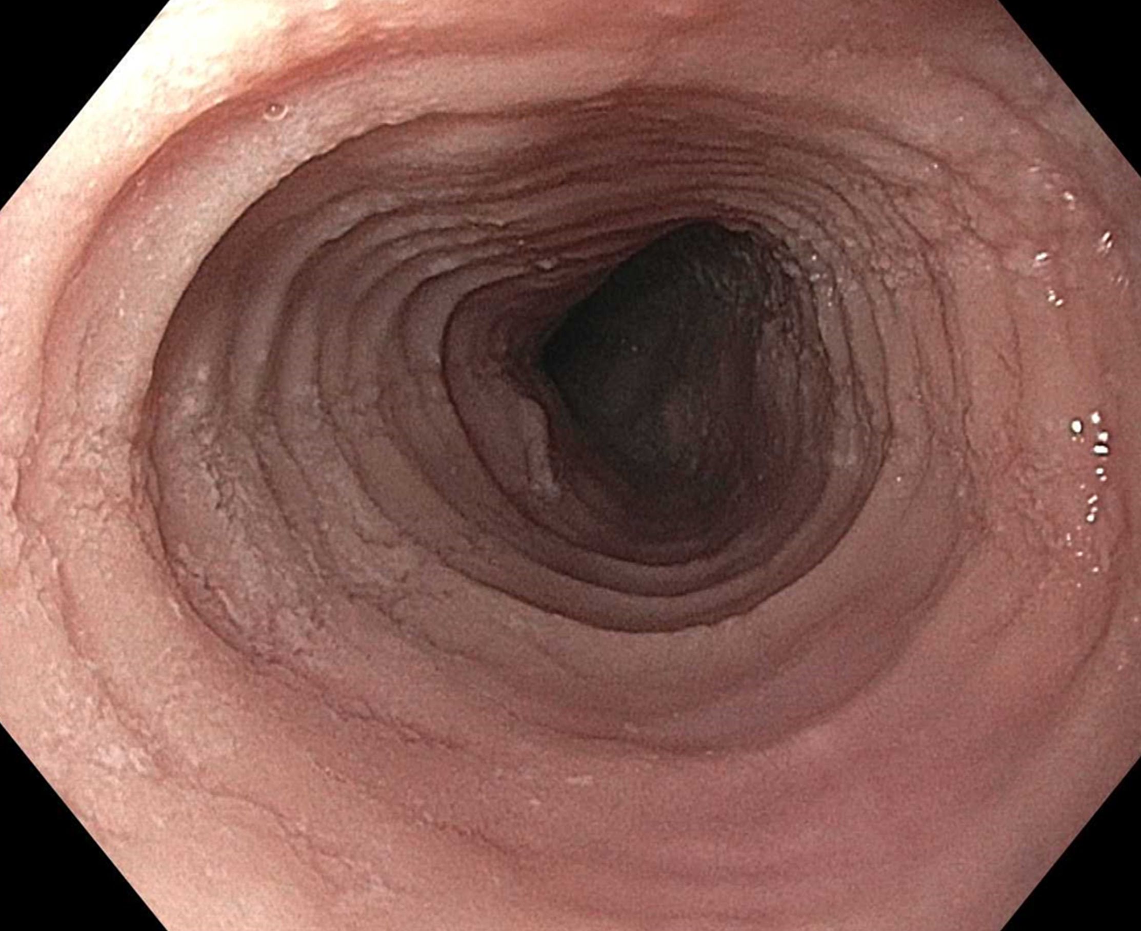
After this, biopsies should be obtained with at least 6 biopsy fragments from different locations in the esophagus. Any visible endoscopic abnormalities should be targeted (the highest yield is in exudates and furrows). The rationale is that EoE is patchy and at least 6 biopsies will maximize diagnostic yield.10 Ideally the initial endoscopy for EoE should be done off of treatments (like PPI or diet restriction) as these could mask the diagnosis. If a patient with suspected EoE has an endoscopy while on PPI, and the endoscopy is normal, a diagnosis of EoE cannot be made. In this case, consideration should be given as to stopping the PPI, allowing a wash out period (at least 1-2 months), and then repeating the endoscopy to confirm the diagnosis. This is important as EoE is a chronic condition necessitating life-long treatment and monitoring, so a definitive diagnosis is critical.
The third and final step in diagnosis is assessing for other conditions that could cause esophageal eosinophilia.9 The most common differential diagnosis is gastroesophageal reflux disease (GERD). In some cases, EoE and GERD overlap or can have a complex relationship.13 Unfortunately the location of the eosinophilia (i.e. distal only) and the level of the eosinophil counts are not useful in making this distinction, so all clinical features (symptoms, presence of erosive esophagitis, or a hiatal hernia endoscopically), and ancillary reflex testing when indicated may be required prior to a formal EoE diagnosis. After the diagnosis is established, there should be direct communication with the patient to review the diagnosis and select treatments. While it is possible to convey results electronically in a messaging portal or with a letter, a more formal interaction, such as a clinic visit, is recommended because this is a new diagnosis of a chronic condition. Similarly, a new diagnosis of inflammatory bowel disease would never be made in a pathology follow-up letter alone.
Treatment of EoE
When it comes to treatment, the new guidelines emphasize several points.10 First, there is the concept that anti-inflammatory treatment should be paired with assessment of fibrostenosis and esophageal dilation; to do either in isolation is incomplete treatment. It is safe to perform dilation both prior to anti-inflammatory treatment (for example, with a critical stricture in a patient with dysphagia) and after anti-inflammatory treatment has been prescribed (for example, during an endoscopy to assess treatment response).
Second, proton pump inhibitors (PPIs), swallowed topical corticosteroids (tCS), or dietary elimination are all acceptable first-line treatment options for EoE. A shared decision-making framework should be used for this discussion. If dietary elimination is selected,14 based on new clinical trial data, guidelines recommend using empiric elimination and starting with a less restrictive diet (either a one-food elimination diet with dairy alone or a two-food elimination with dairy and wheat elimination). If PPIs are selected, the dose should be double the standard reflux dose. Data are mixed as to whether to use twice daily dosing (i.e., omeprazole 20 mg twice daily) or once a day dosing (i.e., omeprazole 40 mg daily), but total dose and adherence may be more important than frequency.10
For tCS use, either budesonide or fluticasone can be selected, but budesonide oral suspension is the only FDA-approved tCS for EoE.15 Initial treatment length is usually 6-8 weeks for diet elimination and, 12 weeks for PPI and tCS. In general, it is best to pick a single treatment to start, and reserve combining therapies for patients who do not have a complete response to a single modality as there are few data to support combination therapy.
After initial treatment, it is critical to assess for treatment response.16 Goals of EoE treatment include improvement in symptoms, but also improvement in endoscopic and histologic features to prevent complications. Symptoms in EoE do not always correlate with underlying biologic disease activity: patients can minimize symptoms with careful eating; they may perceive no difference in symptoms despite histologic improvement if a stricture persists; and they may have minimal symptoms after esophageal dilation despite ongoing inflammation. Because of this, performing a follow-up endoscopy after initial treatment is guideline-recommended.10, 17 This allows assessing for endoscopic improvement, re-assessing for fibrostenosis and performing dilation if indicated, and obtaining esophageal biopsies. If there is non-response, options include switching between other first line treatments or considering “stepping-up” to dupilumab which is also an FDA-approved option for EoE that is recommended in the guidelines.10, 18 In some cases where patients have multiple severe atopic conditions such as asthma or eczema that would warrant dupilumab use, or if patients are intolerant to PPIs or tCS, dupilumab could be considered as an earlier treatment for EoE.
Long-Term Maintenance
If a patient has a good response (for example, improved symptoms, improved endoscopic features, and <15 eos/hpf on biopsy), treatment can be maintained long-term. In almost all cases, if treatment is stopped, EoE disease activity recurs.19 Patients could be seen back in clinic in 6-12 months, and then a discussion can be conducted about a follow-up endoscopy, with timing to be determined based on their individual disease features and severity.17
Patients with more severe strictures, however, may have to be seen in endoscopy for serial dilations. Continued follow-up is essential for optimal care. Just as patients can progress in their disease course with diagnostic delay, there are data that show they can also progress after diagnosis when there are gaps in care without regular follow-up.20 Unlike other chronic esophageal disorders such as GERD and Barrett’s esophagus and other chronic GI inflammatory conditions like inflammatory bowel disease, however, EoE is not associated with an increased risk of esophageal cancer.21, 22
Given its increasing frequency, EoE will be commonly encountered by gastroenterologists both new and established. Having a systematic approach for diagnosis, understanding how to elicit subtle symptoms, implementing a shared decision-making framework for treatment with a structured algorithm for assessing response, performing follow-up, maintaining treatment, and monitoring patients long-term will allow the large majority of EoE patients to be successfully managed.
Dr. Dellon is based at the Center for Esophageal Diseases and Swallowing, Center for Gastrointestinal Biology and Disease, Division of Gastroenterology and Hepatology, University of North Carolina School of Medicine, Chapel Hill. He disclosed research funding, consultant fees, and educational grants from multiple companies.
References
1. Thel HL, et al. Prevalence and Costs of Eosinophilic Esophagitis in the United States. Clin Gastroenterol Hepatol. 2025 Feb. doi: 10.1016/j.cgh.2024.09.031.
2. Lam AY, et al. Epidemiologic Burden and Projections for Eosinophilic Esophagitis-Associated Emergency Department Visits in the United States: 2009-2030. Clin Gastroenterol Hepatol. 2023 Nov. doi: 10.1016/j.cgh.2023.04.028.
3. Schoepfer AM, et al. Delay in diagnosis of eosinophilic esophagitis increases risk for stricture formation in a time-dependent manner. Gastroenterology. 2013 Dec. doi: 10.1053/j.gastro.2013.08.015.
4. Dellon ES, Hirano I. Epidemiology and Natural History of Eosinophilic Esophagitis. Gastroenterology. 2018 Jan. doi: 10.1053/j.gastro.2017.06.067.
5. Chang JW, et al. Loss to follow-up after food impaction among patients with and without eosinophilic esophagitis. Dis Esophagus. 2019 Dec. doi: 10.1093/dote/doz056.
6. Aceves SS, et al. Endoscopic approach to eosinophilic esophagitis: American Society for Gastrointestinal Endoscopy Consensus Conference. Gastrointest Endosc. 2022 Aug. doi: 10.1016/j.gie.2022.05.013.
7. Leiman DA, et al. Quality Indicators for the Diagnosis and Management of Eosinophilic Esophagitis. Am J Gastroenterol. 2023 Jun. doi: 10.14309/ajg.0000000000002138.
8. Hirano I, Furuta GT. Approaches and Challenges to Management of Pediatric and Adult Patients With Eosinophilic Esophagitis. Gastroenterology. 2020 Mar. doi: 10.1053/j.gastro.2019.09.052.
9. Dellon ES, et al. Updated international consensus diagnostic criteria for eosinophilic esophagitis: Proceedings of the AGREE conference. Gastroenterology. 2018 Oct. doi: 10.1053/j.gastro.2018.07.009.
10. Dellon ES, et al. ACG Clinical Guideline: Diagnosis and Management of Eosinophilic Esophagitis. Am J Gastroenterol. 2025 Jan. doi: 10.14309/ajg.0000000000003194.
11. Dellon ES. Optimizing the Endoscopic Examination in Eosinophilic Esophagitis. Clin Gastroenterol Hepatol. 2021 Dec. doi: 10.1016/j.cgh.2021.07.011.
12. Hirano I, et al. Endoscopic assessment of the oesophageal features of eosinophilic oesophagitis: validation of a novel classification and grading system. Gut. 2012 May. doi: 10.1136/gutjnl-2011-301817.
13. Spechler SJ, et al. Thoughts on the complex relationship between gastroesophageal reflux disease and eosinophilic esophagitis. Am J Gastroenterol. 2007 Jun. doi: 10.1111/j.1572-0241.2007.01179.x.
14. Chang JW, et al. Development of a Practical Guide to Implement and Monitor Diet Therapy for Eosinophilic Esophagitis. Clin Gastroenterol Hepatol. 2023 Jul. doi: 10.1016/j.cgh.2023.03.006.
15. Hirano I, et al. Budesonide Oral Suspension Improves Outcomes in Patients With Eosinophilic Esophagitis: Results from a Phase 3 Trial. Clin Gastroenterol Hepatol. 2022 Mar. doi: 10.1016/j.cgh.2021.04.022.
16. Dellon ES, Gupta SK. A conceptual approach to understanding treatment response in eosinophilic esophagitis. Clin Gastroenterol Hepatol. 2019 Oct. doi: 10.1016/j.cgh.2019.01.030.
17. von Arnim U, et al. Monitoring Patients With Eosinophilic Esophagitis in Routine Clinical Practice - International Expert Recommendations. Clin Gastroenterol Hepatol. 2023 Sep. doi: 10.1016/j.cgh.2022.12.018.
18. Dellon ES, et al. Dupilumab in Adults and Adolescents with Eosinophilic Esophagitis. N Engl J Med. 2022 Dec. doi: 10.1056/NEJMoa220598.
19. Dellon ES, et al. Rapid Recurrence of Eosinophilic Esophagitis Activity After Successful Treatment in the Observation Phase of a Randomized, Double-Blind, Double-Dummy Trial. Clin Gastroenterol Hepatol. 2020 Jun. doi: 10.1016/j.cgh.2019.08.050.
20. Chang NC, et al. A Gap in Care Leads to Progression of Fibrosis in Eosinophilic Esophagitis Patients. Clin Gastroenterol Hepatol. 2022 Aug. doi: 10.1016/j.cgh.2021.10.028.
21. Syed A, et al. The relationship between eosinophilic esophagitis and esophageal cancer. Dis Esophagus. 2017 Jul. doi: 10.1093/dote/dox050.
22. Albaneze N, et al. No Association Between Eosinophilic Oesophagitis and Oesophageal Cancer in US Adults: A Case-Control Study. Aliment Pharmacol Ther. 2025 Jan. doi: 10.1111/apt.18431.
Eosinophilic esophagitis (EoE) can be considered a “young” disease, with initial case series reported only about 30 years ago. Since that time, it has become a commonly encountered condition in both emergency and clinic settings. The most recent prevalence study estimates that 1 in 700 people in the U.S. have EoE,1 the volume of EoE-associated ED visits tripped between 2009 and 2019 and is projected to double again by 2030,2 and “new” gastroenterologists undoubtedly have learned about and seen this condition. As a chronic disease, EoE necessitates longitudinal follow-up and optimization of care to prevent complications. With increasing diagnostic delay, EoE progresses in most, but not all, patients from an inflammatory- to fibrostenotic-predominant condition.3
Diagnosis of EoE
The most likely area that you will encounter EoE is during an emergent middle-of-the-night endoscopy for food impaction. If called in for this, EoE will be the cause in more than 50% of patients.4 However, the diagnosis can only be made if esophageal biopsies are obtained at the time of the procedure. This is a critical time to decrease diagnostic delay, as half of patients are lost to follow-up after a food impaction.5 Unfortunately, although taking biopsies during index food impaction is guideline-recommended, a quality metric, and safe to obtain after the food bolus is cleared, this is infrequently done in practice.6, 7
The next most likely area for EoE detection is in the endoscopy suite where 15-23% of patients with dysphagia and 5-7% of patients undergoing upper endoscopy for any indication will have EoE.4 Sometimes EoE will be detected “incidentally” during an open-access case (for example, in a patient with diarrhea undergoing evaluation for celiac). In these cases, it is important to perform a careful history (as noted below) as subtle EoE symptoms can frequently be identified. Finally, when patients are seen in clinic for solid food dysphagia, EoE is clearly on the differential. A few percent of patients with refractory heartburn or chest pain will have EoE causing the symptoms rather than reflux,4 and all patients under consideration for antireflux surgery should have an endoscopy to assess for EoE.
When talking to patients with known or suspected EoE, the history must go beyond general questions about dysphagia or trouble swallowing. Many patients with EoE have overtly or subconsciously modified their eating behaviors over many years to minimize symptoms, may have adapted to chronic dysphagia, and will answer “no” when asked if they have trouble swallowing. Instead, use the acronym “IMPACT” to delve deeper into possible symptoms.8 Do they “Imbibe” fluids or liquids between each bite to help get food down? Do they “Modify” the way they eat (cut food into small bites; puree foods)? Do they “Prolong” mealtimes? Do they “Avoid” certain foods that stick? Do they “Chew’ until their food is a mush to get it down? And do they “Turn away” tablets or pills? Pill dysphagia is often a subtle symptom, and sometimes the only symptom elicited.
Additionally, it may be important to ask a partner or family member (if present) about their observations. They may provide insight (e.g. “yes – he chokes with every bite but never says it bothers him”) that the patient might not otherwise provide. The suspicion for EoE should also be increased in patients with concomitant atopic diseases and in those with a family history of dysphagia or who have family members needing esophageal dilation. It is important to remember that EoE can be seen across all ages, sexes, and races/ethnicities.
Diagnosis of EoE is based on the AGREE consensus,9 which is also echoed in the recently updated American College of Gastroenterology (ACG) guidelines.10 Diagnosis requires three steps. First, symptoms of esophageal dysfunction must be present. This will most typically be dysphagia in adolescents and adults, but symptoms are non-specific in children (e.g. poor growth and feeding, abdominal pain, vomiting, regurgitation, heartburn).
Second, at least 15 eosinophils per high-power field (eos/hpf) are required on esophageal biopsy, which implies that an endoscopy be performed. A high-quality endoscopic exam in EoE is of the utmost importance. The approach has been described elsewhere,11 but enough time on insertion should be taken to fully insufflate and examine the esophagus, including the areas of the gastroesophageal junction and upper esophageal sphincter where strictures can be missed, to gently wash debris, and to assess the endoscopic findings of EoE. Endoscopic findings should be reported using the validated EoE Endoscopy Reference Score (EREFS),12 which grades five key features. EREFS is reproducible, is responsive to treatment, and is guideline-recommended (see Figure 1).6, 10 The features are edema (present=1), rings (mild=1; moderate=2; severe=3), exudates (mild=1; severe=2), furrows (mild=1; severe=2), and stricture (present=1; also estimate diameter in mm) and are incorporated into many endoscopic reporting programs. Additionally, diffuse luminal narrowing and mucosal fragility (“crepe-paper” mucosa) should be assessed.
After this, biopsies should be obtained with at least 6 biopsy fragments from different locations in the esophagus. Any visible endoscopic abnormalities should be targeted (the highest yield is in exudates and furrows). The rationale is that EoE is patchy and at least 6 biopsies will maximize diagnostic yield.10 Ideally the initial endoscopy for EoE should be done off of treatments (like PPI or diet restriction) as these could mask the diagnosis. If a patient with suspected EoE has an endoscopy while on PPI, and the endoscopy is normal, a diagnosis of EoE cannot be made. In this case, consideration should be given as to stopping the PPI, allowing a wash out period (at least 1-2 months), and then repeating the endoscopy to confirm the diagnosis. This is important as EoE is a chronic condition necessitating life-long treatment and monitoring, so a definitive diagnosis is critical.
The third and final step in diagnosis is assessing for other conditions that could cause esophageal eosinophilia.9 The most common differential diagnosis is gastroesophageal reflux disease (GERD). In some cases, EoE and GERD overlap or can have a complex relationship.13 Unfortunately the location of the eosinophilia (i.e. distal only) and the level of the eosinophil counts are not useful in making this distinction, so all clinical features (symptoms, presence of erosive esophagitis, or a hiatal hernia endoscopically), and ancillary reflex testing when indicated may be required prior to a formal EoE diagnosis. After the diagnosis is established, there should be direct communication with the patient to review the diagnosis and select treatments. While it is possible to convey results electronically in a messaging portal or with a letter, a more formal interaction, such as a clinic visit, is recommended because this is a new diagnosis of a chronic condition. Similarly, a new diagnosis of inflammatory bowel disease would never be made in a pathology follow-up letter alone.
Treatment of EoE
When it comes to treatment, the new guidelines emphasize several points.10 First, there is the concept that anti-inflammatory treatment should be paired with assessment of fibrostenosis and esophageal dilation; to do either in isolation is incomplete treatment. It is safe to perform dilation both prior to anti-inflammatory treatment (for example, with a critical stricture in a patient with dysphagia) and after anti-inflammatory treatment has been prescribed (for example, during an endoscopy to assess treatment response).
Second, proton pump inhibitors (PPIs), swallowed topical corticosteroids (tCS), or dietary elimination are all acceptable first-line treatment options for EoE. A shared decision-making framework should be used for this discussion. If dietary elimination is selected,14 based on new clinical trial data, guidelines recommend using empiric elimination and starting with a less restrictive diet (either a one-food elimination diet with dairy alone or a two-food elimination with dairy and wheat elimination). If PPIs are selected, the dose should be double the standard reflux dose. Data are mixed as to whether to use twice daily dosing (i.e., omeprazole 20 mg twice daily) or once a day dosing (i.e., omeprazole 40 mg daily), but total dose and adherence may be more important than frequency.10
For tCS use, either budesonide or fluticasone can be selected, but budesonide oral suspension is the only FDA-approved tCS for EoE.15 Initial treatment length is usually 6-8 weeks for diet elimination and, 12 weeks for PPI and tCS. In general, it is best to pick a single treatment to start, and reserve combining therapies for patients who do not have a complete response to a single modality as there are few data to support combination therapy.
After initial treatment, it is critical to assess for treatment response.16 Goals of EoE treatment include improvement in symptoms, but also improvement in endoscopic and histologic features to prevent complications. Symptoms in EoE do not always correlate with underlying biologic disease activity: patients can minimize symptoms with careful eating; they may perceive no difference in symptoms despite histologic improvement if a stricture persists; and they may have minimal symptoms after esophageal dilation despite ongoing inflammation. Because of this, performing a follow-up endoscopy after initial treatment is guideline-recommended.10, 17 This allows assessing for endoscopic improvement, re-assessing for fibrostenosis and performing dilation if indicated, and obtaining esophageal biopsies. If there is non-response, options include switching between other first line treatments or considering “stepping-up” to dupilumab which is also an FDA-approved option for EoE that is recommended in the guidelines.10, 18 In some cases where patients have multiple severe atopic conditions such as asthma or eczema that would warrant dupilumab use, or if patients are intolerant to PPIs or tCS, dupilumab could be considered as an earlier treatment for EoE.
Long-Term Maintenance
If a patient has a good response (for example, improved symptoms, improved endoscopic features, and <15 eos/hpf on biopsy), treatment can be maintained long-term. In almost all cases, if treatment is stopped, EoE disease activity recurs.19 Patients could be seen back in clinic in 6-12 months, and then a discussion can be conducted about a follow-up endoscopy, with timing to be determined based on their individual disease features and severity.17
Patients with more severe strictures, however, may have to be seen in endoscopy for serial dilations. Continued follow-up is essential for optimal care. Just as patients can progress in their disease course with diagnostic delay, there are data that show they can also progress after diagnosis when there are gaps in care without regular follow-up.20 Unlike other chronic esophageal disorders such as GERD and Barrett’s esophagus and other chronic GI inflammatory conditions like inflammatory bowel disease, however, EoE is not associated with an increased risk of esophageal cancer.21, 22
Given its increasing frequency, EoE will be commonly encountered by gastroenterologists both new and established. Having a systematic approach for diagnosis, understanding how to elicit subtle symptoms, implementing a shared decision-making framework for treatment with a structured algorithm for assessing response, performing follow-up, maintaining treatment, and monitoring patients long-term will allow the large majority of EoE patients to be successfully managed.
Dr. Dellon is based at the Center for Esophageal Diseases and Swallowing, Center for Gastrointestinal Biology and Disease, Division of Gastroenterology and Hepatology, University of North Carolina School of Medicine, Chapel Hill. He disclosed research funding, consultant fees, and educational grants from multiple companies.
References
1. Thel HL, et al. Prevalence and Costs of Eosinophilic Esophagitis in the United States. Clin Gastroenterol Hepatol. 2025 Feb. doi: 10.1016/j.cgh.2024.09.031.
2. Lam AY, et al. Epidemiologic Burden and Projections for Eosinophilic Esophagitis-Associated Emergency Department Visits in the United States: 2009-2030. Clin Gastroenterol Hepatol. 2023 Nov. doi: 10.1016/j.cgh.2023.04.028.
3. Schoepfer AM, et al. Delay in diagnosis of eosinophilic esophagitis increases risk for stricture formation in a time-dependent manner. Gastroenterology. 2013 Dec. doi: 10.1053/j.gastro.2013.08.015.
4. Dellon ES, Hirano I. Epidemiology and Natural History of Eosinophilic Esophagitis. Gastroenterology. 2018 Jan. doi: 10.1053/j.gastro.2017.06.067.
5. Chang JW, et al. Loss to follow-up after food impaction among patients with and without eosinophilic esophagitis. Dis Esophagus. 2019 Dec. doi: 10.1093/dote/doz056.
6. Aceves SS, et al. Endoscopic approach to eosinophilic esophagitis: American Society for Gastrointestinal Endoscopy Consensus Conference. Gastrointest Endosc. 2022 Aug. doi: 10.1016/j.gie.2022.05.013.
7. Leiman DA, et al. Quality Indicators for the Diagnosis and Management of Eosinophilic Esophagitis. Am J Gastroenterol. 2023 Jun. doi: 10.14309/ajg.0000000000002138.
8. Hirano I, Furuta GT. Approaches and Challenges to Management of Pediatric and Adult Patients With Eosinophilic Esophagitis. Gastroenterology. 2020 Mar. doi: 10.1053/j.gastro.2019.09.052.
9. Dellon ES, et al. Updated international consensus diagnostic criteria for eosinophilic esophagitis: Proceedings of the AGREE conference. Gastroenterology. 2018 Oct. doi: 10.1053/j.gastro.2018.07.009.
10. Dellon ES, et al. ACG Clinical Guideline: Diagnosis and Management of Eosinophilic Esophagitis. Am J Gastroenterol. 2025 Jan. doi: 10.14309/ajg.0000000000003194.
11. Dellon ES. Optimizing the Endoscopic Examination in Eosinophilic Esophagitis. Clin Gastroenterol Hepatol. 2021 Dec. doi: 10.1016/j.cgh.2021.07.011.
12. Hirano I, et al. Endoscopic assessment of the oesophageal features of eosinophilic oesophagitis: validation of a novel classification and grading system. Gut. 2012 May. doi: 10.1136/gutjnl-2011-301817.
13. Spechler SJ, et al. Thoughts on the complex relationship between gastroesophageal reflux disease and eosinophilic esophagitis. Am J Gastroenterol. 2007 Jun. doi: 10.1111/j.1572-0241.2007.01179.x.
14. Chang JW, et al. Development of a Practical Guide to Implement and Monitor Diet Therapy for Eosinophilic Esophagitis. Clin Gastroenterol Hepatol. 2023 Jul. doi: 10.1016/j.cgh.2023.03.006.
15. Hirano I, et al. Budesonide Oral Suspension Improves Outcomes in Patients With Eosinophilic Esophagitis: Results from a Phase 3 Trial. Clin Gastroenterol Hepatol. 2022 Mar. doi: 10.1016/j.cgh.2021.04.022.
16. Dellon ES, Gupta SK. A conceptual approach to understanding treatment response in eosinophilic esophagitis. Clin Gastroenterol Hepatol. 2019 Oct. doi: 10.1016/j.cgh.2019.01.030.
17. von Arnim U, et al. Monitoring Patients With Eosinophilic Esophagitis in Routine Clinical Practice - International Expert Recommendations. Clin Gastroenterol Hepatol. 2023 Sep. doi: 10.1016/j.cgh.2022.12.018.
18. Dellon ES, et al. Dupilumab in Adults and Adolescents with Eosinophilic Esophagitis. N Engl J Med. 2022 Dec. doi: 10.1056/NEJMoa220598.
19. Dellon ES, et al. Rapid Recurrence of Eosinophilic Esophagitis Activity After Successful Treatment in the Observation Phase of a Randomized, Double-Blind, Double-Dummy Trial. Clin Gastroenterol Hepatol. 2020 Jun. doi: 10.1016/j.cgh.2019.08.050.
20. Chang NC, et al. A Gap in Care Leads to Progression of Fibrosis in Eosinophilic Esophagitis Patients. Clin Gastroenterol Hepatol. 2022 Aug. doi: 10.1016/j.cgh.2021.10.028.
21. Syed A, et al. The relationship between eosinophilic esophagitis and esophageal cancer. Dis Esophagus. 2017 Jul. doi: 10.1093/dote/dox050.
22. Albaneze N, et al. No Association Between Eosinophilic Oesophagitis and Oesophageal Cancer in US Adults: A Case-Control Study. Aliment Pharmacol Ther. 2025 Jan. doi: 10.1111/apt.18431.
WATS-3D Biopsy Increases Detection of Barrett’s Esophagus in GERD
, new research showed.

Compared with forceps biopsies (FB) alone, the addition of WATS-3D led to confirmation of BE in an additional one fifth of patients, roughly doubled dysplasia diagnoses, and influenced clinical management in the majority of patients.
“The big take-home point here is that the use of WATS-3D brushing along with conventional biopsies increases the likelihood that intestinal metaplasia will be identified,” first author Nicholas Shaheen, MD, MPH, AGAF, with the Center for Esophageal Diseases and Swallowing, University of North Carolina School of Medicine at Chapel Hill, North Carolina, told GI & Hepatology News.
“Almost 20% of patients who harbor BE were only identified by WATS-3D and might have otherwise gone undiagnosed had only forceps biopsies been performed,” Shaheen said.
The study was published in The American Journal of Gastroenterology.
Beyond Traditional Biopsies
BE develops as a complication of chronic GERD and is the chief precursor to esophageal adenocarcinoma. Early detection of BE and dysplasia is crucial to enable timely intervention.
The current gold standard for BE screening involves upper endoscopy with FB following the Seattle protocol, which consists of four-quadrant biopsies from every 1-2 cm of areas of columnar-lined epithelium (CLE) to confirm the presence of intestinal metaplasia. However, this protocol is prone to sampling errors and high false-negative rates, leading to repeat endoscopy, the study team pointed out.
WATS-3D (CDx Diagnostics) is a complementary technique designed to improve diagnostic yield by using brush biopsy to sample more tissue than routine biopsies.
WATS-3D has been shown to increase detection of dysplasia in patients with BE undergoing surveillance for BE, but less is known about the value of WATS-3D for BE screening in a community-based cohort of patients with GERD.
To investigate, Shaheen and colleagues studied 23,933 consecutive patients enrolled in a prospective observational registry assessing the utility of WATS-3D in the screening of symptomatic GERD patients for BE.
Patients had both WATS-3D and FB in the same endoscopic session. No patient had a history of BE, intestinal metaplasia or dysplasia in esophageal mucosa, or esophageal surgery, endoscopic ablation or endoscopic mucosal resection prior to enrollment.
Overall, 6829 patients (29%) met endoscopic criteria for BE (≥ 1 cm esophageal CLE with accompanying biopsies showing intestinal metaplasia).
Of these, 2878 (42%) had intestinal metaplasia identified by either FB or WATS-3D, but 19.3% had their BE diagnosis confirmed solely on the basis of WATS-3D findings.
Among patients who fulfilled the endoscopic criteria for BE, the adjunctive yield of WATS-3D was 76.5% and the absolute yield was 18.1%.
Of the 240 (1.0%) patients with dysplasia, 107 (45%) were found solely by WATS-3D.
‘Clinically Valuable Adjunct’
Among patients with positive WATS-3D but negative FB results, clinical management changed in 90.7% of cases, mostly involving initiation or modification of surveillance and proton pump inhibitor therapy.
These results suggest that WATS-3D is a “clinically valuable adjunct” to FB for the diagnosis of BE when used as a screening tool in symptomatic GERD patients and particularly in patients with endoscopic evidence of > 1 cm esophageal columnar-lined epithelium, the study team wrote.
Adjunctive use of WATS-3D when BE is suspected “may save endoscopies and lead to quicker, more accurate diagnoses,” they added.
The investigators said a limitation of the study is the lack of central pathology review, potentially leading to diagnostic variability. They also noted that over half of the detected dysplasia cases were crypt dysplasia or indefinite for dysplasia, raising concerns about clinical significance.
Reached for comment, Philip O. Katz, MD, AGAF, professor of medicine and director of the GI Function Laboratories, Weill Cornell Medicine in New York, said he’s been using WATS for more than a decade as an adjunct to standard biopsy in patients undergoing screening and surveillance for BE and finds it clinically helpful in managing his patients.
This new study provides “further information that WATS added to biopsy that has been traditionally done with the Seattle protocol increases the yield of intestinal metaplasia and likely dysplasia in patients being screened for Barrett’s,” Katz, who wasn’t involved in the study, told GI & Hepatology News.
Funding for the study was provided by CDx Diagnostics. Shaheen and several coauthors disclosed relationships with the company. Katz disclosed relationships (consultant/advisor) for Phathom Pharmaceuticals and Sebella.
A version of this article appeared on Medscape.com.
, new research showed.
Compared with forceps biopsies (FB) alone, the addition of WATS-3D led to confirmation of BE in an additional one fifth of patients, roughly doubled dysplasia diagnoses, and influenced clinical management in the majority of patients.
“The big take-home point here is that the use of WATS-3D brushing along with conventional biopsies increases the likelihood that intestinal metaplasia will be identified,” first author Nicholas Shaheen, MD, MPH, AGAF, with the Center for Esophageal Diseases and Swallowing, University of North Carolina School of Medicine at Chapel Hill, North Carolina, told GI & Hepatology News.
“Almost 20% of patients who harbor BE were only identified by WATS-3D and might have otherwise gone undiagnosed had only forceps biopsies been performed,” Shaheen said.
The study was published in The American Journal of Gastroenterology.
Beyond Traditional Biopsies
BE develops as a complication of chronic GERD and is the chief precursor to esophageal adenocarcinoma. Early detection of BE and dysplasia is crucial to enable timely intervention.
The current gold standard for BE screening involves upper endoscopy with FB following the Seattle protocol, which consists of four-quadrant biopsies from every 1-2 cm of areas of columnar-lined epithelium (CLE) to confirm the presence of intestinal metaplasia. However, this protocol is prone to sampling errors and high false-negative rates, leading to repeat endoscopy, the study team pointed out.
WATS-3D (CDx Diagnostics) is a complementary technique designed to improve diagnostic yield by using brush biopsy to sample more tissue than routine biopsies.
WATS-3D has been shown to increase detection of dysplasia in patients with BE undergoing surveillance for BE, but less is known about the value of WATS-3D for BE screening in a community-based cohort of patients with GERD.
To investigate, Shaheen and colleagues studied 23,933 consecutive patients enrolled in a prospective observational registry assessing the utility of WATS-3D in the screening of symptomatic GERD patients for BE.
Patients had both WATS-3D and FB in the same endoscopic session. No patient had a history of BE, intestinal metaplasia or dysplasia in esophageal mucosa, or esophageal surgery, endoscopic ablation or endoscopic mucosal resection prior to enrollment.
Overall, 6829 patients (29%) met endoscopic criteria for BE (≥ 1 cm esophageal CLE with accompanying biopsies showing intestinal metaplasia).
Of these, 2878 (42%) had intestinal metaplasia identified by either FB or WATS-3D, but 19.3% had their BE diagnosis confirmed solely on the basis of WATS-3D findings.
Among patients who fulfilled the endoscopic criteria for BE, the adjunctive yield of WATS-3D was 76.5% and the absolute yield was 18.1%.
Of the 240 (1.0%) patients with dysplasia, 107 (45%) were found solely by WATS-3D.
‘Clinically Valuable Adjunct’
Among patients with positive WATS-3D but negative FB results, clinical management changed in 90.7% of cases, mostly involving initiation or modification of surveillance and proton pump inhibitor therapy.
These results suggest that WATS-3D is a “clinically valuable adjunct” to FB for the diagnosis of BE when used as a screening tool in symptomatic GERD patients and particularly in patients with endoscopic evidence of > 1 cm esophageal columnar-lined epithelium, the study team wrote.
Adjunctive use of WATS-3D when BE is suspected “may save endoscopies and lead to quicker, more accurate diagnoses,” they added.
The investigators said a limitation of the study is the lack of central pathology review, potentially leading to diagnostic variability. They also noted that over half of the detected dysplasia cases were crypt dysplasia or indefinite for dysplasia, raising concerns about clinical significance.
Reached for comment, Philip O. Katz, MD, AGAF, professor of medicine and director of the GI Function Laboratories, Weill Cornell Medicine in New York, said he’s been using WATS for more than a decade as an adjunct to standard biopsy in patients undergoing screening and surveillance for BE and finds it clinically helpful in managing his patients.
This new study provides “further information that WATS added to biopsy that has been traditionally done with the Seattle protocol increases the yield of intestinal metaplasia and likely dysplasia in patients being screened for Barrett’s,” Katz, who wasn’t involved in the study, told GI & Hepatology News.
Funding for the study was provided by CDx Diagnostics. Shaheen and several coauthors disclosed relationships with the company. Katz disclosed relationships (consultant/advisor) for Phathom Pharmaceuticals and Sebella.
A version of this article appeared on Medscape.com.
ACG Issues First Guidance on Diagnosis and Management of Gastric Premalignant Conditions
including atrophic gastritis, gastric intestinal metaplasia, dysplasia, and certain gastric epithelial polyps, all of which have an increased risk of progressing to gastric cancer.
The guideline was published online in The American Journal of Gastroenterology.
GPMCs are “common in gastroenterology practices, but in the US, at least, we’ve not had concrete guidance,” first author Douglas Morgan, MD, MPH, AGAF, Division of Gastroenterology, The University of Alabama at Birmingham, noted in an interview.
With these guidelines, we hope there “will be a paradigm shift to finally establish surveillance in the stomach, much like we’ve been doing for decades in the colon and the esophagus,” Morgan said.
Gastric cancer is a common cancer in the United States with disproportionately higher incidence rates in immigrants from countries with a high incidence of gastric cancer and certain non-White populations.
In addition, the 5-year survival rate in the United States for gastric cancer is 36%, which falls short of global standards and is driven by the fact that only a small percentage of these cancers are diagnosed in the early, curable stage.
These guidelines will help address this marked disparity and the burden on minority and marginalized populations, the guideline authors wrote. “The overarching goals are to reduce [gastric cancer] incidence in the United States, increase the detection of early-stage disease (early gastric cancer), and to significantly increase the 5-year survival rates in the near term.”
Key Recommendations
The guideline includes recommendations on endoscopic surveillance for high-risk patients, the performance of high-quality endoscopy and image-enhanced endoscopy (IEE) for diagnosis and surveillance, GPMC histology criteria and reporting, endoscopic treatment of dysplasia, the role of Helicobacter pylori eradication, general risk reduction measures, and the management of autoimmune gastritis and gastric epithelial polyps.
In terms of screening, the guidelines recommend against routine upper endoscopy screening for gastric cancer and GPMC in the general US population (low quality of evidence; conditional recommendation).
They also noted that there is “insufficient” direct US evidence to make a recommendation on opportunistic endoscopy screening for gastric cancer and GPMC in patients deemed at high risk based on race/ethnicity and family history. In addition, they recommend against the use of noninvasive biomarkers for screening or surveillance of GPMC or gastric cancer.
In terms of endoscopic and histologic diagnosis of GPMC, high-quality endoscopic evaluation is crucial to detect premalignant conditions or gastric cancer, the authors said. This includes adequate mucosal cleansing and insufflation, and photodocumentation of anatomic landmarks, as well as use of high-definition white light endoscopy (HDWLE) and IEE.
Systematic gastric biopsies should follow the updated Sydney protocol, with at least two separate containers for antrum/incisura and corpus biopsies. Histology should document the subtype of gastric intestinal metaplasia — incomplete, complete, or mixed — and severity and extent of atrophic gastritis and metaplasia.
Morgan emphasized the importance of coordination between the gastroenterologist and pathologist. “Several of these measures are not routinely reported, so we need to be in conversations with our collaborating pathologists,” he told this news organization.
In terms of GPMC surveillance, the authors suggest surveillance endoscopy every 3 years in high-risk patients with gastric intestinal metaplasia who meet one of the following criteria: High-risk histology (incomplete vs complete subtype, extending into the corpus); family history of gastric cancer in a first-degree relative; foreign-born individuals from high-gastric cancer incidence nations; or high-risk race/ethnicity (East Asian, Latino/a, Black, American Indian, or Alaska Native).
Individuals with multiple risk factors for gastric cancer may be considered for shorter than 3-year intervals.
Low-risk gastric intestinal metaplasia (limited to antrum, mild atrophy, and complete gastric intestinal metaplasia subtype) does not require routine endoscopic surveillance.
In terms of endoscopic management of dysplastic GPMC, endoscopic resection is suggested for dysplasia with visible margins. If dysplasia is not visible, repeat endoscopy with HDWLE and IEE by an experienced endoscopist is advised.
In patients appropriate for endoscopic resection of dysplasia, particularly endoscopic submucosal dissection, referral to a high-volume center with appropriate expertise in the diagnosis and therapeutic resection of gastric neoplasia is strongly recommended.
In patients with confirmed complete resection of dysplasia, endoscopic surveillance is also strongly recommended. Surveillance examinations should be performed by an experienced endoscopist using HDWLE and IEE, with biopsies according to the systematic biopsy protocol in addition to targeted biopsies.
In terms of nonendoscopic GPMC management, testing for H pylori (and eradication treatment if possible) is strongly recommended for patients with GPMC and those with a history of resected early gastric cancer.
Aspirin, nonsteroidal anti-inflammatory drugs, cyclooxygenase 2 inhibitors, or antioxidants are not recommended for patients with GPMC for the purpose of preventing gastric cancer.
In patients with autoimmune gastritis, testing for H pylori with a nonserological test, eradication treatment if positive, and posttreatment testing to confirm eradication is advised.
There is not enough evidence to make a formal recommendation on endoscopic surveillance in those with autoimmune gastritis; surveillance should be individualized, considering the risk for neuroendocrine tumors and possibly gastric cancer.
In terms of gastric epithelial polyps, endoscopic resection of all gastric adenomas is recommended, regardless of size, to exclude or prevent dysplasia and early gastric cancer. Adenomas that are not amenable to endoscopic resection should be referred for surgical resection, if clinically appropriate.
Morgan noted that the ACG GPMC guideline aligns with the updated ACG/American Society for Gastrointestinal Endoscopy upper endoscopy quality indicators released earlier this year.
Implementation of the ACG GPMC guideline and “change in clinical practice will require concrete targets and include training and quality initiatives,” the authors said.
This research received no commercial support. Morgan disclosed research support from Panbela Therapeutics, Thorne, and American Molecular Laboratories.
A version of this article first appeared on Medscape.com.
Safety Profile of GLP-1s ‘Reassuring’ in Upper Endoscopy
according to a meta-analysis of more than 80,000 patients.
Safety profiles, however, were comparable across groups, suggesting that prolonged fasting may be a sufficient management strategy, instead of withholding GLP-1RAs, lead author Antonio Facciorusso, MD, PhD, of the University of Foggia, Italy, and colleagues reported.
“The impact of GLP-1RAs on slowing gastric motility has raised concerns in patients undergoing endoscopic procedures, particularly upper endoscopies,” the investigators wrote in Clinical Gastroenterology and Hepatology. “This is due to the perceived risk of aspiration of retained gastric contents in sedated patients and the decreased visibility of the gastric mucosa, which can reduce the diagnostic yield of the examination.”
The American Society of Anesthesiologists (ASA) recommends withholding GLP-1RAs before procedures or surgery, whereas AGA suggests an individualized approach, citing limited supporting data.
A previous meta-analysis reported that GLP-1RAs mildly delayed gastric emptying, but clinical relevance remained unclear.
The present meta-analysis aimed to clarify this uncertainty by analyzing 13 retrospective studies that involved 84,065 patients undergoing upper endoscopy. Outcomes were compared among GLP-1RA users vs non-users, including rates of retained gastric contents, aborted procedures, and adverse events.
Patients on GLP-1RAs had significantly higher rates of retained gastric contents than non-users (odds ratio [OR], 5.56), a finding that held steady (OR, 4.20) after adjusting for age, sex, diabetes, body mass index, and other therapies.
GLP-1RAs were also associated with an increased likelihood of aborted procedures (OR, 5.13; 1% vs. 0.3%) and a higher need for repeat endoscopies (OR, 2.19; 1% vs 2%); however, Facciorusso and colleagues noted that these events, in absolute terms, were relatively uncommon.
“The rate of aborted and repeat procedures in the included studies was low,” the investigators wrote. “This meant that only for every 110 patients undergoing upper endoscopy while in GLP-1RA therapy would we observe an aborted procedure and only for every 120 patients would we need to repeat the procedure.”
The overall safety profile of GLP-1RAs in the context of upper endoscopy remained largely reassuring, they added. Specifically, rates of bronchial aspiration were not significantly different between users and non-users. What’s more, no single study reported a statistically significant increase in major complications, including pulmonary adverse events, among GLP-1RA users.
According to Facciorusso and colleagues, these findings suggest that retained gastric contents do not appear to substantially heighten the risk of serious harm, though further prospective studies are needed.
“Our comprehensive analysis indicates that, while the use of GLP-1RA results in higher rates of [retained gastric contents], the actual clinical impact appears to be limited,” they wrote. “Therefore, there is no strong evidence to support the routine discontinuation of the drug before upper endoscopy procedures.”
Instead, they supported the AGA task force’s recommendation for an individualized approach, and not withholding GLP-1RAs unnecessarily, calling this “the best compromise.”
“Prolonging the duration of fasting for solids could represent the optimal approach in these patients, although this strategy requires further evaluation,” the investigators concluded.
The investigators disclosed no conflicts of interest.
FROM CLINICAL GASTROENTEROLOGY AND HEPATOLOGY