User login
BET inhibitors could improve production of iPSCs

Image from Salk Institute
A study published in Cell Reports indicates that BET inhibitors can improve the reprogramming of human fibroblasts to create induced pluripotent stem cells (iPSCs).
According to researchers, this improvement in reprogramming can increase the yield of iPSCs from fibroblasts and enhance the quality of the iPSCs by ensuring that more somatic genes are efficiently turned down or turned off during reprogramming.
Study author Kejin Hu, PhD, of the University of Alabama at Birmingham, said the factors that are commonly used to create iPSCs from fibroblasts face a reprogramming barrier.
“If we can lower the barrier, we can enhance the reprogramming efficiency,” he explained. “My strategy is to use chemicals to erase the transcriptional program specific to the starting cells.”
Dr Hu and his colleagues found that BET-specific chemical inhibitors were effective in this regard.
For example, a low concentration of the BET inhibitor JQ1:
- Downregulated 390 fibroblast-specific genes when applied to naïve human fibroblasts
- Downregulated 651 fibroblast-specific genes when applied to human fibroblasts during reprogramming
- Increased the efficiency of successful reprogramming of human fibroblasts to iPSCs by 20-fold.
The researchers also found that fibroblasts change shape when treated with JQ1.
The cells transform from a long spindle shape to polygonal or rounded cells, which shows loss of fibroblast identity and transition to pluripotent stem cells. Presumably, genes that are needed to maintain the spindle shape are downregulated by JQ1.
Dr Hu proposed the following model to explain his team’s findings.
During normal cell division, active fibroblast genes are “bookmarked” by the attachment of BET proteins to acetylated chromatin during the mitotic phases, while RNA Polymerase II drops off of the chromatin.
At the start of interphase, these bookmarks guide the polymerase back to the genes, and they are transcribed by RNA Polymerase II.
In contrast, when JQ1 is added, the active fibroblast genes are de-bookmarked by the interaction of JQ1 with the BET proteins during the mitotic phases of cell division.
This “erases” the epigenetic memory of fibroblast gene expression, which, in turn, results in loss of fibroblast gene transcription when interphase returns.
This also increases the success of reprogramming into iPSCs. ![]()
Therapy can provide clinical benefit in haplo-HSCT, data suggest

Photo by Chad McNeeley
BARCELONA—Results from 2 studies suggest an immunogene therapy can provide a clinical benefit in adults with high-risk hematologic malignancies undergoing haploidentical hematopoietic stem cell transplant (haplo-HSCT).
When compared to historical controls, patients who received the immunogene therapy, Zalmoxis, had lower rates of non-relapse mortality (NRM) and chronic graft-vs-host disease (GVHD), as well as improved overall survival (OS).
The only adverse event related to Zalmoxis was GVHD, which was resolved.
These data were presented at the EBMT International Transplant Course. The studies were funded by MolMed S.p.A., the company developing Zalmoxis.
About the therapy
Zalmoxis is a treatment consisting of allogeneic, genetically modified T cells. The cells are intended to be given to haplo-HSCT recipients to help fight off infection, enhance the success of the transplant, and support long-lasting anticancer effects.
Because the genetically modified T cells can also cause GVHD, they are equipped with a suicide gene, which makes them susceptible to treatment with ganciclovir or valganciclovir. So if a patient develops GVHD, he or she can receive ganciclovir/valganciclovir, which should kill the modified T cells and prevent further development of GVHD.
Trial data
The data presented at the EBMT International Transplant Course were from the phase 1/2 TK007 trial and the ongoing phase 3 TK008 trial.
The TK007 trial included haplo-HSCT recipients with various high-risk hematologic malignancies, while the TK008 trial is enrolling patients with high-risk acute leukemia who are undergoing haplo-HSCT.
Researchers have compared 37 Zalmoxis-treated patients from these trials to 140 contemporaneous control patients from the database of the EBMT registry.
Results of this pair-matched analysis showed an OS improvement in Zalmoxis-treated patients, which was driven by a reduction in NRM. The OS was 49% in the Zalmoxis group and 37% in controls (P=0.01), and the NRM was 22% and 43%, respectively (P=0.014).
Among controls dying from non-relapse causes, the majority (78%) died from either infection (56%) or GVHD (22%), while the only adverse event related to Zalmoxis treatment was GVHD. And this GVHD was fully resolved by activating the suicide-gene system with ganciclovir treatment, without any GVHD-related death.
Furthermore, the incidence of chronic GVHD was lower in Zalmoxis-treated patients than in controls—6% and 25%, respectively (P=0.04).
Therefore, the researchers concluded that the protective effects of Zalmoxis in controlling infection and GVHD mainly drove the decreased NRM in the Zalmoxis group.
These data supported the European Commission’s recent decision to grant conditional marketing authorization for Zalmoxis. ![]()
FDA approves cord blood product
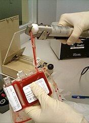
The US Food and Drug Administration (FDA) has issued a biologics license to the Cleveland Cord Blood Center (CCBC) for Clevecord™ (HPC, Cord Blood), a hematopoietic progenitor cell product derived from umbilical cord blood.
Under this license, CCBC is authorized to manufacture Clevecord at its facility in Warrensville Heights, Ohio.
Clevecord is indicated for use in unrelated donor transplant procedures, in conjunction with an appropriate preparative regimen.
The product can be used for hematopoietic and immunologic reconstitution in patients with disorders that affect the hematopoietic system, whether they are inherited, acquired, or result from myeloablative treatment.
Each Clevecord unit contains a minimum of 5 x 10⁸ total nucleated cells with at least 1.25 x 10⁶ viable CD34+ cells at the time of cryopreservation. The recommended minimum dose is 2.5 x 10⁷ nucleated cells/kg at cryopreservation.
Clevecord has been approved with a black box warning, which states that use of the product may result in fatal infusion reactions, graft-vs-host disease, engraftment syndrome, and graft failure.
For more details on Clevecord, see the package insert on the FDA website.
“Obtaining FDA licensure for Clevecord is reflective of the Cleveland Cord Blood Center’s dedication to meeting the highest quality standards in the industry for distribution of our cord blood products to transplant centers throughout the US and around the world,” said Wouter Van’t Hof, cord blood bank director at CCBC.
CCBC collects, processes, stores, and distributes umbilical cord blood units for use in transplants and advanced research in cellular therapy.
The organization says its cord blood collections represent a diverse cross-section of donor ethnicity to support transplant needs, particularly in the underserved African-American population.
“Up to 50% of parents giving birth in our partner hospitals donate their baby’s umbilical cord blood, a rate well above the national average,” said Marcie Finney, executive director of CCBC.
CCBC cord blood units can be searched and accessed through registries, including the National Bone Marrow Donor Program and Bone Marrow Donors Worldwide. ![]()
CBT may be best option for pts with MRD, doc says
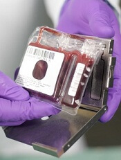
Photo courtesy of NHS
A cord blood transplant (CBT) may be the best option for patients with acute leukemia or myelodysplastic syndrome who have minimal residual disease (MRD) and no related donor, according to the senior author of a study published in NEJM.
This retrospective study showed that patients with MRD at the time of transplant were less likely to relapse if they received CBT rather than a graft from an unrelated adult donor, whether HLA-matched or mismatched.
In addition, the risk of death was significantly higher for patients with a mismatched donor than for CBT recipients, although there was no significant difference between those with a matched donor and CBT recipients.
Among patients without MRD, there were no significant differences between the transplant types for the risk of relapse or death.
“This paper shows that if you’ve got high-risk disease and are at high risk for relapse post-transplant, transplant with a cord blood donor may be the best option,” said Colleen Delaney, MD, of Fred Hutchinson Cancer Research Center in Seattle, Washington.
Dr Delaney and her colleagues analyzed data on 582 patients—300 with acute myeloid leukemia, 185 with acute lymphoblastic leukemia, and 97 with myelodysplastic syndromes.
Most patients received a transplant from an HLA-matched unrelated donor (n=344), 140 received a CBT from an unrelated donor, and 98 received a transplant from an HLA-mismatched unrelated donor.
The researchers calculated the relative risks of death and relapse for each transplant group, and they found that a patient’s MRD status prior to transplant played a role.
Presence of MRD
Among patients with MRD, the risk of death was significantly higher for recipients of mismatched grafts than for CBT recipients, with a hazard ratio (HR) of 2.92 (P=0.001).
However, the risk of death was not significantly different for recipients of matched grafts compared to CBT recipients. The HR was 1.69 (P=0.08).
The risk of relapse was about 3 times higher for recipients of mismatched grafts (HR=3.01, P=0.02) or matched grafts (HR=2.92, P=0.007) than for CBT recipients.
No MRD
Among patients without MRD, there was no significant difference in the risk of death for recipients of CBT, mismatched grafts (HR=1.36, P=0.30), or matched grafts (HR=0.78, P=0.33).
And there was no significant difference in the risk of relapse for recipients of CBT, mismatched grafts (HR=1.28, P=0.60), or matched grafts (HR=1.30, P=0.46).
“This brings home the point that cord blood shouldn’t be called an alternative donor,” Dr Delaney said. “The outcomes are the same as a conventional donor.” ![]()
HSCT may age T cells as much as 30 years

Photo by Chad McNeeley
New research suggests hematopoietic stem cell transplant (HSCT) may increase the molecular age of peripheral blood T cells.
The study showed an increase in peripheral blood T-cell senescence in patients with hematologic malignancies who were treated with autologous (auto-) or allogeneic (allo-) HSCT.
The patients had elevated levels of p16INK4a, a known marker of cellular senescence.
Auto-HSCT in particular had a strong effect on p16INK4a, increasing the expression of this marker to a degree comparable to 30 years of chronological aging.
Researchers reported these findings in EBioMedicine.
“We know that transplant is life-prolonging, and, in many cases, it’s life-saving for many patients with blood cancers and other disorders,” said study author William Wood, MD, of the University of North Carolina School of Medicine in Chapel Hill.
“At the same time, we’re increasingly recognizing that survivors of transplant are at risk for long-term health problems, and so there is interest in determining what markers may exist to help predict risk for long-term health problems or even in helping choose which patients are best candidates for transplantation.”
With this in mind, Dr Wood and his colleagues looked at levels of p16INK4a in 63 patients who underwent auto- or allo-HSCT to treat myeloma, lymphoma, or leukemia. The researchers assessed p16INK4a expression in T cells before HSCT and 6 months after.
Among auto-HSCT recipients, there were no baseline characteristics associated with pre-transplant p16INK4a expression.
However, allo-HSCT recipients had significantly higher pre-transplant p16INK4a levels the more cycles of chemotherapy they received before transplant (P=0.003), if they had previously undergone auto-HSCT (P=0.01), and if they had been exposed to alkylating agents (P=0.01).
After transplant, allo-HSCT recipients had a 1.93-fold increase in p16INK4a expression (P=0.0004), and auto-HSCT recipients had a 3.05-fold increase (P=0.002).
The researchers said the measured change in p16INK4a from pre- to post-HSCT in allogeneic recipients likely underestimates the age-promoting effects of HSCT, given that the pre-HSCT levels were elevated in the recipients from prior therapeutic exposure.
The researchers also pointed out that this study does not show a clear connection between changes in p16INK4a levels and the actual function of peripheral blood T cells, but they did say that p16INK4a is “arguably one of the best in vivo markers of cellular senescence and is directly associated with age-related deterioration.”
So the results of this research suggest the forced bone marrow repopulation associated with HSCT accelerates the molecular aging of peripheral blood T cells.
“Many oncologists would not be surprised by the finding that stem cell transplant accelerates aspects of aging,” said study author Norman Sharpless, MD, of the University of North Carolina School of Medicine.
“We know that, years after a curative transplant, stem cell transplant survivors are at increased risk for blood problems that can occur with aging, such as reduced immunity, increased risk for bone marrow failure, and increased risk of blood cancers. What is important about this work, however, is that it allows us to quantify the effect of stem cell transplant on molecular age.” ![]()
Drug granted orphan designation for GVHD
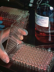
Photo by Linda Bartlett
The European Commission has granted orphan drug designation to ALXN1007 for the treatment of graft-versus-host disease (GVHD).
ALXN1007 is an anti-inflammatory monoclonal antibody targeting complement protein C5a.
The drug is currently under investigation in a phase 2 trial of patients with newly diagnosed acute GVHD of the lower gastrointestinal tract (GI-GVHD).
ALXN1007 is being developed by Alexion Pharmaceuticals, Inc.
About orphan designation
Orphan designation from the European Commission provides regulatory and financial incentives for companies to develop and market therapies that treat a life-threatening or chronically debilitating condition affecting no more than 5 in 10,000 people in the European Union, and where no satisfactory treatment is available.
Orphan designation provides a 10-year period of marketing exclusivity in the European Union if the drug receives regulatory approval. The designation also provides incentives for companies seeking protocol assistance from the European Medicines Agency during the product development phase and direct access to the centralized authorization procedure.
Phase 2 trial of ALXN1007
Results from the phase 2 trial of ALXN1007 in patients with newly diagnosed, acute GI-GVHD were presented at the 21st Congress of the European Hematology Association (abstract LB2269).
The presentation included 15 patients with biopsy-confirmed acute GI-GVHD. The patients had a median age of 60 (range, 25-69), and 60% were male.
Patients had acute myeloid leukemia/myelodysplastic syndrome (n=8), acute lymphoblastic leukemia (n=2), acute lymphocytic leukemia (n=1), acute myeloblastic leukemia (n=1), aplastic anemia (n=1), cutaneous T-cell lymphoma (n=1), or mantle cell lymphoma (n=1).
Most patients received transplants from matched, unrelated donors (n=11), 3 had matched, related donors, and 1 had a mismatched donor. Ten patients received peripheral blood grafts, 4 received cord blood, and 1 received a bone marrow transplant.
Patients had grade 1 (n=7), grade 2 (n=2), and grade 3 acute GI-GVHD (n=6).
The patients received weekly doses of ALXN1007 at 10 mg/kg, in combination with methylprednisolone at an initial dose of 2 mg/kg, through day 56.
Thirteen patients were evaluable for efficacy. One patient experienced leukemia relapse at day 18, and 1 withdrew from the study early.
The overall acute GVHD response rate was 77% (10/13), both at day 28 and day 56. The complete GI-GVHD response rate was 69% at day 28 and 77% at day 56.
At day 180, the nonrelapse mortality rate was 12.5%, and the overall survival rate was 69.2%.
All of the patients had treatment-emergent adverse events (AEs), and 11 patients (69%) had serious treatment-emergent AEs.
Five patients experienced a total of 12 treatment-related AEs (1 case each)—adenovirus infection, bronchopulmonary aspergillosis, chills, corona virus infection, viral cystitis, Epstein-Barr virus infection, hypersensitivity, influenza, influenza-like illness, infusion-related reaction, respiratory syncytial virus infection, and tremor.
There were 6 deaths, but none were considered treatment-related. ![]()
BSIs costly for pediatric transplant, cancer patients

Staphylococcus infection
Photo by Bill Branson
Ambulatory bloodstream infections (BSIs) can be costly in young cancer patients and recipients of hematopoietic stem cell transplants, according to research published in Pediatric Blood & Cancer.
Among the 61 patients studied, the median cost for an ambulatory BSI was $40,852, and the median length of hospital stay was 7 days.
For patients who were hospitalized for BSI and other medical issues, the cost and length of stay were much higher.
“This issue has resonance beyond the pediatric stem cell transplant and oncology patient population,” said study author Amy Billett, MD, of the Dana–Farber Cancer Institute and Boston Children’s Hospital in Massachusetts.
“At a time when many aspects of care are being shifted to the home and of heightened attention to safety and cost, this is the new frontier. What we learn about preventing outpatient bloodstream infections in these patients could have broad relevance.”
To determine the economic and hospitalization impact of ambulatory BSIs, Dr Billet and her colleagues retrospectively analyzed data on outpatient BSIs at Dana-Farber/Boston Children’s that occurred between January 1, 2012, and December 31, 2013, and resulted in hospitalization.
The team identified 74 BSIs in 61 patients. Sixty-nine percent of these infections were classified as central-line-associated bloodstream infections.
In 43% of BSIs, the patient’s central line had to be surgically removed. In 15% of cases, the child was transferred to the intensive care unit. Four patients died during hospitalization, and 3 of these deaths were associated with the infections.
Most of the hospitalizations analyzed—62—were due solely to BSIs. The remainder involved at least 1 other medical issue.
The median total cost of BSIs was $40,852, and the median length of hospital stay was 7 days.
The median cost was $36,611 among patients who were hospitalized for BSIs alone (n=62) and $89,935 for patients who were hospitalized for other medical issues as well. The median lengths of hospital stay were 6 days and 15 days, respectively.
The top 3 drivers of cost for all BSIs were room and board (43%), non-chemotherapy medications (22%), and procedures (11%).
Room and board accounted for 42% of charges among patients who were hospitalized for BSIs alone and 44% among the other patients. Non-chemotherapy medications accounted for 20% and 25%, respectively. And procedures accounted for 11% and 10%, respectively.
“Behind these metrics are real and serious risks to patients’ health,” said study author Chris Wong, MD, of Dana-Farber/Boston Children’s.
“The bottom line is that the dollar cost and lengthy hospital stays signal complications that could become life-threatening or delay treatment of the children’s cancer. Reducing these infections is important both for cost containment and quality of care.” ![]()

Staphylococcus infection
Photo by Bill Branson
Ambulatory bloodstream infections (BSIs) can be costly in young cancer patients and recipients of hematopoietic stem cell transplants, according to research published in Pediatric Blood & Cancer.
Among the 61 patients studied, the median cost for an ambulatory BSI was $40,852, and the median length of hospital stay was 7 days.
For patients who were hospitalized for BSI and other medical issues, the cost and length of stay were much higher.
“This issue has resonance beyond the pediatric stem cell transplant and oncology patient population,” said study author Amy Billett, MD, of the Dana–Farber Cancer Institute and Boston Children’s Hospital in Massachusetts.
“At a time when many aspects of care are being shifted to the home and of heightened attention to safety and cost, this is the new frontier. What we learn about preventing outpatient bloodstream infections in these patients could have broad relevance.”
To determine the economic and hospitalization impact of ambulatory BSIs, Dr Billet and her colleagues retrospectively analyzed data on outpatient BSIs at Dana-Farber/Boston Children’s that occurred between January 1, 2012, and December 31, 2013, and resulted in hospitalization.
The team identified 74 BSIs in 61 patients. Sixty-nine percent of these infections were classified as central-line-associated bloodstream infections.
In 43% of BSIs, the patient’s central line had to be surgically removed. In 15% of cases, the child was transferred to the intensive care unit. Four patients died during hospitalization, and 3 of these deaths were associated with the infections.
Most of the hospitalizations analyzed—62—were due solely to BSIs. The remainder involved at least 1 other medical issue.
The median total cost of BSIs was $40,852, and the median length of hospital stay was 7 days.
The median cost was $36,611 among patients who were hospitalized for BSIs alone (n=62) and $89,935 for patients who were hospitalized for other medical issues as well. The median lengths of hospital stay were 6 days and 15 days, respectively.
The top 3 drivers of cost for all BSIs were room and board (43%), non-chemotherapy medications (22%), and procedures (11%).
Room and board accounted for 42% of charges among patients who were hospitalized for BSIs alone and 44% among the other patients. Non-chemotherapy medications accounted for 20% and 25%, respectively. And procedures accounted for 11% and 10%, respectively.
“Behind these metrics are real and serious risks to patients’ health,” said study author Chris Wong, MD, of Dana-Farber/Boston Children’s.
“The bottom line is that the dollar cost and lengthy hospital stays signal complications that could become life-threatening or delay treatment of the children’s cancer. Reducing these infections is important both for cost containment and quality of care.”
Discovery in mice may have implications for HSCT

Preclinical research has shown how a cell surface molecule, Lymphotoxin β receptor, controls the entry of T cells into the thymus.
Researchers believe this might represent a pathway that could be targeted to reboot the immune system after hematopoietic stem cell transplant (HSCT).
Graham Anderson, PhD, of the University of Birmingham in the UK, and his colleagues conducted this research and reported their findings in the Journal of Immunology.
“The thymus is often something of an ignored organ, but it plays a crucial role in maintaining an effective immune system,” Dr Anderson said.
“Post-transplantation, T-cell progenitors derived from the bone marrow transplant can struggle to enter the thymus, as if the doorway to the thymus is closed. Identifying molecular regulators that can ‘prop open’ the door and allow these cells to enter and mature could well be a means to help reboot the immune system.”
Conducting experiments in mice, Dr Anderson and his colleagues found that Lymphotoxin β receptor was required to allow the entry of T-cell progenitors to the thymus both in a healthy state and during immune recovery after HSCT.
When the researchers used an antibody to stimulate Lymphotoxin β receptor after HSCT, they observed enhanced thymus recovery and an increase in the number of transplant-derived T cells, which correlated with increased adhesion molecule expression by thymic stroma.
The team said this study has revealed a novel link between Lymphotoxin β receptor and thymic stromal cells in thymus colonization and highlights its potential as an immunotherapeutic target to boost T-cell reconstitution after HSCT.
“This is just one piece of the puzzle,” said study author Beth Lucas, PhD, also of the University of Birmingham.
“It may be that there are adverse effects to opening the door to the thymus, but identifying a pathway that regulates this process is a significant step.”
For their next step, the researchers plan to study in vitro samples of the human thymus to examine the role Lymphotoxin β receptor might play in regulating thymus function in humans.
‘Barcoding’ reveals insights regarding HSCs

in the bone marrow
By assigning a “barcode” to hematopoietic stem cells (HSCs), researchers have found they can monitor the cells and study changes that occur over
time.
In tracking the barcoded HSCs, the team discovered why B-1a cells develop primarily during fetal and neonatal life, while adult bone marrow HSCs preferentially give rise to B-2 cells.
Joan Yuan, PhD, of Lund University in Sweden, and her colleagues described this discovery in Immunity.
“By assigning a barcode to the stem cells, we were able to track their performance over long periods of time and see which cells in the blood and the immune system they can induce,” Dr Yuan explained.
“Without the barcode, we only see a bunch of red and white blood cells, without knowing how they are related. This allows us to track which stem cell has given rise to which subsidiary cells and thereby distinguish the ‘family tree’ in the blood.”
In this way, the researchers found that B-1a cells and B-2 cells have a shared precursor in the fetal liver. And definitive fetal liver HSCs gave rise to both B-1a and B-2 cells. However, over time, the HSCs were not able to maintain B-1a output.
“The same stem cells exist within adults [and fetuses], but they have lost their ability to regenerate the entire immune system [in adulthood],” said study author Trine Kristiansen, a doctoral student at Lund University.
“By adding a protein normally only found in the stem cells of a fetus, we were able to reconstruct [the HSCs’] capacity to produce white blood cells.”
The researchers restored the HSC’s ability to produce B-1a cells by inducing expression of the RNA binding protein LIN28B, which regulates fetal hematopoiesis.
The team said these results suggest the decline in regenerative potential is a reversible state for HSCs. The researchers believe this finding could have implications for the treatment of blood disorders and particularly for HSC transplant.
“In this treatment, the patient’s blood system is replaced with that of an adult donor, which could mean losing the B cells that are only produced in fetuses,” Kristiansen said.
Without these cells, a person is at risk of developing immune system disorders that can lead to severe infections and autoimmune diseases.
“Every day, millions of blood cells die, and they can emit DNA and other debris that cause inflammation if not taken care of by the white blood cells,” said study author Elin Jaensson Gyllenbäck, PhD, of Lund University.
“The discovery is a step towards understanding which processes create a proper immune system for those who suffer from blood diseases.”
Immunogene therapy granted conditional authorization
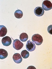
Image by NIAID
The European Commission (EC) has granted conditional marketing authorization for an immunogene therapy known as Zalmoxis.
This means Zalmoxis can be marketed in the European Economic Area as an adjunctive therapy to aid immune reconstitution and help treat graft-versus-host disease (GVHD) in adults with high-risk hematologic malignancies who are receiving a haploidentical hematopoietic stem cell transplant (haplo-HSCT).
Zalmoxis consists of allogeneic T cells genetically modified to express both a truncated form of the human low-affinity nerve growth factor receptor (ΔLNGFR), as a cell-surface selectable marker, and the suicide gene herpes simplex I virus thymidine kinase (HSV-TK Mut2).
The modified T cells are given to haplo-HSCT recipients to help fight off infection, enhance the success of the transplant, and support long-lasting anticancer effects.
Because the T cells can also cause GVHD, they are equipped with the suicide gene, which makes them susceptible to treatment with ganciclovir or valganciclovir. So if a patient develops GVHD, he or she can receive ganciclovir/valganciclovir, which should kill the modified T cells and prevent further development of the disease.
Zalmoxis is being developed by MolMed S.p.A. The company said the treatment should become available in the first European market during the first half of 2017.
About conditional marketing authorization
Conditional marketing authorization represents an expedited path for approval. The EC grants this type of authorization before pivotal registration studies are completed.
Conditional marketing authorization is granted to products whose benefits are thought to outweigh their risks, products that address unmet needs, and products that are expected to provide a significant public health benefit.
Under the provisions of the conditional marketing authorization for Zalmoxis, MolMed will be required to complete a post-marketing study aimed at confirming the clinical benefit of the treatment.
The European Medicines Agency’s Committee for Medicinal Products for Human Use has accepted the ongoing phase 3 TK008 trial as a post-marketing confirmatory study.
Trials of Zalmoxis
The EC’s decision to grant Zalmoxis conditional marketing authorization was based on cumulative efficacy and safety data collected from patients enrolled in a phase 1/2 trial (TK007) and an ongoing phase 3 trial (TK008).
The Zalmoxis group comprised 30 patients from the TK007 trial and 15 patients from the experimental arm of the TK008 trial. The TK007 trial included haplo-HSCT recipients with various high-risk hematologic malignancies, and the TK008 trial is enrolling haplo-HSCT recipients with high-risk acute leukemia.
The data thus far have indicated that Zalmoxis can provide rapid immune reconstitution, an anti-leukemia effect, and complete control of GVHD, in the absence of any post-transplant immunosuppression.
Overall, these effects led to a clinically meaningful increase in survival rates in Zalmoxis-treated patients, when compared to historical controls from the European Society for Blood and Marrow Transplantation (EBMT) database.
The only adverse event related to Zalmoxis treatment was GVHD, which was fully resolved by the activation of the suicide gene system with ganciclovir treatment, without any GVHD-related death.
Detailed results of this analysis are set to be presented during the MolMed-sponsored symposium “A new era of haplo-transplantation” at the EBMT International Transplant Course, which is scheduled to take place September 9-11 in Barcelona, Spain.