User login
Company discontinues phase 3 ADAPT for mRCC
A second interim analysis of
In ADAPT, 462 patients with previously untreated advanced or metastatic renal cell carcinoma (mRCC) were randomized 2:1 between combination treatment with Rocapuldencel-T and sunitinib versus sunitinib monotherapy, after undergoing cytoreductive nephrectomy.
In February 2017, the trial’s Independent Data Monitoring Committee had reviewed the data and concluded that the trial was unlikely to demonstrate a statistically significant improvement in median overall survival in the combination arm and recommended halting the trial. However, the principal investigators and the company, Argos Therapeutics, considered the data too immature to observe the delayed effects associated with immunotherapy and decided to continue the trial. They submitted a protocol amendment to the Food and Drug Administration adding additional co-primary endpoints, and in April of last year, met with the Food and Drug Administration, which accepted the amendment and agreed to continuation of the trial, according to a company press release issued in November.
In the latest interim analysis, which was conducted following an additional 51 deaths, median overall survival for the intent-to-treat patient population was 28.2 months for the combination arm (95% confidence interval, 23.4, 35.2) compared with 31.2 months (95% CI, 23.0, 44.5) for the control arm; this was one of four new co-primary endpoints. The hazard ratio was 1.10 (95% CI, 0.85, 1.42).
Other co-primary endpoints that were evaluated, including overall survival for the patients who remained alive at the time of the February 2017 interim analysis and overall survival for all patients for whom at least 12 months of follow-up was available, did not demonstrate a favorable result, Argos Therapeutics said in a recent press release.
Rocapuldencel-T “consists of autologous dendritic cells programmed with amplified RNA from a patient’s primary tumor” and is “designed to overcome immunosuppression and induce broadly reactive, long-lasting anti-tumor memory T cells” according to the early interim analysis presented at the European Society for Medical Oncology (ESMO) 2017. The drug is also being evaluated in non–small cell lung cancer and bladder cancer.
FDA approves immunotherapy combo for advanced RCC
The Food and Drug Administration has granted approvals to
The approvals were based on statistically significant improvements in overall survival (OS) and objective response rate (ORR) for patients receiving the combination of nivolumab and ipilimumab (n = 425), compared with those receiving sunitinib (n = 422) in CheckMate 214, the FDA said in a press statement.
Median OS was not yet reached in the combination arm at follow-up of 32 months, compared with 25.9 months in the sunitinib arm (hazard ratio, 0.63; 95% confidence interval, 0.44-0.89; P less than .0001). The ORR was 41.6% (95% CI, 36.9-46.5) for the combination versus 26.5% (95% CI, 22.4-31) in the sunitinib arm (P less than .0001).
Efficacy of the combination was not established for patients with favorable-risk disease.
The most common adverse reactions were fatigue, rash, diarrhea, musculoskeletal pain, pruritus, nausea, cough, pyrexia, arthralgia, and decreased appetite.
The recommended schedule and dose is 3 mg/kg nivolumab, followed by 1 mg/kg ipilimumab, on the same day every 3 weeks for four doses, then 240 mg nivolumab every 2 weeks or 480 mg every 4 weeks, the FDA said.
Nivolumab is marketed as Opdivo and ipilimumab as Yervoy by Bristol-Myers Squibb.
Do industry payments increase prescribing for some targeted therapies?
Physicians receiving general payments from the company marketing a targeted cancer therapy were more likely to prescribe it in three out of six drugs evaluated, researchers reported.
Prescribing of sunitinib, dasatinib, and nilotinib was increased for physicians receiving such payments versus not receiving them, while prescribing of imatinib, sorafenib, and pazopanib were not, according to the analysis by Aaron P. Mitchell, MD, of the Lineberger Comprehensive Cancer Center, UNC School of Medicine, University of North Carolina at Chapel Hill, and his coauthors.
In previous studies, pharmaceutical industry payments to physicians have been associated with “higher-cost, brand-name pharmaceutical prescribing,” Dr. Mitchell and his colleagues wrote. The report was published in JAMA Internal Medicine.
“Whether industry payments are associated with physician treatment choice in oncology is uncertain,” they said.
To evaluate the association between payments to oncologists and drug selection, Dr. Mitchell and his colleagues linked Open Payments data from the Centers for Medicare & Medicaid Services to data from Medicare Part D Prescriber Public Use File for the years 2013-2014.
The primary variable in the study was payments received during 2013, according to investigators, and the primary outcome of the analysis was prescriptions filled during 2014.
Open Payments reported in 2013 had a total dollar value of $4.08 billion, including $1.20 billion paid to physicians, according to CMS data.
The researchers focused on targeted therapies for two therapeutic areas: metastatic renal cell carcinoma (RCC), including sorafenib, sunitinib, and pazopanib; and chronic myeloid leukemia (CML), including imatinib, dasatinib, and nilotinib.
They limited their analysis to physicians listed as oncologists who filled at least 20 prescriptions for each of the three drugs in metastatic RCC (n = 354) or in CML (n = 2,225).
Receiving payments categorized as “general,” such as gifts, speaker fees, meals, and travel, increased the odds of prescribing drugs for both metastatic RCC (odds ratio, 2.05; 95% confidence interval, 1.34-3.14; P = .001) and for CML (odds ratio, 1.29; 95% CI, 1.13-1.47; P less than .001).
By contrast, research payments did not increase the odds of prescribing those drugs, the investigators reported.
Looking at specific drugs, they found that receipt of general payments from a drug’s manufacturer was associated with increased prescribing of sunitinib (50.5% versus 34.4%, P = .01), dasatinib (13.8% versus 11.4%, P = .02), and nilotinib (15.4% vs 12.5%, P = .01).
However, no such association was found for sorafenib or pazopanib.
For imatinib, by contrast, investigators said industry payments were associated with a prescribing decrease.
“This may reflect a strategy by the manufacturer of imatinib, which also produces nilotinib, to promote switching to nilotinib before the patent expiration of imatinib in 2015,” the researchers wrote.
Dr. Mitchell and his coauthors reported no conflict of interest disclosures related to the study.
SOURCE: Mitchell AP, et al. JAMA Intern Med. 2018 Apr 9. doi: 0.1001/jamainternmed.2018.0776.
FROM JAMA INTERNAL MEDICINE
Key clinical point: Oncologists receiving general payments from the company marketing a cancer drug were more likely to prescribe it in three out of six drugs evaluated.
Major finding: Prescribing was significantly increased for sunitinib (50.5% versus 34.4%, P = .01), dasatinib (13.8% versus 11.4%, P = .02), and nilotinib (15.4% vs. 12.5%, P = .01), but not for imatinib, sorafenib, or pazopanib.
Study details: An analysis of Centers for Medicare & Medicaid Services Open Payments data and Medicare Part D Prescriber Public Use File for the years 2013 to 2014.
Disclosures: The authors reported no conflict of interest disclosures related to the study.
Source: Mitchell AP et al. JAMA Intern Med. 2018 Apr 9. doi: 0.1001/jamainternmed.2018.0776.
Diabetes from checkpoint inhibitors probably means lifelong insulin
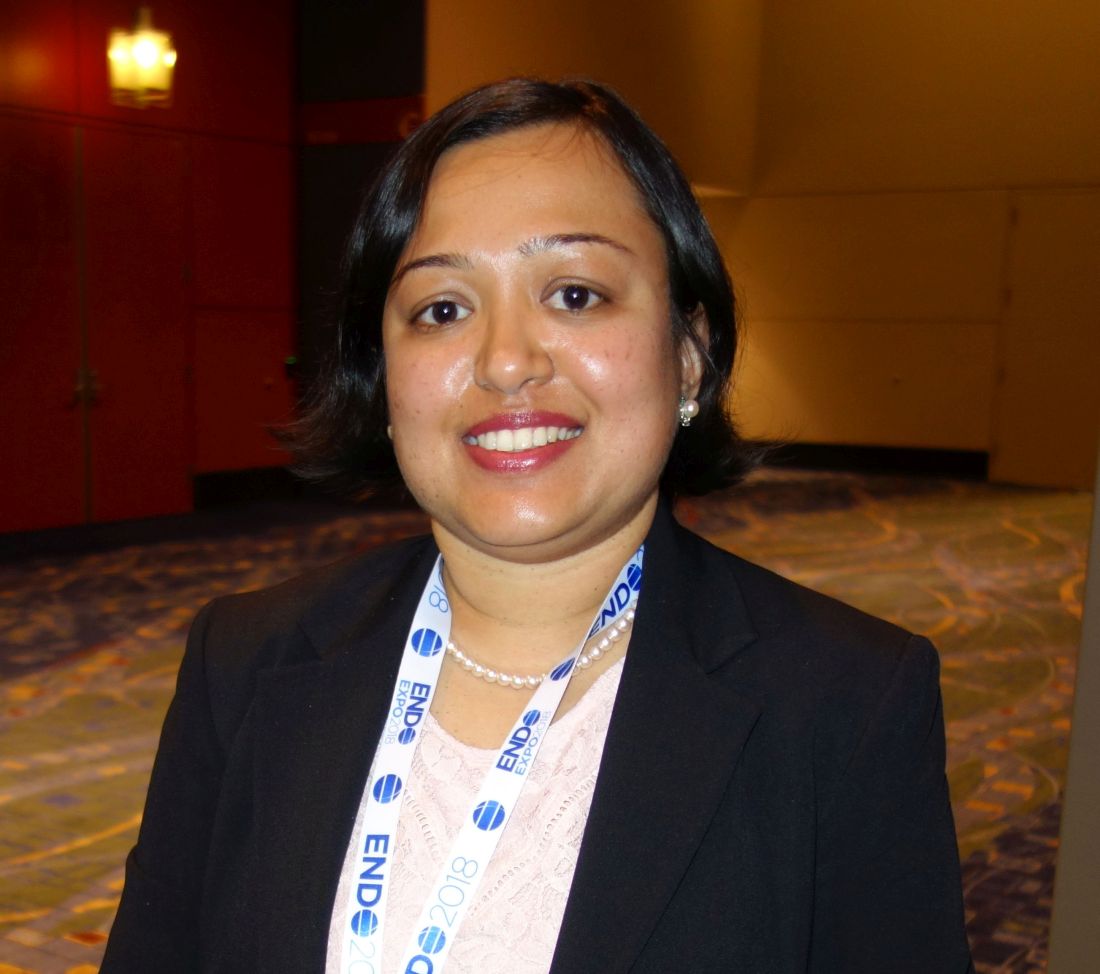
CHICAGO – , according to Priyanka Iyer, MD, an endocrinology fellow at MD Anderson Cancer Center, Houston.
“As long as we get glycemic control, they can continue,” she said at the annual meeting of the Endocrine Society.
Diabetes is a known side effect of immune checkpoint inhibitors (ICIs) but it’s rare, occurring in maybe 0.17% of patients, and its natural history and risk factors are unknown.
ICIs are fairly new agents, and as their use expands beyond clinical trials, “we anticipate seeing larger numbers of cases. Patients should be educated about the symptoms of uncontrolled blood sugars while on ICIs,” and endocrinologists “have to get involved and recognize this entity sooner,” Dr. Iyer said.
In short, her team found that ICI-mediated diabetes can occur in patients with or without preexisting diabetes, and that most patients have evidence of beta-cell failure, likely T-cell mediated destruction due to immune activation. In all but one case, patients remained on insulin at a median follow-up of 44 weeks, even after stopping ICIs. For most, ICI-mediated diabetes likely means lifelong insulin.
They were all on the programmed cell death protein (PD-1) inhibitors nivolumab (Opdivo) or pembrolizumab (Keytruda), or the PD-1 ligand (PD-L) inhibitor durvalumab (Imfinzi). The agents are used for a range of cancers, including renal cell, melanoma, and Hodgkin lymphoma. There were no diabetes cases in patients on single-agent ipilimumab (Yervoy) or tremelimumab, which target cytotoxic T-lymphocyte associated antigen-4 and are used for melanoma and mesothelioma.
Median time to diabetes presentation after the start of ICI treatment was 12.3 weeks but ranged from 1 to 67.2 weeks. Half of the cases presented in diabetic ketoacidosis (DKA). Patients had upward trending hyperglycemia and most had diabetes symptoms for a while before diagnosis. They presented with a blood glucose above 250 mg/dL, and more than half above 500 mg/dL. Median hemoglobin A1c at presentation was 8%, but ranged up to 12.5%.
Every patient required insulin, including the six that discontinued ICIs after developing diabetes. Diabetes resolved in just one patient at 10.2 months; she presented with DKA.
There were no obvious predisposing factors. None of the patients had histories of type 1 diabetes or other autoimmune disease. Five patients had well-controlled type 2 diabetes prior to ICI initiation; four had prediabetes. Some had family members with type 2 diabetes, but not type 1. Four had prior ICI exposure. Just three patients were on concomitant steroids.
A few patients also developed thyroid or pituitary dysfunction, which are more common side effects of ICIs.
The median age at diabetes presentation was 61 years and ranged from 32 to 82 years. The majority of patients were men, which reflects MD Anderson demographics, not a predisposing risk factor, Dr. Iyer said.
Melanoma was the most common cancer, followed by renal cell and prostate; patients had stage 2-4 disease. About half the subjects were on single agent anti-PD-1 treatment, about a third on anti-PD-1 combination treatment, and the rest on anti-PD-L1 combination therapy. C-peptide levels were below 0.9 ng/mL at diabetes diagnosis in most of the patients. Eleven of the 20 tested (55%) were positive for the pancreatic islet cell antibody GAD65.
The investigators had no disclosures. A funding source was not reported.
SOURCE: Iyer PC et al. Abstract OR05-5.
REPORTING FROM ENDO 2018
Key clinical point: Be on the lookout for new-onset diabetes when patients start immune checkpoint inhibitors.
Major finding: In all but one case, patients remained on insulin at a median follow-up of 44 weeks, even after stopping ICIs.
Study details: Review of 24 cases.
Disclosures: The investigators had no disclosures. A funding source was not reported.
Source: Iyer PC et al. Abstract OR05-5.
Tivozanib after sorafenib promising in patients with advanced RCC
For patients with advanced renal cell carcinoma (RCC) progressing after sorafenib treatment, tivozanib was well tolerated and provided promising survival outcomes, investigators in a phase 2 study have reported.
Incidence of adverse events on tivozanib was low, and the safety profile was favorable in comparison with other agents in its class, the investigators said in the European Journal of Cancer.
The findings also help clarify results of a previous randomized phase 3 trial of tivozanib versus sorafenib where the investigators say crossover may have confounded overall survival results to the detriment of the tivozanib arm.
“Collectively, these data provide evidence of the anti-tumor activity of tivozanib and may be used to help frame future studies in recurrent disease,” wrote Ana M. Molina, MD, of Weill Cornell Medicine, New York, and her coauthors.
Tivozanib, recently approved in Europe for untreated RCC, is characterized by highly potent and selective inhibition of the three known vascular endothelial growth factor (VEGF) receptors.
Dr. Molina and her colleagues reported a single-arm crossover study of patients who were previously enrolled in the randomized phase 3 TIVO-1 trial of tivozanib versus sorafenib.
They enrolled a total of 161 patients who were randomized to the sorafenib arm of TIVO-1 and went on to receive tivozanib after disease progression.
Median progression-free survival was 11.0 months and median overall survival was 21.6 months for these crossover patients, Dr. Molina and co-investigators reported.
No patients in the study had a complete response, while 29 (18%) had a partial response and 83 (52%) had stable disease.
“These data compare favorably with other second-line therapies for RCC,” Dr. Molina and co-authors said.
Grade 3 or greater adverse events occurred in 48% of patients, including 24% that were treatment related. The most common grade 3 treatment-related adverse event was hypertension in 11%.
Approximately 4% of patients discontinued tivozanib due to adverse events.
“This study also provided clarity of the TIVO-1 trial, in which patient crossover was thought to have confounded the overall survival results,” Dr. Molina and colleagues said.
In TIVO-1, the primary end point of progression-free survival was improved for tivozanib versus sorafenib (median of 11.9 vs 9.1 months; P = .042), they noted.
However, median overall survival was not statistically different between arms, possibly because 74% of patients randomized to sorafenib were treated with next-line therapy, mainly tivozanib, investigators said.
AVEO Oncology and Astellas Pharma US, Inc. funded the study. Dr. Molina reported receiving honoraria from AVEO, Novartis, and Eisai.
SOURCE: Molina AM, et al. Eur J Cancer. 2018 Mar 13. doi: 10.1016/j.ejca.2018.02.009.
FROM THE EUROPEAN JOURNAL OF CANCER
Key clinical point: Tivozanib has potent antitumor activity in patients with advanced renal cell carcinoma (RCC) who previously progressed on sorafenib.
Major finding: Median progression-free survival was 11.0 months, and median overall survival was 21.6 months for patients receiving tivozanib.
Study details: A single-arm, phase 2 crossover study of patients previously randomized to the sorafenib arm of the phase 3 TIVO-1 study.
Disclosures: AVEO Oncology and Astellas Pharma US, Inc. funded the study. Investigators reported potential conflict of interests related to AVEO, Novartis, Eisai, Pfizer, and others.
Source: Molina AM, et al. Eur J Cancer. 2018 Mar 13. doi: 10.1016/j.ejca.2018.02.009.
Axitinib/avelumab combo shows preliminary efficacy in RCC
The combination of axitinib and avelumab had manageable toxicity and demonstrated preliminary efficacy as a front-line treatment of advanced renal cell carcinoma (RCC), according to results of a recent study.
More than half of patients had a response on the combination of axitinib (Inlyta), a receptor inhibitor of vascular endothelial growth factor (VEGF), and avelumab (Bavencio), an anti-PD-L1 monoclonal antibody, investigators reported in The Lancet Oncology.
The most common treatment-related adverse event was hypertension, wrote lead author Toni K. Choueiri, MD, of the Lank Center for Genitourinary Oncology at the Dana-Farber/Brigham and Women’s Cancer Center in Boston, and his colleagues.
“The combination of avelumab and axitinib in treatment-naive patients with advanced RCC had a manageable safety profile consistent with the profiles of the individual agents when administered as monotherapy, and antitumor activity was encouraging,” said Dr. Choueiri, and his colleagues.
The study, known as JAVELIN Renal 100, was a phase 1b investigation that included 55 patients with advanced clear-cell RCC. A total of 6 patients were enrolled in a smaller dose-finding cohort, and 49 were enrolled in a dose-expansion cohort.
In the dose-finding phase, patients received oral axitinib 5 mg twice a day for 7 days, at which point they started intravenous avelumab 10 mg/kg every 2 weeks, according to the study description. In the dose-expansion phase, patients were directly started on the combination.
Out of 55 patients enrolled, 26 (53%) had a response on the axitinib-avelumab combination, including 3 (6%) who had complete responses, investigators reported.
Grade 3 or greater treatment-related adverse events were reported in 32 patients (58%). The most common of those was hypertension, occurring in 16 patients (29%), followed by ALT increase and palmar-plantar erythrodysesthesia syndrome in 4 patients each (7%), Dr. Choueiri and colleagues wrote.
In the dose-finding phase, investigators reported one dose-limiting toxicity, which was grade 3 proteinuria due to axitinib.
. In that randomized trial, the axitinib-avelumab combination is being compared to sunitinib as a first-line approach in patients with advanced RCC.
“The combination of an antibody that inhibits PD-L1 and PD-1 interactions with a targeted antiangiogenic agent might take advantage of complementary mechanisms of action to provide clinical benefit in patients with advanced renal-cell carcinoma that exceeds the effects of the respective drugs alone without increasing associated toxicity,” Dr. Choueiri and colleagues wrote.
Pfizer and Merck funded the study. Dr. Choueiri declared interests related to several companies, including AstraZeneca, Bristol-Myers Squibb, Eisai, Exelixis, GlaxoSmithKline, Merck, Novartis, Pfizer, Peloton, and Roche/Genentech.
SOURCE: Choueiri TK et al. Lancet Oncol. 2018 Mar 9. doi: 10.1016/S1470-2045(18)30107-4.
FROM THE LANCET ONCOLOGY
Key clinical point: The targeted-immune combination of axitinib and avelumab had manageable toxicity and encouraging preliminary efficacy as a front-line treatment of advanced renal cell carcinoma (RCC).
Major finding: Out of 55 patients enrolled, 26 (53%) had a response, including 3 (6%) who had complete responses.
Study details: A dose-expansion and dose-finding phase 1b study including 55 patients with advanced clear-cell RCC.
Disclosures: Funding for the study came from Pfizer and Merck. Study authors declared interests related to several companies, including Pfizer, Merck, Bristol-Myers Squibb, Novartis, Roche/Genentech.
Source: Choueiri TK et al. Lancet Oncol. 2018 Mar 9. doi: 10.1016/S1470-2045(18)30107-4.
views
The antitumor activity of axitinib and avelumab in this study indicate the potential clinical benefit of targeted-immune combinations for advanced renal cell carcinoma (RCC), according to Viktor Grünwald, MD, PhD.
“Further studies are warranted to explore whether a first-line targeted-immune combination might overcome the standard of sequential targeted and immune therapies,” Dr. Grünwald said in an editorial.
The current absence of long-term safety data for the combination approach is limiting in terms of making definitive conclusions about the toxicity of axitinib and avelumab as a first line combination therapy for advanced clear-cell RCC, Dr. Grünwald wrote.
Even so, axitinib has a “low” incidence of hepatic toxicity, making it a “preferable” agent to evaluate in targeted-immune combination trials such as JAVELIN Renal 100, he added.
Objective responses in JAVELIN Renal 100 were seen in 32 out of 55 patients (58%), of which 3 patients (6%) were complete responses, according to reported data.
Similar findings previously reported for the immune-immune combination of ipilimumab and nivolumab versus sunitinib. In that study, patients receiving the immune-immune combination had an overall response rate of 42% and complete response rate of 9%, while the group patients receiving sunitinib had overall and complete response rates of 27% and 1%, respectively, he said.
“With the dawn of immune-immune and targeted-immune combinations, for the first time in a decade, major progress towards improving the median overall survival and possibly delivering cure to some of our patients with metastatic renal-cell carcinoma seems to have been made,” Dr. Grünwald said.
Viktor Grünwald, MD, PhD, is with the department of hematology, haemostasis, oncology, and stem cell transplantation at Hannover Medical School, Germany. These comments are based on an editorial accompanying the report (Lancet Oncol. 2018 Mar 9. doi: 10.1016/S1470-2045[18]30126-8). Dr. Grünwald disclosed ties to Bristol-Myers Squibb, Merck Sharp and Dohme, Ipsen, Novartis, Roche, AstraZeneca, Pfizer, Cerulean, Eisai, and EUSA Pharma.
Excellent prognosis for cystic RCC evaluated with radiologic threshold of greater than 50%
When a standardized radiologic threshold of greater than 50% is used, patients with unifocal cystic renal cell carcinoma (cRCC) have an excellent prognosis with active surveillance and following surgical resection, findings from a new study show.
At a median follow-up of 5.4 years (IQR 2.8-7.8), none of the 138 patients in the cohort experienced a tumor recurrence or metastasis from cRCC, and 7 (5.1%) died from non–renal-related causes. When comparing patients who initially underwent surgery to those who were initially managed with active surveillance, the researchers found that there was no significant difference in overall survival (P = .07). There were no deaths due to kidney cancer in the entire cohort.
The terminology of cRCC has historically been used to describe an indolent version of RCC that consists primarily of cysts, and the threshold of cystic involvement has traditionally been greater than 75% cystic on pathologic examination.
However, this classification does not contribute to the preoperative decision-making process, the study authors noted in Journal of Urology.
“Cross-sectional imaging provides the benefit of assessing tumor morphology without surgical manipulation, allowing for an accurate assessment of the solid and cystic components and classification of cRCC ,” write Ari Hakimi, MD, of Memorial Sloan Kettering Cancer Center in New York, and his colleagues.
Studies evaluating radiologic criteria for cRCC diagnosis have suggested that cystic changes in 5%-45% of the total mass observed on imaging are associated with favorable survival. Thus, the authors sought to improve the preoperative assessment of cystic renal masses by evaluating cRCC as an enhancing renal lesion that is greater than 50% cystic on cross-sectional imaging. The goal of the current study was then to compare the long-term outcomes of patients with cRCC who underwent surgery and active surveillance using this hypothesized threshold.
The cohort included 138 patients who underwent surgery at Memorial Sloan Kettering Cancer Center for a renal mass from January 2000 to December 2015, and of this group, 102 (73.9%) had renal cell carcinoma and 36 (26.1%) had benign masses. Most of the tumors were Fuhrman grade 1-2 (77.5%), ≤pT2 stage (83.4%) and clear cell histology (65.9%), while the majority of cRCC lesions were Bosniak 3 and 4 (93.5%) and had a solid component of less than 25% (83.3%). On multivariate analysis, men (P = .007) were more likely to have malignant lesions and Bosniak 3 lesions were more likely to be malignant (P = .01).
In the subgroup of 38 active surveillance patients, 27 (71.1%) remained on active surveillance while 11 (28.9%) subsequently had surgery, of which all underwent partial nephrectomy. The median overall growth rate for lesions was 1.0 mm/year (IQR 0-2.8) over 25.3 months (IQR 16.3-44.8), and no evidence of recurrence or metastasis was reported in any of these patients at a median follow-up of 4.3 years (IQR 2.1-5.7) from first imaging diagnosis or 6.9 years (IQR 4.9-8.5) after surgery.
“We believe that our radiologic definition allows for more inclusive criteria of cRCC and would encourage kidney sparing approaches or implementation of AS protocols when feasible,” the authors concluded.
SOURCE: Hakimi A et al. J Urol. 2018 Feb 26 doi: 10.1016/j.juro.2018.02.3087
FROM THE JOURNAL OF UROLOGY
Key clinical point: Patients with unifocal cystic renal cell carcinoma have an excellent prognosis for both active surveillance and following surgery when evaluated with a standardized radiologic threshold of greater than 50% cystic.
Major finding: There was no evidence of tumor recurrence or metastasis from cRCC at a median follow-up of 5.4 years, and seven patients died of other causes.
Study details: Retrospective single-center study that looked at outcomes and clinicopathologic and oncologic features of 138 cases of cystic renal cell carcinoma.
Disclosures: The study was supported by the Sidney Kimmel Center for Prostate and Urologic Cancers and an NIH/NCI Cancer Center Support Grant. There were no author disclosures listed.
Source: Hakimi A et al. J Urol. 2018 Feb 26 doi: 10.1016/j.juro.2018.02.3087.
Immunotherapy regimen influences inflammatory arthritis presentation
Variations in the clinical presentation of immunotherapy-induced inflammatory arthritis is partly explained by which treatment regimen was used to treat the cancer, a single-center study suggests.
While immune checkpoint inhibitors (ICI) have revolutionized the field of oncology, their use for an ever-widening range of indications had created an increasing population of patients referred to rheumatologists for the management of immune-related adverse events (IrAEs), according to Laura C. Cappelli, MD, and her colleagues at John Hopkins University, Baltimore.
Well-established guidelines exist for managing adverse events such as colitis and pneumonitis, but there are only preliminary guidelines for evaluating and treating immunotherapy-induced inflammatory arthritis (IA). “This may stem from a lack of consistent reporting of rheumatologic IrAEs in clinical trials, the non–life threatening nature of [inflammatory arthritis], or lack of recognition of musculoskeletal symptoms by treating providers,” they wrote in Seminars in Arthritis and Rheumatism.
Clinical trials have reported ranges of arthralgia in 1%-43% of patients treated with ICIs, but no accurate estimate of the incidence of IA exists.
The researchers noted that treating patients with ICI-induced IA is complicated by a history of active or recently treated cancer and concerns over using immunosuppression in the context of ICI therapy.
They set out to evaluate the clinical presentations of 30 patients seen in their clinic with ICI-induced IA. Patients were a median of 59 years old and 12 (40%) were female. Tumor types included metastatic melanoma, non–small cell lung cancer, small cell lung cancer, colorectal cancer, Hodgkin lymphoma, cutaneous lymphoma, renal cell carcinoma, duodenal carcinoma, Merkel cell carcinoma, cutaneous basal cell carcinoma, and cutaneous squamous cell carcinoma.
Sixteen patients were treated with anti–programmed cell death protein 1 (PD-1)/programmed death ligand 1 monotherapy, and 14 were treated with combination anti–CTLA-4/PD-1 therapy.
Patients on combination therapy were significantly younger (7.5 years, P = 0.01) and were more likely to have metastatic melanoma as their underlying cancer.
Patients who received combination therapy were more likely to present first with knee IA (n = 10) and none had small joint involvement. In contrast, initial small joint involvement was more common in the monotherapy group (n = 6).
C-reactive protein levels were significantly higher in the combination therapy group (4mg/dL vs. 0.5mg/dL, P = 0.03). Only monotherapy patients were positive for anti–citrullinated peptide antibodies, rheumatoid factor, or antinuclear antibodies.
Most of the patients in the study had an additional IrAE, with colitis being the most common (n=10), followed by thyroid disease, pneumonitis, and rash. Patients on PD-1 or programmed death ligand 1 monotherapy were more likely to have IA as their first IrAE.
The research team noted that the median time to symptom onset was 5 months after ICI initiation.
Diagnosis of IA following patient-reported symptoms was an average of 5.2 months, with a significant difference in lag time to diagnosis depending on initial joint presentation. For example, patients with initial small joint involvement had a 10 month longer lag time to IA diagnosis than those with knees as the initial joint involved.
In terms of treatment, 24 patients were treated with systemic corticosteroids and 10 required additional immunosuppression. The need for corticosteroids did not differ by ICI treatment regimen, but those treated with combination therapy were more likely to require additional immunosuppression (P = 0.02).
Tumor necrosis factor inhibitors with or without methotrexate were prescribed for seven patients. All of the patients had a clinical improvement in their arthritis symptoms. Four had a complete tumor response at the time of tumor necrosis factor inhibitor initiation with none having tumor progression.
The three patients treated with methotrexate monotherapy had a complete or sustained partial tumor response to ICI therapy and their cancer did not develop during IA management follow-up.
The authors went on to look at the persistence of IA after cessation of therapy in a subset of 21 patients. They found that 18 of these patients still had IA symptoms months after stopping treatment. They suggested that the delay in diagnosis and treatment seen in their study might explain the finding.
The study provides “critical information, not just for rheumatologists as they try to recognize subgroups in ICI-induced IA and diagnose patients with this new entity, but also for oncology providers who are usually first to encounter patients with ICI-induced IA and subsequently refer patients to rheumatology,” Dr. Cappelli and colleagues wrote.
The experience so far with using immunosuppression in ICI-induced IA “has been reassuring in terms of cancer outcomes, but more studies are needed to confirm this finding,” they concluded.
SOURCE: Cappelli LC et al. Semin Arthritis Rheum. doi: 10.1016/j.semarthrit. 2018.02.011.
FROM SEMINARS IN ARTHRITIS AND RHEUMATISM
Key clinical point: The clinical features of patients with immunotherapy-induced inflammatory arthritis differ according to the treatment regimen used.
Major findings: Combination immune checkpoint inhibitor therapy was associated with higher C-reactive protein levels and a higher likelihood of having a large joint affected first.
Study details: A single-center, retrospective cohort study of 30 patients with rheumatologist-confirmed inflammatory arthritis after receiving immune checkpoint inhibitor therapy.
Disclosures: The study was funded by the National Institute of Arthritis and Musculoskeletal and Skin Disease and the Jerome L. Greene Foundation.
Source: Cappelli LC et al. Semin Arthritis Rheum. doi: 10.1016/j.semarthrit. 2018.02.011.
Axitinib/pembrolizumab combo safe, effective against mRCC
A combination of the tyrosine kinase inhibitor axitinib (Inlyta) and the immune checkpoint inhibitor pembrolizumab (Keytruda) was associated with acceptable toxicities and showed promising activity against advanced renal cell carcinoma (RCC) in the first-line setting, results of a phase 1b trial indicate.
Of 11 patients enrolled in a dose-finding study and 41 enrolled in the expansion phase of that study, 38 had an objective response (complete or partial response), for an overall response rate of 73%, reported Michael B. Atkins, MD, of Georgetown-Lombardi Comprehensive Cancer Center, Washington, and his colleagues.
“This phase 1b study showed that the combination of axitinib and pembrolizumab at nearly the full planned doses of each drug is tolerable in patients with treatment-naive advanced renal cell carcinoma,” they wrote. The report was published in The Lancet Oncology.
Previous studies of programmed death-1 (PD-1) checkpoint inhibitors such as pembrolizumab or nivolumab (Opdivo) combined with inhibitors of vascular endothelial growth factor (VEGF) have resulted in excessive toxicities attributed to off-target effects of the VEGF inhibitors used.
The investigators reasoned that because axitinib is more selective and specific for targets in the VEGF pathway, it might make a safer and more effective partner to a PD-1 inhibitor than the multikinase inhibitors sunitinib (Sutent) or pazopanib (Votrient).
“A formal systematic review was not done before doing this trial because most of the work combining VEGF pathway inhibitors with checkpoint inhibitors is new and not yet published,” Dr. Atkins and his associates explained.
As of the March 31, 2017, data cutoff, 52 patients from 10 U.S. centers had been treated with the same dose and schedule and were included in the analysis. All patients had tumors with clear cell renal carcinoma histologies; one also had sarcomatoid features.
There were three investigator-assessed dose-limiting toxicities (DLT, the primary endpoint) in the 11 patients treated in the dose-finding phase. One of the patients had a transient ischemic attack and two completed less than 75% of the planned axitinib dose because of treatment-related toxicities.
Of the 52 patients, 25 were still on treatment at the time of data cutoff: 22 who were still receiving both axitinib and pembrolizumab and 3 who were receiving only the PD-1 inhibitor. Eight of the patients continued on therapy despite disease progression.
Of the 27 patients who discontinued both drugs, 10 did so because of adverse events, 9 for disease progression, and others for various reasons such as mixed adverse events and disease progression, investigator discretion, global deterioration, or protocol violation.
Grade 3 or greater adverse events occurred in 34 patients (65%), and included hypertension, diarrhea, fatigue, and elevated alanine aminostransferase (ALT) levels.
The most common potentially immune-related adverse events were diarrhea, ALT elevations, hypothyroidism, and fatigue.
At a median follow-up of 20.4 months, 4 patients had a complete response, and 34 had a partial response. An additional eight patients had stable disease. Responses were seen in 18 of 24 patients with favorable-risk disease and in 18 of 26 patients with intermediate- or poor-risk disease. The median time to response was 2.8 months, and the median duration of response was 18.6 months.
“Future research should focus on investigating the mechanism of the potential synergistic effects of axitinib and pembrolizumab, and whether an immunotherapy-only approach (including combinations) enriched by the appropriate biomarkers, followed by VEGFR TKI salvage, might produce more durable off-treatment responses or whether administering VEGFR TKI monotherapy followed by PD-1 and PD-L1 pathway blockade might produce superior or equivalent results,” the investigators concluded.
Pfizer, in collaboration with Merck, sponsored the study. Dr. Atkins and several coauthors disclosed consulting fees from Pfizer, Merck, and other companies. Four of the coauthors are Pfizer employees and stockholders.
SOURCE: Atkins MB et al. Lancet Oncol 2018 Feb. 10. doi: 10.1016/S1470-2045(18)30081-0.
This phase 1 trial highlights some issues that merit discussion. First, the eligibility criteria of the study population should be considered. Renal cell carcinoma is a very heterogeneous disease with a natural history that could range from an indolent clinical course to a slow progressive or an aggressive behavior. The prognostic models proposed by Memorial Sloan-Kettering Cancer Center and the International Metastatic Renal Cell Carcinoma Database Consortium are now validated for stratification of patients in clinical trials. By contrast, patients treated in clinical practice are often excluded from clinical trials and have a poor prognosis and derive less benefit from standard treatments. Atkins and colleagues treated a small number of patients from a highly selected study population, with no patients with poor clinical conditions or who had not undergone nephrectomy. Considering these aspects, how have the favorable prognostic features of the population affected the feasibility and efficacy of the treatment proposed? In this regard, only assessment of a larger number of patients in a less selected population can confirm these activity results.
Second, the absence of central radiological review in the trial could represent a major limitation in the interpretation of treatment response and the evaluation of progression-free survival. The Response Evaluation Criteria in Solid Tumors version 1.1 criteria used to assess response are often unable to distinguish between pseudoprogression, hyperprogression, and late response to immunotherapies. Therefore, the assessment of response during treatment with immune checkpoint inhibitors remains debated, and physicians often carefully consider the opportunity to continue treatment in cases with doubtful or mixed response and must decide whether to change the therapy on the basis of the clinical benefit being received by patients.
Third, another issue concerns the potential benefit of the combination therapy proposed. Axitinib is a multikinase inhibitor able to act on a broad spectrum of kinases related to angiogenesis. Axitinib monotherapy is considered a therapeutic option after an angiogenesis inhibitor for patients with metastatic renal cell carcinoma. Pembrolizumab is a PD-1 inhibitor under evaluation in combination strategies for the treatment of metastatic renal cell carcinoma. The combination of pembrolizumab and axitinib was safe and feasible, which is in contrast to results previously reported for other combinations, such as nivolumab plus pazopanib or sunitinib and pembrolizumab plus pazopanib. This evidence suggests that similar drugs might have different toxicity profiles when used in combination, and axitinib remains one the most tolerable tyrosine kinase inhibitors.
In summary, the combination of pembrolizumab and axitinib is very promising and the outcomes of Atkins and colleagues’ study could become the first evidence in favor of a combination of two drugs with different mechanisms of action for the treatment of metastatic renal cell carcinoma. Future research should attempt to select more patients who will respond to treatment on the basis of their clinical and molecular features.
Giuseppe Procopio, MD, Raffaele Ratta, MD, Filippo de Braud, MD, and Elena Verzoni, MD, are with the medical oncology department of Fondazione IRCCS Istituto Nazionale dei Tumori, Milan, and the medical oncology department of the University of Milan. The commentary was adapted and condensed from an editorial (Lancet Oncol. 2018 Feb 10. doi: 10.1016/S1470-2045[18]30092-5).
A combination of the tyrosine kinase inhibitor axitinib (Inlyta) and the immune checkpoint inhibitor pembrolizumab (Keytruda) was associated with acceptable toxicities and showed promising activity against advanced renal cell carcinoma (RCC) in the first-line setting, results of a phase 1b trial indicate.
Of 11 patients enrolled in a dose-finding study and 41 enrolled in the expansion phase of that study, 38 had an objective response (complete or partial response), for an overall response rate of 73%, reported Michael B. Atkins, MD, of Georgetown-Lombardi Comprehensive Cancer Center, Washington, and his colleagues.
“This phase 1b study showed that the combination of axitinib and pembrolizumab at nearly the full planned doses of each drug is tolerable in patients with treatment-naive advanced renal cell carcinoma,” they wrote. The report was published in The Lancet Oncology.
Previous studies of programmed death-1 (PD-1) checkpoint inhibitors such as pembrolizumab or nivolumab (Opdivo) combined with inhibitors of vascular endothelial growth factor (VEGF) have resulted in excessive toxicities attributed to off-target effects of the VEGF inhibitors used.
The investigators reasoned that because axitinib is more selective and specific for targets in the VEGF pathway, it might make a safer and more effective partner to a PD-1 inhibitor than the multikinase inhibitors sunitinib (Sutent) or pazopanib (Votrient).
“A formal systematic review was not done before doing this trial because most of the work combining VEGF pathway inhibitors with checkpoint inhibitors is new and not yet published,” Dr. Atkins and his associates explained.
As of the March 31, 2017, data cutoff, 52 patients from 10 U.S. centers had been treated with the same dose and schedule and were included in the analysis. All patients had tumors with clear cell renal carcinoma histologies; one also had sarcomatoid features.
There were three investigator-assessed dose-limiting toxicities (DLT, the primary endpoint) in the 11 patients treated in the dose-finding phase. One of the patients had a transient ischemic attack and two completed less than 75% of the planned axitinib dose because of treatment-related toxicities.
Of the 52 patients, 25 were still on treatment at the time of data cutoff: 22 who were still receiving both axitinib and pembrolizumab and 3 who were receiving only the PD-1 inhibitor. Eight of the patients continued on therapy despite disease progression.
Of the 27 patients who discontinued both drugs, 10 did so because of adverse events, 9 for disease progression, and others for various reasons such as mixed adverse events and disease progression, investigator discretion, global deterioration, or protocol violation.
Grade 3 or greater adverse events occurred in 34 patients (65%), and included hypertension, diarrhea, fatigue, and elevated alanine aminostransferase (ALT) levels.
The most common potentially immune-related adverse events were diarrhea, ALT elevations, hypothyroidism, and fatigue.
At a median follow-up of 20.4 months, 4 patients had a complete response, and 34 had a partial response. An additional eight patients had stable disease. Responses were seen in 18 of 24 patients with favorable-risk disease and in 18 of 26 patients with intermediate- or poor-risk disease. The median time to response was 2.8 months, and the median duration of response was 18.6 months.
“Future research should focus on investigating the mechanism of the potential synergistic effects of axitinib and pembrolizumab, and whether an immunotherapy-only approach (including combinations) enriched by the appropriate biomarkers, followed by VEGFR TKI salvage, might produce more durable off-treatment responses or whether administering VEGFR TKI monotherapy followed by PD-1 and PD-L1 pathway blockade might produce superior or equivalent results,” the investigators concluded.
Pfizer, in collaboration with Merck, sponsored the study. Dr. Atkins and several coauthors disclosed consulting fees from Pfizer, Merck, and other companies. Four of the coauthors are Pfizer employees and stockholders.
SOURCE: Atkins MB et al. Lancet Oncol 2018 Feb. 10. doi: 10.1016/S1470-2045(18)30081-0.
FROM THE LANCET ONCOLOGY
Key clinical point: A combination of axitinib and pembrolizumab was tolerable and showed activity against advanced/metastatic renal cell carcinoma.
Major finding: At a median of 20.4 months of follow-up, the overall response rate was 73%.
Study details: Open-label phase 1b dose-finding trial and expansion in 52 patients with advanced RCC.
Disclosures: Pfizer, in collaboration with Merck, sponsored the study. Dr. Atkins and several coauthors disclosed consulting fees from Pfizer, Merck, and other companies. Four of the coauthors are Pfizer employees and stockholders.
Source: Atkins MB et al. Lancet Oncol 2018 Feb 10. doi: 10.1016/S1470-2045(18)30081-0.
ERV expression may predict response to immune checkpoint blockade in ccRCC, other solid tumors
SAN FRANCISCO – The (ccRCC), according to findings from an analysis of nearly 5,000 tumor samples.
Similar associations may exist in other solid tumors, Shridar Ganesan, MD, reported at the ASCO-SITC Clinical Immuno-Oncology Symposium.
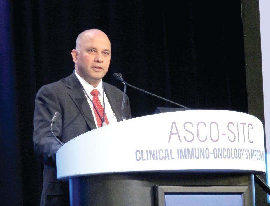
Merkel cell carcinoma, for example, has a high response rate despite its relatively low mutation burden. This is explained by the presence of infection with the Merkel cell polyomavirus in a low mutation burden subset, he said, noting that expression of the virus likely leads to expression of antigens that makes the disease both highly immunogenic and responsive to immune checkpoint therapy.
Expression of exogenous viruses is associated with response to immune checkpoint therapy in other cancers with low mutation burden, such as Epstein-Barr virus-positive gastric cancer, natural killer lymphoma, and Hodgkin disease.
“Intriguingly, renal cell carcinoma also has a relatively higher response rate to immune checkpoint therapy than would be anticipated by its relatively low mutation burden. However, this has no evidence of exogenous virus infection. Therefore, we turned our attention to endogenous retroviruses, which are an abundant potential stimulant of innate immunity in cancer,” he said.
Endogenous retroviruses (ERVs) now comprise about 5-8% of the human genome, and almost all of them have mutations that disable key coding genes of the retrovirus, meaning most cannot produce all the proteins necessary for viral replication.
“They’re essentially genomic fossils,” Dr. Ganesan said, adding that they all are silenced epigenetically in most normal adult tissues, both by DNA methylation and histone methylation. “However, inappropriate expression of endogenous retroviruses has been reported by multiple groups in some cancers and has been associated with evidence of immune activation. One can imagine that endogenous retroviral expression can be immunogenic both by expression of some [open reading frames] that are not disabled ... or just the viral RNA itself, which can be detected by cytoplasmic sensors ... and activate innate immunity,” he said.
To assess whether ERV expression corresponds with evidence of immune checkpoint activation in cancer, he and his colleagues conducted a pan-cancer analysis of more than 4,900 tumors across 21 cancer types using data from the Cancer Genome Atlas (TCGA).
“When we did this for about 66 annotated endogenous retroviruses in the TCGA database, we could see that different cancers had different amounts of retrovirus associated with immune activation ... in fact the strongest signal seen in this analysis was in clear cell renal cell carcinoma,” he said.
Signals were also seen in estrogen receptor–positive/human epidermal growth factor receptor 2-negative breast cancer, head and neck squamous cell carcinoma, and colon cancer.
A variety of ERVs were upregulated in RCC; different panels were associated with different cancer types. But two – ERVK.2 and ERV3.2 – were consistently upregulated across all the tumor types.
A closer look at ERV expression showed three clear clusters: extremely-high, intermediate, and low expression of ERV, and the very-high expression cluster had increased expression of numerous checkpoint genes, including CD8A, PD1, CTLA4, and LAG3 among others, compared with the low expression cluster.
This leads to the question of why a subset of RCCs have ERV expression.
In an attempt to answer that question, a transcript analysis was conducted to determine which transcripts are differentially regulated between the high and low ERV-expressing groups, and gene ontology analysis was performed.
“The results were quite striking. If you look at the modules that are differentially expressed between the high-ERV and the low-ERV groups, what pops up is really a lot of histone methyltransferase modules and chromatin regulation modules, ” he said, noting that this makes sense because of the silencing of ERV expression by histone methylation and DNA methylation, and suggests that “there is some deep abnormality in chromatin modulation in this subset of RCCs.
“This is intriguing, because RCCs are known to have mutations in chromatin modifying genes,” he said.
A look into whether the high-ERV–expressing group was enriched in any of these mutations showed some enrichment of BAP1 in the high- vs. low-expressing group, and that is currently being looked at further, he noted.
The next question was whether ERV expression correlated with response to immune checkpoint blockade in ccRCC, and this was looked at in a nonrandomized group of 15 patients with metastatic ccRCC who were treated with single-agent immune checkpoint therapy who had either clearly documented partial response or progressive disease. In 13 patient samples for which RNA expression of ERV3.2 was successfully measured by quantitative real-time polymerase chain reaction using two primer sets, ERV3.2 expression was significantly higher in responders vs. nonresponders in both primer sets (P less than .05 and .005).
“In summary, we have shown that expression of ERVs correlates with immune activation and increased expression of immune checkpoint genes in a subset of ccRCC, and perhaps several other solid tumor classes, and the expression of ERV3.2 is perhaps associated with response to PD1 blockade in this small preliminary cohort of ccRCC patients,” he said, noting that abnormal expression of ERV may be a biomarker of immune checkpoint therapy response in some cancers with a low mutation burden. “Mechanisms underlying ERV expression need to be investigated and may reflect underlying chromatin alterations or epigenetic abnormalities.”
Dr. Ganesan reported that his spouse is employed by Merck and that he is a consultant and/or advisory board member for Novartis, Roche, and Inspirata. He also holds patents with Inspirata.
SOURCE: Panda A et al., ASCO-SITC, Abstract #104.
SAN FRANCISCO – The (ccRCC), according to findings from an analysis of nearly 5,000 tumor samples.
Similar associations may exist in other solid tumors, Shridar Ganesan, MD, reported at the ASCO-SITC Clinical Immuno-Oncology Symposium.
Merkel cell carcinoma, for example, has a high response rate despite its relatively low mutation burden. This is explained by the presence of infection with the Merkel cell polyomavirus in a low mutation burden subset, he said, noting that expression of the virus likely leads to expression of antigens that makes the disease both highly immunogenic and responsive to immune checkpoint therapy.
Expression of exogenous viruses is associated with response to immune checkpoint therapy in other cancers with low mutation burden, such as Epstein-Barr virus-positive gastric cancer, natural killer lymphoma, and Hodgkin disease.
“Intriguingly, renal cell carcinoma also has a relatively higher response rate to immune checkpoint therapy than would be anticipated by its relatively low mutation burden. However, this has no evidence of exogenous virus infection. Therefore, we turned our attention to endogenous retroviruses, which are an abundant potential stimulant of innate immunity in cancer,” he said.
Endogenous retroviruses (ERVs) now comprise about 5-8% of the human genome, and almost all of them have mutations that disable key coding genes of the retrovirus, meaning most cannot produce all the proteins necessary for viral replication.
“They’re essentially genomic fossils,” Dr. Ganesan said, adding that they all are silenced epigenetically in most normal adult tissues, both by DNA methylation and histone methylation. “However, inappropriate expression of endogenous retroviruses has been reported by multiple groups in some cancers and has been associated with evidence of immune activation. One can imagine that endogenous retroviral expression can be immunogenic both by expression of some [open reading frames] that are not disabled ... or just the viral RNA itself, which can be detected by cytoplasmic sensors ... and activate innate immunity,” he said.
To assess whether ERV expression corresponds with evidence of immune checkpoint activation in cancer, he and his colleagues conducted a pan-cancer analysis of more than 4,900 tumors across 21 cancer types using data from the Cancer Genome Atlas (TCGA).
“When we did this for about 66 annotated endogenous retroviruses in the TCGA database, we could see that different cancers had different amounts of retrovirus associated with immune activation ... in fact the strongest signal seen in this analysis was in clear cell renal cell carcinoma,” he said.
Signals were also seen in estrogen receptor–positive/human epidermal growth factor receptor 2-negative breast cancer, head and neck squamous cell carcinoma, and colon cancer.
A variety of ERVs were upregulated in RCC; different panels were associated with different cancer types. But two – ERVK.2 and ERV3.2 – were consistently upregulated across all the tumor types.
A closer look at ERV expression showed three clear clusters: extremely-high, intermediate, and low expression of ERV, and the very-high expression cluster had increased expression of numerous checkpoint genes, including CD8A, PD1, CTLA4, and LAG3 among others, compared with the low expression cluster.
This leads to the question of why a subset of RCCs have ERV expression.
In an attempt to answer that question, a transcript analysis was conducted to determine which transcripts are differentially regulated between the high and low ERV-expressing groups, and gene ontology analysis was performed.
“The results were quite striking. If you look at the modules that are differentially expressed between the high-ERV and the low-ERV groups, what pops up is really a lot of histone methyltransferase modules and chromatin regulation modules, ” he said, noting that this makes sense because of the silencing of ERV expression by histone methylation and DNA methylation, and suggests that “there is some deep abnormality in chromatin modulation in this subset of RCCs.
“This is intriguing, because RCCs are known to have mutations in chromatin modifying genes,” he said.
A look into whether the high-ERV–expressing group was enriched in any of these mutations showed some enrichment of BAP1 in the high- vs. low-expressing group, and that is currently being looked at further, he noted.
The next question was whether ERV expression correlated with response to immune checkpoint blockade in ccRCC, and this was looked at in a nonrandomized group of 15 patients with metastatic ccRCC who were treated with single-agent immune checkpoint therapy who had either clearly documented partial response or progressive disease. In 13 patient samples for which RNA expression of ERV3.2 was successfully measured by quantitative real-time polymerase chain reaction using two primer sets, ERV3.2 expression was significantly higher in responders vs. nonresponders in both primer sets (P less than .05 and .005).
“In summary, we have shown that expression of ERVs correlates with immune activation and increased expression of immune checkpoint genes in a subset of ccRCC, and perhaps several other solid tumor classes, and the expression of ERV3.2 is perhaps associated with response to PD1 blockade in this small preliminary cohort of ccRCC patients,” he said, noting that abnormal expression of ERV may be a biomarker of immune checkpoint therapy response in some cancers with a low mutation burden. “Mechanisms underlying ERV expression need to be investigated and may reflect underlying chromatin alterations or epigenetic abnormalities.”
Dr. Ganesan reported that his spouse is employed by Merck and that he is a consultant and/or advisory board member for Novartis, Roche, and Inspirata. He also holds patents with Inspirata.
SOURCE: Panda A et al., ASCO-SITC, Abstract #104.
REPORTING FROM THE CLINICAL IMMUNO-ONCOLOGY SYMPOSIUM
Key clinical point: ERV3.2 expression is associated with immune checkpoint blockade response in ccRCC.
Major finding: ERV3.2 expression was significantly higher in responders vs. nonresponders in two primer sets (P less than .05 and.005).
Study details: A pan-cancer analysis of more 4,900 tumors.
Disclosures: Dr. Ganesan reported that his spouse is employed by Merck and that he is a consultant and/or advisory board member for Novartis, Roche, and Inspirata. He also holds patents with Inspirata
Source: Panda A et al. ASCO-SITC, Abstract #104.