User login
Simulation could help guide MM treatment
A simulation could help personalize therapy for multiple myeloma (MM), according to researchers.
With the help of gene expression signatures, a simulated treatment learning model identified which MM patients would have the greatest progression-free survival (PFS) benefit from treatment with bortezomib or lenalidomide.
Joske Ubels, of University Medical Center Utrecht in the Netherlands, and her colleagues described this research in Nature Communications.
“The key idea of simulated treatment learning is that a patient’s treatment benefit can be estimated by comparing [his or her] survival to a set of genetically similar patients [who] received the comparator treatment,” the researchers noted.
For this study, the team applied a simulated treatment learning model called GESTURE to two MM gene expression datasets. One set included patients who received bortezomib (and other therapies). The other included patients who received lenalidomide (and other therapies).
For the bortezomib dataset, the researchers combined data from three randomized, phase 3 trials of 910 MM patients (total therapy 2, total therapy 3, and HOVON-65/GMMG-HD4).
The model suggested that, in 19.8% of these patients, bortezomib would produce a two-fold greater PFS benefit than in the patient population as a whole.
For the lenalidomide dataset, the researchers obtained data on 662 MM patients in the CoMMpass trial.
The model suggested that, in 31.1% of these patients, lenalidomide would produce a three-fold greater PFS benefit than that observed in the patient population as a whole.
Based on these results, the researchers concluded that GESTURE “can derive clinically actionable gene expression signatures that enable a more personalized approach to treatment.”
The method requires a large dataset but could be useful for trials that have missed their primary endpoint, according to the researchers.
The team’s next step is to see if GESTURE makes useful treatment predictions for other cancers. The code needed to train and validate the model is available at github.com/jubels/GESTURE.
The Van Herk Fellowship provided support for this research. The lenalidomide dataset was created as part of the Multiple Myeloma Research Foundation Personalized Medicine Initiative.
Dr. Ubels and one co-investigator are employees of SkylineDx. Another co-investigator served on the company’s advisory board. All other study authors reported having no relevant conflicts of interest.
Access to care drives disparity between urban, rural cancer patients
New research suggests that better access to quality care may reduce disparities in survival between cancer patients living in rural areas of the US and those living in urban areas.
The study showed that urban and rural cancer patients had similar survival outcomes when they were enrolled in clinical trials.
These results, published in JAMA Network Open, cast new light on decades of research showing that cancer patients living in rural areas don’t live as long as urban cancer patients.
“These findings were a surprise, since we thought we might find the same disparities others had found,” said study author Joseph Unger, PhD, of Fred Hutchinson Cancer Research Center in Seattle, Washington.
“But clinical trials are a key difference here. In trials, patients are uniformly assessed, treated, and followed under a strict, guideline-driven protocol. This suggests that giving people with cancer access to uniform treatment strategies could help resolve the disparities in outcomes that we see between rural and urban patients.”
Dr Unger and his colleagues studied data on 36,995 patients who were enrolled in 44 phase 3 or phase 2/3 SWOG trials from 1986 through 2012. All 50 states were represented.
Patients had 17 different cancer types, including acute myeloid leukemia (AML), non-Hodgkin lymphoma (NHL), and multiple myeloma (MM).
Using US Department of Agriculture population classifications known as Rural-Urban Continuum Codes, the researchers categorized the patients as either rural or urban and analyzed their outcomes.
A minority of patients (19.4%, n=7184) were from rural locations. They were significantly more likely than urban patients to be 65 or older (P<0.001) and significantly less likely to be black (vs all other races; P<0.001).
However, there was no significant between-group difference in sex (P=0.53), and all major US geographic regions (West, Midwest, South, and Northeast) were represented.
Results
The researchers limited their analysis of survival to the first 5 years after trial enrollment to emphasize outcomes related to cancer and its treatment. They looked at overall survival (OS) as well as cancer-specific survival.
The team found no meaningful difference in OS or cancer-specific survival between rural and urban patients for 16 of the 17 cancer types.
The exception was estrogen receptor-negative, progesterone receptor-negative breast cancer. Rural patients with this cancer didn’t live as long as their urban counterparts. The hazard ratio (HR) was 1.27 (95% CI, 1.06-1.51; P=0.008) for OS and 1.26 (95% CI, 1.04-1.52; P=0.02) for cancer-specific survival.
The researchers believe this finding could be attributed to a few factors, including timely access to follow-up chemotherapy after patients’ first round of cancer treatment.
Although there were no significant survival differences for patients with hematologic malignancies, rural patients had slightly better OS if they had advanced indolent NHL or AML but slightly worse OS if they had MM or advanced aggressive NHL. The HRs were as follows:
- Advanced indolent NHL—HR=0.91 (95% CI, 0.64-1.29; P=0.60)
- AML—HR=0.94 (95% CI, 0.83-1.06; P=0.29)
- MM—HR=1.05 (95% CI, 0.93-1.18, P=0.46)
- Advanced aggressive NHL—HR=1.05 (95% CI, 0.87-1.27; P=0.60).
Rural patients had slightly better cancer-specific survival if they had advanced indolent NHL but slightly worse cancer-specific survival if they had AML, MM, or advanced aggressive NHL. The HRs were as follows:
- Advanced indolent NHL—HR=0.98 (95% CI, 0.66-1.45; P=0.90)
- AML—HR=1.01 (95% CI, 0.86-1.20; P=0.87)
- MM—HR=1.04 (95% CI, 0.90-1.20; P=0.60)
- Advanced aggressive NHL—HR=1.08 (95% CI, 0.87-1.34; P=0.50).
The researchers said these findings suggest it is access to care, and not other characteristics, that drive the survival disparities typically observed between urban and rural cancer patients.
“If people diagnosed with cancer, regardless of where they live, receive similar care and have similar outcomes, then a reasonable inference is that the best way to improve outcomes for rural patients is to improve their access to quality care,” Dr Unger said.
This research was supported by the National Cancer Institute and the HOPE Foundation. The researchers reported financial relationships with various pharmaceutical companies.
Drug coverage linked to OS in myeloma
Prescription drug coverage at diagnosis is associated with outcomes among Medicare beneficiaries with myeloma, according to new research.
Patients enrolled in a Medicare Part D plan (PDP) and those with other creditable prescription drug coverage (OCC) at the time of myeloma diagnosis were more likely to receive active care and had better overall survival (OS) than Medicare beneficiaries with no drug coverage.
Adam Olszewski, MD, of Rhode Island Hospital in Providence, and his colleagues reported these findings in the Journal of Clinical Oncology.
The researchers looked at 9755 patients diagnosed with myeloma from 2006 to 2011, assessing the patients’ first-line treatment and OS.
The team classified patients according to the prescription drug coverage they had at myeloma diagnosis. There were 1460 patients with no prescription drug coverage, 3283 with PDP, 3607 with OCC, and 1405 with dual eligibility for Medicare and Medicaid coverage.
Treatment
In a multivariate analysis, PDP beneficiaries were 6% more likely than beneficiaries with no drug coverage to receive active care for myeloma.
However, PDP beneficiaries were 14% less likely to receive parenteral chemotherapy and 38% less likely to receive classic cytotoxic agents. The use of bortezomib-based regimens was similar between PDP patients and those with no drug coverage.
Beneficiaries with OCC were 3% more likely than those with no drug coverage to receive active myeloma care. The use of parenteral chemotherapy, classic cytotoxic agents, and bortezomib-based regimens was similar between the OCC group and the group without drug coverage.
Medicare/Medicaid dual enrollees were about as likely as beneficiaries with no drug coverage to receive active myeloma care. However, the dual enrollees were less likely to receive parenteral chemotherapy, classic cytotoxic agents, and bortezomib-based regimens.
Survival
The median follow-up was 4.9 years. The median survival for all myeloma patients was 2.3 years, and the 3-year OS was 43.1%.
The researchers noted that, among the beneficiaries without drug coverage at diagnosis, 41% had obtained PDP or OCC by the following January. Even so, their OS was significantly worse than beneficiaries who had PDP or OCC at diagnosis.
In a multivariate analysis, OS was 16% longer in both the PDP group and the OCC group than in the group without drug coverage at diagnosis. OS was 8% longer for dual enrollees than for patients without drug coverage.
Dr Olszewski and his colleagues noted that survival differences were largest during the first year and decreased over time as more patients without drug coverage obtained coverage.
The researchers said they couldn’t determine whether the worse OS in the group without drug coverage was the result of not receiving therapy, lack of access to immunomodulatory drugs, or other medical issues.
The team also said their findings should be interpreted with caution because the survival results are “confounded by multiple baseline factors and mediated by the quality of cancer treatment.”
Still, the results “strongly suggest that patients with myeloma without prescription drug coverage may not have received the most effective first-line therapy,” Dr Olszewski and his colleagues wrote.
“Survival for PDP and OCC groups remained identical, which supports the notion that having any prescription drug coverage contributed to optimal treatment and outcomes.”
Having prescription drug coverage is associated with improved myeloma outcomes
Medicare beneficiaries with myeloma who have prescription drug coverage have shown both decreased used of classic cytotoxic chemotherapy and better survival, according to new research.
The findings suggested that prescription drug coverage brings better access to all existing treatment options.
“In this analysis of Medicare beneficiaries with myeloma, the receipt of therapy and survival differed according to prescription drug coverage status,” Adam Olszewski, MD, of the Lifespan Cancer Institute at Rhode Island Hospital in Providence, R.I., and his colleagues noted in the study. “Patients with PDP [prescription drug plan coverage through Medicare Part D] or OCC [other credible prescription drug coverage] more often received active myeloma care, compared to those without coverage,” they wrote in Journal of Clinical Oncology.
The researchers looked at 9,755 patients diagnosed with myeloma during 2006-2011 and examined what was used to treat the myeloma as a first line treatment. The cohort included 1,460 patients with no prescription drug coverage, 3,283 with PDP coverage, 3,607 with OCC, and 1,405 dual eligibility for Medicare and Medicaid coverage.
The study found that, compared with beneficiaries with no coverage, Medicare beneficiaries with PDP coverage “were 14% less likely to be treated with parenteral chemotherapy and 38% less likely to receive classic cytotoxic agents.” Additionally, among the cohort of beneficiaries that were without drug coverage prior to the diagnosis of myeloma, 41% actively obtained coverage, but even then, their survival was “significantly worse, compared with the beneficiaries who had coverage at diagnosis.”
Beneficiaries classified as having other credible coverage were 3% more likely to receive active myeloma care than were those without coverage, but the use of parenteral regimens did not differ between those groups.
Researchers noted that overall survival was 10% higher at 1 year and 6% higher at 3 years for beneficiaries with PDP coverage or OCC than it was for those without coverage, but they added that the analysis required cautious interpretation “as it is confounded by multiple baseline factors and mediated by the quality of cancer treatment. ... We could not discern whether worse survival in the group without coverage was a result of not receiving therapy at all, an inability to access IMiDs [immunomodulatory drugs], or poor control of other medical issues.”
However, a comparison with the control group “strongly suggest[s] that patients with myeloma without prescription drug coverage may not have received the most effective first-line therapy,” Dr. Olszewski and his colleagues added. “Survival for PDP and OCC groups remained identical, which supports the notion that having any prescription drug coverage contributed to optimal treatment and outcomes.”
The study was limited by the fact that unobserved clinical differences between beneficiaries with or without prescription drug coverage could have accounted for differences in mortality and that the comparison of treatments was restricted to parenteral regimens because IMiDs were observed to have been administered only for PDP enrollees.
Dr. Olszewski and study coauthor Amy Davidoff, PhD, of Yale University, New Haven, Conn., disclosed acting in consulting or advisory roles and receiving research funding from several pharmaceutical companies that develop cancer treatments.
SOURCE: Olszewski A et al. J Clin Oncol. 2018 Aug 16. doi: 10.1200/JCO.2018.77.8894.
FROM THE JOURNAL OF CLINICAL ONCOLOGY
Key clinical point: Prescription drug coverage is related to better outcomes for Medicare patients with myeloma.
Major finding: Compared with patients without coverage, patients with prescription drug plan coverage through Medicare Part D were 14% less likely to receive parenteral chemotherapy and 38% less likely to receive classic cytotoxic agents.
Study details: Observational study using SEER-Medicare data for 9,755 beneficiaries diagnosed with myeloma during 2006-2011.
Disclosures: The study was supported by scholar awards from the American Cancer Society and the American Society of Hematology and by a grant from the National Institute of General Medical Sciences. Report authors Dr. Olszewski and one coauthor disclosed receiving research funding and other financial compensation from several pharmaceutical companies that develop cancer treatments.
Source: Olszewski A et al. J Clin Oncol. 2018 Aug 16. doi: 10.1200/JCO.2018.77.8894
Lenalidomide may be best maintenance for MM
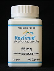
Lenalidomide may be the best maintenance treatment option for patients with newly diagnosed multiple myeloma (MM), according to the authors of a systematic review and meta-analysis.
Francesca M. Gay, MD, PhD, of the University of Torino in Italy, and her coauthors wrote that, despite the well-recognized importance of novel agent–based maintenance therapy for MM, there is a lack of direct or indirect comparisons between the available regimens.
In a paper published in JAMA Oncology, the researchers reported the results of a systematic review and meta-analysis of 11 prospective, phase 3, randomized, controlled trials of 8 varieties of maintenance therapy in 5073 participants with newly diagnosed MM.
The researchers found that lenalidomide-based regimens showed the best progression-free survival rates, compared with placebo. The hazard ratio (HR) was 0.39 for lenalidomide plus prednisone, and the HR was 0.47 for lenalidomide alone.
In 74% of the network meta-analysis simulations, lenalidomide-based regimens were the most effective options.
Four other maintenance treatment options—thalidomide-interferon (HR, 0.50), thalidomide-bortezomib (HR, 0.58), bortezomib-prednisone (HR, 0.72), and thalidomide alone (HR, 0.73)—also showed progression-free survival gains, but interferon therapy (HR, 0.91) failed to show any benefit.
For overall survival, lenalidomide alone (HR, 0.76) was the best option, followed by thalidomide-bortezomib (HR, 0.82) and bortezomib-prednisone (HR, 0.84). None of the other regimens considered showed benefits for overall survival.
“Long-term use of lenalidomide undoubtedly has advantages, owing to the lack of neuropathy, which is the main factor limiting the long-term use of both thalidomide and bortezomib,” the authors wrote.
When the authors restricted their analysis to trials conducted in the setting of autologous stem cell transplant, they found similar results, with lenalidomide-based regimens having the best progression-free and overall survival.
Patients with a good prognosis and standard-risk chromosomal abnormalities also did best with lenalidomide-based maintenance, while those with a poor prognosis—for example, with ISS stage III disease—benefited more from bortezomib-based maintenance.
However, patients with high-risk chromosomal abnormalities gained no advantage from any regimen, which the authors suggested may relate to small sample size, different cut-off points, or the patients’ extremely poor prognosis.
The authors noted that their analysis did not take into account adverse events, drug discontinuations, or quality of life but focused solely on progression-free survival and overall survival.
“An increase in second primary malignant disease with prolonged lenalidomide therapy has been reported, but the survival benefit overcame the risk in all the trials,” they wrote.
The authors also commented that better treatment options are needed for patients with aggressive disease, and there are ongoing trials looking at second-generation proteasome inhibitors, immunomodulatory agents, and monoclonal antibodies for maintenance therapy.
Most authors declared research funding, advisory board positions, fees, and honoraria from the pharmaceutical industry, including lenalidomide manufacturer Celgene.
Lenalidomide best option for myeloma maintenance therapy
Lenalidomide may be the best maintenance treatment option for patients with newly diagnosed multiple myeloma, say the authors of a systematic review and meta-analysis.
Francesca M. Gay, MD, from the division of hematology at the University of Torino (Italy), and her coauthors wrote that despite the well-recognized importance of novel agent–based maintenance therapy for multiple myeloma, there is a lack of direct or indirect comparisons between the available regimens.
In a paper published online in JAMA Oncology, the researchers reported the results of the systematic review and meta-analysis of 11 prospective, phase 3 randomized, controlled trials of eight varieties of maintenance therapy, in 5,073 participants with newly diagnosed multiple myeloma.
Their analysis found that lenalidomide-based regimens showed the best progression-free survival rates (hazard ratio, 0.39 for lenalidomide plus prednisone; HR, 0.47 for lenalidomide alone), compared with placebo, and in 74% of the network meta-analysis simulations, they were the most effective options.
Four other maintenance treatment options - thalidomide-interferon, thalidomide-bortezomib, bortezomib-prednisone, and thalidomide alone – also showed progression-free survival gains – but interferon therapy failed to show any benefit.
However, for overall survival, lenalidomide alone was the best option, followed by thalidomide-bortezomib and bortezomib-prednisone. None of the other regimens considered showed benefits for overall survival.
“Long-term use of lenalidomide undoubtedly has advantages, owing to the lack of neuropathy, which is the main factor limiting the long-term use of both thalidomide and bortezomib,” the authors wrote.
When the authors restricted their analysis to trials conducted in the setting of autologous stem cell transplantation they found similar results, with lenalidomide-based regimens having the best progression-free and overall survival.
Patients with a good prognosis and standard-risk chromosomal abnormalities also did best with lenalidomide-based maintenance, while those with a poor prognosis – for example, with ISS stage III disease – benefited more from bortezomib-based maintenance. However patients with high-risk chromosomal abnormalities gained no advantage from any regimen, which the authors suggested may relate to small sample size, different cut-off points or their extremely poor prognosis.
The authors noted that their analysis did not take into account adverse events, drug discontinuations, or quality of life but focused solely on progression-free survival and overall survival.
“An increase in second primary malignant disease with prolonged lenalidomide therapy has been reported, but the survival benefit overcame the risk in all the trials,” they wrote.
They also commented that better treatment options are needed for patients with aggressive disease, and there are ongoing trials looking at second-generation proteasome inhibitors, immunomodulatory agents, and monoclonal antibodies for maintenance therapy.
Most authors declared research funding, advisory board positions, fees and honoraria from the pharmaceutical industry, including lenalidomide manufacturer Celgene.
SOURCE: Gay F et al. 2018 Aug 9. doi:10.1001/jamaoncol.2018.2961.
FROM JAMA ONCOLOGY
Key clinical point: Lenalidomide is the best option for maintenance therapy in multiple myeloma.
Major finding: Lenalidomide-based maintenance regimens show the best progression-free and overall survival in multiple myeloma.
Study details: Systematic review and network meta-analysis of 11 studies in 5073 participants with newly diagnosed multiple myeloma.
Disclosures: Most authors declared research funding, advisory board positions, fees and honoraria from the pharmaceutical industry, including lenalidomide manufacturer Celgene.
Source: Gay F et al. 2018 Aug 9. doi: 10.1001/jamaoncol.2018.2961.
Team reports ‘simple’ way for GPs to rule out MM
Researchers say they have determined which blood tests can help general practitioners (GPs) rule out a diagnosis of multiple myeloma (MM).
The team discovered that plasma viscosity (PV) and erythrocyte sedimentation rate (ESR) were more helpful in ruling out MM than a C-reactive protein (CRP) test.
Furthermore, the possibility of MM “is extremely low” in patients with normal hemoglobin (Hb) and normal PV or ESR.
“Ordinarily, a GP will see a patient with myeloma every 5 years, and early diagnosis matters,” said study author William Hamilton, MD, of the University of Exeter Medical School in the UK.
“We report a simple way a GP can check patients presenting symptoms such as back, rib, and chest pain or recurrent chest infections and determine whether they have myeloma or not.”
Dr Hamilton and his colleagues reported their findings in the British Journal of General Practice.
The researchers analyzed blood tests performed on 2703 MM patients up to 5 years prior to diagnosis. The team then compared results in the MM cases to blood test results in 12,157 patients without MM, matched for age and other relevant parameters.
The researchers used likelihood ratios (LRs) to classify tests as useful for ruling in or ruling out MM.
The team explained that positive likelihood (LR+) tests indicate how many times more likely a positive test occurs in individuals with MM than in those without the disease. Negative likelihood (LR–) tests indicate how many times less likely a negative result will occur in individuals with MM than in those without MM.
A test was defined as useful for ruling in MM if the LR+ was ≥ 5 and useful for ruling out MM if the LR– was ≤ 0.2.
Results
None of the inflammatory markers analyzed proved useful (LR+ ≥ 5) for ruling in MM.
The LR+ was:
- 2.0 for raised PV
- 1.9 for raised ESR
- 1.2 for raised CRP.
Similarly, none of the tests alone was useful (LR– ≤ 0.2) for ruling out MM.
The LR– was:
- 0.42 for normal Hb
- 0.81 for normal calcium
- 0.80 for normal creatinine
- 0.28 for normal ESR
- 0.32 for normal PV
- 0.87 for normal CRP.
However, several combinations of tests were useful for ruling out MM.
| Tests | LR– (95% CI) |
| Hb and PV | 0.12 (0.07 to 0.23) |
| Hb and ESR | 0.16 (0.13 to 0.21 |
| Hb, creatinine, and PV | 0.09 (0.04 to 0.20) |
| Hb, calcium, and PV | 0.06 (0.02 to 0.18 |
| Hb, creatinine, and ESR | 0.16 (0.12 to 0.21) |
| Hb, calcium, and ESR | 0.16 (0.12 to 0.22) |
| Hb, calcium, creatinine, and PV | 0.06 (0.02 to 0.19) |
| Hb, calcium, creatinine, and ESR | 0.17 (0.12 to 0.24) |
| Hb and CRP* | 0.39 (0.32 to 0.47)* |
| Hb, creatinine, and CRP* | 0.33 (0.26 to 0.42)* |
| Hb, calcium, and CRP* | 0.33 (0.25 to 0.42)* |
| Hb, calcium, creatinine, and CRP* | 0.31 (0.22 to 0.42)* |
| *Not useful (LR– ≤0.2) | |
Conclusions/implications
The researchers concluded that, with normal Hb and normal PV or ESR, the possibility of MM is very low, and assessing CRP or creatinine as well increases the sensitivity of testing only slightly.
“The combination of levels of hemoglobin . . . and 1 of 2 inflammatory markers [ESR or PV] are a sufficient test rule out myeloma,” said study author Constantinos Koshiaris, of the University of Oxford in the UK.
“If abnormalities are detected in this test, it should lead to urgent urine protein tests, which can help speed up diagnosis.”
The researchers also recommend adding calcium tests if patients have certain symptoms, such as back pain, rib pain, joint pain, and fracture.
Study links communication, outcomes in cancer
Better communication between cancer patients and healthcare providers may provide tangible benefits, according to research published in JNCCN.
Cancer survivors who reported greater satisfaction in communicating with healthcare providers had better general health and mental health, fewer doctor visits, and reduced healthcare spending, when compared to patients who were less satisfied with communication.
“Our study suggests that when cancer care providers are more effective communicators, their patients are more likely to follow medical advice and medication protocols,” said study author Ashish Rai, PhD, of the American Cancer Society in Framingham, Massachusetts.
For this study, Dr Rai and his colleagues analyzed data from the Medical Expenditure Panel Survey (MEPS) from 2008 through 2014.
The researchers evaluated 4588 cancer survivors, dividing them into non-elderly and elderly groups. The non-elderly patients (n=2257) had a median age of 54 (range, 18-64), and the elderly patients (n=2331) had a median age of 75.
Communication satisfaction was measured by the Consumer Assessment of Healthcare Providers and Systems (CAHPS), in conjunction with the MEPS data.
Patients used a 4-point scale ranging from “never” to “always” to track whether their providers did the following:
- Listened carefully
- Explained things in a way that was easy to understand
- Showed respect for what the respondent had to say
- Spent enough time with the respondent.
A global satisfaction rating scale (0 to 10) was factored into a composite score and tracked across 12 months.
The researchers then assessed various patient outcomes.
Satisfaction and outcomes
Overall, patients who were the most satisfied with communication had the best outcomes with regard to general, physical, and mental health; fewer emergency department, hospital, and office visits; and reduced drug, out-of-pocket, and total healthcare expenditures.
However, the associations between communication satisfaction and outcomes were not always significant.
In an adjusted analysis, the elderly patients who were more satisfied with communication in year 1 had significantly better outcomes in year 2 for general health, mental health, and total healthcare expenditures.
| Elderly patients 65+ | |||
| Least satisfied
(tertile 1) |
Moderately satisfied (tertile 2) | Most satisfied
(tertile 3) |
|
| Excellent/very good general health | |||
| Unadjusted proportion | 23.6% | 31.8% | 45.8% |
| Predictive margin | 30.3
(95% CI 26.0–34.6) |
32.2
(95% CI 28.9–35.5) |
38.9
(95% CI 35.1–42.7) |
| P value | Reference | 0.466 | 0.007 |
| Highest quartile of mental health | |||
| Unadjusted proportion | 22.9% | 34.8% | 41.7% |
| Predictive margin | 27.1
(95% CI 22.1–32.1) |
35.5
(95% CI 31.5–39.5) |
37.0
(95% CI 32.7–41.4) |
| P value | Reference | 0.013 | 0.005 |
| Total healthcare expenditure | |||
| Unadjusted mean | $33,558 | $27,341 | $29,591 |
| Predictive margin | $34,071 ($29,011–$39,131) | $28,230 ($22,907–$33,553) | $26,995 ($22,568–$31,422) |
| P value | Reference | 0.301 | 0.049 |
For the non-elderly patients, those who were more satisfied with communication in year 1 had significantly better outcomes in year 2 for physician office visits and mental health.
| Non-elderly patients (18-64) | |||
| Least satisfied (tertile 1) | Moderately satisfied (tertile 2) | Most satisfied (tertile 3) | |
| Total physician office visits | |||
| Unadjusted mean | 7.96 | 6.96 | 5.85 |
| Predictive margin | 7.42
(95% CI 6.78–8.06) |
6.60
(95% CI 5.98–7.22) |
6.26
(95% CI 5.47–7.05) |
| P value | Reference | 0.211 | 0.038 |
| Highest quartile of mental health | |||
| Unadjusted proportion | 23.5% | 35.5% | 41.1% |
| Predictive margin | 29.7
(95% CI 25.3–34.1) |
36.0
(95% CI 31.3–40.7) |
34.0
(95% CI 29.5–38.4) |
| P value | Reference | 0.036 | 0.187 |
Baseline health and satisfaction
In both age groups, patients with better baseline health reported higher satisfaction with communication. Conversely, the more comorbidities patients had, the lower their satisfaction rating.
The researchers said this suggests that more complex circumstances negatively impacted patients’ perception of their communication, and the finding highlights the importance of coordinating care across a team of providers.
“The results of this study present an interesting challenge: those survivors most in need of good communication about complex medical issues may not be receiving the information they seek in a manner that they find helpful. That, in turn, results in higher healthcare utilization and expenditure,” said Crystal Denlinger, MD, of Fox Chase Cancer Center in Philadelphia, Pennsylvania, who was not involved in this study.
“This could be due to many factors, including time constraints, competing priorities, and increasingly complex cancer therapies. This study highlights the need for additional research into how to tailor the healthcare experience both during and after cancer treatment in order to communicate more effectively.”
Conclusions
“Communication needs vary from patient to patient,” Dr Rai noted. “While time constraints do pose a challenge, the amount of time spent is only one of the attributes of effective communication. By tailoring their communication strategy to a patient’s specific needs, providers may be able to communicate more effectively in the same amount of time.”
Dr Rai also pointed out the importance of delegating both clinical and communication duties as needed. Dr Rai and his colleagues also cited earlier research demonstrating better outcomes for patients who had the option of communicating with their provider electronically.1,2
Ultimately, the researchers concluded that effective provider communication can improve outcomes by streamlining care, alleviating anxiety, boosting mutual trust, and increasing treatment adherence.
1. Basch E, Deal AM, Dueck AC, et al. Overall survival results of a trial assessing patient-reported outcomes for symptom monitoring during routine cancer treatment. JAMA 2017;318:197–198.
2. Smith AB, Basch E. Role of patient-reported outcomes in postsurgical monitoring in oncology. J Oncol Pract 2017;13:535–538.
Auto-HSCT linked to higher AML, MDS risk
Patients undergoing autologous hematopoietic stem cell transplant (auto-HSCT) for lymphoma or myeloma have an increased risk of acute myeloid leukemia (AML) and myelodysplastic syndromes (MDS), according to a retrospective study.
The study suggested these patients have 10 to 100 times the risk of AML or MDS as the general population.
The elevated risk also exceeds that of similar lymphoma and myeloma patients largely untreated with auto-HSCT.
Tomas Radivoyevitch, PhD, of the Cleveland Clinic Foundation in Ohio, and his colleagues reported these findings in Leukemia Research.
The investigators noted that exposure to DNA-damaging drugs and ionizing radiation—both used in auto-HSCT—is known to increase the risk of AML and MDS.
With this in mind, the team analyzed data on auto-HSCT recipients reported to the Center for International Blood and Marrow Transplant Research (CIBMTR).
Analyses were based on 9028 patients undergoing auto-HSCT from 1995 to 2010 for Hodgkin lymphoma (n=916), non-Hodgkin lymphoma (NHL, n=3546), or plasma cell myeloma (n=4566). Their median duration of follow-up was 90 months, 110 months, and 97 months, respectively.
Overall, 3.7% of the cohort developed AML or MDS after their transplant.
More aggressive transplant protocols increased the likelihood of this outcome. The risk of developing AML or MDS was higher for:
- Hodgkin lymphoma patients who received conditioning with total body radiation versus chemotherapy alone (hazard ratio [HR], 4.0)
- NHL patients who received conditioning with total body radiation (HR, 1.7) or with busulfan and melphalan or cyclophosphamide (HR, 1.8) versus the BEAM regimen (bischloroethylnitrosourea, etoposide, cytarabine, and melphalan)
- NHL or myeloma patients who received 3 or more lines of chemotherapy versus 1 line (HR, 1.9 for NHL and 1.8 for myeloma)
- NHL patients who underwent transplant in 2005 to 2010 versus 1995 to 1999 (HR, 2.1).
Patients reported to the Surveillance, Epidemiology and End Results database with the same lymphoma and myeloma diagnoses, few of whom underwent auto-HSCT, had risks of AML and MDS that were 5 to 10 times higher than the background level in the population.
However, the study auto-HSCT cohort had a risk of AML that was 10 to 50 times higher and a relative risk of MDS that was roughly 100 times higher than the background level.
“These increases may be related to exposure to high doses of DNA-damaging drugs given for [auto-HSCT], but this hypothesis can only be tested in a prospective study,” Dr Radivoyevitch and his coinvestigators wrote.
The reason for the greater elevation of MDS risk, compared with AML risk, is unknown.
“One possible explanation is that many cases of MDS evolve to AML, and that earlier diagnosis from increased post-transplant surveillance resulted in a deficiency of AML,” the investigators wrote. “A second is based on steeper MDS versus AML incidences versus age . . . and the possibility that transplantation recipient marrow ages (ie, marrow biological ages) are perhaps decades older than calendar ages.”
The study authors said they had no relevant conflicts of interest. The CIBMTR is supported by several US government agencies and numerous pharmaceutical companies.
Janssen seeks approval for split dosing of daratumumab
Janssen has applied to the Food and Drug Administration and the European Medicines Agency to allow splitting of the first infusion of daratumumab (Darzalex) in multiple myeloma patients over 2 consecutive days.
The goal is to improve the treatment experience for patients and physicians, according to the announcement from Janssen.
The regulatory submissions are based on the global, multi-arm, phase 1b MMY1001 study (NCT01998971). The study evaluated daratumumab in combination with various other treatments in 240 patients with multiple myeloma. It found that both the safety profile and the pharmacokinetics concentrations seen with either single dosing or split dosing were similar.
Daratumumab is the first approved monoclonal antibody that targets CD38, which is expressed across multiple myeloma cells regardless of disease stage. Daratumumab is currently approved for treatment of multiple myeloma in both the United States and the European Union either as monotherapy or in conjunction with other treatments.
Daratumumab is known to sometimes cause severe/serious infusion reactions, such as anaphylactic reactions; interfere with serological testing; and cause neutropenia or thrombocytopenia.


