User login
VIDEO: Risks for anastomotic leak after colectomy identified
SAN FRANCISCO– A large retrospective study identified risk factors for anastomotic leak within 30 days of colectomy.
The analysis of data on 13,684 patients looked at a multicenter cohort, compared with previous smaller studies that generally focused on single institutions, Dr. Emily F. Midura and her associates reported at the annual clinical congress of the American College of Surgeons.
In a video interview at the meeting, Dr. Midura described the risk factors identified in the study.
The 4% of patients who developed a leak averaged 13 days in the hospital, compared with 8 days for patients with no leak, said Dr. Midura of the University of Cincinnati. A total of 6% of patients who developed an anastomotic leak died, compared with a 2% mortality rate in patients without a leak.
Indications for colectomy included cancer in 42% of patients, diverticulitis in 23%, inflammatory bowel disease in 6%, and other reasons in the rest of the cohort.
Dr. Midura reported having no financial disclosures.
The video associated with this article is no longer available on this site. Please view all of our videos on the MDedge YouTube channel
On Twitter @sherryboschert
AT THE ACS CLINICAL CONGRESS

Colorectal cancer on the increase in young adults
Colorectal cancer rates have declined in both men and women since 1975, particularly in those over age 75 years, but there has also been a significant increase in the incidence among those aged 20-34 years, a retrospective cohort study shows.
Researchers analyzed data from 393,241 patients registered in the Surveillance, Epidemiology, and End Results (SEER) colorectal cancer registry from 1975 to 2010, finding the overall, age-adjusted incidence of colorectal cancer decreased by 0.92% during that time.

They observed a steady decline in the incidence of colorectal cancer in the over 50 age group, with a 1.15% decrease in those aged over 75 years, while in those aged 20-34 years, there was a nearly 2% increase, according to data published online November 5 in JAMA Surgery (doi:10.1001/jamasurg.2014.1756).
“At the present rate, the incidence rate for young patients with newly diagnosed colon or rectal cancer will nearly double by 2030, while it will similarly decline by more than one-third among patients older than the screening age of 50 years,” wrote Dr. Christina E. Bailey of the University of Texas M.D. Anderson Cancer Center, Houston, and colleagues.
The study was supported by grants from the National Institutes of Health and National Cancer Institute. There were no conflicts of interest declared.
Colorectal cancer rates have declined in both men and women since 1975, particularly in those over age 75 years, but there has also been a significant increase in the incidence among those aged 20-34 years, a retrospective cohort study shows.
Researchers analyzed data from 393,241 patients registered in the Surveillance, Epidemiology, and End Results (SEER) colorectal cancer registry from 1975 to 2010, finding the overall, age-adjusted incidence of colorectal cancer decreased by 0.92% during that time.
They observed a steady decline in the incidence of colorectal cancer in the over 50 age group, with a 1.15% decrease in those aged over 75 years, while in those aged 20-34 years, there was a nearly 2% increase, according to data published online November 5 in JAMA Surgery (doi:10.1001/jamasurg.2014.1756).
“At the present rate, the incidence rate for young patients with newly diagnosed colon or rectal cancer will nearly double by 2030, while it will similarly decline by more than one-third among patients older than the screening age of 50 years,” wrote Dr. Christina E. Bailey of the University of Texas M.D. Anderson Cancer Center, Houston, and colleagues.
The study was supported by grants from the National Institutes of Health and National Cancer Institute. There were no conflicts of interest declared.
FROM JAMA SURGERY
Key clinical point: Colorectal cancer rates have declined steadily since 1975 in those over 50, but have increased in the 20- to 34-year-old age group.
Major finding: Colorectal cancer incidence has increased nearly 2% in those aged 20-34 years.
Data source: Retrospective cohort study of data from 393,241 patients registered in the Surveillance, Epidemiology, and End Results (SEER) colorectal cancer registry.
Disclosures: The study was supported by grants from the National Institutes of Health and National Cancer Institute. There were no conflicts of interest declared.

Uncomplicated diverticulitis patients safely avoid routine antibiotics
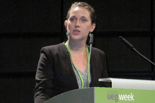
VIENNA – Patients with first-episode, uncomplicated diverticulitis can safely be managed by observation alone without routine antibiotic treatment, based on results from a prospective, controlled, multicenter Dutch trial with 528 patients.
Coupled with similar results from a 2012 Swedish randomized trial, the new findings show that an observational approach without up-front antibiotic treatment is safe and effective for routine practice for patients with a modified Hinchey stage 1 classification, the most common diverticulitis presentation, Dr. Lidewine Daniels said at the United European Gastroenterology Global Congress.

The 2012 trial, done in Sweden and Iceland, randomized 623 patients with acute, uncomplicated diverticulitis, and found no statistically significant difference in the rate of recovery without complications during 12 months of follow-up, regardless of whether or not patients received routine antibiotic treatment at the time of initial diagnosis (Br. J. Surg. 2012;99:532-9).
This approach “can now be adopted into guidelines,” said Dr. Daniels, a researcher in the surgery department at the Academic Medical Center in Amsterdam. This would shift current practice recommendations, such as those published earlier this year by the American College of Colon and Rectal Surgeons (Dis. Colon Rectum 2014;57:284-94), which have generally called for routine antibiotic management of these types of diverticulitis patients, Dr. Daniels said.
The DIABOLO (Multicenter Randomized Clinical Trial Investigating the Cost-effectiveness of Treatment Strategies With or Without Antibiotics for Uncomplicated Acute Diverticulitis) trial enrolled patients with CT-proven, left-sided, first-episode, uncomplicated diverticulitis at 22 Dutch centers. All patients were classified as having stage 1 disease by the modified Hinchey criteria (Int. J. Colorectal Dis. 2012;27:207-14), with about 92% of patients classified as Hinchey 1a.
The 266 patients randomized to initial antibiotic treatment began at least 2 days on intravenous treatment with amoxicillin and clavulanic acid, followed by a switch to oral delivery of the same combination when appropriate for a total of 10 days on the antibiotic.
Patients remained hospitalized until they switched from intravenous to oral treatment. The 262 patients randomized to no initial antibiotic treatment were hospitalized and observed, and received supportive treatment until they were judged ready for discharge by being able to eat a normal diet, had a temperature of less than 38 °C, and had a self-rated pain score of less than 4 on a visual analog scale of 0-10 without use of pain medication; in addition, the patient’s agreement was needed for discharge.
During 6 months of follow-up, the median time to complete recovery – defined as the criteria for hospital discharge plus a return to normal activity – occurred after a median of 12 days among patients in the antibiotic group and 14 days among those in the observation group, a difference that was not statistically significant for the study’s primary endpoint.
The results also showed no statistically significant differences between the two study arms for almost all the other outcomes assessed, including need for readmission, complications, need for surgery, morbidities, serious morbidities, and deaths. The only significant differences in outcomes were a decreased rate of hospitalization at the time of initial treatment among patients in the observation-only arm, and fewer hospitalized days among the observation-only patients.
“Observational treatment is without short- or long-term repercussions,” Dr. Daniels concluded.
Dr. Daniels had no disclosures.
On Twitter @mitchelzoler
VIENNA – Patients with first-episode, uncomplicated diverticulitis can safely be managed by observation alone without routine antibiotic treatment, based on results from a prospective, controlled, multicenter Dutch trial with 528 patients.
Coupled with similar results from a 2012 Swedish randomized trial, the new findings show that an observational approach without up-front antibiotic treatment is safe and effective for routine practice for patients with a modified Hinchey stage 1 classification, the most common diverticulitis presentation, Dr. Lidewine Daniels said at the United European Gastroenterology Global Congress.
The 2012 trial, done in Sweden and Iceland, randomized 623 patients with acute, uncomplicated diverticulitis, and found no statistically significant difference in the rate of recovery without complications during 12 months of follow-up, regardless of whether or not patients received routine antibiotic treatment at the time of initial diagnosis (Br. J. Surg. 2012;99:532-9).
This approach “can now be adopted into guidelines,” said Dr. Daniels, a researcher in the surgery department at the Academic Medical Center in Amsterdam. This would shift current practice recommendations, such as those published earlier this year by the American College of Colon and Rectal Surgeons (Dis. Colon Rectum 2014;57:284-94), which have generally called for routine antibiotic management of these types of diverticulitis patients, Dr. Daniels said.
The DIABOLO (Multicenter Randomized Clinical Trial Investigating the Cost-effectiveness of Treatment Strategies With or Without Antibiotics for Uncomplicated Acute Diverticulitis) trial enrolled patients with CT-proven, left-sided, first-episode, uncomplicated diverticulitis at 22 Dutch centers. All patients were classified as having stage 1 disease by the modified Hinchey criteria (Int. J. Colorectal Dis. 2012;27:207-14), with about 92% of patients classified as Hinchey 1a.
The 266 patients randomized to initial antibiotic treatment began at least 2 days on intravenous treatment with amoxicillin and clavulanic acid, followed by a switch to oral delivery of the same combination when appropriate for a total of 10 days on the antibiotic.
Patients remained hospitalized until they switched from intravenous to oral treatment. The 262 patients randomized to no initial antibiotic treatment were hospitalized and observed, and received supportive treatment until they were judged ready for discharge by being able to eat a normal diet, had a temperature of less than 38 °C, and had a self-rated pain score of less than 4 on a visual analog scale of 0-10 without use of pain medication; in addition, the patient’s agreement was needed for discharge.
During 6 months of follow-up, the median time to complete recovery – defined as the criteria for hospital discharge plus a return to normal activity – occurred after a median of 12 days among patients in the antibiotic group and 14 days among those in the observation group, a difference that was not statistically significant for the study’s primary endpoint.
The results also showed no statistically significant differences between the two study arms for almost all the other outcomes assessed, including need for readmission, complications, need for surgery, morbidities, serious morbidities, and deaths. The only significant differences in outcomes were a decreased rate of hospitalization at the time of initial treatment among patients in the observation-only arm, and fewer hospitalized days among the observation-only patients.
“Observational treatment is without short- or long-term repercussions,” Dr. Daniels concluded.
Dr. Daniels had no disclosures.
On Twitter @mitchelzoler
AT UEG WEEK VIENNA 2014
Key clinical point: Observation-only worked as well as routine antibiotic treatment for patients with uncomplicated diverticulitis.
Major finding: Median time to full recovery was 12 days for antibiotic-treated patients and 14 days for observation-only patients.
Data source: The DIABOLO study, which randomized 528 patients with first-time, CT-proven, acute, uncomplicated diverticulitis at 22 Dutch centers.
Disclosures: Dr. Daniels had no disclosures.
FDA approves budesonide rectal foam for distal UC
A rectal foam formulation of 2% budesonide has been approved by the Food and Drug Administration for treating distal ulcerative colitis, the manufacturer, Salix Pharmaceuticals, has announced.
The approved indication is for the induction of remission in patients with mild to moderate distal ulcerative colitis extending up to 40 cm from the anal verge. The foam is administered rectally and “overcomes treatment limitations associated with currently approved therapies which are often ineffective due to insufficient distribution of active drug to the distal colon,” the company said in a statement announcing the final approval on Oct. 8.
The company will be marketing the product as Uceris.
The company also markets budesonide oral extended-release tablets under the same name.
Transanal extraction found effective in rectal cancer surgery
Patients who underwent laparoscopic total mesorectal excision with coloanal anastomosis for rectal cancer had similar rates of mortality and morbidity, regardless of whether the extraction was performed transanally or transabdominally, a long-term single-center study showed.
“There are few data of full laparoscopic coloanal anastomosis for rectal cancer, including small series and short follow-up,” authors led by Dr. Quentin Denost of the department of surgery at Saint-André Hospital, Bordeau, France, wrote. “Moreover, the risk of anastomotic or perineal recurrence induced by transanal extraction of the rectal specimen is not known.” In addition, they continued, functional outcomes of laparoscopic coloanal anastomosis “have never been reported, and therefore the potential risk of anal incontinence related to transanal specimen extraction has never been discussed.”
In an effort to investigate the long-term outcome of laparoscopic coloanal anastomosis for rectal cancer, the researchers evaluated records of 220 patients who underwent laparoscopic total mesorectal excision and coloanal anastomosis for rectal cancer at Saint-André Hospital between 2000 and 2010 (Ann. Surg. 2014 Sept. 1 [doi:10.1097/SLA.0000000000000855]). Study endpoints of interest were circumferential margin, mesorectal grade, local recurrence, survival, and functional outcome obtained by a questionnaire sent to patients free of disease with at least 1 year of follow-up after stoma closure.
More than half of the patients (63%) were male, their median age was 64, and their median body mass index was 25 kg/m2. The tumors were a median of 4 cm from the anal verge and 1 cm from the anal ring, and 82% of the patients had stage T3 or T4 disease.
The authors reported that the overall mortality and surgical morbidity rates were 0.5% and 17%, respectively, the rate of positive circumferential resection margin was 9%, and the median anal continence score was 6 (range, 0-20). After a median follow-up of 51 months, the local recurrence rate was 4%, while at 5 years, the overall survival and disease-free survival rates were 83% and 70%, respectively.
When the authors evaluated results by extraction site, no significant differences were observed between the transanal extraction and transabdomonal extraction groups in the rate of overall mortality (0.8% vs. 0%, respectively; P = 1.000), overall morbidity (34% vs. 43%), positive circumferential margin (7% vs. 11%; P = .324), mesorectal grade, local recurrence (4% vs. 5%; P = .98), and disease-free survival (72% vs. 68%; P = .63). The median continence score was 6 in both groups (P = .92).
The findings demonstrated that pelvic control and survival “were not compromised by the association between mini invasive surgery and ultralow sphincter preservation,” the authors concluded. “Moreover, we demonstrated the safety and efficacy of transanal extraction of the rectal specimen with similar oncologic and functional outcome than the conventional abdominal extraction. Because of the wound advantages of transanal extraction, in terms of abdominal wall preservation, transanal extraction can be recommended in laparoscopic surgical management of low rectal cancer.”
They acknowledged certain limitations of the study, including the fact that BMI was slightly lower in the transanal group, compared with the transabdomonal group (24.3 vs. 25.8 kg/m2, respectively; P = .01). This suggests “that some obese patients probably received transabdominal instead of transanal extraction,” Dr. Denost and associates wrote. “Therefore, as we recommend preventing excessive stretching of the anal sphincter during rectal extraction, we also recommend to be cautious when performing transanal extraction in obese patients with wide mesorectal specimen, especially to avoid mesorectal injury and tumor spillage.”
The authors reported having no relevant financial disclosures.
On Twitter @dougbrunk
FROM ANNALS OF SURGERY
Key clinical point: Transanal extraction can be recommended in the laparoscopic surgical management of low rectal cancer.
Major finding: During laparoscopic total mesorectal excision, no significant differences were observed between patients who underwent transanal extraction or transabdomonal extraction in the rate of overall mortality (0.8% vs. 0%, respectively; P = 1.000), overall morbidity (34% vs. 43%), or local recurrence (4% vs. 5%; P = .98).
Data source: A single-center study of 220 patients who underwent laparoscopic total mesorectal excision and coloanal anastomosis for rectal cancer at Saint-André Hospital in Bordeaux, France, between 2000 and 2010.
Disclosures: The authors reported having no relevant financial disclosures.
Postoperative complications increase risk of death in CRC patients
Patients who develop infectious complications after undergoing curative surgery for colorectal cancer face a significantly increased risk of death, results from a large retrospective study showed.
“The association of postoperative complications with long-term survival after major surgery has been suggested by several studies in mixed populations and is to some degree expected and intuitive,” authors led by Dr. Avo Artinyan, of the surgery department at Baylor College of Medicine, Houston, wrote online Sept. 1 in Annals of Surgery. “It has been difficult, however, to determine a specific cause-effect relationship, particularly because this association is noted even in patients who suffer late mortality, that is, those who presumably recover from postoperative complications.”

In an effort to investigate the effect of postoperative complications on long-term survival after colorectal cancer resection, the researchers evaluated the records of 12,075 patients from the Veterans Affairs Surgical Quality Improvement Program and the Central Cancer Registry databases who underwent resection for nonmetastatic colorectal cancer from 1999 to 2009 (Ann. Surg. 2014 Sept. 1 [doi: 10.1097/SLA.0000000000000854]). They categorized patients by presence of any complication within 30 days and by type of complication (infectious vs. noninfectious); excluded patients who died within 90 days of the procedure; and performed univariate and multivariate analyses adjusted for patient, disease, and treatment factors.
The average age of the cohort was 69 years, 98% were men, more than two-thirds (69%) had an American Society of Anesthesiologists (ASA) classification score of 3, and 61% had stage 1 or 2 disease. Dr. Artinyan and his associates found that the overall morbidity and infectious complication rates were 27.8% and 22.5%, respectively.
Compared with patients who had no postoperative complications, those who did were older and had lower postoperative serum albumin, worse functional status, and higher ASA scores (P less than .001). Multivariate analysis revealed that the presence of any complication was associated with a 24% increased hazard of death (hazard ratio, 1.24; P less that .001). When the analysis was limited to the type of complication, patients with infectious complications (in particular, surgical site infections) had an increased hazard of death (HR, 1.31), predominately those with severe infections (HR, 1.41).
“To our knowledge, this is the largest single study to examine the association of postoperative complications with long-term survival for CRC,” the authors wrote. “Similar to other groups, we have demonstrated that postoperative complications occur in a significant proportion of patients after CRC resection and that most patients with postoperative morbidity have at least one infectious complication.”
They acknowledged certain limitations of the study, including its retrospective design and the potential for selection bias. “Additional limitations include the absence of margin data – which may have a considerable impact on both the risk of organ-space infections and disease recurrence – and the inability to calculate cancer-specific survival and other cancer-specific outcomes,” they wrote.
“Overall all-cause survival, however, is still a commonly used and useful outcome measure, and we have attempted to mitigate the effect of early non–cancer-related mortality with the exclusion of early deaths.”
The authors reported having no financial disclosures.
On Twitter @dougbrunk
Patients who develop infectious complications after undergoing curative surgery for colorectal cancer face a significantly increased risk of death, results from a large retrospective study showed.
“The association of postoperative complications with long-term survival after major surgery has been suggested by several studies in mixed populations and is to some degree expected and intuitive,” authors led by Dr. Avo Artinyan, of the surgery department at Baylor College of Medicine, Houston, wrote online Sept. 1 in Annals of Surgery. “It has been difficult, however, to determine a specific cause-effect relationship, particularly because this association is noted even in patients who suffer late mortality, that is, those who presumably recover from postoperative complications.”
In an effort to investigate the effect of postoperative complications on long-term survival after colorectal cancer resection, the researchers evaluated the records of 12,075 patients from the Veterans Affairs Surgical Quality Improvement Program and the Central Cancer Registry databases who underwent resection for nonmetastatic colorectal cancer from 1999 to 2009 (Ann. Surg. 2014 Sept. 1 [doi: 10.1097/SLA.0000000000000854]). They categorized patients by presence of any complication within 30 days and by type of complication (infectious vs. noninfectious); excluded patients who died within 90 days of the procedure; and performed univariate and multivariate analyses adjusted for patient, disease, and treatment factors.
The average age of the cohort was 69 years, 98% were men, more than two-thirds (69%) had an American Society of Anesthesiologists (ASA) classification score of 3, and 61% had stage 1 or 2 disease. Dr. Artinyan and his associates found that the overall morbidity and infectious complication rates were 27.8% and 22.5%, respectively.
Compared with patients who had no postoperative complications, those who did were older and had lower postoperative serum albumin, worse functional status, and higher ASA scores (P less than .001). Multivariate analysis revealed that the presence of any complication was associated with a 24% increased hazard of death (hazard ratio, 1.24; P less that .001). When the analysis was limited to the type of complication, patients with infectious complications (in particular, surgical site infections) had an increased hazard of death (HR, 1.31), predominately those with severe infections (HR, 1.41).
“To our knowledge, this is the largest single study to examine the association of postoperative complications with long-term survival for CRC,” the authors wrote. “Similar to other groups, we have demonstrated that postoperative complications occur in a significant proportion of patients after CRC resection and that most patients with postoperative morbidity have at least one infectious complication.”
They acknowledged certain limitations of the study, including its retrospective design and the potential for selection bias. “Additional limitations include the absence of margin data – which may have a considerable impact on both the risk of organ-space infections and disease recurrence – and the inability to calculate cancer-specific survival and other cancer-specific outcomes,” they wrote.
“Overall all-cause survival, however, is still a commonly used and useful outcome measure, and we have attempted to mitigate the effect of early non–cancer-related mortality with the exclusion of early deaths.”
The authors reported having no financial disclosures.
On Twitter @dougbrunk
FROM ANNALS OF SURGERY
Key clinical point: Postoperative complications after colorectal cancer surgery are associated with decreased long-term survival.
Major finding: The presence of any complication after CRC surgery was associated with a 24% increased hazard of death (hazard ratio, 1.24; P less than .001).
Data source: A retrospective evaluation of 12,075 patients who underwent resection for nonmetastatic CRC from 1999-2009.
Disclosures: The authors reported having no financial disclosures.

Implementation of Universal Lynch Syndrome Screening in the VHA
Background: Colorectal cancer (CRC) is a common cancer and tumor analysis to screen for Lynch syndrome is recommended by several organizations. The Genomic Medicine Service (GMS) at the VHA has been charged with implementing a universal Lynch screening program within the VHA. Colorectal cancer is the third most common cancer and second most common cause of cancer death in the United States. Lynch syndrome, the most common inherited colon cancer predisposition syndrome, may be responsible for 3% of all CRCs, and increases an individual’s risks of colorectal, endometrial, ovarian, and stomach cancers. In 2009, the Evaluation of Genomic Applications in Practice and Prevention (EGAPP) working group found sufficient evidence to recommend using tumor analysis to screen for Lynch syndrome in all individuals newly diagnosed with CRC, regardless of age or family history, to reduce cancer morbidity and mortality. The NCCN guidelines now also recommend routine tumor testing for all patients diagnosed with CRC. Universal Lynch syndrome screening is also a goal of the CDC initiative, Healthy People 2020. As an integrated, nationwide health care system with over 7 million users and at least 4,000 CRC diagnoses a year, the VHA is in a unique position to identify an estimated 120 to 150 new cases of Lynch syndrome per year. Following these individuals with Lynch syndrome with appropriate screening will decrease the risk of subsequent cancers. In addition, education and cascade testing for relatives will increase the impact of a screening program. The GMS is a relatively new national service, which provides genetic counseling and diagnostic services to our nation’s veterans, using the VA’s telehealth infrastructure. In 2010, the Genomic Medicine Program Advisory Committee recommended that Lynch screening be implemented in the VHA. In 2011, the VHA and GMS began implementation with the following algorithm: MSI by PCR, with MSI-high tumors moving forward to IHC and BRAF if indicated and finally, germline testing after genetic counseling and informed consent.
Purpose: To evaluate the process of implementing universal Lynch screening in VAMCs with which GMS is aligned.
Methods: Empirical data collected during implementation in 8 sites. As many as 70 different VHA facilities perform colon resections; this translates to a significant coordination of effort to implement screening by 1 centralized program. The GMS is only aligned with about 60 sites nationally, and not all of them perform colon resections; 45 sites in which we are providing service perform colon resections. The GMS reached out to 13 of these sites.
Results: While there are national infrastructure tools, such as a single EHR, which improve care for veterans, the individual culture within each VAMC creates a myriad of challenges in defining one workable national protocol. Unique challenges have been identified in implementing a national program in numerous facilities. The greatest barriers to implementation include funding concerns, coordination of key players in multiple departments (GI, surgery, pathology, and hem/onc) and identification of a local clinical champion to manage the cases. Other barriers include coordination of follow-up after positive tumor screening results and program staff turnover.
Conclusions: Despite these barriers, genomic medicine has facilitated the establishment of universal Lynch screening in 8 VHA facilities. The GMS continues outreach to VAMCs that use our services to expand the Lynch screening program. Increased education regarding national guidelines as well as collaboration between multiple stakeholders will be necessary to accomplish VHA-wide implementation of universal Lynch syndrome screening.
Use of Adjuvant Chemotherapy In Stage III Colon Cancer: Analysis of National and VA Nebraska-Western Iowa Health Care System Data Using National Cancer Database
Purpose: The National Comprehensive Cancer Network recommends chemotherapy for all stage III colon cancer patients. In fact, the American Society of Clinical Oncology quality program and the American College of Surgeons use administration of chemotherapy in stage III colon cancer as a quality measure. Though adjuvant chemotherapy improves overall survival in stage III colon cancer, prior studies have shown that it is underused. Its use was reported around 60%, with the use lower in females, the elderly, and African Americans. Our study uses a larger patient population from more recent years. We also analyzed different factors that may influence its use, including insurance type.
Methods: This is a retrospective study of stage 3 colon cancer patients diagnosed nationwide (n = 207,718) as well as at VA Nebraska-Western Iowa Health Care System (n = 10) in the National Cancer Data Base (NCDB). While the national data were available between 2000 and 2011, the data from the VAMC were available for 2010 and 2011. The NCDB contains about 70% of new cancer diagnosis in the U.S., deriving its data from > 1,500 American College of Surgeons-accredited cancer programs. Chi-square test was used to determine any difference in characteristics of patients who did or did not receive chemotherapy.
Results: One-fourth of all colon cancer patients at the VAMC were in stage III. Ninety percent of these patients received adjuvant chemotherapy. Nationwide, only 65% of such patients received adjuvant chemotherapy, with its use lower in elderly patients, whites, females, patients with one or more comorbidities, those with longer travel to treatment facility, and those with Medicare insurance, lower education, and lower income levels (all P < .05). Nonwhite and uninsured were more likely to be aged < 60 years.
Conclusions: This is the largest study to determine the use of chemotherapy in stage III colon cancer, both nationally and at local VAMC. Though its use nationwide has increased in recent years, the overall use is still low. Patient characteristics such as age, race, and gender, as well as socioeconomic factors influence its use. These findings have important implications for health care reform.
Physician Nonadherence to Repeat Colonoscopy Guidelines for Screening and Polyp Surveillance in an Integrated Managed Care System
Purpose: Colonoscopy can decrease colorectal cancer (CRC) mortality; however, performing colonoscopy more frequently than recommended may increase cost and risk. The primary aim of this study was to determine rates and correlates of physician nonadherence to guidelines for colonoscopy screening and polyp surveillance intervals. The effectiveness of cancer screening and prevention programs will be diminished if capacity is reduced by overutilization of colonoscopy due to physician nonadherence to published guidelines. Results from this study can inform efforts to improve these types of programs, enhance provider education, and better align use of colonoscopy for CRC screening and surveillance with clinical guidelines.
Methods: The VA electronic medical records of 2,443 patients aged 50 to 64 years, with a fiscal year 2008 VA facility-performed colonoscopy, and no colonoscopy in prior 10 years, were reviewed and data were abstracted. Additional physician data were merged from the American Medical Association master file. Patients with incomplete colonoscopies or inadequate bowel preparations were excluded from analysis (n = 988). Nonadherence was defined as any recommendation that did not match the guideline follow-up interval. Data were collected from 25 VA hospitals that were randomly selected from 85 qualifying facilities stratified by academic affiliation (academic/nonacademic), geographic region, resource level (complexity score = high vs medium/low), and subgroups that performed ≥ 500 colonoscopies in fiscal year 2008. Patients were grouped into clinical scenarios based on index colonoscopy results including no polyp/normal tissue (n = 893), hyperplastic polyp only (n = 203), low risk adenoma (1 to 2 adenomas < 1 cm; n = 231), and higher risk adenoma (3-10 adenomas or any adenoma ≥ 1 cm or high grade dysplasia; n=128). The proportion of nonadherent recommendations was calculated for each of these 4 groups. We calculated adjusted odds ratios (OR) and 95% confidence intervals (CI) for the association between nonadherence, risk group, and physician characteristics, using SUDAAN software (v11.0.0) to account for our complex sample design.
Results: The overall nonadherence rate was 36% and ranged from 3% to 80% across medical facilities. Nonadherence varied by colonoscopy results: 28% among normal colonoscopies, 52% for hyperplastic polyps, 45% for low risk adenomas, and 49% for higher risk adenomas. All nonadherent recommendations dictated a shorter interval. In adjusted analyses, nonadherence was significantly higher for colonoscopies with hyperplastic (OR = 3.1, 95% CI [1.7, 5.6], or adenomatous polyps [low risk adenoma OR = 2.1], 95% CI [1.1, 4.2]; high risk adenoma OR = 2.8, 95% CI [1.2, 6.7]) compared with normal colonoscopies. Nonadherence was also significantly associated with geographic region, Charlson comorbidity score, and colonoscopy type. There were higher adjusted odds of nonadherence for colonoscopies performed in the Northeast than those of the Midwest (OR = 4.6, 95% CI [1.8, 11.8]); for increasing comorbidity (OR = 1.2 for a 1-unit Charlson score increase, 95% CI [1.1, 1.]), and for colonoscopies in which the indication was surveillance rather than screening (OR = 2.5, 95% CI [1.1, 5.6]). No other statistically significant associations were noted.
Conclusions: In a managed care setting with salaried physicians, more than one-third of recommendations suggested a need for repeat colonoscopy sooner than guidelines. The strongest predictors of nonadherence were colonoscopy findings and geographic region. Further research should investigate if targeting nonadherence to colonoscopy guidelines can reduce colonoscopy overuse and associated health care costs.
Endorectal Ultrasound (ERUS): Improving Veteran Patient Care and Outcomes While Streamlining Preoperative Evaluation in Rectal Cancer
Purpose: ERUS is the gold standard imaging for staging of rectal neoplasms. It determines location, distance, penetration depth, regional lymph node status, and is critical to guiding treatment of rectal cancer. ERUS allows the evaluation of the tumor postoperatively as a means of minimally invasive and cost-effective local surveillance for local recurrence. ERUS was not available at the Raymond G. Murphy VAMC in Albuquerque, New Mexico before 2010 so veterans were outsourced to other facilities for screening. This resulted in delaying treatment up to 12 weeks. Thirty-nine percent (7/18) of patients were delayed over 30 days to the start of treatment. Cost to the institution was fee for service $2,500 and loss of revenue-specialist consultation/procedure.
Methods: In 2010, the NMVAHCS purchased a BK medical ultrasound with a rigid rectal probe. A standard operating procedure was established as well as training for staff on the actual procedure as well as the cleaning and disinfection of the equipment. The physician, a fellowship trained colorectal surgeon, took a refresher course on ERUS as well.
Results: The first ERUS procedure was done on June 15, 2010. Since that time the outsourcing of rectal ultrasounds has ceased. A total of 78 patients have been referred to colorectal surgery for ERUS staging and surveillance. We have calculated a cost savings to the institution of $195,000 for fee basis of the procedure. In addition the Raymond G. Murphy VAMC has benefited in revenue earned for the specialty consultation and cost of the procedure. Most important, we have decreased the time from the initial consult to ERUS completion to 7-10 days. This gets our veterans to their definitive cancer treatment faster. Additionally, it provides continuity of care by keeping everything in the VA system and making it easier for ongoing surveillance.
Conclusions: ERUS is the imaging modality of choice. It is user dependent. However, it remains the most economical when compared to MRI. It is a relatively simple and inexpensive tool that can be utilized in the clinic setting. The patients benefit from expedited staging and quicker onset of treatment for rectal cancer.