User login
GIs lead the way in value-based payments
CMS has released a report detailing implementation of the value-based payment modifier (VBPM), which was created by the Affordable Care Act, and of the five specialties highlighted as having the largest share of physicians that earned a positive payment adjustment in 2015, gastroenterology led the way.
Within gastroenterology, more than 4,000 physicians were in groups that were subject to the VBPM in 2013, and 5.5 percent of those gastroenterologists were in groups that received a positive payment adjustment in 2015 (for services rendered in 2013). Gastroenterology was followed by dermatology, endocrinology, obstetrics/gynecology, and physical medicine and rehabilitation.
This is the first year that payment adjustments were applied based on cost and quality standards. Only physicians practicing in groups with more than 100 eligible professionals (EPs) were required to submit data for the 2013 calendar year.
Beginning on Jan. 1, 2016, the VBPM payment adjustments will expand to include groups of physicians with 10 or more EPs based on data submitted for the 2014 calendar year. In 2017, the program expands to include all physicians, regardless of group size.
All physicians and physician groups need to register and submit data to CMS using the Physician Quality Reporting System (PQRS) beginning in 2015 to avoid an automatic downward payment adjustment in 2017.
How GIs can participate
Gastroenterologists can use the AGA Digestive Health Recognition Program™ (DHRP) to submit PQRS data to CMS. DHRP is a quality improvement program and clinical data registry that allows clinicians to demonstrate and be recognized for superior quality of care in colorectal cancer screening and surveillance and in the treatment of hepatitis C virus and IBD.
What is the VBPM?
The VBPM program “provides for differential payment to a physician or group of physicians under the Medicare Physician Fee Schedule (PFS) based upon the quality of care furnished compared to the cost of care during a performance period.” The VBPM is “an adjustment made on a per claim basis to Medicare payments for items and services under the Medicare PFS.”
More from the 2015 VBPM Report
According to the 2015 Value-Based Payment Modifier Program Experience Report, there were a total of 1,010 physician groups that were within the scope of the VBPM for the 2013 calendar year, but only 14 groups will receive a positive payment adjustment in 2015. Of the remaining groups, 666 will have no payment adjustment in 2015 and 330 will have downward payment adjustments. Of note, only 11 of the 330 groups are receiving a downward payment adjustment based on poor performance in the quality program; the remaining groups either did not register for PQRS or did not meet minimum reporting requirements. Only 106 of 1,010 VBPM eligible groups chose to participate in the quality-tiering program.
How did you perform?
VPBM performance data can be accessed by obtaining and reviewing your Quality and Resource Use Report, which contains information such as the number of Medicare beneficiaries treated, per-patient costs and admissions. Additional information on the VBPM and PQRS and available on the CMS Value-Based Payment Modifier website, which you can access by visiting CMS.gov and selecting “Medicare”.
Your Research Foundation gifts at work
The AGA Research Foundation is the charitable arm of the AGA. The AGA Research Foundation plays an important role in medical research by providing grants to young scientists at a critical time in their career.
“I received the AGA grant at a very critical stage in my career. This funding will facilitate the further development of my academic career in GI physiology and disease. Thanks to the foundation donors, I have the opportunity to fill important gaps in hopes to provide novel therapeutic targets for colon cancer treatment.” – Xiang Xue, Ph.D., 2015 Research Scholar Award recipient.
In the past decade alone, we’ve witnessed seminal work in colorectal cancer genetics and a renaissance in the understanding of irritable bowel sydrome and the gut microbiome.
However, continued progress in advancing the treatment and cure of digestive diseases is at risk due to cuts in government spending. Since 2005, Congress has slashed research funding by 20% and even greater cuts are on the horizon. Investigators in the early stages of their careers are particularly hard hit. Without help from other funding sources, young investigators are struggling to continue their research, build their research portfolio, and obtain federal funding.
Your contribution makes a difference!
With donations from AGA members, we can provide young researchers with a secure, ongoing stable source of funding that drives advancement in the diagnosis, treatment, and cure of digestive diseases.
Everyone benefits from GI research developed by dedicated investigators.
“The AGA Research Foundation award was critical to jumpstarting the research that led to the development of a novel drug used to treat diabetes. I wouldn’t have been able to do that without AGA’s support. I can’t overemphasize how vital it is to support young talented investigators at a time [when] it’s very difficult to get any kind of federal support.” – Jean-Pierre Raufman, M.D., 1984 AGA Research Scholar Award recipient.
Many breakthroughs have been achieved through gastroenterological and hepatological research over the past century, forming the basis of modern medical practice. We invite you to contribute to this tradition of discovery.
Make a tax-deductible donation to the AGA Research Foundation at www.gastro.org/contribute or by mail to 4930 Del Ray Avenue, Bethesda, MD 20814.
Rare mutation identified in fatal enteropathy
Whole exome sequencing of an infant who died at 5 months of severe protein-losing enteropathy has revealed a rare mutation in the plasmalemma vesicle–associated protein gene.
The infant, who was from consanguineous parents, presented at 8 days of age with secretory diarrhea, metabolic acidosis, lethargy, poor feeding, and severe hyponatremia causing seizures, then developed hematochezia a day later.
Laboratory tests showed undetectable albumin that did not respond to repeated infusions, but there was no evidence of proteinuria, according to a report published in the July issue of Cellular and Molecular Gastroenterology and Hepatology (doi:10.1016/j.jcmgh.2015.05.001).
“Over the next 2 months of life, he developed enteroviral and rhinoviral upper respiratory tract infections,” wrote Dr. Abdul Elkadri of the Hospital for Sick Children, Toronto, and colleagues.
“He persistently had severe anasarca and was found to have venous thrombosis in multiple locations.”
At 5 months, the infant developed sepsis, which resulted in multiorgan failure and death.
Researchers performed whole exome sequencing on samples obtained on two separate occasions during endoscopic investigation for severe protein-losing enteropathy, and used samples from patients with gastrointestinal symptoms but normal endoscopy and histology as controls.
Sequencing identified a homozygous nonsense mutation (1072C>T; p.Arg358*) in the plasmalemma vesicle–associated protein gene (PLVAP); an extremely rare mutation that has been identified only in one heterozygote individual from more than 121,000 sequenced.
“PLVAP is a cationic, integral membrane glycoprotein that is specifically expressed in endothelial cells,” the authors wrote.
“PLVAP forms homodimers and plays a critical role in the formation of the diaphragms of caveolae and fenestrae/transendothelial channels [and] PLVAP positive diaphragms of fenestrated capillaries are essential for the maintenance of blood composition.”
When the researchers examined the patient’s biopsies under electron microscope, they found a complete absence of the fenestrae and caveolae of endothelial cells of all the capillaries in the duodenum villi examined.
They also observed tissue edema, endothelial thickening, and extracellular lipid deposition that were previously reported in PLVAP knockout mice.
“These data suggested that the truncated mutant PLVAP was either not expressed or was incapable of forming diaphragms.”
The authors said this was the first human PLVAP mutation resulting in severe, fatal sieving hypoproteinemia and enteropathy.
“Deletion of fenestral diaphragms causes leakage of plasma proteins into the interstitium of organs provided with fenestrated capillaries (intestine, pancreas, adrenals) and from there into the peritoneal cavity and into the intestine lumen, but not in organs with continuous endothelium (heart, muscle, lung),” they wrote.
The patient also had severely depleted plasma proteins, which the authors suggested occurred because of the loss of fenestral diaphragms in the intestine.
Two broad categories of protein-losing enteropathies have previously been described – the mucosal injury observed with inflammatory bowel disease, and abnormalities of the lymphatic system observed in primary intestinal lymphangiectasia.
However, there has been growing interest in possible genetic causes of these severe intestinal conditions, and causative genetic defects have yet to be identified in many infants with severe intestinal disease, including protein-losing enteropathy.
“Further work in animals modeling the human PLVAP mutation should shed light on the molecular mechanism of PLVAP downregulation, as well as the molecular mechanisms resulting in epithelial barrier disruption prompting interventional strategies.”
No relevant conflicts of interest were declared.

|
Dr. Nan Gao |
Protein-losing enteropathy (PLE) features excessive serum protein loss into the gastrointestinal tract, often eliciting severe hypoproteinemia, edema, ascites, malnutrition, and fatality in affected infants. This rare clinical syndrome is thought to contribute to several erosive or nonerosive GI pathologies, such as the inflammatory bowel diseases; however, its genetic and molecular pathogenesis has not been explored in detail. From an infant who died from a severe PLE, Elkadri et al. isolated a homozygous nonsense mutation in a gene named plasmalemma vesicle–associated protein (PLVAP). This single nucleotide mutation (1072C>T) prematurely truncates PLVAP protein into an unstable form that cannot contribute to proper formation of diaphragms in the fenestrae – small pores in endothelial cells used for rapid exchange of molecules between fenestrated blood vessels.
Interestingly, this primary pathological defect was found in the patient’s intestinal vascular endothelial cells, rather than intestinal epithelial cells that are often affected in some well-known forms of congenital enteropathy, e.g., microvillus inclusion disease or congenital tufting enteropathy. The histopathological features of endothelium in this PLVAP p.R358 patient resembled those of PLVAP knockout mice at ultrastructural and biochemical levels. With genome sequencing, it becomes plausible to screen infants for this genetic mutation and prevent severe complications at an early disease stage. Studies extrapolating genetic causes of rare GI and hepatic disorders are much needed, as we move into the era of precision medicine, to effectively identify and guide treatment of these patients.
Nan Gao, Ph.D., is assistant professor of biological sciences, department of biological sciences, Rutgers University, Newark, N.J. He has no conflicts of interest.
|
|
Dr. Nan Gao |
Protein-losing enteropathy (PLE) features excessive serum protein loss into the gastrointestinal tract, often eliciting severe hypoproteinemia, edema, ascites, malnutrition, and fatality in affected infants. This rare clinical syndrome is thought to contribute to several erosive or nonerosive GI pathologies, such as the inflammatory bowel diseases; however, its genetic and molecular pathogenesis has not been explored in detail. From an infant who died from a severe PLE, Elkadri et al. isolated a homozygous nonsense mutation in a gene named plasmalemma vesicle–associated protein (PLVAP). This single nucleotide mutation (1072C>T) prematurely truncates PLVAP protein into an unstable form that cannot contribute to proper formation of diaphragms in the fenestrae – small pores in endothelial cells used for rapid exchange of molecules between fenestrated blood vessels.
Interestingly, this primary pathological defect was found in the patient’s intestinal vascular endothelial cells, rather than intestinal epithelial cells that are often affected in some well-known forms of congenital enteropathy, e.g., microvillus inclusion disease or congenital tufting enteropathy. The histopathological features of endothelium in this PLVAP p.R358 patient resembled those of PLVAP knockout mice at ultrastructural and biochemical levels. With genome sequencing, it becomes plausible to screen infants for this genetic mutation and prevent severe complications at an early disease stage. Studies extrapolating genetic causes of rare GI and hepatic disorders are much needed, as we move into the era of precision medicine, to effectively identify and guide treatment of these patients.
Nan Gao, Ph.D., is assistant professor of biological sciences, department of biological sciences, Rutgers University, Newark, N.J. He has no conflicts of interest.
Whole exome sequencing of an infant who died at 5 months of severe protein-losing enteropathy has revealed a rare mutation in the plasmalemma vesicle–associated protein gene.
The infant, who was from consanguineous parents, presented at 8 days of age with secretory diarrhea, metabolic acidosis, lethargy, poor feeding, and severe hyponatremia causing seizures, then developed hematochezia a day later.
Laboratory tests showed undetectable albumin that did not respond to repeated infusions, but there was no evidence of proteinuria, according to a report published in the July issue of Cellular and Molecular Gastroenterology and Hepatology (doi:10.1016/j.jcmgh.2015.05.001).
“Over the next 2 months of life, he developed enteroviral and rhinoviral upper respiratory tract infections,” wrote Dr. Abdul Elkadri of the Hospital for Sick Children, Toronto, and colleagues.
“He persistently had severe anasarca and was found to have venous thrombosis in multiple locations.”
At 5 months, the infant developed sepsis, which resulted in multiorgan failure and death.
Researchers performed whole exome sequencing on samples obtained on two separate occasions during endoscopic investigation for severe protein-losing enteropathy, and used samples from patients with gastrointestinal symptoms but normal endoscopy and histology as controls.
Sequencing identified a homozygous nonsense mutation (1072C>T; p.Arg358*) in the plasmalemma vesicle–associated protein gene (PLVAP); an extremely rare mutation that has been identified only in one heterozygote individual from more than 121,000 sequenced.
“PLVAP is a cationic, integral membrane glycoprotein that is specifically expressed in endothelial cells,” the authors wrote.
“PLVAP forms homodimers and plays a critical role in the formation of the diaphragms of caveolae and fenestrae/transendothelial channels [and] PLVAP positive diaphragms of fenestrated capillaries are essential for the maintenance of blood composition.”
When the researchers examined the patient’s biopsies under electron microscope, they found a complete absence of the fenestrae and caveolae of endothelial cells of all the capillaries in the duodenum villi examined.
They also observed tissue edema, endothelial thickening, and extracellular lipid deposition that were previously reported in PLVAP knockout mice.
“These data suggested that the truncated mutant PLVAP was either not expressed or was incapable of forming diaphragms.”
The authors said this was the first human PLVAP mutation resulting in severe, fatal sieving hypoproteinemia and enteropathy.
“Deletion of fenestral diaphragms causes leakage of plasma proteins into the interstitium of organs provided with fenestrated capillaries (intestine, pancreas, adrenals) and from there into the peritoneal cavity and into the intestine lumen, but not in organs with continuous endothelium (heart, muscle, lung),” they wrote.
The patient also had severely depleted plasma proteins, which the authors suggested occurred because of the loss of fenestral diaphragms in the intestine.
Two broad categories of protein-losing enteropathies have previously been described – the mucosal injury observed with inflammatory bowel disease, and abnormalities of the lymphatic system observed in primary intestinal lymphangiectasia.
However, there has been growing interest in possible genetic causes of these severe intestinal conditions, and causative genetic defects have yet to be identified in many infants with severe intestinal disease, including protein-losing enteropathy.
“Further work in animals modeling the human PLVAP mutation should shed light on the molecular mechanism of PLVAP downregulation, as well as the molecular mechanisms resulting in epithelial barrier disruption prompting interventional strategies.”
No relevant conflicts of interest were declared.

Cefazolin ranks sixth as a cause of drug-induced liver injury
A single intravenous infusion of cefazolin can cause drug-induced liver injury, and the antibiotic ranked sixth among pharmacologic causes of hepatic injury in an analysis of 1,212 patients. “Cephalosporins appear to be a relatively common cause of antibiotic-associated liver injury,” said Dr. Saleh Alqahtani at the University of Texas Southwestern in Dallas and his associates in a report in the July issue of Clinical Gastroenterology and Hepatology (doi: 10.1016/j.cgh.2015.01.010).
“The latency period is typically 1-3 weeks after exposure, and patients may not become symptomatic until after the antibiotic is stopped – this is particularly true in the unique clinical syndrome in which a single infusion of cefazolin leads to drug-induced liver injury [DILI].”
Cephalosporins have been reported as rare causes of DILI, but most data come from single case reports, the researchers said. To study causes of DILI, they analyzed cases from the Drug-Induced Livery Injury Network, an ongoing prospective study at eight U.S. medical centers. Enrolled patients had strong clinical suspicion for liver injury caused by a drug or an herbal agent. Liver injury was defined based on specific criteria for liver enzymes, alkaline phosphatase, or total bilirubin levels, or as an international normalized ratio greater than 1.5 that was accompanied by elevated liver enzymes. Patients were followed for at least 6 months after their baseline visit.
Among the 1,212 cases of DILI in the analysis, one-third were linked to antimicrobial therapies, including 41 (3.3%) in which cephalosporins were implicated, the investigators reported. Nineteen of the cases were tied to a single dose of intravenous cefazolin given before surgery. These patients developed cholestatic or mixed hepatocellular-cholestatic injury 1-3 weeks after the cefazolin infusion. They almost always had jaundice and pruritus, and usually also had fever and nausea. Signs and symptoms were self-limiting, resolving within a few days to weeks.
“Because of confusion about the specific diagnosis, patients underwent substantial diagnostic testing (including multiple computed tomography scans, magnetic resonance imaging scans, endoscopic retrograde cholangiopancreatography exams, and liver biopsies),” which in some cases led to severe complications, the investigators said.
The study also identified barriers to identifying cefazolin as a cause of DILI. Patients often did not know they had received the antibiotic and clinicians often did not know that cefazolin could cause DILI. In more than half of cases, DILI was linked to cefazolin only after careful medical record reviews. “For these reasons, we speculate that cefazolin is and has been underappreciated as a cause of DILI,” the researchers noted. “The appearance of jaundice and pruritus 1-3 weeks after minor surgery should lead to a search of surgical records and medications that might have been given during surgery. These results also imply that the merits of routine use of cefazolin at the time of uncomplicated surgery should be reconsidered carefully.”
Two patients died after receiving cephalosporins other than cefazolin, and another patient developed severe liver injury, the researchers said. “However, in each of the fatal cases, patients had a complicated clinical course, with a severe hypersensitivity reaction on top of an underlying liver disease. Therefore, we urge caution in concluding that non-cefazolin cephalosporin-induced DILI may be severe or fatal,” they said.
The study was funded by the National Institute of Diabetes and Digestive and Kidney Diseases, the National Institutes of Health, the National Cancer Institute, and by six Clinical and Translational Science Award grants. The investigators reported no conflicts of interest.
The investigators from the Drug-Induced Liver Injury Network (DILIN) report a previously unrecognized phenomenon: Patients who receive a single dose of an IV cephalosporin prior to an operative procedure may develop jaundice and biochemical cholestasis 1-3 weeks later. Remarkably, every single patient in this case series also presented with pruritus, suggesting an allergic reaction. Nearly half of the patients reported a previous “drug allergy” (although it is uncertain whether this was disclosed before their procedures), and some were in fact penicillin allergic, so likely should not have ever received cefazolin.

|
Dr. Stuart C. Gordon |
Some of the late-onset cases described in this series could have justified a misdiagnosis of “postoperative cholestasis” or have led to a fishing expedition for various “zebra” diagnoses. What is instructive in this report is that, for the most part, neither the patients nor the doctors evaluating their unexplained hepatitis ever suspected that an antibiotic had even been given. This observation highlights the fact that often medications that are used just once in the surgical suite will then disappear from a patient’s medications list and are often difficult to subsequently identify in the electronic medical record.Cefazolin is the workhorse for preoperative prophylaxis in cardiac and orthopedic surgery, and most operations involving skin, such as plastic surgery. Such antibiotic prophylaxis is generally used very appropriately and according to evidence-based clinical guidelines and it is a closely monitored and audited quality indicator at hospitals and surgical centers. The use of intravenous cefazolin as preoperative prophylaxis will likely not be diminished by these reports, but this case series again emphasizes the need to avoid cephalosporins among patients who report previous beta-lactam allergies. Early recognition of this culprit in cases of unexplained cholestatic hepatitis, especially in patients who recently underwent operative procedures, may obviate hospitalization.
Dr. Stuart Gordon is professor of medicine at Wayne State University School of Medicine and director of the Division of Hepatology at Henry Ford Health Systems, Detroit. He has no relevant conflicts of interest.
The investigators from the Drug-Induced Liver Injury Network (DILIN) report a previously unrecognized phenomenon: Patients who receive a single dose of an IV cephalosporin prior to an operative procedure may develop jaundice and biochemical cholestasis 1-3 weeks later. Remarkably, every single patient in this case series also presented with pruritus, suggesting an allergic reaction. Nearly half of the patients reported a previous “drug allergy” (although it is uncertain whether this was disclosed before their procedures), and some were in fact penicillin allergic, so likely should not have ever received cefazolin.
|
|
Dr. Stuart C. Gordon |
Some of the late-onset cases described in this series could have justified a misdiagnosis of “postoperative cholestasis” or have led to a fishing expedition for various “zebra” diagnoses. What is instructive in this report is that, for the most part, neither the patients nor the doctors evaluating their unexplained hepatitis ever suspected that an antibiotic had even been given. This observation highlights the fact that often medications that are used just once in the surgical suite will then disappear from a patient’s medications list and are often difficult to subsequently identify in the electronic medical record.Cefazolin is the workhorse for preoperative prophylaxis in cardiac and orthopedic surgery, and most operations involving skin, such as plastic surgery. Such antibiotic prophylaxis is generally used very appropriately and according to evidence-based clinical guidelines and it is a closely monitored and audited quality indicator at hospitals and surgical centers. The use of intravenous cefazolin as preoperative prophylaxis will likely not be diminished by these reports, but this case series again emphasizes the need to avoid cephalosporins among patients who report previous beta-lactam allergies. Early recognition of this culprit in cases of unexplained cholestatic hepatitis, especially in patients who recently underwent operative procedures, may obviate hospitalization.
Dr. Stuart Gordon is professor of medicine at Wayne State University School of Medicine and director of the Division of Hepatology at Henry Ford Health Systems, Detroit. He has no relevant conflicts of interest.
A single intravenous infusion of cefazolin can cause drug-induced liver injury, and the antibiotic ranked sixth among pharmacologic causes of hepatic injury in an analysis of 1,212 patients. “Cephalosporins appear to be a relatively common cause of antibiotic-associated liver injury,” said Dr. Saleh Alqahtani at the University of Texas Southwestern in Dallas and his associates in a report in the July issue of Clinical Gastroenterology and Hepatology (doi: 10.1016/j.cgh.2015.01.010).
“The latency period is typically 1-3 weeks after exposure, and patients may not become symptomatic until after the antibiotic is stopped – this is particularly true in the unique clinical syndrome in which a single infusion of cefazolin leads to drug-induced liver injury [DILI].”
Cephalosporins have been reported as rare causes of DILI, but most data come from single case reports, the researchers said. To study causes of DILI, they analyzed cases from the Drug-Induced Livery Injury Network, an ongoing prospective study at eight U.S. medical centers. Enrolled patients had strong clinical suspicion for liver injury caused by a drug or an herbal agent. Liver injury was defined based on specific criteria for liver enzymes, alkaline phosphatase, or total bilirubin levels, or as an international normalized ratio greater than 1.5 that was accompanied by elevated liver enzymes. Patients were followed for at least 6 months after their baseline visit.
Among the 1,212 cases of DILI in the analysis, one-third were linked to antimicrobial therapies, including 41 (3.3%) in which cephalosporins were implicated, the investigators reported. Nineteen of the cases were tied to a single dose of intravenous cefazolin given before surgery. These patients developed cholestatic or mixed hepatocellular-cholestatic injury 1-3 weeks after the cefazolin infusion. They almost always had jaundice and pruritus, and usually also had fever and nausea. Signs and symptoms were self-limiting, resolving within a few days to weeks.
“Because of confusion about the specific diagnosis, patients underwent substantial diagnostic testing (including multiple computed tomography scans, magnetic resonance imaging scans, endoscopic retrograde cholangiopancreatography exams, and liver biopsies),” which in some cases led to severe complications, the investigators said.
The study also identified barriers to identifying cefazolin as a cause of DILI. Patients often did not know they had received the antibiotic and clinicians often did not know that cefazolin could cause DILI. In more than half of cases, DILI was linked to cefazolin only after careful medical record reviews. “For these reasons, we speculate that cefazolin is and has been underappreciated as a cause of DILI,” the researchers noted. “The appearance of jaundice and pruritus 1-3 weeks after minor surgery should lead to a search of surgical records and medications that might have been given during surgery. These results also imply that the merits of routine use of cefazolin at the time of uncomplicated surgery should be reconsidered carefully.”
Two patients died after receiving cephalosporins other than cefazolin, and another patient developed severe liver injury, the researchers said. “However, in each of the fatal cases, patients had a complicated clinical course, with a severe hypersensitivity reaction on top of an underlying liver disease. Therefore, we urge caution in concluding that non-cefazolin cephalosporin-induced DILI may be severe or fatal,” they said.
The study was funded by the National Institute of Diabetes and Digestive and Kidney Diseases, the National Institutes of Health, the National Cancer Institute, and by six Clinical and Translational Science Award grants. The investigators reported no conflicts of interest.

Mixed results for fecal transplants in ulcerative colitis
Two studies of using fecal microbiota transplants to treat ulcerative colitis have had mixed results.
Researchers conducted a double-blind parallel study of 75 individuals with active ulcerative colitis (UC); 38 received fecal microbiota transplants from healthy anonymous donors, and 37 received a placebo dose of water, once a week for 6 weeks.
While the trial was terminated early for reasons of futility, among the patients who completed treatment, the authors observed a 7-week remission rate of 24% among the patients who received the fecal transplants, compared with a 5% remission rate in the placebo group – a statistically significant difference – according to the study published in the July issue of Gastroenterology (doi:10.1053/j.gastro.2015.04.001).
“The benefit was relatively modest, but our endpoint for treatment success was more stringent than most trials in UC and remission rates seen with FMT [fecal microbiota transplants] were consistent with a similar endpoint for a novel biologic therapy,” wrote Dr. Paul Moayyedi of the Farncombe Family Digestive Health Research Institute, Ontario, and coauthors.
Seven of the nine patients who achieved remission – defined as a Mayo score ≤ 2 with an endoscopic Mayo score of 0 – after the FMTs were all treated with transplants from a single donor. “The efficacy of this approach may also be donor dependent and this may explain why some case series have shown promise, and others have had disappointing results,” the authors wrote.
Those who had UC for less than a year had a significantly higher rate of remission than those who had the disease for more than a year, while patients who received the transplants showed a greater microbial diversity in their stool, compared with baseline, than did the patients in the control group.
A second study found no significant different in remission rates between patients who received donor FMTs and a control group who received autologous transplants.
In the double-blind randomized trial, 50 patients with moderately active UC were administered two transplants via nasoduodenal tube, 3 weeks apart, according to the paper published in the July issue of Gastroenterology (doi:10.1053/j.gastro.2015.03.045).
Among the 37 patients who completed the study, 30.4% of those who received transplants from donors and 20% of controls met the primary endpoint of clinical remission (P = .51), according to the intention-to-treat analysis.
In the per-protocol analysis, the difference between the active and control group was slightly greater but still did not reach statistical significance.
The authors suggested that low numbers may have contributed to the lack of effect of the donor FMTs, but the study did find that, at 12 weeks, those who had received the donor FMTs and responded to it had a microbiota similar to that of their healthy donors.
“Therefore we may assume that even though both treatment groups included responders, the subsequent effects on microbiota composition are different in the FMT-D (donor) and FMT-A (autologous) groups,” wrote Dr. Noortje G. Rossen of the Academic Medical Center, Amsterdam, and coauthors.
The first study was funded by Hamilton Academic Health Sciences Organization, and Crohn’s and Colitis Canada. Some authors declared honoraria, speaking engagements, advisory board positions, consultancies, and research funding from pharmaceutical companies. The second study was supported by MLDS and NWO-Spinoza, and no conflicts of interest were declared.
Though it has not yet been established whether dysbiosis is a cause or consequence of inflammation in inflammatory bowel disease, excitement among patients and clinicians around fecal microbiota transplantation in IBD has grown rapidly. Online success stories tout the “Power of Poop,” and patients don’t even need to wait for a new drug to come down the pipeline. For those ready to pursue FMT without hard efficacy and safety data, there are DIY guidebooks, YouTube videos, and websites to help them find donors.
|
Dr. Colleen R. Kelly |
Both of these studies were underpowered, but they represent an improvement compared with previous case series and cohort studies, which were limited by small numbers of patients, open-label design, vague FMT protocols, incomplete reporting of IBD-specific data, and poorly defined outcomes. Neither showed FMT to be overwhelmingly beneficial, though there may have been signals of benefit in some subjects, in whom microbiome engraftment led to clinical remission and endoscopic response. Why was one study positive and the other negative, despite enrolling a similar patient population? Perhaps a lower GI route of administration and a more intensive dosing regimen are required to effectively alter the distal gut microbiome. Alternatively, there may be an optimal donor microbial profile. Anti-TNF agents may have impacted response through immunologic factors. Or were Moayeddi’s results only a statistical anomaly, driven by the unusually low 5% response in the placebo group?
What is clear is that these two studies are not game-changers. We have been manipulating the microbiome in IBD with antibiotics and probiotics for decades. Microbiota-based therapies may prove to be beneficial in some patients, but IBD is heterogeneous. If FMT can make some better, can it make others worse? Larger clinical trials to establish both efficacy and safety are critical before FMT can be considered ready for prime time.
Dr. Colleen Kelly is a gastroenterologist at the Women’s Medicine Collaborative and an assistant professor of medicine (clinical) at The Warren Alpert Medical School of Brown University, Providence, R.I. She is a consultant and site investigator for clinical trials at Seres Health, and has received research support from Assembly Biosciences.
Though it has not yet been established whether dysbiosis is a cause or consequence of inflammation in inflammatory bowel disease, excitement among patients and clinicians around fecal microbiota transplantation in IBD has grown rapidly. Online success stories tout the “Power of Poop,” and patients don’t even need to wait for a new drug to come down the pipeline. For those ready to pursue FMT without hard efficacy and safety data, there are DIY guidebooks, YouTube videos, and websites to help them find donors.
|
|
Dr. Colleen R. Kelly |
Both of these studies were underpowered, but they represent an improvement compared with previous case series and cohort studies, which were limited by small numbers of patients, open-label design, vague FMT protocols, incomplete reporting of IBD-specific data, and poorly defined outcomes. Neither showed FMT to be overwhelmingly beneficial, though there may have been signals of benefit in some subjects, in whom microbiome engraftment led to clinical remission and endoscopic response. Why was one study positive and the other negative, despite enrolling a similar patient population? Perhaps a lower GI route of administration and a more intensive dosing regimen are required to effectively alter the distal gut microbiome. Alternatively, there may be an optimal donor microbial profile. Anti-TNF agents may have impacted response through immunologic factors. Or were Moayeddi’s results only a statistical anomaly, driven by the unusually low 5% response in the placebo group?
What is clear is that these two studies are not game-changers. We have been manipulating the microbiome in IBD with antibiotics and probiotics for decades. Microbiota-based therapies may prove to be beneficial in some patients, but IBD is heterogeneous. If FMT can make some better, can it make others worse? Larger clinical trials to establish both efficacy and safety are critical before FMT can be considered ready for prime time.
Dr. Colleen Kelly is a gastroenterologist at the Women’s Medicine Collaborative and an assistant professor of medicine (clinical) at The Warren Alpert Medical School of Brown University, Providence, R.I. She is a consultant and site investigator for clinical trials at Seres Health, and has received research support from Assembly Biosciences.
Two studies of using fecal microbiota transplants to treat ulcerative colitis have had mixed results.
Researchers conducted a double-blind parallel study of 75 individuals with active ulcerative colitis (UC); 38 received fecal microbiota transplants from healthy anonymous donors, and 37 received a placebo dose of water, once a week for 6 weeks.
While the trial was terminated early for reasons of futility, among the patients who completed treatment, the authors observed a 7-week remission rate of 24% among the patients who received the fecal transplants, compared with a 5% remission rate in the placebo group – a statistically significant difference – according to the study published in the July issue of Gastroenterology (doi:10.1053/j.gastro.2015.04.001).
“The benefit was relatively modest, but our endpoint for treatment success was more stringent than most trials in UC and remission rates seen with FMT [fecal microbiota transplants] were consistent with a similar endpoint for a novel biologic therapy,” wrote Dr. Paul Moayyedi of the Farncombe Family Digestive Health Research Institute, Ontario, and coauthors.
Seven of the nine patients who achieved remission – defined as a Mayo score ≤ 2 with an endoscopic Mayo score of 0 – after the FMTs were all treated with transplants from a single donor. “The efficacy of this approach may also be donor dependent and this may explain why some case series have shown promise, and others have had disappointing results,” the authors wrote.
Those who had UC for less than a year had a significantly higher rate of remission than those who had the disease for more than a year, while patients who received the transplants showed a greater microbial diversity in their stool, compared with baseline, than did the patients in the control group.
A second study found no significant different in remission rates between patients who received donor FMTs and a control group who received autologous transplants.
In the double-blind randomized trial, 50 patients with moderately active UC were administered two transplants via nasoduodenal tube, 3 weeks apart, according to the paper published in the July issue of Gastroenterology (doi:10.1053/j.gastro.2015.03.045).
Among the 37 patients who completed the study, 30.4% of those who received transplants from donors and 20% of controls met the primary endpoint of clinical remission (P = .51), according to the intention-to-treat analysis.
In the per-protocol analysis, the difference between the active and control group was slightly greater but still did not reach statistical significance.
The authors suggested that low numbers may have contributed to the lack of effect of the donor FMTs, but the study did find that, at 12 weeks, those who had received the donor FMTs and responded to it had a microbiota similar to that of their healthy donors.
“Therefore we may assume that even though both treatment groups included responders, the subsequent effects on microbiota composition are different in the FMT-D (donor) and FMT-A (autologous) groups,” wrote Dr. Noortje G. Rossen of the Academic Medical Center, Amsterdam, and coauthors.
The first study was funded by Hamilton Academic Health Sciences Organization, and Crohn’s and Colitis Canada. Some authors declared honoraria, speaking engagements, advisory board positions, consultancies, and research funding from pharmaceutical companies. The second study was supported by MLDS and NWO-Spinoza, and no conflicts of interest were declared.

Apply for AGA Fellowship by July 24
The deadline to apply for the 2016 AGA Fellowship, July 24, 2015, is approaching fast. Don’t wait to submit your application for fellowship, a step that can help you gain recognition for your accomplishments in clinical private or academic practice, or basic or clinical research. AGA Fellows are presented with a special certificate and are honored for their commitment to the GI field with the prestigious AGAF designation to use in professional activities. They also receive recognition at Digestive Disease Week® and on the AGA website.
Learn more about the criteria for becoming an AGA Fellow and apply online today: http://www.gastro.org/about/aga-fellows-program. Questions? Contact AGA at [email protected].
Get personal with the new gastro.org
AGA recently updated its website, gastro.org, to take advantage of mobile-first re-engineering and make it simpler, more useful, and more enjoyable. The new site gives you:
• Streamlined design tailored to smartphones and tablets.
• Faster load times and improved search.
• Personalized content that keeps you up-to-date with what you want and need to know.
• Easier and quicker event registration, downloads, and product purchasing.
But to make the website as effective as possible for you, AGA needs you to update your current member profile and fully update your professional and demographic information so that AGA can provide you with the news, products, and services that suit your professional needs. This information will be used to tailor a more customized member experience for you by ensuring you receive content that best aligns with your interests. Updating your information online takes only minutes and will not prevent you from accessing any AGA content.
Log in to your member account and update your profile today: http://www.gastro.org/my-aga.
Questions? Contact AGA at [email protected] or 301-941-2651.
New AGA Institute President will guide implementation of Strategic Plan
Dr. Michael Camilleri, MPhil, MRCP, FACP, AGAF, of the Mayo Clinic in Rochester, Minn., began his term as the 110th president of AGA Institute immediately following Digestive Disease Week® (DDW) 2015.
Dr. Camilleri’s longtime commitment to mentoring and leadership will translate into his new role at AGA, where he will work to ensure that the organization is a home away from one’s own institution or practice – providing gastroenterologists with career support, research funding, a voice on Capitol Hill, practice resources, and opportunities for educational advancement.

“I’m excited to embark on this leadership position and look forward to helping AGA members navigate the clinical landscape, from trainees to seasoned physicians,” said Dr. Camilleri.
As president, Dr. Camilleri will guide AGA in implementing the 2015-2020: AGA Strategic Plan, which is focused on advancing the science and practice of gastroenterology through three fundamental areas: practice and quality, research and innovation, and education and training, all supported by strong advocacy efforts, prestigious publications, and organizational support. These key initiatives will include a focus on advocating for fair reimbursement, working effectively with FDA and industry, and further developing AGA’s relations with patients.
“Together with the AGA Governing Board and leadership cabinet, I will work tirelessly to implement the new AGA Strategic Plan, with the goal of continuing the advancement of education, research and innovation in the GI community,” he added.
Dr. Camilleri has served AGA in many capacities throughout the past 25 years, particularly shining in the editorial realm where he has helped to establish and advance AGA’s publications. Among his many contributions, he was the creator and first editor of AGA’s flagship clinical journal, Clinical Gastroenterology and Hepatology, the associate editor of Gastroenterology from 1996 to 2001, and the editor of AGA Perspectives from 2007 to 2010. He also served as chair of the AGA Institute Council Neurogastroenterology Section, after which he received AGA’s 2015 Neurogastroenterology Section Research Mentor award.
In 1977, after attending the University of Malta Medical School and completing 2 years of residency at St. Luke’s Hospital in Malta, where he grew up, Dr. Camilleri attended the Hammersmith Hospital Royal Postgraduate School of Medicine in London, for both research and clinical training in internal medicine and gastroenterology. He also received a master’s degree in physiology and medicine at the University of London and became a member of the Royal College of Physicians. In 1983, following his work in London, Dr. Camilleri took a research fellow position in the division of gastroenterology and hepatology at Mayo Clinic, where he still works today. Although he briefly left Mayo to serve as deputy director in the Department of Medicine in Malta for 2 years, Dr. Camilleri returned to Mayo Clinic in 1985. Currently, he is the Atherton and Winifred W. Bean professor of medicine, physiology, and pharmacology, and the executive dean of development at Mayo Clinic, where he specializes in gastrointestinal motility.
Dr. Camilleri and his wife Josephine, who have been married for nearly 40 years, have four children: Michael, Christopher, Alex, and Hannah.
To learn more about Dr. Camilleri, read the Gastroenterology article detailing his early life, academic background, practice history, and awards at AGA and beyond.
Dr. Michael Camilleri, MPhil, MRCP, FACP, AGAF, of the Mayo Clinic in Rochester, Minn., began his term as the 110th president of AGA Institute immediately following Digestive Disease Week® (DDW) 2015.
Dr. Camilleri’s longtime commitment to mentoring and leadership will translate into his new role at AGA, where he will work to ensure that the organization is a home away from one’s own institution or practice – providing gastroenterologists with career support, research funding, a voice on Capitol Hill, practice resources, and opportunities for educational advancement.
“I’m excited to embark on this leadership position and look forward to helping AGA members navigate the clinical landscape, from trainees to seasoned physicians,” said Dr. Camilleri.
As president, Dr. Camilleri will guide AGA in implementing the 2015-2020: AGA Strategic Plan, which is focused on advancing the science and practice of gastroenterology through three fundamental areas: practice and quality, research and innovation, and education and training, all supported by strong advocacy efforts, prestigious publications, and organizational support. These key initiatives will include a focus on advocating for fair reimbursement, working effectively with FDA and industry, and further developing AGA’s relations with patients.
“Together with the AGA Governing Board and leadership cabinet, I will work tirelessly to implement the new AGA Strategic Plan, with the goal of continuing the advancement of education, research and innovation in the GI community,” he added.
Dr. Camilleri has served AGA in many capacities throughout the past 25 years, particularly shining in the editorial realm where he has helped to establish and advance AGA’s publications. Among his many contributions, he was the creator and first editor of AGA’s flagship clinical journal, Clinical Gastroenterology and Hepatology, the associate editor of Gastroenterology from 1996 to 2001, and the editor of AGA Perspectives from 2007 to 2010. He also served as chair of the AGA Institute Council Neurogastroenterology Section, after which he received AGA’s 2015 Neurogastroenterology Section Research Mentor award.
In 1977, after attending the University of Malta Medical School and completing 2 years of residency at St. Luke’s Hospital in Malta, where he grew up, Dr. Camilleri attended the Hammersmith Hospital Royal Postgraduate School of Medicine in London, for both research and clinical training in internal medicine and gastroenterology. He also received a master’s degree in physiology and medicine at the University of London and became a member of the Royal College of Physicians. In 1983, following his work in London, Dr. Camilleri took a research fellow position in the division of gastroenterology and hepatology at Mayo Clinic, where he still works today. Although he briefly left Mayo to serve as deputy director in the Department of Medicine in Malta for 2 years, Dr. Camilleri returned to Mayo Clinic in 1985. Currently, he is the Atherton and Winifred W. Bean professor of medicine, physiology, and pharmacology, and the executive dean of development at Mayo Clinic, where he specializes in gastrointestinal motility.
Dr. Camilleri and his wife Josephine, who have been married for nearly 40 years, have four children: Michael, Christopher, Alex, and Hannah.
To learn more about Dr. Camilleri, read the Gastroenterology article detailing his early life, academic background, practice history, and awards at AGA and beyond.

Rapid antibody assay is sensitive for celiac disease
A point-of-care test for deamidated gliadin peptide antibodies was as sensitive as standard serology in detecting celiac disease, according to a single-center prospective study reported in the July issue of Clinical Gastroenterology and Hepatology.
The results suggest that the new test could be used as a rapid serologic assay in outpatient care, which could save costs by reducing office visits and negating the need for duodenal biopsy in some patients, said Dr. Peter Mooney of Royal Hallamshire Hospital in Sheffield, England, and his associates. “Endoscopy is performed in both primary and secondary care in the United States, and the use of this point-of-care test before all procedures has the potential to reduce the numbers of missed cases at endoscopy significantly,” the investigators said.
Celiac disease affects about 0.2%-1.2% of the global population, and about three to four cases go undetected for every case that is diagnosed. Part of the problem is that endoscopists often do not recognize signs of celiac disease and therefore miss the chance to perform duodenal biopsy, the investigators said. A previous algorithm for when to perform duodenal biopsy detected 100% of cases of celiac disease, but relied on serology, which often is not available in clinical practice, the researchers noted (Clin. Gastroenterol. Hepatol. 2015 Jan. 2 [doi: 10.1016/j.cgh.2015.01.010]).
Therefore, they prospectively compared three point-of-care, finger prick–based assays for biomarkers of celiac disease to standard serology and duodenal biopsy. The study groups included 55 endoscopy patients who had tested positive for endomysial antibody and were considered at high risk for celiac disease, and a separate group of 508 patients who were undergoing endoscopy for any reason.
The rapid assay for deamidated gliadin peptide antibodies was the most sensitive of the point-of-case tests, detecting 63 of 68 previously undiagnosed cases of celiac disease (92.7%; 95% confidence interval [CI], 83%-97.3%), the researchers reported. This sensitivity was very similar to that for standard serology for anti-tissue transglutaminase antibodies (91.2, 95% CI: 81.1%-96.4%), they said>. Among the five patients with false-negative results, one was positive for antibodies to both tissue transglutaminase and endomysial antibodies, one was positive only for endomysial antibodies, and three had seronegative celiac disease that was confirmed by a compatible human leukocyte antigen genotype. Notably, four of the five cases that the deamidated gliadin peptide antibody test failed to detect had symptoms such as weight loss, chronic diarrhea, and anemia, meaning that they would have been very likely to be detected by an algorithm that incorporated both rapid assay results and clinical findings, the investigators emphasized.
The deamidated gliadin peptide antibody assay costs about $28 per test and is not yet available in the United States, said the researchers. Using the rapid assay as a point-of-care diagnostic test for celiac disease would save about $9,500 per 1,000 endoscopies, but would miss about 7% of cases, they added. “The prevalence of celiac disease in our cohort was 13%, and represents referral bias to a tertiary celiac center,” they said. “In lower-prevalence populations, such as the 2% of 4% expected for all-comers to endoscopy, the positive predictive value inevitably will decrease and this will have an impact on cost savings.” But negative predictive value also would improve when testing lower-prevalence populations, and negative predictive value is crucial when screening in order to avoid missing true cases of the disease, they emphasized.
Tillotts Pharma makes the deamidated gliadin peptide antibody test and funded the study. One coauthor reported having received relevant research support from Tillotts Pharma, BHR Pharmaceuticals, and Coeliac UK. The other investigators reported having no conflicts of interest.
Split dose best for colon cleansing before colonoscopy Split-dose regimens were more than twice as effective for colon cleansing as day-before preparations, and colonoscopy patients preferred them, a meta-analysis of 47 randomized trials showed.
Split-dose regimens might be superior for colon cleaning because of the shorter interval between the last preparation product taken and undergoing colonoscopy, said Myriam Martel at McGill University, Montreal, and her associates in a report in the July issue of Gastroenterology (doi:10.1053/j.gastro.2015.04.004).
Repeating a colonoscopy because of inadequate preparation of the colon is expensive. Colon-cleansing preparations include polyethylene glycol (PEG), sodium phosphate, picosulfate, and oral sulfate solutions, the reviewers noted.
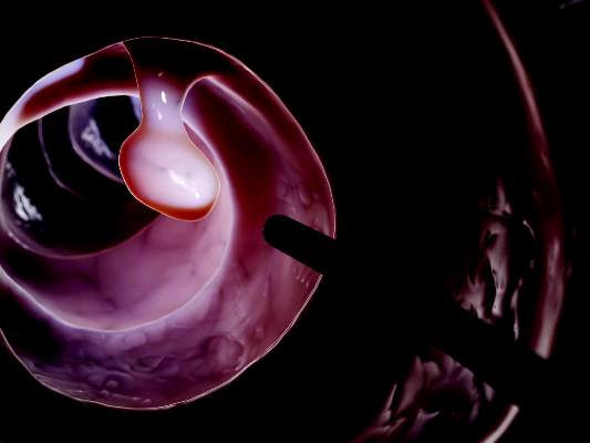
To compare the efficacy of split-dose and other schedules and to determine the best products and volumes, they reviewed 47 randomized trials found by systematically searching MEDLINE, EMBASE, Scopus, CENTRAL, and the ISI Web of Knowledge database. The studies were published between 1980 and 2014 and did not include pediatric, hospitalized, or inflammatory bowel disease patients, the reviewers said.
Split-dose preparations provided significantly better colon cleansing than did day-before preparations (odds ratio [OR], 2.51; 95% confidence interval [CI], 1.86-3.39), regardless of whether the day-before preparation was PEG (OR, 2.6; 95% CI, 1.46-4.63), sodium phosphate (OR, 9.34; 95% CI, 2.12-41.11), or picosulfate (OR, 3.54; 95% CI, 1.95-6.45), Ms. Martel and her associates reported.
In the intention-to-treat analysis, the highest volumes of PEG (3 L or more) led to greater bowel cleanliness than did lower split-dose volumes (OR, 1.89; 95% CI, 1.01-3.46). Patients were almost twice as likely to report being willing to repeat split-dose preparations than day-before cleansing, and preferred low-volume preparations over high-volume ones, they said.
Secondary outcomes reported in the 47 trials included side effects, detection of polyps and adenomas, and resumption of daily activities, but definitions of these measures varied too much for the reviewers to reliably compare them. And since other studies are assessing the role of specific diets and adjuvants in colon preparation, they did not include those variables in their analyses.
Ms. Martel and her associates received no funding for the study. One coauthor reported consulting relationships with Pendopharm, Boston Scientific, Olympus Canada, and Cook, and grant funding from Boston Scientific and Cook.
Split-dose regimens might be superior for colon cleaning because of the shorter interval between the last preparation product taken and undergoing colonoscopy, said Myriam Martel at McGill University, Montreal, and her associates in a report in the July issue of Gastroenterology (doi:10.1053/j.gastro.2015.04.004).
Repeating a colonoscopy because of inadequate preparation of the colon is expensive. Colon-cleansing preparations include polyethylene glycol (PEG), sodium phosphate, picosulfate, and oral sulfate solutions, the reviewers noted.
To compare the efficacy of split-dose and other schedules and to determine the best products and volumes, they reviewed 47 randomized trials found by systematically searching MEDLINE, EMBASE, Scopus, CENTRAL, and the ISI Web of Knowledge database. The studies were published between 1980 and 2014 and did not include pediatric, hospitalized, or inflammatory bowel disease patients, the reviewers said.
Split-dose preparations provided significantly better colon cleansing than did day-before preparations (odds ratio [OR], 2.51; 95% confidence interval [CI], 1.86-3.39), regardless of whether the day-before preparation was PEG (OR, 2.6; 95% CI, 1.46-4.63), sodium phosphate (OR, 9.34; 95% CI, 2.12-41.11), or picosulfate (OR, 3.54; 95% CI, 1.95-6.45), Ms. Martel and her associates reported.
In the intention-to-treat analysis, the highest volumes of PEG (3 L or more) led to greater bowel cleanliness than did lower split-dose volumes (OR, 1.89; 95% CI, 1.01-3.46). Patients were almost twice as likely to report being willing to repeat split-dose preparations than day-before cleansing, and preferred low-volume preparations over high-volume ones, they said.
Secondary outcomes reported in the 47 trials included side effects, detection of polyps and adenomas, and resumption of daily activities, but definitions of these measures varied too much for the reviewers to reliably compare them. And since other studies are assessing the role of specific diets and adjuvants in colon preparation, they did not include those variables in their analyses.
Ms. Martel and her associates received no funding for the study. One coauthor reported consulting relationships with Pendopharm, Boston Scientific, Olympus Canada, and Cook, and grant funding from Boston Scientific and Cook.
