User login
SHM Surveys Revisited
The Benchmarks Committee has had a busy 2006 so far. Many thanks to the 400-plus hospital medicine groups that completed the 2005-06 SHM survey, “The Authoritative Source on the State of Hospital Medicine,” as well as those of you who participated in the follow-up survey to clarify questions around additional revenue or supplements to program income.
We had some behind-the-scenes work on the survey. After the first pass of data analysis, it became clear that there was something amiss with the responses pertaining to the supplemental income questions. Only 47% of programs reported that they received additional income from hospitals or other sources. This seemed incompatible with what we know about current payment rates for fee-for-service work. A quick discussion took place among the committee and we decided to resurvey the group with more precise questions on exactly how finances flow into a program. As you have seen, the additional results bore out our suspicions. With the revised survey tool, 97% of the programs (when re-queried) reported receiving income above and beyond fees from direct patient care.
It was a Herculean task of digesting and formatting the data into usable form. SHM Senior Vice President Joe Miller deserves many thanks for poring through the data and creating something that all of you can use to better your programs.
Now, a few words about the recent annual meeting in Washington, DC: The work of the Benchmarks Committee was highlighted by SHM CEO Larry Wellikson, MD, in a plenary session on the first day of the meeting. An insightful panel discussion followed in which panel members Bob Wachter, MD (representing academic hospital medicine), Mary Jo Gorman, MD (corporate hospital medicine), and John Nelson, MD (community hospital medicine) gave some insightful commentary on the results of the survey and then fielded questions from the audience.
In between sessions at the conference, the committee held its annual face-to-face meeting and discussed plans for the year.
Under the guidance and editorial excellence of committee member Leslie Flores, the Benchmarks Committee continues to make strong progress with the dashboard project. As you may recall, this project will document and describe 10 commonly used dashboard metrics, including sample reports. There are plans for this material to be published as a supplement to The Hospitalist later this year.
The committee discussed the type of surveys conducted by SHM. A recommendation came out of our meeting to change from large-scale, all-encompassing, biannual surveys to smaller annual surveys. The annual survey would alternate its focus every other year between evaluating individuals’ productivity and compensation, and the characteristics of hospital medicine groups. Each survey would also have the flexibility to address specific timely questions that might focus on a specific research or hot topic of the day. The goal is to get our first trial survey out by the end of 2006, which would focus on group characteristics.
Lastly, a big thank you to our outgoing chairperson, Teresa Jones. Her hard work and organizational skills are going to be a tough act to follow.
Dr. Kealey is chair of SHM’s Benchmarks Committee
Advocacy Day Educates Lawmakers about Hospital Medicine
By Eric Siegal, MD
As reported in the SHM Meeting Reporter (see July The Hospitalist, supplement 1), SHM’s first Legislative Advocacy Day was an overwhelming success. More than 70 members from 29 states visited Capitol Hill on May 3 to educate lawmakers and their staffs about hospital medicine and SHM’s proposals to improve the quality of care in our nation’s hospitals.
Advocacy Day participants attended a morning briefing that covered procedural tips on how to have a successful meeting and the health issues currently before Congress—and SHM’s positions—before leaving for previously scheduled appointments on Capitol Hill.
Once there, hospitalists took time to educate legislators and their staff members about the fastest growing medical specialty in the United States, and then turned to SHM’s recommendations for improving the quality, safety, and cost-effectiveness of inpatient medical care.
Among the issues discussed with lawmakers: the need for more funding for the Agency for Healthcare Research and Quality (AHRQ); SHM support for federal initiatives to measure resource use and improve quality, including a CMS demonstration project to assess the contributions of hospital medicine programs to improve patient care and more efficient management of hospital resources; and the importance of gainsharing demonstration projects that focus on inpatient medical services such as those provided by hospital medicine programs.
More than 130 appointments were scheduled with House and Senate offices, many with members of Congress who sit on the key health committees with jurisdiction over Medicare as well as on committees responsible for determining funding levels for the National Institutes of Health, the CDC, and the Agency for Healthcare Research and Quality.
SHM encourages those of you who participated in Advocacy Day to thank the lawmakers and staff with whom you visited (view the sample thank-you letter and instructions online at www.hospitalmedicine.org under “Advocacy & Policy”).
Advocacy Day opened doors to congressional offices; follow-up from SHM members will keep hospital medicine’s issues before lawmakers as they vote on legislation of importance to our specialty. The more SHM members interact with members of Congress and their staff, the louder our voices and the more effective our messages. Advocacy Day is just the beginning of regular contact by hospitalists with their elected representatives in Washington.
A New Way to Get Everything SHM
SHM has launched the SHM Store, a new Internet-based marketplace that will redefine the way SHM educational products and apparel are distributed. The online store will be a convenient option for hectic lifestyles because it will be open 24 hours a day and can be accessed from anywhere there is an Internet connection.
The SHM Store is user-friendly and allows you to easily and quickly purchase the quality products and apparel that you have come to expect from SHM. Apparel and accessories include men’s and women’s wind jackets, golf shirts, scrub tops, baseball caps, and lapel pins—all with the SHM logo.
You’ll also find the most up-to-date educational materials from SHM. The 2005-2006 results of the SHM survey, “The Authoritative Source on the State of Hospital Medicine,” can be ordered in CD and print format. The Practice Management Course CD with binder is also available. This combination package includes all of the materials distributed at an onsite presentation of the course. And, last but not least, get your very own copy or additional copies of The Core Competencies in Hospital Medicine: A Framework For Curriculum Development.
The ordering process is simple. Access the SHM Store at www.hospitalmedicine.org/shmstore or navigate from www.hospitalmedicine.org and click on SHM Store. The store allows you to browse available products and descriptions. When you see something you want, select the quantity you’d like to purchase and add the items to your shopping cart. Once you have completed your shopping trip, click “Check Out.” A confirmation of your transaction will be sent to your e-mail inbox and your products will be shipped to your attention.
It’s that easy. Make sure you go online to browse the new SHM Store. If you have any questions or would like to learn more about the SHM Store, contact SHM at CustomerService@hospitalmedicine. org.
SHM, Health Groups Urge House to Boost Discretionary Funding
SHM joined more than 800 health, education, and labor organizations in writing a May 8 letter to the House leadership. Organized by the Coalition for Health Funding and the Committee on Education Funding, the letter calls on Congress to add $7 billion to the budget in discretionary funding for health, education, and labor programs. It asks lawmakers to ensure that the final allocation for the House and Senate Labor, Health and Human Services, Education Appropriations Subcommittees reflects a $7 billion increase above the President’s request for these critically important programs in FY 2007.
“While our organizations represent a wide array of domestic priorities,” the letter states, “we are united in our effort to advance the bipartisan goal of adding $7 billion in discretionary funding for health, education, labor enforcement, job training, and social services programs as the budget process moves forward. On behalf of our millions of constituents, we strongly urge you to provide at least $7 billion in additional federal support to restore funding levels to FY 2005. This is essential to sustain the well-being and prosperity of our nation.”
Agencies whose funding could be boosted by the additional spending called for in the coalition letter include the Agency for Healthcare Research and Quality, the National Institutes of Health, and the Centers for Disease Control and Prevention.
For full coverage of May 2006 advocacy and policy news, visit www.hospitalmedicine.org. TH
Dr. Siegal is chair of SHM’s Public Policy Committee.
SHM: BEHIND THE SCENES
Education and Quality Initiatives: SHM’s Heart and Soul
By Geri Barnes

This month’s SHM staff article highlights the department responsible for SHM’s education and quality initiatives. While my fellow senior managers may disagree, I believe that we are the heart and soul of SHM in meeting members’ needs. So important is this organizational focus that the Education Committee, chaired by Preetha Basaviah, MD, has its own mission, which parallels and provides detail to the organizational mission of promoting excellence in the practice of hospital medicine. And the Hospital Quality and Patient Safety Committee, chaired by Mahalakshmi Halasyamani, MD, is one of the most dynamic and prolific of SHM’s committees.
We know that members join SHM for the opportunity to effect change and network with colleagues. Equally strong is the quest for educational resources that will facilitate their efforts in improving quality inpatient care. SHM’s education plan focuses and organizes our energy on developing those resources.
SHM’s resources come in many forms. The most visible is our annual meeting. By all accounts, the 2006 annual meeting was extremely successful. It was our largest meeting to date, with almost 1,200 attendees, 99% of whom were members. That number represents more than 21% of our total membership. I wonder how many other organizations can boast of such a high percentage of member attendance at their annual meeting. The attendees actively participated in the educational sessions and the Research, Innovations, and Clinical Vignettes poster session, visited the exhibits, and networked during breaks and social events. To enhance the educational experience, we created a password-protected Web site where registrants could access the handouts for sessions they planned to attend. Because of the value of these materials, we are currently determining ways to utilize the handouts to expand our educational resources.
For several years, SHM has presented the Leadership Academy and “Best Practices in Managing a Hospital Medicine Program” course. These courses are so popular that they have become a mainstay of SHM educational programs and are offered on an annual basis. In September we will present the first Level II Leadership Academy designed to build upon and expand Leadership Academy I. Additionally, “Fundamentals in Billing and Coding” a pre-course to the University of California San Francisco “Management of the Hospitalized Patient” course will be offered in October for the first time. This outgrowth of the Best Practices course was identified as a need by attendees and other members. We anticipate that we will continue to offer these important programs for many years to come.
While we reach a significant number of hospitalists and other healthcare providers through our annual meeting and other face-to-face meetings, we reach many more through other educational and quality improvement efforts. Hospitalists, in their unique role as providers responsible for the general medical care of hospitalized patients, find themselves filling roles as key players and drivers of institutional quality improvement. To aid them in this effort, we’ve developed Web-based resource rooms designed to facilitate the implementation of a quality improvement program for specific disease state/clinical condition, including antimicrobial resistance, stroke, venous thromboembolism, and heart failure. Resource rooms on glycemic control and discharge planning are currently under development.
For most resource rooms, the key is the QI Workbook, a resource built on proven principles of quality improvement, personal experiences, and evidence-based medicine. Resources in addition to the workbook include slide sets that can be used in training sessions, “Teaching Pearls,” patient education resources, and clinical tools in the form of algorithms, order sets, and discharge planning sheets. Most of the resource rooms have online CME programs and each room has a well-developed bibliography.
What will likely prove to be SHM’s most far-reaching resource is The Core Competencies in Hospital Medicine: A Framework For Curriculum Development. Published earlier this year as a supplement to the Journal of Hospital Medicine, the Core Competencies provides a structure for professional and curricular development based on a shared understanding of the essential knowledge, skills, and attitudes expected of physicians working as hospitalists. While the target audience is the directors of hospitalist programs and fellowships and residency programs (and medical school internal medicine clerkships), imagine the number of people reached when the Core Competencies touch hospitalist students across the board. Additionally, the Core Competencies are the focal point for every educational and quality improvement initiative that we undertake. This document will continue to drive the content of each annual meeting, provide direction for the organization, and help us prioritize the development of resources rooms and other educational and quality improvement resources.
SHM is lucky to have partners who understand the importance of hospitalists as key members of the multidisciplinary team and the army that will lead the charge in implementing protocols to improve patient care. From foundations and sister associations, to government and industry, we partner with organizations that appreciate the role that hospitalists play in improving the healthcare system. The partnerships have led to the development of toolkits, resource rooms, CME modules, and other important resources. New initiatives soon to be launched are mentored implementation programs and demonstration projects that will provide us with increased knowledge of how our resources are used and identify areas in which they can be improved.
What is required on a staff level to make this happen? A commitment to do whatever it takes to get the job done. Six of us are equally dedicated to leading, managing, and supporting these efforts. In addition to me, staff members Tina Budnitz, Erica Pearson, Joy Wittnebert, Lillian Higgins, and Theresa Jones chart new ground and are actively engaged in a dynamic learning process on a daily basis. But it is our members and experts that drive this process, and we are grateful for their patience and guidance.
The Education and Quality Initiatives staff members do not work in a vacuum in the national office. We have the support and brainpower of the rest of the staff, with whom we collaborate daily. Membership and marketing, information technology, and business operations staff all play a role in our success.
What makes the national office run as a well-oiled machine? Next month you’ll hear from Steve Poitras, director, Business Operations, who is instrumental in making that happen.
Barnes is director, Education & Quality Initiatives, for SHM.
The Benchmarks Committee has had a busy 2006 so far. Many thanks to the 400-plus hospital medicine groups that completed the 2005-06 SHM survey, “The Authoritative Source on the State of Hospital Medicine,” as well as those of you who participated in the follow-up survey to clarify questions around additional revenue or supplements to program income.
We had some behind-the-scenes work on the survey. After the first pass of data analysis, it became clear that there was something amiss with the responses pertaining to the supplemental income questions. Only 47% of programs reported that they received additional income from hospitals or other sources. This seemed incompatible with what we know about current payment rates for fee-for-service work. A quick discussion took place among the committee and we decided to resurvey the group with more precise questions on exactly how finances flow into a program. As you have seen, the additional results bore out our suspicions. With the revised survey tool, 97% of the programs (when re-queried) reported receiving income above and beyond fees from direct patient care.
It was a Herculean task of digesting and formatting the data into usable form. SHM Senior Vice President Joe Miller deserves many thanks for poring through the data and creating something that all of you can use to better your programs.
Now, a few words about the recent annual meeting in Washington, DC: The work of the Benchmarks Committee was highlighted by SHM CEO Larry Wellikson, MD, in a plenary session on the first day of the meeting. An insightful panel discussion followed in which panel members Bob Wachter, MD (representing academic hospital medicine), Mary Jo Gorman, MD (corporate hospital medicine), and John Nelson, MD (community hospital medicine) gave some insightful commentary on the results of the survey and then fielded questions from the audience.
In between sessions at the conference, the committee held its annual face-to-face meeting and discussed plans for the year.
Under the guidance and editorial excellence of committee member Leslie Flores, the Benchmarks Committee continues to make strong progress with the dashboard project. As you may recall, this project will document and describe 10 commonly used dashboard metrics, including sample reports. There are plans for this material to be published as a supplement to The Hospitalist later this year.
The committee discussed the type of surveys conducted by SHM. A recommendation came out of our meeting to change from large-scale, all-encompassing, biannual surveys to smaller annual surveys. The annual survey would alternate its focus every other year between evaluating individuals’ productivity and compensation, and the characteristics of hospital medicine groups. Each survey would also have the flexibility to address specific timely questions that might focus on a specific research or hot topic of the day. The goal is to get our first trial survey out by the end of 2006, which would focus on group characteristics.
Lastly, a big thank you to our outgoing chairperson, Teresa Jones. Her hard work and organizational skills are going to be a tough act to follow.
Dr. Kealey is chair of SHM’s Benchmarks Committee
Advocacy Day Educates Lawmakers about Hospital Medicine
By Eric Siegal, MD
As reported in the SHM Meeting Reporter (see July The Hospitalist, supplement 1), SHM’s first Legislative Advocacy Day was an overwhelming success. More than 70 members from 29 states visited Capitol Hill on May 3 to educate lawmakers and their staffs about hospital medicine and SHM’s proposals to improve the quality of care in our nation’s hospitals.
Advocacy Day participants attended a morning briefing that covered procedural tips on how to have a successful meeting and the health issues currently before Congress—and SHM’s positions—before leaving for previously scheduled appointments on Capitol Hill.
Once there, hospitalists took time to educate legislators and their staff members about the fastest growing medical specialty in the United States, and then turned to SHM’s recommendations for improving the quality, safety, and cost-effectiveness of inpatient medical care.
Among the issues discussed with lawmakers: the need for more funding for the Agency for Healthcare Research and Quality (AHRQ); SHM support for federal initiatives to measure resource use and improve quality, including a CMS demonstration project to assess the contributions of hospital medicine programs to improve patient care and more efficient management of hospital resources; and the importance of gainsharing demonstration projects that focus on inpatient medical services such as those provided by hospital medicine programs.
More than 130 appointments were scheduled with House and Senate offices, many with members of Congress who sit on the key health committees with jurisdiction over Medicare as well as on committees responsible for determining funding levels for the National Institutes of Health, the CDC, and the Agency for Healthcare Research and Quality.
SHM encourages those of you who participated in Advocacy Day to thank the lawmakers and staff with whom you visited (view the sample thank-you letter and instructions online at www.hospitalmedicine.org under “Advocacy & Policy”).
Advocacy Day opened doors to congressional offices; follow-up from SHM members will keep hospital medicine’s issues before lawmakers as they vote on legislation of importance to our specialty. The more SHM members interact with members of Congress and their staff, the louder our voices and the more effective our messages. Advocacy Day is just the beginning of regular contact by hospitalists with their elected representatives in Washington.
A New Way to Get Everything SHM
SHM has launched the SHM Store, a new Internet-based marketplace that will redefine the way SHM educational products and apparel are distributed. The online store will be a convenient option for hectic lifestyles because it will be open 24 hours a day and can be accessed from anywhere there is an Internet connection.
The SHM Store is user-friendly and allows you to easily and quickly purchase the quality products and apparel that you have come to expect from SHM. Apparel and accessories include men’s and women’s wind jackets, golf shirts, scrub tops, baseball caps, and lapel pins—all with the SHM logo.
You’ll also find the most up-to-date educational materials from SHM. The 2005-2006 results of the SHM survey, “The Authoritative Source on the State of Hospital Medicine,” can be ordered in CD and print format. The Practice Management Course CD with binder is also available. This combination package includes all of the materials distributed at an onsite presentation of the course. And, last but not least, get your very own copy or additional copies of The Core Competencies in Hospital Medicine: A Framework For Curriculum Development.
The ordering process is simple. Access the SHM Store at www.hospitalmedicine.org/shmstore or navigate from www.hospitalmedicine.org and click on SHM Store. The store allows you to browse available products and descriptions. When you see something you want, select the quantity you’d like to purchase and add the items to your shopping cart. Once you have completed your shopping trip, click “Check Out.” A confirmation of your transaction will be sent to your e-mail inbox and your products will be shipped to your attention.
It’s that easy. Make sure you go online to browse the new SHM Store. If you have any questions or would like to learn more about the SHM Store, contact SHM at CustomerService@hospitalmedicine. org.
SHM, Health Groups Urge House to Boost Discretionary Funding
SHM joined more than 800 health, education, and labor organizations in writing a May 8 letter to the House leadership. Organized by the Coalition for Health Funding and the Committee on Education Funding, the letter calls on Congress to add $7 billion to the budget in discretionary funding for health, education, and labor programs. It asks lawmakers to ensure that the final allocation for the House and Senate Labor, Health and Human Services, Education Appropriations Subcommittees reflects a $7 billion increase above the President’s request for these critically important programs in FY 2007.
“While our organizations represent a wide array of domestic priorities,” the letter states, “we are united in our effort to advance the bipartisan goal of adding $7 billion in discretionary funding for health, education, labor enforcement, job training, and social services programs as the budget process moves forward. On behalf of our millions of constituents, we strongly urge you to provide at least $7 billion in additional federal support to restore funding levels to FY 2005. This is essential to sustain the well-being and prosperity of our nation.”
Agencies whose funding could be boosted by the additional spending called for in the coalition letter include the Agency for Healthcare Research and Quality, the National Institutes of Health, and the Centers for Disease Control and Prevention.
For full coverage of May 2006 advocacy and policy news, visit www.hospitalmedicine.org. TH
Dr. Siegal is chair of SHM’s Public Policy Committee.
SHM: BEHIND THE SCENES
Education and Quality Initiatives: SHM’s Heart and Soul
By Geri Barnes
This month’s SHM staff article highlights the department responsible for SHM’s education and quality initiatives. While my fellow senior managers may disagree, I believe that we are the heart and soul of SHM in meeting members’ needs. So important is this organizational focus that the Education Committee, chaired by Preetha Basaviah, MD, has its own mission, which parallels and provides detail to the organizational mission of promoting excellence in the practice of hospital medicine. And the Hospital Quality and Patient Safety Committee, chaired by Mahalakshmi Halasyamani, MD, is one of the most dynamic and prolific of SHM’s committees.
We know that members join SHM for the opportunity to effect change and network with colleagues. Equally strong is the quest for educational resources that will facilitate their efforts in improving quality inpatient care. SHM’s education plan focuses and organizes our energy on developing those resources.
SHM’s resources come in many forms. The most visible is our annual meeting. By all accounts, the 2006 annual meeting was extremely successful. It was our largest meeting to date, with almost 1,200 attendees, 99% of whom were members. That number represents more than 21% of our total membership. I wonder how many other organizations can boast of such a high percentage of member attendance at their annual meeting. The attendees actively participated in the educational sessions and the Research, Innovations, and Clinical Vignettes poster session, visited the exhibits, and networked during breaks and social events. To enhance the educational experience, we created a password-protected Web site where registrants could access the handouts for sessions they planned to attend. Because of the value of these materials, we are currently determining ways to utilize the handouts to expand our educational resources.
For several years, SHM has presented the Leadership Academy and “Best Practices in Managing a Hospital Medicine Program” course. These courses are so popular that they have become a mainstay of SHM educational programs and are offered on an annual basis. In September we will present the first Level II Leadership Academy designed to build upon and expand Leadership Academy I. Additionally, “Fundamentals in Billing and Coding” a pre-course to the University of California San Francisco “Management of the Hospitalized Patient” course will be offered in October for the first time. This outgrowth of the Best Practices course was identified as a need by attendees and other members. We anticipate that we will continue to offer these important programs for many years to come.
While we reach a significant number of hospitalists and other healthcare providers through our annual meeting and other face-to-face meetings, we reach many more through other educational and quality improvement efforts. Hospitalists, in their unique role as providers responsible for the general medical care of hospitalized patients, find themselves filling roles as key players and drivers of institutional quality improvement. To aid them in this effort, we’ve developed Web-based resource rooms designed to facilitate the implementation of a quality improvement program for specific disease state/clinical condition, including antimicrobial resistance, stroke, venous thromboembolism, and heart failure. Resource rooms on glycemic control and discharge planning are currently under development.
For most resource rooms, the key is the QI Workbook, a resource built on proven principles of quality improvement, personal experiences, and evidence-based medicine. Resources in addition to the workbook include slide sets that can be used in training sessions, “Teaching Pearls,” patient education resources, and clinical tools in the form of algorithms, order sets, and discharge planning sheets. Most of the resource rooms have online CME programs and each room has a well-developed bibliography.
What will likely prove to be SHM’s most far-reaching resource is The Core Competencies in Hospital Medicine: A Framework For Curriculum Development. Published earlier this year as a supplement to the Journal of Hospital Medicine, the Core Competencies provides a structure for professional and curricular development based on a shared understanding of the essential knowledge, skills, and attitudes expected of physicians working as hospitalists. While the target audience is the directors of hospitalist programs and fellowships and residency programs (and medical school internal medicine clerkships), imagine the number of people reached when the Core Competencies touch hospitalist students across the board. Additionally, the Core Competencies are the focal point for every educational and quality improvement initiative that we undertake. This document will continue to drive the content of each annual meeting, provide direction for the organization, and help us prioritize the development of resources rooms and other educational and quality improvement resources.
SHM is lucky to have partners who understand the importance of hospitalists as key members of the multidisciplinary team and the army that will lead the charge in implementing protocols to improve patient care. From foundations and sister associations, to government and industry, we partner with organizations that appreciate the role that hospitalists play in improving the healthcare system. The partnerships have led to the development of toolkits, resource rooms, CME modules, and other important resources. New initiatives soon to be launched are mentored implementation programs and demonstration projects that will provide us with increased knowledge of how our resources are used and identify areas in which they can be improved.
What is required on a staff level to make this happen? A commitment to do whatever it takes to get the job done. Six of us are equally dedicated to leading, managing, and supporting these efforts. In addition to me, staff members Tina Budnitz, Erica Pearson, Joy Wittnebert, Lillian Higgins, and Theresa Jones chart new ground and are actively engaged in a dynamic learning process on a daily basis. But it is our members and experts that drive this process, and we are grateful for their patience and guidance.
The Education and Quality Initiatives staff members do not work in a vacuum in the national office. We have the support and brainpower of the rest of the staff, with whom we collaborate daily. Membership and marketing, information technology, and business operations staff all play a role in our success.
What makes the national office run as a well-oiled machine? Next month you’ll hear from Steve Poitras, director, Business Operations, who is instrumental in making that happen.
Barnes is director, Education & Quality Initiatives, for SHM.
Virus Alerts
CDC Urges Awareness of Measles in Americans Returning from Germany
By Alfred Valles, resident, internal medicine, Mayo Clinic College of Medicine
The Centers for Disease Control and Prevention (CDC) has issued an alert urging American travelers to remain aware of the possibility of measles exposure. Many Americans have traveled to and from Germany for the World Cup soccer championship games. Three of the twelve hosting cities—Cologne, Dortmund, and Gelsenkirchen—are of particular concern, given the recent measles outbreaks that have been reported in those cities and their surrounding areas.
Since January 1 of this year, some 1,200 cases have been identified in or near these cities. American travelers were undoubtedly among the large crowds of people gathered to pay homage to their favorite sport, and many others will visit Germany on vacation or business, making transmission of this respiratory droplet-born pathogen a very real threat.
The CDC recommends the following precautions:
- Travelers who plan to go to Germany should check their immunization records and visit their doctors if they are not immune to measles or are not sure they are.
- People returning from Germany, especially those who went to see the World Cup, should see a healthcare provider if they develop the symptoms of measles, including a fever, a raised rash that begins on the face and spreads to the arms and legs, a cough, red eyes, or a runny nose.
- People with these symptoms should limit their contact with others.
- Clinicians seeing patients with these symptoms should inquire about travel history and immunization status.
This warning is not to be taken lightly. Approximately two of every 1,000 patients infected with measles will die of the disease. Complications such as encephalitis are of particular concern for those who are malnourished or immunosuppressed.
Remember, live virus measles vaccine given within 72 hours of exposure may prevent the disease, while immune globulin given up to six days after exposure may prevent complications of measles in those who are at risk, including pregnant women, people with weak immune systems, and children.
The World cup can be dangerous, even for non-players. For more information about the measles outbreak and travel precautions, visit www.cdc.gov.
Lymphocytic Choriomeningitis Virus: Facts and Prevention
By the Special Pathogens Branch, CDC
In May 2005, the CDC investigated a cluster of lymphocytic choriomeningitis virus (LCMV) illnesses in four solid organ transplant recipients from a common donor, three of whom died. The source of the LCMV was traced to a hamster that had recently been acquired by a member of the donor’s household. It was subsequently determined that several LCMV-infected pet rodents had originated from a single distributor, who may have distributed other infected rodents to pet stores in the northeastern and midwestern United States.1 However, the risk of contracting LCMV from rodent exposure is not limited only to this outbreak, nor is the danger confined only to patients undergoing organ transplant.
Clinicians need to be familiar with LCMV because of its potential to cause meningitis, its teratogenicity, and the risk that it may bring about serious disease in immunocompromised individuals.
LCMV is normally carried by wild house mice, but can be transmitted to laboratory and pet rodents at breeding facilities, in pet stores, and in homes. Humans become infected in one of the following ways:
- Through direct contact with the secretions or excretions of infected rodents;
- By inhalation of dust or droplets containing LCMV from rodents;
- As a result of transplacental spread from an infected pregnant woman to her fetus; and
- By receipt of an organ transplant from an infected donor.2
Among those tested, about 5% have shown serologic evidence of previous infection with LCMV.3,4 In healthy adults, LCMV is typically a nonspecific viral syndrome sometimes followed by aseptic meningitis or other neurologic signs. Patients with weakened immune systems can suffer severe, possibly fatal systemic illness.2,5 Maternal infection with LCMV during pregnancy can result in spontaneous abortion or early neonatal death, as well as in defects similar to those of other congenital infections such as toxoplasmosis and cytomegalovirus (“TORCH” infections).6-10 The proportion of developmental defects caused by LCMV is not known.
Clinicians should consider LCMV in the differential diagnosis of patients with aseptic meningitis; in cases of fetal demise or congenital defects, including congenital hydrocephalus, chorioretinitis, blindness, or mental retardation; or in recent transplant recipients who present with signs of post-transplant infection. A detailed rodent exposure history should be taken. Contact your state health department or Special Pathogens Branch, CDC (404-639-1510), for information about testing for LCMV.
Currently, there is no specific treatment for LCMV infection other than supportive care. Ribavirin inhibits LCMV multiplication in laboratory experiments but has not been tested in clinical trials.
Clinicians should counsel their patients about the risks of contracting LCMV from laboratory, pet, and wild rodents. Wild mice in the home should be controlled and removed promptly. Immunocompromised individuals and women who are pregnant or planning to become pregnant should avoid any contact with wild or pet rodents, their excretions, and their nesting materials. While a woman is pregnant, pet rodents should be housed outside the home or in a separate part of the home where other individuals can care for the pets and clean their cages. Counseling a woman already exposed to rodents during pregnancy can be challenging; for assistance, contact your state health department.
Further information about LCMV infection and its prevention, including management and prevention of rodent infestation in the home, is available from Special Pathogens Branch, CDC, at www.cdc.gov/ncidod/dvrd/spb/mnpages/dispages/lcmv.htm. For more information about diseases commonly carried by rodents, please visit www.cdc.gov/rodents. TH
References
- Centers for Disease Control and Prevention. Update: interim guidance for minimizing risk for human lymphocytic choriomeningitis virus infection associated with pet rodents. MMWR. Aug 19, 2005;54(32):799-801.
- Fischer SA, Graham MB, Kuehnert MJ, et al. Transmission of lymphocytic choriomeningitis virus by organ transplantation. N Engl J Med. 2006;354(21):2235-2249.
- Childs JE, Glass GE, Ksiazek TG, et al. Human-rodent contact and infection with lymphocytic choriomeningitis and Seoul viruses in an inner-city population. Am J Trop Med Hyg. 1991 Feb;44(2):117–121.
- Park JY, Peters CJ, Rollin PE, et al. Age distribution of lymphocytic choriomeningitis virus serum antibody in Birmingham, Alabama: evidence of a decreased risk of infection. Am J Trop Med Hyg. 1997 Jul;57(1):37–41.
- Horton J, Hotchin JE, Olson KB, et al. The effects of MP virus infection in lymphoma. Cancer Res. 1971 Aug;31(8):1066–1068.
- Barton LL, Mets MB. Congenital lymphocytic choriomeningitis virus infection: decade of rediscovery. Clin Infect Dis. 2001 Aug 1;33(3):370–374.
- Barton LL, Mets MB, Beauchamp CL. Lymphocytic choriomeningitis virus: emerging fetal teratogen. Am J Obstet Gynecol. 2002 Dec ;187(6):1715–1716.
- Wright R, Johnson D, Neumann M, et al. Congenital lymphocytic choriomeningitis virus syndrome: a disease that mimics congenital toxoplasmosis or cytomegalovirus infection. Pediatrics. 1997 Jul;100(1):E9.
- Ford-Jones EL, Ryan G . Implications for the fetus of maternal infections in pregnancy. In: Cohen J, Powderly WG, eds. Infectious Diseases, 2nd ed. New York, NY: Mosby; 2004: 709–723.
- Greenhow TL, Weintrub PS. Your diagnosis, please. Neonate with hydrocephalus. Pediatr Infect Dis J. 2003 Dec;22(12):1099, 1111–1112.
ECT wipes out 30 years of memories
Woman loses 30 years of memories after electroconvulsive therapy
Richland County (SC) Circuit Court
A 55-year old woman with a history of depression underwent successful electroconvulsive therapy (ECT) after her husband and father died. Six months later she became depressed, and a new psychiatrist referred her to his partner for additional ECT treatments.
The partner administered outpatient ECT at a hospital daily for 10 days. The referring psychiatrist wrote in the patient’s chart that the patient experienced memory loss and severe cognitive problems during the initial ECT regimen but did not report this development to his partner and allegedly encouraged the patient to continue ECT.
After the second round of ECT treatments, the patient suffered brain damage and lost all her memories from the past 30 years—including the births of her children and her job skills—leaving her unable to work.
In court, the patient claimed ECT should be administered no more than three times a week, and the referring psychiatrist should have told his partner about the patient’s memory problems.
- The case was settled for $18,000
Dr. Grant’s observations
Although this case concerns ECT, the claim is based on negligence—that is, the psychiatrist did not fulfill his duty to care for the patient. The negligence claim focused on how the treatment was implemented, not whether ECT was appropriate for this woman’s depression.
ECT’s response rate ranges from 50% to 60%1 among patients who did not respond to one or more antidepressant trials. Symptomatic improvement usually is faster with ECT than with pharmacotherapy2 when ECT is administered three times per week. Mortality rates with ECT are similar to those associated with minor surgery.1
In addition to being an effective and safe treatment for depression, ECT rarely is a basis for malpractice. One study found that only 4 (0.2%) of 1,700 psychiatric malpractice claims filed between 1984 and 1990 concerned ECT’s side effects, complications, or appropriateness.3 Few patients who receive ECT file a malpractice claim because most are satisfied with the treatment; approximately 80% of ECT patients say they would consent to ECT again.4,5 In fact, one might consider withholding ECT from severely depressed patients grounds for malpractice.
Although safe and effective, ECT could present health risks that you need to discuss with patients. In particular, cognitive problems such as delirium and impaired attention and memory may result.1
Cognitive impairment risk in ect
ECT’s more severe cognitive side effects stem from:
- bilateral electrode placement
- sine wave stimulation
- suprathreshold stimulus intensity
- administration >3 times per week
- large numbers of treatments, usually >20 in an acute treatment course
- some medications, such as lithium carbonate and anticholinergics6
- pre-existing neurologic diseases such as Alzheimer’s or Parkinson’s disease.1
The magnitude of retrograde amnesia often is greatest immediately after treatment. Patients are more likely to forget public information such as current events than personal information.10 The effects usually subside over time, and older memories are more likely to be recovered than more recent ones. ECT can cause permanent memory loss, particularly after bilateral electrode placement, suprathreshold stimulus intensity, sine wave stimulation, or large numbers of treatments—usually more than 20.
Ensuring adequate informed consent when delivering ECT or before referring a patient for treatment can help prevent a malpractice claim. Although specific requirements for ECT consent vary by jurisdiction, follow these general principles:1
- Provide the patient adequate information. Explain the reasons for ECT, describe the procedure including choice of stimulus electrode placement, offer alternative treatments, and explain the risks, benefits, anticipated number of treatments, relapse risk, and need for continuing treatment.
- Make sure the patient is capable of understanding and acting reasonably on this information and knows he or she can refuse treatment at any time.
- Tell the patient that a successful outcome is not guaranteed.
- Describe the likelihood and potential severity of major risks associated with ECT, including mortality, cardiovascular and CNS problems, and minor side effects such as headache, muscle aches, or nausea.
- Be sure the patient understands that consent is voluntary and can be withdrawn. The patient should know that he or she is also consenting to emergency treatment.
- Tell patients about possible behavioral restrictions—such as needing a friend or family member to monitor the patient or not being able to drive a car—that may be necessary during evaluation, treatment, and recuperation.
Although ECT might impair memory, it can improve neuropsychological domains such as global cognitive status and measures of general intelligence.11 Also, there is no evidence that ECT causes lasting problems in executive functioning, abstract reasoning, creativity, semantic memory, implicit memory, or skill acquisition or retention. Long-term negative effects on ability to learn and retain new information are unlikely.1
Avoiding an ect related malpractice claim
To reduce the possibility of a malpractice claim after ECT:
- Inform the patient about the risk of cognitive side effects as part of the informed consent process (Box).
- Assess the patient’s orientation and memory functions before and throughout ECT. In the above case, the referring psychiatrist had a duty to inform the psychiatrist administering ECT about the patient’s memory problems and recommend decreasing or discontinuing ECT.
- Consider a patient’s mood state, which may influence how ECT patients rate their memory.12 Ask about symptoms of depression. Patients with cognitive complaints such as subjective memory loss are more likely than those without such problems to have depression symptoms.1
- Do not administer ECT more than 3 times per week. No evidence supports more frequent use, and daily ECT may increase cognitive problems.1 The psychiatrist in the above case was negligent in providing a treatment frequency with no scientific support or medical rationale.
- Verify that the physician is qualified to perform ECT. Hospitals must ensure ECT quality and safety and should have a written plan for providing and maintaining ECT privileges.
- Involve the family when appropriate. Family members often care for patients during outpatient ECT. Give patients and family members literature describing ECT. Allow them time to consider the procedure, then schedule an appointment to answer questions.
1. American Psychiatric Association. The practice of electroconvulsive therapy: recommendations for treatment, training, and privileging, 2nd ed. Washington, DC: American Psychiatric Publishing; 2001.
2. Nobler MS, Sackeim HA, Moeller JR, et al. Quantifying the speed of symptomatic improvement with electroconvulsive therapy: comparison of alternative statistical methods. Convuls Ther 1997;13:208-21.
3. Slawson P. Psychiatric malpractice and ECT: a review of 1,700 claims. Convuls Ther 1991;7:255-61.
4. Freeman CP, Cheshire KE. Attitude studies on electroconvulsive therapy. Convuls Ther. 1986;2:31-42.
5. Pettinati HM, Tanburello TA, Ruetsch CR, et al. Patient attitudes toward electroconvulsive therapy. Psychopharmacol Bull. 1994;30:471-5.
6. Small JG, Kellams JJ, Milstein V, et al. Complications with electroconvulsive treatment combined with lithium. Biol Psychiatry 1980;15:103-12.
7. Sobin C, Sackeim HA, Prudic J, et al. Predictors of retrograde amnesia following ECT. Am J Psychiatry 1995;152:995-1001.
8. Donahue JC. Electroconvulsive therapy and memory loss: anatomy of a debate. J ECT 2000;16:133-43.
9. Sackeim HA. Memory and ECT: from polarization to reconciliation. J ECT 2000;16:87-96.
10. Lisanby SH, Maddox JH, Prudic J, et al. The effects of electroconvulsive therapy on memory of autobiographical and public events. Arch Gen Psychiatry 2000;57:581-90.
11. Sackeim HA, Prudic J, Devanand DP, et al. Effects of stimulus intensity and electrode placement on the efficacy and cognitive effects of electroconvulsive therapy. N Engl J Med 1993;328:839-46.
12. Coleman EA, Sackeim HA, Prudic J, et al. Subjective memory complaints before and after electroconvulsive therapy. Biol Psychiatry 1996;39:346-56.
Woman loses 30 years of memories after electroconvulsive therapy
Richland County (SC) Circuit Court
A 55-year old woman with a history of depression underwent successful electroconvulsive therapy (ECT) after her husband and father died. Six months later she became depressed, and a new psychiatrist referred her to his partner for additional ECT treatments.
The partner administered outpatient ECT at a hospital daily for 10 days. The referring psychiatrist wrote in the patient’s chart that the patient experienced memory loss and severe cognitive problems during the initial ECT regimen but did not report this development to his partner and allegedly encouraged the patient to continue ECT.
After the second round of ECT treatments, the patient suffered brain damage and lost all her memories from the past 30 years—including the births of her children and her job skills—leaving her unable to work.
In court, the patient claimed ECT should be administered no more than three times a week, and the referring psychiatrist should have told his partner about the patient’s memory problems.
- The case was settled for $18,000
Dr. Grant’s observations
Although this case concerns ECT, the claim is based on negligence—that is, the psychiatrist did not fulfill his duty to care for the patient. The negligence claim focused on how the treatment was implemented, not whether ECT was appropriate for this woman’s depression.
ECT’s response rate ranges from 50% to 60%1 among patients who did not respond to one or more antidepressant trials. Symptomatic improvement usually is faster with ECT than with pharmacotherapy2 when ECT is administered three times per week. Mortality rates with ECT are similar to those associated with minor surgery.1
In addition to being an effective and safe treatment for depression, ECT rarely is a basis for malpractice. One study found that only 4 (0.2%) of 1,700 psychiatric malpractice claims filed between 1984 and 1990 concerned ECT’s side effects, complications, or appropriateness.3 Few patients who receive ECT file a malpractice claim because most are satisfied with the treatment; approximately 80% of ECT patients say they would consent to ECT again.4,5 In fact, one might consider withholding ECT from severely depressed patients grounds for malpractice.
Although safe and effective, ECT could present health risks that you need to discuss with patients. In particular, cognitive problems such as delirium and impaired attention and memory may result.1
Cognitive impairment risk in ect
ECT’s more severe cognitive side effects stem from:
- bilateral electrode placement
- sine wave stimulation
- suprathreshold stimulus intensity
- administration >3 times per week
- large numbers of treatments, usually >20 in an acute treatment course
- some medications, such as lithium carbonate and anticholinergics6
- pre-existing neurologic diseases such as Alzheimer’s or Parkinson’s disease.1
The magnitude of retrograde amnesia often is greatest immediately after treatment. Patients are more likely to forget public information such as current events than personal information.10 The effects usually subside over time, and older memories are more likely to be recovered than more recent ones. ECT can cause permanent memory loss, particularly after bilateral electrode placement, suprathreshold stimulus intensity, sine wave stimulation, or large numbers of treatments—usually more than 20.
Ensuring adequate informed consent when delivering ECT or before referring a patient for treatment can help prevent a malpractice claim. Although specific requirements for ECT consent vary by jurisdiction, follow these general principles:1
- Provide the patient adequate information. Explain the reasons for ECT, describe the procedure including choice of stimulus electrode placement, offer alternative treatments, and explain the risks, benefits, anticipated number of treatments, relapse risk, and need for continuing treatment.
- Make sure the patient is capable of understanding and acting reasonably on this information and knows he or she can refuse treatment at any time.
- Tell the patient that a successful outcome is not guaranteed.
- Describe the likelihood and potential severity of major risks associated with ECT, including mortality, cardiovascular and CNS problems, and minor side effects such as headache, muscle aches, or nausea.
- Be sure the patient understands that consent is voluntary and can be withdrawn. The patient should know that he or she is also consenting to emergency treatment.
- Tell patients about possible behavioral restrictions—such as needing a friend or family member to monitor the patient or not being able to drive a car—that may be necessary during evaluation, treatment, and recuperation.
Although ECT might impair memory, it can improve neuropsychological domains such as global cognitive status and measures of general intelligence.11 Also, there is no evidence that ECT causes lasting problems in executive functioning, abstract reasoning, creativity, semantic memory, implicit memory, or skill acquisition or retention. Long-term negative effects on ability to learn and retain new information are unlikely.1
Avoiding an ect related malpractice claim
To reduce the possibility of a malpractice claim after ECT:
- Inform the patient about the risk of cognitive side effects as part of the informed consent process (Box).
- Assess the patient’s orientation and memory functions before and throughout ECT. In the above case, the referring psychiatrist had a duty to inform the psychiatrist administering ECT about the patient’s memory problems and recommend decreasing or discontinuing ECT.
- Consider a patient’s mood state, which may influence how ECT patients rate their memory.12 Ask about symptoms of depression. Patients with cognitive complaints such as subjective memory loss are more likely than those without such problems to have depression symptoms.1
- Do not administer ECT more than 3 times per week. No evidence supports more frequent use, and daily ECT may increase cognitive problems.1 The psychiatrist in the above case was negligent in providing a treatment frequency with no scientific support or medical rationale.
- Verify that the physician is qualified to perform ECT. Hospitals must ensure ECT quality and safety and should have a written plan for providing and maintaining ECT privileges.
- Involve the family when appropriate. Family members often care for patients during outpatient ECT. Give patients and family members literature describing ECT. Allow them time to consider the procedure, then schedule an appointment to answer questions.
1. American Psychiatric Association. The practice of electroconvulsive therapy: recommendations for treatment, training, and privileging, 2nd ed. Washington, DC: American Psychiatric Publishing; 2001.
2. Nobler MS, Sackeim HA, Moeller JR, et al. Quantifying the speed of symptomatic improvement with electroconvulsive therapy: comparison of alternative statistical methods. Convuls Ther 1997;13:208-21.
3. Slawson P. Psychiatric malpractice and ECT: a review of 1,700 claims. Convuls Ther 1991;7:255-61.
4. Freeman CP, Cheshire KE. Attitude studies on electroconvulsive therapy. Convuls Ther. 1986;2:31-42.
5. Pettinati HM, Tanburello TA, Ruetsch CR, et al. Patient attitudes toward electroconvulsive therapy. Psychopharmacol Bull. 1994;30:471-5.
6. Small JG, Kellams JJ, Milstein V, et al. Complications with electroconvulsive treatment combined with lithium. Biol Psychiatry 1980;15:103-12.
7. Sobin C, Sackeim HA, Prudic J, et al. Predictors of retrograde amnesia following ECT. Am J Psychiatry 1995;152:995-1001.
8. Donahue JC. Electroconvulsive therapy and memory loss: anatomy of a debate. J ECT 2000;16:133-43.
9. Sackeim HA. Memory and ECT: from polarization to reconciliation. J ECT 2000;16:87-96.
10. Lisanby SH, Maddox JH, Prudic J, et al. The effects of electroconvulsive therapy on memory of autobiographical and public events. Arch Gen Psychiatry 2000;57:581-90.
11. Sackeim HA, Prudic J, Devanand DP, et al. Effects of stimulus intensity and electrode placement on the efficacy and cognitive effects of electroconvulsive therapy. N Engl J Med 1993;328:839-46.
12. Coleman EA, Sackeim HA, Prudic J, et al. Subjective memory complaints before and after electroconvulsive therapy. Biol Psychiatry 1996;39:346-56.
Pediatric Special Section
Meningococcal Disease in Peds
By J. Christopher Day, MD
Thompson, MJ, Ninis N, Perera R, et al. Clinical recognition of meningococcal disease in children and adolescents. Lancet. 2006 Feb 6;367(9508)397-403.
Data from medical records and parental questionnaires were examined to determine the time course and prevalence of signs and symptoms in meningococcal disease (sepsis and meningitis) in 448 children in Wales, England, and Northern Ireland.
The authors note that classic clinical features of the disease appear late in the illness. The symptoms that appear earliest (fever, poor feeding, nausea, vomiting, coryza, sore throat) are very non-specific, but somewhat later symptoms of sepsis including leg pains, cold hands and feet, and abnormal skin color appear before the classical clinical features in 72% of the children studied. The authors hope that recognition of these features can lead to earlier diagnosis and treatment of this severe disease.
Treatment of Infant Botulism
Arnon SS, Schechter R, Maslanka SE, et al. Human botulism immune globulin for the treatment of infant botulism. N Engl J Med. 2006 Feb 2;354((5):462-471.
This is is a report of a randomized, double-blind, placebo controlled trial of the use of human botulism immune globulin intravenous (BIG-IV) used for the treatment of infant botulism. The study was performed on 122 infants with (initially) suspected and (later) laboratory confirmed infant botulism. BIG-IV was given within three days of hospital admission. The authors also performed an open-label study of 382 laboratory confirmed cases. Some of these began treatment as late as 18 days after hospital admission.
In the randomized trial, outcomes compared with placebo included a reduction in hospital stay from 5.7 weeks to 2.6 weeks (P<0.001); a reduction in the mean duration of intensive care by 3.2 weeks (P<0.001); the mean duration of mechanical ventilation by 2.6 weeks (P= 0.01); the mean duration of tube or intravenous feeding by 6.4 weeks; and the mean hospital charges per patient by $88,600 (P<0.001).
The only notable adverse event perhaps related to treatment was a transient blush-like erythematous rash. In the open-label study, treatment given within three days of hospital admission shortened the mean length of stay by approximately one week more than did treatment given four to seven days after admission. BIG-IV is now licensed as BabyBIG. The authors recommend treatment as soon as possible after hospital admission.—JCD
Eczema: Wet Wraps Versus Conventional Treatment
Hindley D, Galloway G, Murray J, et al. A randomised study of “wet wraps” versus conventional treatment for atopic eczema. Arch Dis Child. 2006 Feb;91(2):164-168.
The authors were interested in comparing the efficacy and parent perception of ease of application of conventional treatment (emulsifying ointments and topical steroids) with wet wraps (tubular retention bandages applied wet as occlusive dressings over emulsifying ointments) and topical steroids for exacerbations of eczema.
Fifty children were enrolled and randomized to either treatment group. Treatment was continued and patients were followed for one month. The change in eczema severity was scored using a previously published scale (SCORAD) for assessing severity. No differences were noted between the two groups, though the study had a number of limitations including use of only one potency of steroid (1% hydrocortisone) limiting generalizability, the length of treatment (regimens vary in length), and patient drop-outs (five dropped out—all in the wet wrap group).
Parents felt wet wraps were harder to use. The authors do note that there may be clinical situations (they mention short term treatment of acute erythrodermic eczema) where wet wraps are a useful option. There appear to be no advantages in the group studied over a four-week period and the disadvantages likely include parental perception of difficulty of use and a possibility of increased skin infections.—JCD
A Study of Dehydrated Peds
Mallory MD, Kadish H, Zebrack M, Nelson D. Use of a pediatric observation unit for treatment of children with dehydration caused by gastroenteritis. Pediatr Emerg Care. 2006 Jan;22(1):1-6.
A case-control study was performed to examine 467 admissions to an observation unit in patients with dehydration from vomiting or diarrhea from presumed gastroenteritis. Patients were presumably admitted to the observation unit (staffed by emergency department personnel) if they were believed to require more rehydration than could be reasonably offered in the emergency department itself.
Controls were defined as those patients able to be discharged home from the observation unit within 24 hours and cases as those patients that required subsequent inpatient admission. Nineteen percent of the patients studied required inpatient admission.
One of the goals of the study was to determine variables useful in distinguishing patients who would require subsequent inpatient admission from those who would be discharged home from the observation unit. No such variables were found.
Variables studied included oral intake prior to presentation, urine output at presentation, symptoms at presentation (vomiting versus diarrhea versus both), predominant symptoms (vomiting versus diarrhea versus no predominance), duration of symptoms, abdomen tender to palpation (yes/no), previous visit within five days (yes/no), age, vital signs, bicarbonate levels, and mean sodium levels.—JCD
Routine Neuroimaging: Little Value When Evaluating Febrile Seizures
By Stevie Wilson, RN, CPNP
Teng D, Dayan P, Tyler S, et al. Risk of intracranial pathologic conditions requiring emergency intervention after a first complex febrile seizure episode among children. Pediatrics. 2006;117(2):304-308.
Febrile seizures occur in up to 5% of all children. One-third of febrile seizures are classified as complex (i.e., having multiple seizures, prolonged duration, or focal seizures). Complex febrile seizures have been associated with an increased risk of recurrent febrile seizures and epilepsy. However, they have not been associated with pathologic intracranial lesions that require emergency surgical intervention.
Previous studies suggest that intracranial abnormalities are rare among simple febrile seizures. The American Academy of Pediatrics (AAP) recommends against the use of neuroimaging with simple seizures, however, practice guidelines for emergency neuroimaging for complex seizures do not exist. The objective of this study was to determine the likelihood of a significant intracranial condition requiring emergency intervention in children with the first complex febrile seizure.
A retrospective review of prospectively collected data was performed for 71 children who presented to the pediatric emergency department after a first complex febrile seizure. None of the 71 patients had a pathologic intracranial lesion that required emergency intervention. The authors therefore concluded that the risk of pathologic intracranial conditions requiring emergency intervention is low, suggesting that routine emergency neuroimaging for this population is unnecessary.
Managing Bronchiolitis above Sea Level
Choudhuri JA, Ogden LG, Ruttenber JA, et al. Effect of altitude on hospitalizations for respiratory syncytial virus infection. Pediatrics. 2006;117(2):349-356.
This interesting study sought to evaluate the effect altitude has on hospitalizations for RSV infection. A multivariate analysis was obtained and suggests that the rate of hospitalization for RSV increased by 25% among infants who were younger than one year of age for every 1,000 meters of altitude. The risk of RSV-associated hospitalization was highest at elevations above 2,500 meters.—SW
Dilemmas for Wheezing Peds
By Chris Miller, MD
Oymar K, Halvorsen T, Aksnes L. Mast cell activation and leukotriene secretion in wheezing infants. Relation to respiratory syncytial virus and outcome. Pediatr Allergy Immunol. 2006 Feb;17(1):37-42.
The wheezing infant is a common problem in pediatric medicine; however, the long-term outcome and risk for recurrent wheezing remains unclear. The authors of this study assert that the risk for recurrent wheezing may be related to the type of inflammation and specific underlying virus during the initial wheezing episode.
Several studies have evaluated the specific contribution of eosinophils to respiratory inflammation in the wheezing child. These studies have found that eosinophilic inflammation may play a role in the airway hyper-reactivity in a child with persistent wheezing. Conversely, little information is available regarding mast cell involvement. This prospective study was designed to assess mast cell activation, in relation to respiratory syncytial virus (RSV) infection and persistent wheezing in wheezing infants.
Researchers enrolled 106 wheezing infants who were subsequently admitted to the hospital. None of these infants had a prior history of wheezing. Infants with prior history of atopic disease, lung disease, or signs of bacterial infection were excluded. RSV testing of nasopharyngeal mucous was performed. Sixty-seven (63%) of the wheezing infants had RSV. An additional 23 healthy infants were selected as controls.
To assess the extent of mast cell activation, urinary prostaglandin F2 (U-PGF2) was measured. To assess for leukotriene secretion urinary leukotriene 4 (U-LTE4) was measured. To evaluate for persistence or reoccurrence of wheezing, a 20-month post-hospitalization follow-up was done. One hundred and three (97%) of the previously hospitalized wheezing infants were available for the follow-up.
The authors used the term “persistent wheezing” to describe those children who experienced at least three wheezing episodes including the first hospitalization. These recurrences were identified either by parental report utilizing a standardized questionnaire or physician/hospitalization records. The remaining children were considered “transient wheezers.”
Review of the data regarding U-PGF2, a specific marker for mast cell activation, showed U-PGF2 to be greatest in the RSV positive patients, followed by the RSV negative patients and finally by the control group. U-LTE4, and thus leukotriene involvement, was also found to be significantly elevated in both the RSV positive and negative wheezing infants compared with the controls. No significant difference was found in U-LTE4 between RSV-positive infants and RSV-negative infants. The transient wheezing infant had a higher U-PGF2 than the persistent wheezing infant. Transient wheezers also had a higher U-LTE4 compared with persistent wheezers, although this was not statistically significant.
With univariate regression analysis positive predictive factors for persistent wheezing included increasing age, RSV negative disease, parental atopy, and male gender. Negative predictive factors for persistent wheezing included an elevated U-PGF2 level.
In discussion the authors conclude that mast cell activation and leukotriene secretion is present in a wheezing infant as noted by increased U-PGF2 and U-LTE4 levels, respectively. Higher U-PGF2 levels in RSV-positive patients indicates that mast cell activation may play a bigger part in the inflammatory process of RSV-induced wheezing compared with non-RSV induced wheezing. Leukotrienes appear to be involved in both RSV and non-RSV wheezing infants. Despite these results, mast cell activation and leukotriene secretion do not appear to be associated with persistent wheezing.
Limitations of this interesting study include the small sample size, apparent lack of follow-up with the control patients, and the diagnosis of recurrent wheezing episodes being characterized by the parents.
Influenza Pneumonia in Pediatric Patients
Lahti E, Peltola V, Virkki R, et al. Influenza pneumonia. Pediatr Infect Dis J. 2006 Feb;25(2):160-164.
Influenza and pneumonia are common in children. The objective of this study was to describe the frequency and characteristics of laboratory-documented and radiographically proven influenza-associated pneumonia in children.
This retrospective, single site study was conducted at Turku University Hospital (Finland). Chart reviews of both inpatient and outpatient visits of children younger than 16 were performed for a 24-year period between 1980 through 2003. Children with influenza A or B antigen detected nasopharyngeal aspirates were identified. Also children with chest radiographs obtained during their influenza infection were identified. Clinical findings upon initial presentation were summarized, in addition to laboratory values for white blood cell count (WBC) and C-reactive protein (CRP).
A total of 936 patients with virologically confirmed influenza were identified. Of this total, 79% had influenza A and the remaining had influenza B. Chest radiographs were reported on 400 (43%) of these patients. Of the 400 films, 228 were initially read as having infiltrates. However, during over-reading of the films 70 chest radiographs did not have an infiltrate and an additional 24 films could not be found. In summary, 134 children had both confirmed influenza and radiographically proven pneumonia. Thus 14% of the children with influenza who also had a chest radiograph had pneumonia.
Clinical findings of the 134 patients with influenza and pneumonia were similar among those with influenza A or B. The most common symptoms were fever (98%), cough (84%), and rhinorrhea (65%). The classical findings of headaches and myalgias were difficult to uncover due to the young age of the patients (median age 2.2 years). Of the 134 identified patients with influenza and pneumonia only 27% presented with dyspnea and 22% with tachypnea. Crackles were heard in 22%, rhonchi in 43%, and decreased breath sounds in 10%. No abnormal auscultation findings were found in 32% of the children. Sixty-eight percent of the study population was admitted to the hospital. The rate of hospitalization was greater for the younger patients. One-third of the patients had received antibiotics prior to enrollment, and 80% of the children received antibiotics during the hospitalization or at time of discharge. Four children required ventilator therapy. One patient with muscular dystrophy died of severe pneumonia.
Laboratory assessment found that 89% of the children had WBC less than 15 x 109/liter. CRP values were normal or only slightly increased in 55% of the children. Three children (2%) had laboratory-documented concomitant bacterial infections. Of these three cases two were due to bacteremia and one to a positive tracheal aspirate. Five children (7%) had double viral infections.
In this hospital-based study 14% of the patients with influenza had radiographically proven pneumonia. The authors found that in contrast with what we know about adults with influenza pneumonia, data shows influenza pneumonia in children is generally a benign disease. The greater burden of disease tends to be in the children younger than three years of age. Respiratory clinical findings may be absent in light of radiographically proven pneumonia. Laboratory findings show that most children with influenza pneumonia have normal WBC counts and normal to only slightly elevated CRP levels.
Shortcomings of this informative study include the lack of direct evidence in proving that the presence of influenza in the upper respiratory tract is directly associated with the infiltrate of the lower respiratory tract. The data may also be skewed because fewer than one-half of the patients with influenza had chest radiographs. The authors’ conclusions demonstrated that clinical findings did not correlate well with radiographically proven infiltrates.—CM
Bradycardia During Methadone Therapy in an Infant
By Lisa Carney, MD
Wheeler AD, Tobias JD. Bradycardia during methadone therapy in an infant. Pediatr Crit Care Med. 2006;7(1):83-85.
This retrospective case report demonstrates the occurrence of bradycardia associated with the use of methadone administered to prevent withdrawal in an infant with physical tolerance following long-term opioid therapy in the PICU setting.
The authors describe the onset of sinus bradycardia in an infant following the initiation of methadone therapy as a transition for intravenous fentanyl administration. The onset of bradycardia was temporally related to standard doses of methadone. These episodes resolved with tactile stimulation. No other pathologic conditions were noted that could have been responsible for the bradycardia. Additionally, the episodes resolved with cessation of methadone therapy. It is unlikely that the bradycardia was merely a manifestation of deep sedation with methadone because the infant’s sedation scores were the same as when he had been receiving fentanyl, a time during which no bradycardia was noted.
When administering opioids, the clinician generally focuses on adverse effects such as respiratory depression, slowing of gastrointestinal motility, and physical dependence. But because methadone’s three-dimensional structure shares similarities with calcium channel antagonists, bradycardia may occur—especially at higher doses. This effect has been reported in the adult literature; however, this is the first report in an infant.
As pediatric hospitalists, we may receive a patient in transfer from the PICU who was recently started on methadone therapy. Given the relatively high frequency of this scenario, it is unclear why bradycardia has not been previously reported in the pediatric population. It may be that the effect was not attributed to methadone and in the majority of cases the slowing of the heart rate was likely to have been innocuous from a physiologic standpoint. What may be more significant is the unnecessary investigation into the etiology of the bradycardia if its relationship to methadone is not appreciated. However, there may be a subset of patients who will not tolerate the bradycardia. Thus, close monitoring is suggested during methadone therapy.
Innoculation Conundrums
Halperin SA, Sweet L, Baxendale D, et al. How soon after a prior tetanus-diphtheria vaccination can one give adult formulation tetanus-diphtheria-acellular pertussis vaccine? Pediatr Infect Dis J. 2006 Mar;25(3):195-200.
Adult tetanus/diphtheria toxoids and acellular pertussis vaccines (Tdap) have been developed to prevent pertussis in adolescents and adults. There are concerns that unacceptable rates of severe injection site reactions, including Arthus-type reactions might occur if Tdap is administered too soon after a previous tetanus/diphtheria toxoid vaccine, such as TD or Td.
This study was conducted via a school-based program where more than 7,000 children/adolescents in grades three-12 were enrolled. These students received Tdap vaccine at intervals from previous vaccination with TD or Td of either 18 months-nine years or greater than/equal to 10 years. The 18 month-nine year interval was further divided into eight cohorts. One cohort per year two to nine (+/- 0.5 years) since receipt of the last TD/Td. Approximately 85% of the students provided accurate documentation of adverse events. There were no serious/major adverse events. There were no differences in reports of fever. Injection site erythema and swelling were slightly and statistically significantly increased with those participants with the most recent prior TD/Td. The increase in these localized injection site events ranged from 3.75%-10.3%.
In summary, although there is a slight increase in injection site events with decreasing interval since a previous immunization, Tdap can be safely administered at intervals of greater than or equal to 18 months since a previous TD/Td vaccine.—LC
Viral Occurrences in Young Children
Wolf DG, Greenberg D, Kalkstein D, et al. Comparison of human metapneumovirus, respiratory syncytial virus and influenza A virus lower respiratory tract infections in hospitalized young children. Pediatr Infect Dis J. 2006;25(4):320-324.
The authors compared the clinical and demographic features of children with lower respiratory tract infection (LRI) caused by human metapneumovirus (HMPV), respiratory syncytial virus (RSV), and influenza A virus and sought to determine whether coinfection by HMPV and other respiratory viruses leads to increased disease severity.
This prospective study enrolled 516 children <5 years old who were admitted with LRI at the Soroka University Medical Center in Israel during a one-year period from November 2001 through October 2002. At least one virus was detected in 57% of the enrolled patients.
Of those 293 patients, the viral breakdown was as follows: HMPV (13%), RSV (20%), influenza A (15%), parainfluenza (7%), and adenovirus (2%). The seasonal distribution of HMPV infections resembled those of RSV and influenza with peak incidence between November and March. Twenty-four percent of the HMPV children had co-infections with other respiratory viruses, most commonly RSV.
HMPV patients were older than RSV patients (17.6 +/- 16.8 months versus 10.5 +/- 11.8 months). HMPV was associated with wheezing and hypoxemia at a rate similar to that of RSV and higher than that of influenza A. Atelectasis was more common among HMPV (40%) than among RSV and influenza patients (13%). HMPV was more often associated with a diagnosis of pneumonia than RSV and influenza. HMPV was also more often associated with a diagnosis of asthma and less often with a diagnosis of bronchiolitis than RSV, even when corrected for age. Children who had a co-infection with HMPV had a higher rate of gastrointestinal symptoms but did not show a more severe respiratory picture.
In conclusion, the clinical pattern of HMPV (wheezing, hypoxemia) more closely resembles that of RSV than that of influenza A LRI. Additionally, there was no difference in disease severity between children with HMPV and RSV infection. However, the differences in age, radiographic findings, and clinical diagnosis suggest that HMPV pathogenesis may differ from that of RSV.—LC
Kawasaki Disease in Infants Younger Than Six Months
By Judith Waldman, DO
Chang FY, Hwang B, Chen SJ, et al. Characteristics of Kawasaki disease in infants younger than six months of age. Pediatr Infect Dis J. 2006 Mar;25(3):241-244.
Pi-Chang Lee, MD, and colleagues compared the clinical manifestations, laboratory results, echocardiographic findings, treatment, and outcome between two groups of patients hospitalized with Kawasaki disease. Group one consisted of 20 patients younger than six months. Group two consisted of 100 patients older than six months. The data was collected by chart review.
Researchers found that the time from onset of disease to diagnosis was longer in infants younger than six months. Patients less than six months of age had a higher incidence of incomplete Kawasaki disease. A presentation of full diagnostic criteria within 10 days occurred more frequently in patients greater than six months of age. There was no significant difference in major clinical manifestations between the two groups. However, the frequency of gallbladder hydrops was higher in patients older than six months of age.
Group one had higher white blood cell counts, platelet counts, and triglyceride concentrations and lower hemoglobin values. Group one had higher incidence of coronary involvement, including irregular surface and/or perivascular brightness of coronary artery wall, coronary dilatation, and aneurysm formation. There was no significant difference in giant aneurysm formation between the two groups. Group one had a higher incidence of pericardial effusion and tricuspid regurgitation/mitral regurgitation. Fewer patients received IVIG within 10 days in the younger age group. Diagnostic delay and incomplete presentation postponed IVIG in the patients less than six months of age.
New recommendations pertaining to the diagnosis and treatment of Kawasaki disease in the American Academy of Pediatric’s Red Book in 2006 should be beneficial in making a more rapid diagnosis and thus more timely treatment. In the past, diagnosis of Kawasaki required five full days of fever. Now, IVIG can be started after four days of fever if the patient meets four out of the five total criteria for disease. In cases of incomplete Kawasaki disease, physicians should base diagnosis and treatment on laboratory test results instead of visible symptoms. Obtain a baseline echo as usual. The laboratory criteria are albumin 3 g/dL or less, anemia, high alanine amino transferase, platelets after 7 days to 450,000/mm3 or greater, WBC count is 15,000 mm3 or greater, and urine 10 WBC/high powered field or greater. For patients who do not respond to 2g/kg of IVIG, the new recommendation is to retreat these patients with another 2g/kg of IVIG or use pulse steroid therapy. Look for these recommendations in the new 2006 Red Book. TH
In the Literature
Repeat Testing for C. Diff?
By Jeff Glasheen, MD
Mohan SS, McDermott BP, Parchuri S, et al. Lack of value of repeat stool testing for Clostridium difficile toxin. Am J Med. 2006 Apr;119(4):356.e7-356.e8
Clostridium difficile is a common complication of antibiotic and chemotherapeutic use, especially in hospitalized patients. Yet most nosocomial diarrhea is not caused by C. difficile. Most antibiotics can cause loose stools through changes in the gastrointestinal flora that result in inadequate digestion and absorption of carbohydrates and a resultant osmotic diarrhea. Further, antibiotics such as erythromycin and amoxicillin/clavulanate may result in diarrhea via increases in GI tract motility. While osmotic and motility causes of diarrhea tend to improve with antibiotic discontinuation, C. difficile-associated diarrhea is associated with significant morbidity that often continues until adequately treated.
Thus having a test that differentiates between C. difficile and non-clostridial diarrhea is essential. The most commonly used test is the enzyme immunoassay (EIA) that detects toxins A and B. The sensitivity and specificity of this test has been reported to range between 50%-90% and 70%-95%, respectively. The authors of this paper evaluated the utility of repeat EIA testing in patients with a one negative test in the setting of nosocomial diarrhea associated with antibiotic use.
The authors reviewed 474 sequential EIA tests for C. difficile in 396 patients over a 10-month period at a large university-affiliated community hospital with an EIA sensitivity and specificity of 80%-90% and 80%-95%, respectively. Tests were considered to be “repeat” if they occurred within seven days of the original negative test. Of the 78 repeat tests (16.5% of all tests), only one was positive, resulting in a 0.8% conversion rate. At an institutional cost of $128 per test the total cost of EIA testing over the 10-month period was $60,672. The cost of repeat testing alone was $9,984. The authors conclude that there is limited value—and high cost—in repeat EIA testing and that alternative sources of diarrhea should be sought or we should repeat EIA testing in patients with continued nosocomial diarrhea and a negative EIA test.
While prior studies have shown incremental benefit of retesting for C. difficile with the EIA assay, this study’s authors conclude that repeat “C. difficile testing is not clinically justified and is economically wasteful.” Unfortunately, the authors did not utilize a strong enough research design to defend this statement. From the data presented, all we can conclude is that repeat testing with an EIA assay did not add significant value to the diagnostic workup. The lack of a negative gold standard test, such as the cytotoxin assay or follow up outcomes, such as resolution of diarrhea, leaves the reader pondering if the repeat EIA assays were negative because the patients did not have C. difficile or because the test was not sensitive enough to detect the toxin. The reported sensitivity for the EIA assay used was 80-90%, meaning that 10%-20% of patients with C. difficile disease would have had a false negative test. While a second negative test would lower the likelihood of true disease, it would not lower it enough in a patient with a high pre-test probability of disease to sufficiently rule out the disease such that further testing is indicated.
Additionally, while we can extrapolate that the EIA test was of little utility to the patients studied here, no patient-specific data is presented. Thus it is difficult to determine if our patient population is represented in the study. More knowledge about the patients would allow the reader to use published prediction rules to better delineate how likely it was that this cohort was at high risk for having toxin-mediated diarrhea. Perhaps a more reasonable approach to this clinical conundrum would be to send the repeat stool test for a cytotoxin assay or to treat the patient empirically in situations where either the likelihood of disease or the disease burden is high.
Stroke after MI
By Ethan Cumbler, MD
Witt BJ, Ballman KV, Brown RD Jr, et al. The incidence of stroke after myocardial infarction: a meta-analysis. Am J Med. 2006 Apr;119(4):354.e1-9.
Stroke and myocardial infarction (MI) share many of the risk factors leading to atherosclerosis, including hypertension, hyperlipidemia, diabetes, tobacco abuse, and age. Logically, patients at risk for one event would also be at risk for the other, yet this interaction appears to be more complex. The study by Witt, et al., aims to elucidate the rate of in-hospital stroke in patients initially admitted with an MI.
The authors analyzed 22 observational studies of myocardial infarction that recorded the incidence of cerebrovascular accidents after acute MI. Clinical trials were excluded from the analysis in order to provide representation of an unselected population. Of the trials reviewed, 11 were included in the analysis of in-hospital strokes, three for the 30-day time point, and two for the one-year time point. The other trials used different time points and were not included in the analysis. The patients had a mean age ranging between 59 and 72.7 years, and all had a predominance of males. The rate of in-hospital stroke was 11.1 events per 1,000 hospitalizations. This incidence rose to 12.2/1,000 at 30 days and 21.4/1,000 at one year.
Plausible hypotheses for why the rate of cerebrovascular accident would be particularly high in the post-MI period include the potential for localized wall motion dysmotility or low flow leading to intracardiac thrombosis, event-related arrhythmia, or procedure-related embolic events. The studies from which this meta-analysis was derived were not designed in such a way for a causal relationship to be identified. However, age, diabetes, hypertension, prior stroke, anterior location of MI, atrial fibrillation, heart failure, and nonwhite race were all found to have an association with increased risk for stroke. Interestingly, angina on presentation was associated with an apparent decreased risk, theorized to potentially be due to ischemic preconditioning.
While this study shares the usual limitations of meta-analyses of observational studies, the authors have done an excellent summation of the data available including both English and non-English language articles in the analysis. Notably, the review included studies spanning more than 25 years and, thus, included data from studies done in the era prior to modern therapy for cardiac ischemia including potent antiplatelet and statin therapy. The three studies with in-hospital time points started in the 1990s had a lower average rate of stroke, which may reflect the effect of more potent anti-platelet agents used in today’s therapy for acute coronary syndromes.
The implication for the hospitalist is to recognize that patients admitted for MI are at high risk for stroke during the index hospitalization. A low threshold for suspicion of a cerebrovascular event needs to be maintained for post-MI patients with new neurologic symptoms. Future studies will be needed to address the risk/benefit of anticoagulation in high-risk patients for stroke following a myocardial infarction.
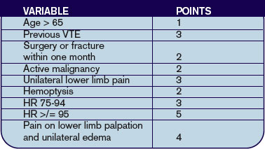
Predicting PE in the ED Using Revised Geneva Score
By Jeffrey Carter, MD
Le Gal G, Righini M, Roy PM, et al. Prediction of pulmonary embolism in the emergency department: the revised Geneva score. Ann Intern Med. 2006 Feb 7;144(3):165-171.
Introduction: Pulmonary embolism remains a common life-threatening disorder with imperfect diagnostic modalities and strategies. Much of the current literature focuses on the development and validation of clinical probability assessments that identify low-risk patients who can be safely managed without invasive testing or lung scanning.
Two common scoring systems include the Geneva and Wells criteria, which employ a combination of historical information, clinical and laboratory data to stratify patients into three risk groups.1,2 It has recently been shown that a single negative D-dimer test safely excludes patients with suspected VTE in a large cohort, with a three-month follow-up rate of VTE of 0%.3 Criticism of current clinical probability assessment strategies include the inclusion of subjective criteria or the need for blood gas values.1,2 In this paper, the authors sought to derive and validate a scoring system based on readily available objective clinical data.
Methods: The cohort evaluated in this study consists of the same 965 patients used to prospectively evaluate an emergency department diagnostic strategy of VTE.3 Patients presenting to the emergency ward with complaints of chest pain or shortness of breath were evaluated with a standard diagnostic workup that included a checklist of demographic and historical information, signs and symptoms possibly consistent with VTE, ABG, chest X-ray, and EKG results, as well as the likelihood of another diagnosis. However, the goal was to create a scoring system based on readily available objective data, so ABG, chest X-ray and electrocardiogram results were not evaluated as possible components of the score. Clinical variables associated with pulmonary embolism were evaluated for statistical significance in both a univariate and multivariate model. These variables were then validated both internally and with an external cohort.
Results: The incidence of VTE was 23% (222/965). Ten clinical variables were found to be statistically significantly associated with VTE; these comprise the Revised Geneva Score.
Clinical probability is based on points: 0-3 is low probability; 4-10 is intermediate probability, and >10 is high probability. Low, intermediate, and high clinical probabilities had respective rates of VTE of 8%, 28%, and 74%. These percentages were found in the validation cohort and were similar to those in the derivation cohort.
Discussion: This study focuses on the derivation and validation of a clinical scoring system that can provide a numerical estimate of likelihood of VTE. The diagnostic strategy by which VTE is confirmed or excluded is from derivation cohort and is published elsewhere.3 In that study patients classified as no VTE and not treated had a 90-day risk of VTE of 1%. The nine-point revised Geneva score does indeed provide an accurate assessment of risk of VTE, and can thus help guide clinical decision-making. However it is not clear that the revised Geneva score will help decrease invasive diagnostic tests. In the low-risk group, 8% is too great a risk of a life-threatening illness to forego further diagnostic evaluation. Coupled with a negative ELISA D-dimer, exclusion of these patients is safe, but these patients cannot be safely excluded without risk stratification.3
References
- Wells PS, Anderson DR, Rodger M, et al. Derivation of a simple clinical model to categorize patients’ probability of pulmonary embolism: increasing the model’s utility with the SimpliRED D-dimer. Thromb Haemost. 2000 Mar;83:416-420.
- Wicki J, Perneger TV, Junof AF, et al. Assessing clinical probability of pulmonary embolism in the emergency ward: a simple score. Arch Intern Med. 2001 Jan 8;161(1):92-97.
- Perrier A, Roy PM, Aujesky D, et al. Diagnosing pulmonary embolism in outpatients with clinical assessment, D-dimer measurement, venous ultrasound and helical computed tomography: a multicenter management study. Am J Med. 2004 Mar 1;116(5):291-299.
LMWH for Inpatient Palliative Care
By Jeanie Youngwerth, MD
Noble SI, Nelson A, Turner C, et al. Acceptability of low molecular weight heparin thromboprophylaxis for patients receiving palliative care: qualitative study. BMJ. 2006 Mar 11:332(7541);577-580.ePub 2006 Feb 3.
Venous thromboembolism is a major risk factor for patients with malignancy. VTE may reduce survival time in patients receiving palliative care, with one in seven inpatients with cancer dying from pulmonary embolism. The American College of Chest Physicians recommend low molecular weight heparin (LMWH) in hospitalized patients with cancer as level 1A evidence in their 2004 consensus statement on VTE. There are no thromboprophylaxis guidelines in the United Kingdom. Many physicians view daily injections of LMWH as unnecessary distress for palliative care patients. This study focused on what inpatients with advanced cancer receiving palliative care thought about the effect of thromboprophylaxis on overall quality of life.
This was a qualitative study of 28 inpatients receiving palliative care who had advanced metastatic cancer and who were in a regional cancer center in Wales. The patients had received LMWH for at least five consecutive days. The patients were audiotaped and then had semi-structured interviews transcribed regarding cancer treatments they had received. These interviews covered the patients’ insight into prognosis, their understanding of thromboprophylaxis, their beliefs concerning the effects of thromboprophylaxis on overall quality of life, and the negative aspects of heparin treatment.
The main outcome measures were recurring themes of the effect of thromboprophylaxis on overall quality of life. Major themes identified were insight into prognosis, knowledge and understanding of thromboprophylaxis, acceptability of heparin thromboprophylaxis, reassurance, and optimism. Minor themes identified were bruising, negative impact of antiembolic stockings, and anger at paternalistic views toward terminally ill patients.
Most patients showed clear insight into the nature of their condition and understood heparin prophylaxis for VTE. Many patients identified immobility and surgery as VTE risk factors, with little understanding of cancer as a risk factor. All knew that VTE could cause death, but none were aware of the common symptoms. All patients found LMWH thromboprophylaxis acceptable. Patients believed taking measures to prevent symptoms might improve their quality of life and felt that the medical team had not given up on them.
The only negative experiences of LMWH thromboprophylaxis was bruising. All of the patients who wore antiembolic stockings during previous admissions found them uncomfortable. Patients expressed their need to be involved in decision-making, particularly with respect to withdrawal or non-administration of treatment.
This small, qualitative study showed that, contrary to many physicians’ beliefs, patients in palliative care units believe that LMWH injections are acceptable as thromboprophylaxis, but antiembolic stockings are not. The effect of daily injections had little or no effect on the quality of life, with many patients having positive feelings that things were being done to prevent new problems from occurring.
This study was limited by the small sample size, the qualitative nature that could introduce interpretation bias, and the fact that only patients using LMWH were included. Additionally, the United Kingdom has aggressively educated the public on the risks of VTE associated with long flights such that baseline knowledge may differ in other parts of the world. Resource utilization, including drug costs and length of stay, as well as effect on mortality were not studied.
The implications for hospitalists are that many inpatients with advanced cancer receiving palliative care may find LMWH thromboprophylaxis acceptable therapy, and that discussions with the patient regarding this option should be explored together.
Cost Analysis: Intensive Glycemic Control in Critically Ill Adults
By Whitney Woodmansee, MD
Krinsley JS, Jones, RL. Cost analysis of intensive glycemic control in critically ill adult patients. Chest. 2006;129:644-650.
Several studies have demonstrated improved outcomes and decreased mortality in ICU patients treated with intensive control of blood glucose levels. This study sought to identify the costs associated with intensive glycemic control in the ICU.
An ICU patient database was analyzed for cost data related to intensive glycemic control. A baseline group of 800 consecutive ICU patients admitted prior to initiation (baseline) of an intensive glucose management (blood glucose levels between 80-140 mg/dl protocol were compared with a treatment group of 800 consecutive patients admitted after initiation of the protocol). Previously reported outcomes of these patients demonstrated significant improvement in mortality with tight glycemic control. Costs related to ICU and non-ICU length of stay; duration of mechanical ventilation; and all laboratory, pharmacy, and radiology services were analyzed between groups. Resource utilization was determined by assessing charges from the database and adjusting for inflation and applying Medicare cost, charge ratios for each category, and fiscal year. Costs associated with the intensive glycemic control protocol were determined. Unfortunately, only cost estimates for insulin and associated disposable supplies for each group were available for analysis.
Baseline and treatment populations did not differ significantly regarding demographics such as age, gender, race, admitting diagnosis, diabetes prevalence, or APACHE II scores. There were fewer patients in the treatment group that required mechanical ventilation during their ICU stay (40.6% versus 33.6%). Intensive glucose management was associated with a 13.9% reduction in total ICU days and duration of mechanical ventilation (median of two days decreased to 1.7 days p=0.045). There was a $1,580 adjusted cost savings per patient in the intensive treatment group compared with the baseline group (p<0.001). This reduction in cost was primarily driven by a decrease in laboratory and radiology costs in the ventilated patients. There were nonsignificant cost reductions in the unventilated patients.
Intensive control of hyperglycemia in the hospitalized ICU patient appears to be associated with reduction of morbidity and mortality. This suggests that tight glycemic control also leads to reductions in overall patient care costs—particularly in the ventilated ICU patient. Although not a randomized control trial, database analysis of costs and resource utilization demonstrated an overall cost savings in the treatment group (after initiation of an intensive glycemic control protocol) compared with the baseline group (before protocol initiation). One caveat is that the authors used estimates when determining the costs associated with the implementation of the intensive glucose management protocol. Nevertheless, intensive glycemic control was associated with an overall reduction in patient costs related to decreased ICU days and mechanical ventilation as well as resource utilization in a patient population already shown to have improved mortality. These results, if confirmed, suggest that tight glycemic control in the ICU is cost effective and should become standard medical practice. TH
Repeat Testing for C. Diff?
By Jeff Glasheen, MD
Mohan SS, McDermott BP, Parchuri S, et al. Lack of value of repeat stool testing for Clostridium difficile toxin. Am J Med. 2006 Apr;119(4):356.e7-356.e8
Clostridium difficile is a common complication of antibiotic and chemotherapeutic use, especially in hospitalized patients. Yet most nosocomial diarrhea is not caused by C. difficile. Most antibiotics can cause loose stools through changes in the gastrointestinal flora that result in inadequate digestion and absorption of carbohydrates and a resultant osmotic diarrhea. Further, antibiotics such as erythromycin and amoxicillin/clavulanate may result in diarrhea via increases in GI tract motility. While osmotic and motility causes of diarrhea tend to improve with antibiotic discontinuation, C. difficile-associated diarrhea is associated with significant morbidity that often continues until adequately treated.
Thus having a test that differentiates between C. difficile and non-clostridial diarrhea is essential. The most commonly used test is the enzyme immunoassay (EIA) that detects toxins A and B. The sensitivity and specificity of this test has been reported to range between 50%-90% and 70%-95%, respectively. The authors of this paper evaluated the utility of repeat EIA testing in patients with a one negative test in the setting of nosocomial diarrhea associated with antibiotic use.
The authors reviewed 474 sequential EIA tests for C. difficile in 396 patients over a 10-month period at a large university-affiliated community hospital with an EIA sensitivity and specificity of 80%-90% and 80%-95%, respectively. Tests were considered to be “repeat” if they occurred within seven days of the original negative test. Of the 78 repeat tests (16.5% of all tests), only one was positive, resulting in a 0.8% conversion rate. At an institutional cost of $128 per test the total cost of EIA testing over the 10-month period was $60,672. The cost of repeat testing alone was $9,984. The authors conclude that there is limited value—and high cost—in repeat EIA testing and that alternative sources of diarrhea should be sought or we should repeat EIA testing in patients with continued nosocomial diarrhea and a negative EIA test.
While prior studies have shown incremental benefit of retesting for C. difficile with the EIA assay, this study’s authors conclude that repeat “C. difficile testing is not clinically justified and is economically wasteful.” Unfortunately, the authors did not utilize a strong enough research design to defend this statement. From the data presented, all we can conclude is that repeat testing with an EIA assay did not add significant value to the diagnostic workup. The lack of a negative gold standard test, such as the cytotoxin assay or follow up outcomes, such as resolution of diarrhea, leaves the reader pondering if the repeat EIA assays were negative because the patients did not have C. difficile or because the test was not sensitive enough to detect the toxin. The reported sensitivity for the EIA assay used was 80-90%, meaning that 10%-20% of patients with C. difficile disease would have had a false negative test. While a second negative test would lower the likelihood of true disease, it would not lower it enough in a patient with a high pre-test probability of disease to sufficiently rule out the disease such that further testing is indicated.
Additionally, while we can extrapolate that the EIA test was of little utility to the patients studied here, no patient-specific data is presented. Thus it is difficult to determine if our patient population is represented in the study. More knowledge about the patients would allow the reader to use published prediction rules to better delineate how likely it was that this cohort was at high risk for having toxin-mediated diarrhea. Perhaps a more reasonable approach to this clinical conundrum would be to send the repeat stool test for a cytotoxin assay or to treat the patient empirically in situations where either the likelihood of disease or the disease burden is high.
Stroke after MI
By Ethan Cumbler, MD
Witt BJ, Ballman KV, Brown RD Jr, et al. The incidence of stroke after myocardial infarction: a meta-analysis. Am J Med. 2006 Apr;119(4):354.e1-9.
Stroke and myocardial infarction (MI) share many of the risk factors leading to atherosclerosis, including hypertension, hyperlipidemia, diabetes, tobacco abuse, and age. Logically, patients at risk for one event would also be at risk for the other, yet this interaction appears to be more complex. The study by Witt, et al., aims to elucidate the rate of in-hospital stroke in patients initially admitted with an MI.
The authors analyzed 22 observational studies of myocardial infarction that recorded the incidence of cerebrovascular accidents after acute MI. Clinical trials were excluded from the analysis in order to provide representation of an unselected population. Of the trials reviewed, 11 were included in the analysis of in-hospital strokes, three for the 30-day time point, and two for the one-year time point. The other trials used different time points and were not included in the analysis. The patients had a mean age ranging between 59 and 72.7 years, and all had a predominance of males. The rate of in-hospital stroke was 11.1 events per 1,000 hospitalizations. This incidence rose to 12.2/1,000 at 30 days and 21.4/1,000 at one year.
Plausible hypotheses for why the rate of cerebrovascular accident would be particularly high in the post-MI period include the potential for localized wall motion dysmotility or low flow leading to intracardiac thrombosis, event-related arrhythmia, or procedure-related embolic events. The studies from which this meta-analysis was derived were not designed in such a way for a causal relationship to be identified. However, age, diabetes, hypertension, prior stroke, anterior location of MI, atrial fibrillation, heart failure, and nonwhite race were all found to have an association with increased risk for stroke. Interestingly, angina on presentation was associated with an apparent decreased risk, theorized to potentially be due to ischemic preconditioning.
While this study shares the usual limitations of meta-analyses of observational studies, the authors have done an excellent summation of the data available including both English and non-English language articles in the analysis. Notably, the review included studies spanning more than 25 years and, thus, included data from studies done in the era prior to modern therapy for cardiac ischemia including potent antiplatelet and statin therapy. The three studies with in-hospital time points started in the 1990s had a lower average rate of stroke, which may reflect the effect of more potent anti-platelet agents used in today’s therapy for acute coronary syndromes.
The implication for the hospitalist is to recognize that patients admitted for MI are at high risk for stroke during the index hospitalization. A low threshold for suspicion of a cerebrovascular event needs to be maintained for post-MI patients with new neurologic symptoms. Future studies will be needed to address the risk/benefit of anticoagulation in high-risk patients for stroke following a myocardial infarction.
Predicting PE in the ED Using Revised Geneva Score
By Jeffrey Carter, MD
Le Gal G, Righini M, Roy PM, et al. Prediction of pulmonary embolism in the emergency department: the revised Geneva score. Ann Intern Med. 2006 Feb 7;144(3):165-171.
Introduction: Pulmonary embolism remains a common life-threatening disorder with imperfect diagnostic modalities and strategies. Much of the current literature focuses on the development and validation of clinical probability assessments that identify low-risk patients who can be safely managed without invasive testing or lung scanning.
Two common scoring systems include the Geneva and Wells criteria, which employ a combination of historical information, clinical and laboratory data to stratify patients into three risk groups.1,2 It has recently been shown that a single negative D-dimer test safely excludes patients with suspected VTE in a large cohort, with a three-month follow-up rate of VTE of 0%.3 Criticism of current clinical probability assessment strategies include the inclusion of subjective criteria or the need for blood gas values.1,2 In this paper, the authors sought to derive and validate a scoring system based on readily available objective clinical data.
Methods: The cohort evaluated in this study consists of the same 965 patients used to prospectively evaluate an emergency department diagnostic strategy of VTE.3 Patients presenting to the emergency ward with complaints of chest pain or shortness of breath were evaluated with a standard diagnostic workup that included a checklist of demographic and historical information, signs and symptoms possibly consistent with VTE, ABG, chest X-ray, and EKG results, as well as the likelihood of another diagnosis. However, the goal was to create a scoring system based on readily available objective data, so ABG, chest X-ray and electrocardiogram results were not evaluated as possible components of the score. Clinical variables associated with pulmonary embolism were evaluated for statistical significance in both a univariate and multivariate model. These variables were then validated both internally and with an external cohort.
Results: The incidence of VTE was 23% (222/965). Ten clinical variables were found to be statistically significantly associated with VTE; these comprise the Revised Geneva Score.
Clinical probability is based on points: 0-3 is low probability; 4-10 is intermediate probability, and >10 is high probability. Low, intermediate, and high clinical probabilities had respective rates of VTE of 8%, 28%, and 74%. These percentages were found in the validation cohort and were similar to those in the derivation cohort.
Discussion: This study focuses on the derivation and validation of a clinical scoring system that can provide a numerical estimate of likelihood of VTE. The diagnostic strategy by which VTE is confirmed or excluded is from derivation cohort and is published elsewhere.3 In that study patients classified as no VTE and not treated had a 90-day risk of VTE of 1%. The nine-point revised Geneva score does indeed provide an accurate assessment of risk of VTE, and can thus help guide clinical decision-making. However it is not clear that the revised Geneva score will help decrease invasive diagnostic tests. In the low-risk group, 8% is too great a risk of a life-threatening illness to forego further diagnostic evaluation. Coupled with a negative ELISA D-dimer, exclusion of these patients is safe, but these patients cannot be safely excluded without risk stratification.3
References
- Wells PS, Anderson DR, Rodger M, et al. Derivation of a simple clinical model to categorize patients’ probability of pulmonary embolism: increasing the model’s utility with the SimpliRED D-dimer. Thromb Haemost. 2000 Mar;83:416-420.
- Wicki J, Perneger TV, Junof AF, et al. Assessing clinical probability of pulmonary embolism in the emergency ward: a simple score. Arch Intern Med. 2001 Jan 8;161(1):92-97.
- Perrier A, Roy PM, Aujesky D, et al. Diagnosing pulmonary embolism in outpatients with clinical assessment, D-dimer measurement, venous ultrasound and helical computed tomography: a multicenter management study. Am J Med. 2004 Mar 1;116(5):291-299.
LMWH for Inpatient Palliative Care
By Jeanie Youngwerth, MD
Noble SI, Nelson A, Turner C, et al. Acceptability of low molecular weight heparin thromboprophylaxis for patients receiving palliative care: qualitative study. BMJ. 2006 Mar 11:332(7541);577-580.ePub 2006 Feb 3.
Venous thromboembolism is a major risk factor for patients with malignancy. VTE may reduce survival time in patients receiving palliative care, with one in seven inpatients with cancer dying from pulmonary embolism. The American College of Chest Physicians recommend low molecular weight heparin (LMWH) in hospitalized patients with cancer as level 1A evidence in their 2004 consensus statement on VTE. There are no thromboprophylaxis guidelines in the United Kingdom. Many physicians view daily injections of LMWH as unnecessary distress for palliative care patients. This study focused on what inpatients with advanced cancer receiving palliative care thought about the effect of thromboprophylaxis on overall quality of life.
This was a qualitative study of 28 inpatients receiving palliative care who had advanced metastatic cancer and who were in a regional cancer center in Wales. The patients had received LMWH for at least five consecutive days. The patients were audiotaped and then had semi-structured interviews transcribed regarding cancer treatments they had received. These interviews covered the patients’ insight into prognosis, their understanding of thromboprophylaxis, their beliefs concerning the effects of thromboprophylaxis on overall quality of life, and the negative aspects of heparin treatment.
The main outcome measures were recurring themes of the effect of thromboprophylaxis on overall quality of life. Major themes identified were insight into prognosis, knowledge and understanding of thromboprophylaxis, acceptability of heparin thromboprophylaxis, reassurance, and optimism. Minor themes identified were bruising, negative impact of antiembolic stockings, and anger at paternalistic views toward terminally ill patients.
Most patients showed clear insight into the nature of their condition and understood heparin prophylaxis for VTE. Many patients identified immobility and surgery as VTE risk factors, with little understanding of cancer as a risk factor. All knew that VTE could cause death, but none were aware of the common symptoms. All patients found LMWH thromboprophylaxis acceptable. Patients believed taking measures to prevent symptoms might improve their quality of life and felt that the medical team had not given up on them.
The only negative experiences of LMWH thromboprophylaxis was bruising. All of the patients who wore antiembolic stockings during previous admissions found them uncomfortable. Patients expressed their need to be involved in decision-making, particularly with respect to withdrawal or non-administration of treatment.
This small, qualitative study showed that, contrary to many physicians’ beliefs, patients in palliative care units believe that LMWH injections are acceptable as thromboprophylaxis, but antiembolic stockings are not. The effect of daily injections had little or no effect on the quality of life, with many patients having positive feelings that things were being done to prevent new problems from occurring.
This study was limited by the small sample size, the qualitative nature that could introduce interpretation bias, and the fact that only patients using LMWH were included. Additionally, the United Kingdom has aggressively educated the public on the risks of VTE associated with long flights such that baseline knowledge may differ in other parts of the world. Resource utilization, including drug costs and length of stay, as well as effect on mortality were not studied.
The implications for hospitalists are that many inpatients with advanced cancer receiving palliative care may find LMWH thromboprophylaxis acceptable therapy, and that discussions with the patient regarding this option should be explored together.
Cost Analysis: Intensive Glycemic Control in Critically Ill Adults
By Whitney Woodmansee, MD
Krinsley JS, Jones, RL. Cost analysis of intensive glycemic control in critically ill adult patients. Chest. 2006;129:644-650.
Several studies have demonstrated improved outcomes and decreased mortality in ICU patients treated with intensive control of blood glucose levels. This study sought to identify the costs associated with intensive glycemic control in the ICU.
An ICU patient database was analyzed for cost data related to intensive glycemic control. A baseline group of 800 consecutive ICU patients admitted prior to initiation (baseline) of an intensive glucose management (blood glucose levels between 80-140 mg/dl protocol were compared with a treatment group of 800 consecutive patients admitted after initiation of the protocol). Previously reported outcomes of these patients demonstrated significant improvement in mortality with tight glycemic control. Costs related to ICU and non-ICU length of stay; duration of mechanical ventilation; and all laboratory, pharmacy, and radiology services were analyzed between groups. Resource utilization was determined by assessing charges from the database and adjusting for inflation and applying Medicare cost, charge ratios for each category, and fiscal year. Costs associated with the intensive glycemic control protocol were determined. Unfortunately, only cost estimates for insulin and associated disposable supplies for each group were available for analysis.
Baseline and treatment populations did not differ significantly regarding demographics such as age, gender, race, admitting diagnosis, diabetes prevalence, or APACHE II scores. There were fewer patients in the treatment group that required mechanical ventilation during their ICU stay (40.6% versus 33.6%). Intensive glucose management was associated with a 13.9% reduction in total ICU days and duration of mechanical ventilation (median of two days decreased to 1.7 days p=0.045). There was a $1,580 adjusted cost savings per patient in the intensive treatment group compared with the baseline group (p<0.001). This reduction in cost was primarily driven by a decrease in laboratory and radiology costs in the ventilated patients. There were nonsignificant cost reductions in the unventilated patients.
Intensive control of hyperglycemia in the hospitalized ICU patient appears to be associated with reduction of morbidity and mortality. This suggests that tight glycemic control also leads to reductions in overall patient care costs—particularly in the ventilated ICU patient. Although not a randomized control trial, database analysis of costs and resource utilization demonstrated an overall cost savings in the treatment group (after initiation of an intensive glycemic control protocol) compared with the baseline group (before protocol initiation). One caveat is that the authors used estimates when determining the costs associated with the implementation of the intensive glucose management protocol. Nevertheless, intensive glycemic control was associated with an overall reduction in patient costs related to decreased ICU days and mechanical ventilation as well as resource utilization in a patient population already shown to have improved mortality. These results, if confirmed, suggest that tight glycemic control in the ICU is cost effective and should become standard medical practice. TH
Case Managers Offer Options
Jonathan P. Weisul, MD, FACEP, has direct responsibility over both case management and contracted hospitalist services at CHRISTUS St. Frances Cabrini Hospital in Alexandria, La., where he is regional chief medical officer and vice president of medical affairs. To him, the two most important aspects of the relationship between hospitalists and case managers involve communication and respect.
“In our system it’s a very collaborative approach where the case manager presents options to the physician as discharge planning progresses and the physician explains the progression of the patient’s treatment to the case manager,” he says.
In the early days of managed care, Dr. Weisul says, case management received some bad press in that “it was perceived that their interest was [just] getting patients out of the hospital. And I would say that one area where the communication fails is the oftentimes misheld belief on the physician’s part that case managers are trying to practice medicine,” he explains. “Communication usually involves the case manager presenting options to a physician in a way that would be in the best interest of the patient—not trying to usurp any authority from the physician-patient relationship.”
The greatest partnership between hospitalists and case managers occurs when they both share options and needs, and perhaps the best venue for sharing occurs at interdisciplinary meetings. All case managers interviewed for this article stress the importance of daily meetings as a clear advantage for an effective discharge plan.
L. Greg Cunningham, MHA, CEO of the American Case Management Association (ACMA), headquartered in Little Rock, Ark., says the best thing hospitalists and case managers can do to improve their working relationship is “communicate about their patient caseload first thing in the morning.” Both are sharing information. “One is sharing more expectations of what needs to be done for the patient,” he says. “The other is sharing more expectations about what the physician needs to do in terms of decision-making such as getting signatures on forms and communicating with the patient and family.”

Be Proactive
Theresa Brocato, RN, BSN, CPUR, manager of case management and social work at CHRISTUS St. Frances Cabrini Hospital, has been in that position for almost four years. For seven years before that she worked in managed care for Ochsner Health Plan of Louisiana. In her opinion hospitalists can be most helpful to case managers by thinking proactively.
“We have a very proactive discharge planning department,” she says of her department of nine case managers (all nurses) and five social workers who work directly with patients and families, “because as soon as the patient gets to the hospital we start the discharge planning.”
The case managers interview patients and their family and let them know what their estimated time to wellness will be. “We base the estimate on a working DRG [drug-related group],” she says. “We talk to the hospitalist up front, and [we] say, for instance, ‘For this patient, the average time to wellness is about six days, and we need to start preparing for that ultimate discharge. Is there anything you foresee that you might need on discharge, such as equipment or home health?’ And then we start coordinating with the physicians to get the orders in place and get everything set up so that they have a smooth transition at the time of discharge.”
But Dr. Weisul says this kind of communication can be misinterpreted. “Communication seems to fail in the most difficult cases where the physician seems to perceive that the progress of the medical care might not be going on [in] as timely [a manner] as he or she might expect and feels that the case manager, in … looking at when the appropriate discharge time might be, is pressuring the physician,” he says. “But the reverse is true: What they’re doing is just asking for input from the hospitalist [or other] physician as to progress on the path of returning the patient to health and just looking to the future.”
Brocato says the three hospitalists at Cabrini have been easy to work with, sometimes seeking out the case managers and social workers to ask for their help on difficult cases, working with their fellow interdisciplinary-team members to design discharge plans from innovative ideas that solve patient’s challenges, and including the patient in their planning.
Appropriate Levels of Care
At Cabrini Hospital, the case managers and social workers hold daily interdisciplinary discharge planning meetings. Each case is reviewed according to nationally accepted criteria to ensure that the care provided meets standards appropriate for the acute inpatient level of care. Other treatment levels identified during discharge planning include rehabilitation, long-term acute care, and outpatient levels of care.
One recommendation that Brocato would make to hospitalists to better help the work of the case managers, the hospital, and the patients is to recognize earlier which patients will require a longer length of stay (more than three weeks) so that those individuals can be transitioned into a more appropriate level of care.
Some examples of diagnoses in which long-term acute care might be called for, she says, include “osteomyelitis, where a patient will be on a course of antibiotics for six weeks and may require extensive wound therapy. In that case, as soon as we get the results of the bone scan and we see that, we immediately ask the physician to think about moving the patient.”
Other examples include those patients who will need a long time to recover, such as those in the ICU. “Maybe they’ve have been on a ventilator for a long time and they get debilitated,” says Brocato. “Or if they need to be weaned from the ventilator and need some intensive respiratory toilet. The long-term acute care setting is the more appropriate setting to work on trying to rehabilitate the patient.”
Follow-up
Another important element to a good discharge plan is follow-up. Cabrini Hospital has initiated a program whereby a nurse has been hired to call on patients within two days of discharge to check on how things are going. That is, Brocato says, “whether they understood the discharge instructions, to make sure that they got their prescriptions, and [to ensure] they have some kind of follow-up appointment made and are planning to go to that.”
A 2001 study conducted by the section of general internal medicine in the department of medicine at West Virginia University (Morgantown) showed that the effect of employing a nurse discharge planner to work with the hospitalist service had a positive effect on outcomes in an academic teaching hospital.1 When a general medicine service, specialist-staffed service, and a hospitalist service with nurse discharge planner were compared, the hospitalist-discharge planner group was associated with a lower average cost and shorter average length of hospital stay. There was no apparent compromise in clinical outcomes and patient satisfaction with care.
Competent Colleagues
The American Case Management Association (ACMA), begun in 1999, is the first and only nonprofit hospital-based case management organization in the United States. It represents nurses, social workers, physicians, and other professionals who practice hospital case management. The physicians whom ACMA represents are primarily medical directors hired as the catalyst for attendings who are less than cooperative and are impeding discharge (typically not the hospitalists). “The organization is growing at an average annual growth rate of 25%,” says Cunningham. “We’ve just started a new certification process for hospital-based case managers—one for nurses and one for social workers.”
There is a core portion to the exam that tests for knowledge, and a specialty portion of the exam in which “they have to validate those skill sets. The specialty portion of includes a clinical simulation, which is the application of their skills and knowledge,” explains Cunningham. “They have to [show that they can] make not only a decision, but sequential decisions. So we’re testing their ability to take a case and work through it.”
The hospitalist should expect that high level of competency from a case manager, just as the case manager should be able to expect the highest competency of the hospitalist. “The hospitalist should not lessen their expectation of the clinical competency of the case managers,” says Cunningham. “We are advocating that the physicians … increase their expectations of the clinical competency of those individuals.”
Cunningham recommends that hospitalists discuss their case manager’s background if they suspect there is a diminished competency. “Competent case managers “not only make it better for patients,” he says, “but practitioners’ lives are made much easier when competent case managers are hired.”
All the case managers at Cabrini Hospital have a strong clinical background. “It is really important that the case managers are competent in the field they are working in,” says Brocato, “so that the physicians can trust that they understand the clinical side as well as what might be needed at discharge planning. [At Cabrini Hospital] they are placed in the units where they work based on their careers as nurses. The case manager that works in ICU, for instance, was an ICU nurse for many years.”
Brocato believes “hospitalists need to feel confident that the case managers—or discharge planners as they’re called at some hospitals—have a strong clinical background. In that way, when the hospitalists “are discussing their cases, they feel that we know what they’re talking about. For us, it means that we feel we are all on the same page when we’re dealing with the physicians so that we know what the course of treatment will be. Then the case managers are able to make a better discharge plan based on what the expectations of the hospital stay are going to be, so we can plan ahead.”
Dr. Weisul, who oversees three healthcare facilities in the central Louisiana region, knows that the relationship between hospitalists and case managers can be a fruitful one for all concerned. Cabrini has achieved the lowest case mix-adjusted length of stay in its healthcare system. In addition, when physicians were surveyed regarding the discharge planning process provided to their patients, the hospital achieved a combined rate of 97% “satisfied” or “very satisfied.”
“The idea that case management can achieve, with the physician, a low length of stay does not necessarily have to be in an environment of contention,” says Dr. Weisul.
Conclusion
Realize that case managers are there to assist hospitalists meet patients’ care goals. Watch for patients who a need longer length of stay and alert case managers in those cases to ensure moving them to appropriate levels of care, such as long-term acute care setting as soon as possible. Let case managers know how patients and families can reach you post-discharge. Expect the highest standard of competency from case managers and work with hospital administrators and case management to consistently make this a reality. TH
Andrea Sattinger writes regularly for The Hospitalist.
Reference
- Palmer HC Jr, Armistead NS, Elnicki DM, et al. The effect of a hospitalist service with nurse discharge planner on patient care in an academic teaching hospital. Am J Med. 2001;111:627-632.
Resources
- Case Management Society of America (CMSA): www.cmsa.org/
- American Case Management Association (ACMA): www.acmaweb.org/
Jonathan P. Weisul, MD, FACEP, has direct responsibility over both case management and contracted hospitalist services at CHRISTUS St. Frances Cabrini Hospital in Alexandria, La., where he is regional chief medical officer and vice president of medical affairs. To him, the two most important aspects of the relationship between hospitalists and case managers involve communication and respect.
“In our system it’s a very collaborative approach where the case manager presents options to the physician as discharge planning progresses and the physician explains the progression of the patient’s treatment to the case manager,” he says.
In the early days of managed care, Dr. Weisul says, case management received some bad press in that “it was perceived that their interest was [just] getting patients out of the hospital. And I would say that one area where the communication fails is the oftentimes misheld belief on the physician’s part that case managers are trying to practice medicine,” he explains. “Communication usually involves the case manager presenting options to a physician in a way that would be in the best interest of the patient—not trying to usurp any authority from the physician-patient relationship.”
The greatest partnership between hospitalists and case managers occurs when they both share options and needs, and perhaps the best venue for sharing occurs at interdisciplinary meetings. All case managers interviewed for this article stress the importance of daily meetings as a clear advantage for an effective discharge plan.
L. Greg Cunningham, MHA, CEO of the American Case Management Association (ACMA), headquartered in Little Rock, Ark., says the best thing hospitalists and case managers can do to improve their working relationship is “communicate about their patient caseload first thing in the morning.” Both are sharing information. “One is sharing more expectations of what needs to be done for the patient,” he says. “The other is sharing more expectations about what the physician needs to do in terms of decision-making such as getting signatures on forms and communicating with the patient and family.”
Be Proactive
Theresa Brocato, RN, BSN, CPUR, manager of case management and social work at CHRISTUS St. Frances Cabrini Hospital, has been in that position for almost four years. For seven years before that she worked in managed care for Ochsner Health Plan of Louisiana. In her opinion hospitalists can be most helpful to case managers by thinking proactively.
“We have a very proactive discharge planning department,” she says of her department of nine case managers (all nurses) and five social workers who work directly with patients and families, “because as soon as the patient gets to the hospital we start the discharge planning.”
The case managers interview patients and their family and let them know what their estimated time to wellness will be. “We base the estimate on a working DRG [drug-related group],” she says. “We talk to the hospitalist up front, and [we] say, for instance, ‘For this patient, the average time to wellness is about six days, and we need to start preparing for that ultimate discharge. Is there anything you foresee that you might need on discharge, such as equipment or home health?’ And then we start coordinating with the physicians to get the orders in place and get everything set up so that they have a smooth transition at the time of discharge.”
But Dr. Weisul says this kind of communication can be misinterpreted. “Communication seems to fail in the most difficult cases where the physician seems to perceive that the progress of the medical care might not be going on [in] as timely [a manner] as he or she might expect and feels that the case manager, in … looking at when the appropriate discharge time might be, is pressuring the physician,” he says. “But the reverse is true: What they’re doing is just asking for input from the hospitalist [or other] physician as to progress on the path of returning the patient to health and just looking to the future.”
Brocato says the three hospitalists at Cabrini have been easy to work with, sometimes seeking out the case managers and social workers to ask for their help on difficult cases, working with their fellow interdisciplinary-team members to design discharge plans from innovative ideas that solve patient’s challenges, and including the patient in their planning.
Appropriate Levels of Care
At Cabrini Hospital, the case managers and social workers hold daily interdisciplinary discharge planning meetings. Each case is reviewed according to nationally accepted criteria to ensure that the care provided meets standards appropriate for the acute inpatient level of care. Other treatment levels identified during discharge planning include rehabilitation, long-term acute care, and outpatient levels of care.
One recommendation that Brocato would make to hospitalists to better help the work of the case managers, the hospital, and the patients is to recognize earlier which patients will require a longer length of stay (more than three weeks) so that those individuals can be transitioned into a more appropriate level of care.
Some examples of diagnoses in which long-term acute care might be called for, she says, include “osteomyelitis, where a patient will be on a course of antibiotics for six weeks and may require extensive wound therapy. In that case, as soon as we get the results of the bone scan and we see that, we immediately ask the physician to think about moving the patient.”
Other examples include those patients who will need a long time to recover, such as those in the ICU. “Maybe they’ve have been on a ventilator for a long time and they get debilitated,” says Brocato. “Or if they need to be weaned from the ventilator and need some intensive respiratory toilet. The long-term acute care setting is the more appropriate setting to work on trying to rehabilitate the patient.”
Follow-up
Another important element to a good discharge plan is follow-up. Cabrini Hospital has initiated a program whereby a nurse has been hired to call on patients within two days of discharge to check on how things are going. That is, Brocato says, “whether they understood the discharge instructions, to make sure that they got their prescriptions, and [to ensure] they have some kind of follow-up appointment made and are planning to go to that.”
A 2001 study conducted by the section of general internal medicine in the department of medicine at West Virginia University (Morgantown) showed that the effect of employing a nurse discharge planner to work with the hospitalist service had a positive effect on outcomes in an academic teaching hospital.1 When a general medicine service, specialist-staffed service, and a hospitalist service with nurse discharge planner were compared, the hospitalist-discharge planner group was associated with a lower average cost and shorter average length of hospital stay. There was no apparent compromise in clinical outcomes and patient satisfaction with care.
Competent Colleagues
The American Case Management Association (ACMA), begun in 1999, is the first and only nonprofit hospital-based case management organization in the United States. It represents nurses, social workers, physicians, and other professionals who practice hospital case management. The physicians whom ACMA represents are primarily medical directors hired as the catalyst for attendings who are less than cooperative and are impeding discharge (typically not the hospitalists). “The organization is growing at an average annual growth rate of 25%,” says Cunningham. “We’ve just started a new certification process for hospital-based case managers—one for nurses and one for social workers.”
There is a core portion to the exam that tests for knowledge, and a specialty portion of the exam in which “they have to validate those skill sets. The specialty portion of includes a clinical simulation, which is the application of their skills and knowledge,” explains Cunningham. “They have to [show that they can] make not only a decision, but sequential decisions. So we’re testing their ability to take a case and work through it.”
The hospitalist should expect that high level of competency from a case manager, just as the case manager should be able to expect the highest competency of the hospitalist. “The hospitalist should not lessen their expectation of the clinical competency of the case managers,” says Cunningham. “We are advocating that the physicians … increase their expectations of the clinical competency of those individuals.”
Cunningham recommends that hospitalists discuss their case manager’s background if they suspect there is a diminished competency. “Competent case managers “not only make it better for patients,” he says, “but practitioners’ lives are made much easier when competent case managers are hired.”
All the case managers at Cabrini Hospital have a strong clinical background. “It is really important that the case managers are competent in the field they are working in,” says Brocato, “so that the physicians can trust that they understand the clinical side as well as what might be needed at discharge planning. [At Cabrini Hospital] they are placed in the units where they work based on their careers as nurses. The case manager that works in ICU, for instance, was an ICU nurse for many years.”
Brocato believes “hospitalists need to feel confident that the case managers—or discharge planners as they’re called at some hospitals—have a strong clinical background. In that way, when the hospitalists “are discussing their cases, they feel that we know what they’re talking about. For us, it means that we feel we are all on the same page when we’re dealing with the physicians so that we know what the course of treatment will be. Then the case managers are able to make a better discharge plan based on what the expectations of the hospital stay are going to be, so we can plan ahead.”
Dr. Weisul, who oversees three healthcare facilities in the central Louisiana region, knows that the relationship between hospitalists and case managers can be a fruitful one for all concerned. Cabrini has achieved the lowest case mix-adjusted length of stay in its healthcare system. In addition, when physicians were surveyed regarding the discharge planning process provided to their patients, the hospital achieved a combined rate of 97% “satisfied” or “very satisfied.”
“The idea that case management can achieve, with the physician, a low length of stay does not necessarily have to be in an environment of contention,” says Dr. Weisul.
Conclusion
Realize that case managers are there to assist hospitalists meet patients’ care goals. Watch for patients who a need longer length of stay and alert case managers in those cases to ensure moving them to appropriate levels of care, such as long-term acute care setting as soon as possible. Let case managers know how patients and families can reach you post-discharge. Expect the highest standard of competency from case managers and work with hospital administrators and case management to consistently make this a reality. TH
Andrea Sattinger writes regularly for The Hospitalist.
Reference
- Palmer HC Jr, Armistead NS, Elnicki DM, et al. The effect of a hospitalist service with nurse discharge planner on patient care in an academic teaching hospital. Am J Med. 2001;111:627-632.
Resources
- Case Management Society of America (CMSA): www.cmsa.org/
- American Case Management Association (ACMA): www.acmaweb.org/
Liability, Medical Error Legislation
Liability. To a physician, the word conjures the threat of a malpractice lawsuit and the reality of escalating insurance costs. But some protection may be at hand in the form of several laws recently passed by Congress that aim to relieve both threat and reality.
Hospitalists and Liability
For the time being, the majority of hospitalists are covered by their employer’s liability insurance, according to the cover article in the December 2005 issue of The Hospitalist (“A Malpractice Primer” p. 1). However, that doesn’t mean they’re unaffected by risk of malpractice suits.
—Mary A. Germann, RN, MN, CHE
Mary A. Germann, RN, MN, CHE, certified healthcare compliance officer and founder of Operations Solutions for Healthcare, Inc., a consulting firm based in Atlanta, has answered many liability questions from hospitalists and believes they are at least as worried about the issue as other physicians.
“Hospitalists tend to be concerned about their risk,” she says. “They want to know how to protect themselves. Even though they may not be ‘personally’ at risk, their medical license is still on the line."
Germann explains that hospitalists may in fact be more vulnerable to liability suits than other physicians. “In some ways [hospitalists] are more at risk because they are usually new to their patients,” she says. “They have not had the opportunity to build a relationship with a patient, and must create trust and open communications in a very short time. This weak link can increase a hospitalist’s vulnerability. Lack of information and communication is one of the biggest reasons reported that patients choose to sue.”
With this in mind, let’s take a look at current and pending legislation that will shape liability risk and claims for years to come.
The HEALTH Act of 2005
Passed into law in July of last year, the HEALTH Act (or Help Efficient, Accessible, Low-cost, Timely Healthcare Act), places multiple limits on liability claims. The law caps noneconomic damages in medical malpractice suits at $250,000 for compensating patient injury, limits attorneys’ contingency fees, and requires a finding of malicious intent to support an award of punitive damages. The law also exempts manufacturers and distributors of medical products from punitive damage awards if the U.S. Food and Drug Administration approved the product.
The Patient Safety and Quality Improvement Act of 2005
Also signed into law last July, this act establishes a voluntary, confidential reporting structure for use by physicians, hospitals, and other healthcare professional and entities. This law renders reported medical errors into confidential, privileged data and allows healthcare providers to report their medical errors under a “patient safety activity” umbrella that prohibits the information from being used in a civil action (i.e. liability case). All medical errors reported are covered by the law and not subject to subpoena, Freedom of Information Act request, or use in a disciplinary proceeding.
On reporting medical errors within a hospital system, Germann says, “This is really a joint effort; I don’t think any one entity or organization can do it by themselves. Hospitals have to have a system in place for reporting errors and near misses. Studies have shown that organizations that aggressively support error disclosure have a decreased incidence in the number of suits and a decrease in the compensation payouts.”
The National Medical Error Disclosure and Compensation Act of 2005
Also known as the MEDiC Act, this bill was introduced in the Senate in September 2005 by Senator Hillary Rodham Clinton (D-N.Y.) and Senator Barack Obama (D-Ill.).
Designed to extend the Patient Safety and Quality Improvement Act of 2005 and “promote a culture of safety within hospitals, health systems, clinics, and other sites of healthcare,” this act would establish a federal Office of Patient Safety and Health Care Quality to implement and oversee a new national patient safety database, as well as the MEDiC Program. This program would provide funding to those healthcare providers with systems to disclose medical errors to patients and offer fair compensation to patients if the provider is at fault.
In reducing administrative and legal costs for medical malpractice claims, the MEDiC Act would require participating medical liability insurance companies and healthcare providers to apply a percentage of their savings toward reducing medical errors. The bill also requires that, to the extent possible, some of these cost savings be passed along to providers as lower malpractice insurance premiums.
Although not specifically stated in the bill, a goal of the MEDiC Act is to provide an interim solution to the escalating costs of liability lawsuits.
“I think [medical error reporting] is a very good direction to take,” says Germann. “One of the major barriers to disclosing errors is fear of malpractice suits. To improve quality and decrease medical errors, it’s important for physicians to be able to disclose errors.”
In addition, she stresses that hospitalists and other physicians have little to fear in disclosure. “The majority of errors are not caused by incompetent physicians,” notes Germann. “They are secondary to system failures. Physicians must be able to expose these. And hospitalists see more system errors because they live within the system; they can be a great asset in helping hospital administrators improve quality and systems.”
The MEDiC Act has been under review by the Senate Committee on Health, Education, Labor, and Pensions since September.
So far, 2005 and 2006 have seen big changes in liability reform and in medical error reporting. Together, this legislation—perhaps in conjunction with future laws—will change the risks of liability faced by hospitalists. "No one law is going to solve the entire problem,” says Germann. “All of these together will make the improvements." TH
Jane Jerrard writes “Public Policy” every month for The Hospitalist.
Urine for a Surprise
A58-year-old white female presented with a two-week history of severe fatigue. She was admitted for acute renal failure (creatinine of 3.0 mg/dL with baseline 0.9 mg/dL two months prior) and anemia (hematocrit of 27.9 with baseline 37.5 two month prior). She had no prior history of renal failure. Her review of systems was otherwise negative. Her urine was orange and dipstick was positive for bilirubin but serum bilirubin was normal. (See photo at right.) Blood smear showed schistocytes and haptoglobin was undetectable. Physical exam showed yellow sclera, but was otherwise normal.

What is the most appropriate treatment for this patient?
- Draw serum complement levels and spin urine for sediment analysis;
- Order a STAT renal ultrasound;
- Ask the patient about over-the-counter medication use;
- Check urine porphyrin levels; or
- Order immediate referral for plasmapheresis.
Discussion
The answer is C: Ask the patient about over-the-counter (OTC) medication use. This patient’s presentation was consistent with overuse of phenazopyridine. Phenazopyridine is an azo dye, which appears to exert a local anesthetic action on urinary tract mucosa. It’s reported adverse reactions include acute renal failure, hemolytic anemia, hepatitis, and methemoglobinemia, which have been reported after acute ingestions, chronic overdoses, and in chronically appropriate doses.1-7 The mechanism of these adverse reactions is not well understood. Although the differential diagnosis for renal failure and hemolytic anemia is extensive, yellow sclera and orange urine in the setting of a normal bilirubin level raised the suspicion of phenazopyridine use in this patient.
This case highlights the common overuse of over the counter medications, as well as global lack of knowledge of their potential adverse reactions. Although phenazopyridine is widely used, 50% of product consumers do not know that it is a urinary tract analgesic, and 80% do not know either the cause of their symptoms or the action of the drug.8
Additionally, although OTC medication use is reported by two-thirds of all hospitalized patients, documentation of them is present in only 10% of admission paperwork.9-10 Given that drug related hospitalizations account for 5%-8% of all hospital stays, it is essential that a complete OTC medication list be included as a routine part of the history obtained from all patients at the time of hospital admission.11-12 With history taking vigilance and patient education, adverse events from OTC medications can be minimized. TH
References
- Gabor EP, Lowenstein L, De Leeuw NK. Hemolytic anemia induced by Phenylazo-Diamino-Pyridine (Pyridium). Can Med Assoc J. 1964 Oct;91:756-759.
- Nathan DM, Siegel AJ, Bunn HF. Acute methemoglobinemia and hemolytic anemia with phenazopyridine: possible relation to acute renal failure. Arch Int Med. 1977 Nov;137(11):1636-1638.
- Gavish D, Knobler H, Gottehrer N, et al. Methemoglobinemia, muscle damage and renal failure complicating phenazopyridine overdose. Isr J Med Sci. 1986 Jan;22(1):45-47.
- Vega J. Acute Renal Failure caused by phenazopyridine. Rev Med Chil. 2003 May;131(5):541-544.
- Kornowski R, Averbuch M, Jaffe A, et al. Sedural toxicity. Harefuah. 1991 Mar 15;120(6):324-325.
- Thomas RJ, Doddabele S, Karnad AB. Chronic severe hemolytic anemia related to surreptitious phenazopyridine abuse. Ann Int Med. 1994;121:308.
- Landman J, Kavaler E, Waterhouse R. Acquired methemoglobinemia possibly related to phenazopyridine in a woman with normal renal function. J Urol. 1997 Oct;158(4):1520-1521.
- Chih-Wen S, Asch SM, Fielder E, et al. Consumer knowledge of over-the-counter phenazopyridine. Ann Fam Med. 2004 May-Jun;2(3):240-244.
- Chrischilles EA, Foley DJ, Wallace RB, et al. Use of medications by persons 65 and over; data from the established populations for epidemiologic studies of the elderly. J Geront. 1992 Sep;47(5):M137-144.
- Simons LA, Tett S, Simons J, et al. Multiple medication use in the elderly. Use of prescription and non-prescription drugs in an Australian community setting. Med J Aust. 1992 Aug 17;157(4):242-246.
- Hallas J, Jensen KB, Grodum E, et al. Drug-related admissions to a department of medical gastroenterology. The role of self-medicated and prescribed drugs. Scand J Gastroenterol. 1991 Feb;26(2):174-180.
- Pirmohamed M, James S, Meakin S, et al. Adverse drug reactions as cause of admission to hospital: prospective analysis of 18,820 patients. BMJ. 2004 Jul;329(7456):15-19.
A58-year-old white female presented with a two-week history of severe fatigue. She was admitted for acute renal failure (creatinine of 3.0 mg/dL with baseline 0.9 mg/dL two months prior) and anemia (hematocrit of 27.9 with baseline 37.5 two month prior). She had no prior history of renal failure. Her review of systems was otherwise negative. Her urine was orange and dipstick was positive for bilirubin but serum bilirubin was normal. (See photo at right.) Blood smear showed schistocytes and haptoglobin was undetectable. Physical exam showed yellow sclera, but was otherwise normal.
What is the most appropriate treatment for this patient?
- Draw serum complement levels and spin urine for sediment analysis;
- Order a STAT renal ultrasound;
- Ask the patient about over-the-counter medication use;
- Check urine porphyrin levels; or
- Order immediate referral for plasmapheresis.
Discussion
The answer is C: Ask the patient about over-the-counter (OTC) medication use. This patient’s presentation was consistent with overuse of phenazopyridine. Phenazopyridine is an azo dye, which appears to exert a local anesthetic action on urinary tract mucosa. It’s reported adverse reactions include acute renal failure, hemolytic anemia, hepatitis, and methemoglobinemia, which have been reported after acute ingestions, chronic overdoses, and in chronically appropriate doses.1-7 The mechanism of these adverse reactions is not well understood. Although the differential diagnosis for renal failure and hemolytic anemia is extensive, yellow sclera and orange urine in the setting of a normal bilirubin level raised the suspicion of phenazopyridine use in this patient.
This case highlights the common overuse of over the counter medications, as well as global lack of knowledge of their potential adverse reactions. Although phenazopyridine is widely used, 50% of product consumers do not know that it is a urinary tract analgesic, and 80% do not know either the cause of their symptoms or the action of the drug.8
Additionally, although OTC medication use is reported by two-thirds of all hospitalized patients, documentation of them is present in only 10% of admission paperwork.9-10 Given that drug related hospitalizations account for 5%-8% of all hospital stays, it is essential that a complete OTC medication list be included as a routine part of the history obtained from all patients at the time of hospital admission.11-12 With history taking vigilance and patient education, adverse events from OTC medications can be minimized. TH
References
- Gabor EP, Lowenstein L, De Leeuw NK. Hemolytic anemia induced by Phenylazo-Diamino-Pyridine (Pyridium). Can Med Assoc J. 1964 Oct;91:756-759.
- Nathan DM, Siegel AJ, Bunn HF. Acute methemoglobinemia and hemolytic anemia with phenazopyridine: possible relation to acute renal failure. Arch Int Med. 1977 Nov;137(11):1636-1638.
- Gavish D, Knobler H, Gottehrer N, et al. Methemoglobinemia, muscle damage and renal failure complicating phenazopyridine overdose. Isr J Med Sci. 1986 Jan;22(1):45-47.
- Vega J. Acute Renal Failure caused by phenazopyridine. Rev Med Chil. 2003 May;131(5):541-544.
- Kornowski R, Averbuch M, Jaffe A, et al. Sedural toxicity. Harefuah. 1991 Mar 15;120(6):324-325.
- Thomas RJ, Doddabele S, Karnad AB. Chronic severe hemolytic anemia related to surreptitious phenazopyridine abuse. Ann Int Med. 1994;121:308.
- Landman J, Kavaler E, Waterhouse R. Acquired methemoglobinemia possibly related to phenazopyridine in a woman with normal renal function. J Urol. 1997 Oct;158(4):1520-1521.
- Chih-Wen S, Asch SM, Fielder E, et al. Consumer knowledge of over-the-counter phenazopyridine. Ann Fam Med. 2004 May-Jun;2(3):240-244.
- Chrischilles EA, Foley DJ, Wallace RB, et al. Use of medications by persons 65 and over; data from the established populations for epidemiologic studies of the elderly. J Geront. 1992 Sep;47(5):M137-144.
- Simons LA, Tett S, Simons J, et al. Multiple medication use in the elderly. Use of prescription and non-prescription drugs in an Australian community setting. Med J Aust. 1992 Aug 17;157(4):242-246.
- Hallas J, Jensen KB, Grodum E, et al. Drug-related admissions to a department of medical gastroenterology. The role of self-medicated and prescribed drugs. Scand J Gastroenterol. 1991 Feb;26(2):174-180.
- Pirmohamed M, James S, Meakin S, et al. Adverse drug reactions as cause of admission to hospital: prospective analysis of 18,820 patients. BMJ. 2004 Jul;329(7456):15-19.
Expert Testimony and the Hospitalist
An attorney approaches you about testifying as an expert witness on behalf of a patient against a physician in your area. How do you decide whether to testify?
The decision of whether to testify in a malpractice case is one of the most difficult, yet one of the most important non-patient care decisions a physician can make. Expert testimony is essential to medical malpractice litigation.
The physician expert, however, is often caught in the middle of conflicting tensions. The obligation to promote fairness, justice, and self-regulation of the profession are balanced against the professional and social pressure not to testify against colleagues and not to participate in a legal system that many physicians feel victimizes members of the profession. Nonetheless, the legal system relies on competent medical expertise to be just and fair, and relies on medical professionals to provide that expertise. An individual physician’s decision to participate in a medical malpractice case should be guided by careful consideration of their duties as applied to the specific situation.
What Is Expert Testimony, and Who Is an Expert?
A medical malpractice claim requires that the plaintiff show that the defendant(s) breached a duty to the plaintiff by failing to perform to the standard of care. The central issue in many malpractice cases involves defining the standard of care and determining whether the defendant(s) deviated from it. The only way for a jury to determine what is the standard of care is to listen to the opinions of experts and make a decision based on the persuasiveness and credibility of the experts.
Expert witnesses differ from other witnesses because expert witnesses can offer opinions while other witnesses can only testify to facts or their own personal experience.1 Expert witnesses must have specialized knowledge or experience to be allowed to offer opinion testimony. The U.S. Supreme Court has required that scientific testimony be relevant and reliable, and requires the judge presiding over a case to determine the validity of scientific testimony.2 If the judge decides that scientific testimony to be offered by an expert is not valid or reliable, the judge may refuse to allow it. Thus, the judge determines who may serve as an expert in front of the jury.
Licensed physicians are usually considered experts on the standard of care, regardless of the specialty or area of practice of the testifying expert. Tort reform in many states is focusing on expert testimony, including limiting judicial discretion in qualifying experts. For example, in Pennsylvania, only an expert in the field of the defendant may give expert testimony against him or her.
Given that most physicians are allowed by most judges to testify as to the standard of care, an expert may be testifying outside their scope of practice or the area in which they have actual specialized knowledge and experience. Further, once an expert is allowed to testify, there are no consequences for offering opinions that are unsupported by evidence or patently inaccurate. These are the areas where professional integrity is crucial, and professional societies can play a role in regulation and oversight of physicians serving as experts.3
Self-Regulation of Expert Witnesses
What do individuals and professional societies need to contribute to oversight of expert testimony? A physician’s obligation to participate in malpractice cases arises from the privilege of self-regulation enjoyed by the medical profession. As a result of the degree of specialized knowledge and skill needed to practice medicine, physicians have a duty to take action against other physicians who are impaired or incompetent.3 Professional societies promote the highest ethical and professional standards for members and therefore have a responsibility to discipline members who are irresponsible or unqualified in their expert legal involvement.
Physicians also have a duty to patients to provide expert testimony. In the event of harm caused by negligence, patients are denied any compensation at all if responsible physicians are unwilling to become involved as plaintiff’s experts. Physicians must uphold the duty to act in the best interest of patients and society and to promote justice in the system by rendering fair and honest opinions—even if that results in liability for a fellow physicians and despite the current atmosphere of negativity toward physicians testifying against other physicians.3
Based on these ethical principles, physicians have a duty to provide accurate and responsible expert testimony. Inaccurate expert testimony is damaging to the system in many ways: It increases malpractice costs, injures the reputation of individual practitioners, and adversely affects the standard of care by promoting false standards. Distinguishing irresponsible testimony from reasonable differences of opinion may be challenging. Any valid lawsuit will involve conflicting opinions as to whether the standard of care was met—otherwise there would be no need for expert testimony. To protect against overreaching, physicians should limit themselves to areas of their own academic and experiential expertise, and should limit their testimony to their scope of training or practice. Their opinions should be consistent with prevailing literature and good clinical practice. Offering an unusual opinion that contradicts the literature may be appropriate if the expert has good reason to believe the atypical opinion is correct and can provide evidence to support the claim.
Applying these principles to the practice of hospitalists requires special considerations. Hospital medicine is not a discrete specialty in the sense of having a certifying board or mandatory focused training requirements; however, hospitalists practice in a particular environment and should limit their expert testimony to that environment. Hospitalists have diverse practices and some practice outpatient medicine or primary care as well. Thus, it is particularly important for hospitalists to demonstrate responsibility and integrity in limiting their testimony to areas where they have genuine specialized knowledge and experience. Other physicians, attorneys, and judges must rely on hospitalists to enforce standards themselves because no single standard can be applied to all hospitalists.
As a further consideration in the interests of justice and professionalism, physicians should recuse themselves from any case with an actual or perceived conflict of interest. Prominent physicians, including public figures and society leaders, have the same duty regarding self-regulation in the profession and promotion of justice in the system as any other physician. Any reason to be personally predisposed to one side of the case or the other as a result of personal involvement or professional interests may make the physician an inappropriate expert. Unless testifying on behalf of the position of a society, a physician should not use society membership as direct evidence of expertise. Society leaders should be cautious about the appearance of conflict of interest, specifically that their status in the society confers special expert qualifications.
Professional societies have a special role in ensuring quality healthcare and a special role in the trust of society. Many societies, including the American Medical Association and the American Academy of Pediatrics have created guidelines for expert witnesses, and the AMA has further discussed the need for a more active role in expert oversight by professional societies.3 Societies need to establish standards for members who act as experts and create enforcement mechanisms for those standards. Further, societies should decide whether they intend to undertake formal disciplinary actions against physicians acting improperly by making the state disciplinary boards aware of their findings of improper conduct. Such standards and policies must be explicit, documented, and published. This is an expanded role for professional societies in the arena of explicit regulation and discipline of members, but there has been increasing recognition that professional societies are an ideal forum for increasing regulation and standards for expert testimony.4-5
The Verdict
The decision to participate in a medical malpractice case as an expert witness requires competence, accurate self-assessment, and integrity. A physician should testify only to what he or she genuinely believes to be the standard of care, regardless of which side approaches him or her. The physician should consider his or her current practice and expertise to decide if he or she can provide honest and accurate expert testimony.
He or she should keep in mind the interests of patients in having access to the courts and to fair compensation from injuries, and therefore physicians should have the courage and integrity to testify against colleagues when he or she genuinely believes that the colleague injured the plaintiff through a deviation from the standard of care. Society relies upon physicians to risk disfavor with colleagues in the interest of promoting justice and protecting patients when the physician believes that is the right thing to do. Physicians interested in policy issues around expert testimony should become involved in their professional societies to create and enforce quality standards for expert witnesses.
References
- Federal Rules of Evidence 701, 702, 703.
- Daubert v. Merrell Dow, 509 U.S. 579; 113 S. Ct. 2786; 125 L. Ed. 2d 469 (1993).
- American Medical Association Code of Medical Ethics, Discipline in Medicine, E-9.04 Available at www.ama-assn.org. Last accessed May 25, 2006.
- Feld AD, Carey WD. Expert witness malfeasance: how should specialty societies respond? Am J Gastroenterol. 2005 May;100(5):991-995.
- Gomez JCB. Silencing the hired guns: ensuring honesty in medical testimony. J Leg Med. 2005 Sep;26(3):385-399.
SHM: BEHIND THE SCENES
SHM Online
By Scott Johnson

Last month, my next door neighbor at the office and SHM’s director of membership, Todd Von Deak, wrote a new column meant to share with you a “behind the scenes” look at SHM. I am pleased to carry the torch in this issue of The Hospitalist and give you a sneak peek at what’s next for SHM’s Web and IT initiatives.
First, let’s step back and look at some changes we’ve recently implemented and how each should benefit your membership.
Our database is key in our efforts to keep you up to date on not only the latest news from SHM, but also from the hospital medicine community at large. Over the past couple of months, we have completed a major review of our data-gathering processes and made changes to how we store your information. These changes will not only better protect your privacy, which is a major concern of ours, but also better enable us to reach you at your preferred address—whether at home or work.
Part of great customer service is listening. Many members have shared that they find it cumbersome to have to remember their member number in order to log onto SHM’s Web site. We’re pleased to announce that you can now access the SHM Web site with your own personalized username and password.
To create your own login and password, just visit www.hospitalmedicine.org/activate. Once online, you will be asked to enter your member number and last name, and then asked to choose a new username and password.
In addition to allowing you to create your own username and password, we’ve also streamlined the process to renew your membership or join online. Check it out when you get a chance. I’d love to hear your feedback.
Now that we have had our flashback, let’s discuss what we’ve got planned for the future. Since launching the new Career Center (www.hospitalmedicine.org/careercenter) we have begun working on projects to create a more personal experience for you as you visit the SHM Web site and receive our monthly e-mail newsletter.
The SHM Store, which launched in June, features a variety of SHM products and resources. Key educational products such as the results from our “Survey on the State of the Hospital Medicine Movement” will be available, as will favorites like SHM hats and golf shirts (both men’s and women’s).
The next evolution of SHM’s eNewsletter, our monthly e-mail newsletter, will occur this fall. In addition to a new look and feel, the eNewsletter will contain information unique for members and non-members and will be personalized to your areas of interest.
We are also creating a personally tailored Web site where you can create your own “My SHM” page. Once live, this new benefit will enable you to select topics of interest and have related articles, stories, and links automatically appear on your own “My SHM” page.
Having worked with healthcare associations for almost seven years I recognize how important your time is and the value of having the best and most current information at your fingertips. Our goal is to not only create a place where you can find the information you need to excel in your field, but to find ways in which you can “pull” the information relevant to you to your inbox, PDA, or iPod.
For the moment, we are focused on improving and enhancing the current tools in SHM’s tool belt. But on the not-too-distant horizon you’ll be hearing more about our plans to use new tools, such as PDAs and iPods, to provide you with even more information and resources. We’re confident that these resources will help you enhance the quality of care you provide to your patients and their families.
Steve Jobs, CEO of Apple Computer, once said, “The journey is the reward.” In the time I have spent with SHM it has already been quite a rewarding journey and I am excited about the landscape that lies before us.
Next month, you’ll hear from Geri Barnes, our director of education and quality initiatives. Geri is leading the charge to keep our education program in its current position as the most informative and innovative set of offerings within our specialty.
If you ever have any questions about our Web-based offerings, or feedback on how we can improve existing products and programs, please e-mail me at [email protected].
Johnson is SHM’s director of information services.
New Leadership AcademyOffering to Debut in Nashville
Level II track created in response to demand

SHM is pleased to announce the expansion of its cutting edge leadership program, with the debut of a Level II track as part of the Leadership Academy Sept. 11-14 in Nashville, Tenn.
Over the course of four days, the Leadership Academy will provide hospitalist leaders with the skills and resources required to successfully lead and manage a hospital medicine program now and in the future.
Topics that will be addressed include:
- Leadership challenges in hospital medicine;
- Finance and the hospitalist;
- Leading recruitment, retention, and staff development; and
- Leading and managing change.
This course was created in response to feedback from previous Leadership Academy attendees and is designed to give them the opportunity to build on the time they’ve spent back at the hospital since attending a Leadership Academy. Level II offers an in-depth look at how to finance a hospital, insight on how to lead recruitment, retention, and staff development. It also presents advanced skills in negotiation.
Because Level II is considered an advanced course, attendees must have participated in a previous leadership academy or have completed an MBA program to be accepted.
As with any SHM educational event, ample time will be set aside for interaction with faculty and participants so you can get answers to your pressing questions and make connections for the future.
“The fact that learning takes place both inside and outside of the classroom is one of the things that makes SHM’s Leadership Academies so powerful,” says Larry Wellikson, CEO of SHM.
According to Scott Enderby, a Leadership Academy graduate, “This is the only medical conference I’ve ever gone to where I went to every meeting and still wanted more.”
The September venue—the Gaylord Nashville Resort & Convention Center in Nashville—offers ample opportunity to relax in the midst of your intense learning experience. From tours aboard the hotel’s Delta Flatboats to the chance to play 18 holes at the Grand Ole Opry Course, there is something for everyone, including your family.
To register for the September Leadership Academy, visit www.hospitalmedicine.org or call (800) 843-3360.
Can’t join us in Nashville? Mark your calendars for our winter Leadership Academy, Feb. 26-March 1, 2007, in Orlando, Fla. TH
An attorney approaches you about testifying as an expert witness on behalf of a patient against a physician in your area. How do you decide whether to testify?
The decision of whether to testify in a malpractice case is one of the most difficult, yet one of the most important non-patient care decisions a physician can make. Expert testimony is essential to medical malpractice litigation.
The physician expert, however, is often caught in the middle of conflicting tensions. The obligation to promote fairness, justice, and self-regulation of the profession are balanced against the professional and social pressure not to testify against colleagues and not to participate in a legal system that many physicians feel victimizes members of the profession. Nonetheless, the legal system relies on competent medical expertise to be just and fair, and relies on medical professionals to provide that expertise. An individual physician’s decision to participate in a medical malpractice case should be guided by careful consideration of their duties as applied to the specific situation.
What Is Expert Testimony, and Who Is an Expert?
A medical malpractice claim requires that the plaintiff show that the defendant(s) breached a duty to the plaintiff by failing to perform to the standard of care. The central issue in many malpractice cases involves defining the standard of care and determining whether the defendant(s) deviated from it. The only way for a jury to determine what is the standard of care is to listen to the opinions of experts and make a decision based on the persuasiveness and credibility of the experts.
Expert witnesses differ from other witnesses because expert witnesses can offer opinions while other witnesses can only testify to facts or their own personal experience.1 Expert witnesses must have specialized knowledge or experience to be allowed to offer opinion testimony. The U.S. Supreme Court has required that scientific testimony be relevant and reliable, and requires the judge presiding over a case to determine the validity of scientific testimony.2 If the judge decides that scientific testimony to be offered by an expert is not valid or reliable, the judge may refuse to allow it. Thus, the judge determines who may serve as an expert in front of the jury.
Licensed physicians are usually considered experts on the standard of care, regardless of the specialty or area of practice of the testifying expert. Tort reform in many states is focusing on expert testimony, including limiting judicial discretion in qualifying experts. For example, in Pennsylvania, only an expert in the field of the defendant may give expert testimony against him or her.
Given that most physicians are allowed by most judges to testify as to the standard of care, an expert may be testifying outside their scope of practice or the area in which they have actual specialized knowledge and experience. Further, once an expert is allowed to testify, there are no consequences for offering opinions that are unsupported by evidence or patently inaccurate. These are the areas where professional integrity is crucial, and professional societies can play a role in regulation and oversight of physicians serving as experts.3
Self-Regulation of Expert Witnesses
What do individuals and professional societies need to contribute to oversight of expert testimony? A physician’s obligation to participate in malpractice cases arises from the privilege of self-regulation enjoyed by the medical profession. As a result of the degree of specialized knowledge and skill needed to practice medicine, physicians have a duty to take action against other physicians who are impaired or incompetent.3 Professional societies promote the highest ethical and professional standards for members and therefore have a responsibility to discipline members who are irresponsible or unqualified in their expert legal involvement.
Physicians also have a duty to patients to provide expert testimony. In the event of harm caused by negligence, patients are denied any compensation at all if responsible physicians are unwilling to become involved as plaintiff’s experts. Physicians must uphold the duty to act in the best interest of patients and society and to promote justice in the system by rendering fair and honest opinions—even if that results in liability for a fellow physicians and despite the current atmosphere of negativity toward physicians testifying against other physicians.3
Based on these ethical principles, physicians have a duty to provide accurate and responsible expert testimony. Inaccurate expert testimony is damaging to the system in many ways: It increases malpractice costs, injures the reputation of individual practitioners, and adversely affects the standard of care by promoting false standards. Distinguishing irresponsible testimony from reasonable differences of opinion may be challenging. Any valid lawsuit will involve conflicting opinions as to whether the standard of care was met—otherwise there would be no need for expert testimony. To protect against overreaching, physicians should limit themselves to areas of their own academic and experiential expertise, and should limit their testimony to their scope of training or practice. Their opinions should be consistent with prevailing literature and good clinical practice. Offering an unusual opinion that contradicts the literature may be appropriate if the expert has good reason to believe the atypical opinion is correct and can provide evidence to support the claim.
Applying these principles to the practice of hospitalists requires special considerations. Hospital medicine is not a discrete specialty in the sense of having a certifying board or mandatory focused training requirements; however, hospitalists practice in a particular environment and should limit their expert testimony to that environment. Hospitalists have diverse practices and some practice outpatient medicine or primary care as well. Thus, it is particularly important for hospitalists to demonstrate responsibility and integrity in limiting their testimony to areas where they have genuine specialized knowledge and experience. Other physicians, attorneys, and judges must rely on hospitalists to enforce standards themselves because no single standard can be applied to all hospitalists.
As a further consideration in the interests of justice and professionalism, physicians should recuse themselves from any case with an actual or perceived conflict of interest. Prominent physicians, including public figures and society leaders, have the same duty regarding self-regulation in the profession and promotion of justice in the system as any other physician. Any reason to be personally predisposed to one side of the case or the other as a result of personal involvement or professional interests may make the physician an inappropriate expert. Unless testifying on behalf of the position of a society, a physician should not use society membership as direct evidence of expertise. Society leaders should be cautious about the appearance of conflict of interest, specifically that their status in the society confers special expert qualifications.
Professional societies have a special role in ensuring quality healthcare and a special role in the trust of society. Many societies, including the American Medical Association and the American Academy of Pediatrics have created guidelines for expert witnesses, and the AMA has further discussed the need for a more active role in expert oversight by professional societies.3 Societies need to establish standards for members who act as experts and create enforcement mechanisms for those standards. Further, societies should decide whether they intend to undertake formal disciplinary actions against physicians acting improperly by making the state disciplinary boards aware of their findings of improper conduct. Such standards and policies must be explicit, documented, and published. This is an expanded role for professional societies in the arena of explicit regulation and discipline of members, but there has been increasing recognition that professional societies are an ideal forum for increasing regulation and standards for expert testimony.4-5
The Verdict
The decision to participate in a medical malpractice case as an expert witness requires competence, accurate self-assessment, and integrity. A physician should testify only to what he or she genuinely believes to be the standard of care, regardless of which side approaches him or her. The physician should consider his or her current practice and expertise to decide if he or she can provide honest and accurate expert testimony.
He or she should keep in mind the interests of patients in having access to the courts and to fair compensation from injuries, and therefore physicians should have the courage and integrity to testify against colleagues when he or she genuinely believes that the colleague injured the plaintiff through a deviation from the standard of care. Society relies upon physicians to risk disfavor with colleagues in the interest of promoting justice and protecting patients when the physician believes that is the right thing to do. Physicians interested in policy issues around expert testimony should become involved in their professional societies to create and enforce quality standards for expert witnesses.
References
- Federal Rules of Evidence 701, 702, 703.
- Daubert v. Merrell Dow, 509 U.S. 579; 113 S. Ct. 2786; 125 L. Ed. 2d 469 (1993).
- American Medical Association Code of Medical Ethics, Discipline in Medicine, E-9.04 Available at www.ama-assn.org. Last accessed May 25, 2006.
- Feld AD, Carey WD. Expert witness malfeasance: how should specialty societies respond? Am J Gastroenterol. 2005 May;100(5):991-995.
- Gomez JCB. Silencing the hired guns: ensuring honesty in medical testimony. J Leg Med. 2005 Sep;26(3):385-399.
SHM: BEHIND THE SCENES
SHM Online
By Scott Johnson
Last month, my next door neighbor at the office and SHM’s director of membership, Todd Von Deak, wrote a new column meant to share with you a “behind the scenes” look at SHM. I am pleased to carry the torch in this issue of The Hospitalist and give you a sneak peek at what’s next for SHM’s Web and IT initiatives.
First, let’s step back and look at some changes we’ve recently implemented and how each should benefit your membership.
Our database is key in our efforts to keep you up to date on not only the latest news from SHM, but also from the hospital medicine community at large. Over the past couple of months, we have completed a major review of our data-gathering processes and made changes to how we store your information. These changes will not only better protect your privacy, which is a major concern of ours, but also better enable us to reach you at your preferred address—whether at home or work.
Part of great customer service is listening. Many members have shared that they find it cumbersome to have to remember their member number in order to log onto SHM’s Web site. We’re pleased to announce that you can now access the SHM Web site with your own personalized username and password.
To create your own login and password, just visit www.hospitalmedicine.org/activate. Once online, you will be asked to enter your member number and last name, and then asked to choose a new username and password.
In addition to allowing you to create your own username and password, we’ve also streamlined the process to renew your membership or join online. Check it out when you get a chance. I’d love to hear your feedback.
Now that we have had our flashback, let’s discuss what we’ve got planned for the future. Since launching the new Career Center (www.hospitalmedicine.org/careercenter) we have begun working on projects to create a more personal experience for you as you visit the SHM Web site and receive our monthly e-mail newsletter.
The SHM Store, which launched in June, features a variety of SHM products and resources. Key educational products such as the results from our “Survey on the State of the Hospital Medicine Movement” will be available, as will favorites like SHM hats and golf shirts (both men’s and women’s).
The next evolution of SHM’s eNewsletter, our monthly e-mail newsletter, will occur this fall. In addition to a new look and feel, the eNewsletter will contain information unique for members and non-members and will be personalized to your areas of interest.
We are also creating a personally tailored Web site where you can create your own “My SHM” page. Once live, this new benefit will enable you to select topics of interest and have related articles, stories, and links automatically appear on your own “My SHM” page.
Having worked with healthcare associations for almost seven years I recognize how important your time is and the value of having the best and most current information at your fingertips. Our goal is to not only create a place where you can find the information you need to excel in your field, but to find ways in which you can “pull” the information relevant to you to your inbox, PDA, or iPod.
For the moment, we are focused on improving and enhancing the current tools in SHM’s tool belt. But on the not-too-distant horizon you’ll be hearing more about our plans to use new tools, such as PDAs and iPods, to provide you with even more information and resources. We’re confident that these resources will help you enhance the quality of care you provide to your patients and their families.
Steve Jobs, CEO of Apple Computer, once said, “The journey is the reward.” In the time I have spent with SHM it has already been quite a rewarding journey and I am excited about the landscape that lies before us.
Next month, you’ll hear from Geri Barnes, our director of education and quality initiatives. Geri is leading the charge to keep our education program in its current position as the most informative and innovative set of offerings within our specialty.
If you ever have any questions about our Web-based offerings, or feedback on how we can improve existing products and programs, please e-mail me at [email protected].
Johnson is SHM’s director of information services.
New Leadership AcademyOffering to Debut in Nashville
Level II track created in response to demand
SHM is pleased to announce the expansion of its cutting edge leadership program, with the debut of a Level II track as part of the Leadership Academy Sept. 11-14 in Nashville, Tenn.
Over the course of four days, the Leadership Academy will provide hospitalist leaders with the skills and resources required to successfully lead and manage a hospital medicine program now and in the future.
Topics that will be addressed include:
- Leadership challenges in hospital medicine;
- Finance and the hospitalist;
- Leading recruitment, retention, and staff development; and
- Leading and managing change.
This course was created in response to feedback from previous Leadership Academy attendees and is designed to give them the opportunity to build on the time they’ve spent back at the hospital since attending a Leadership Academy. Level II offers an in-depth look at how to finance a hospital, insight on how to lead recruitment, retention, and staff development. It also presents advanced skills in negotiation.
Because Level II is considered an advanced course, attendees must have participated in a previous leadership academy or have completed an MBA program to be accepted.
As with any SHM educational event, ample time will be set aside for interaction with faculty and participants so you can get answers to your pressing questions and make connections for the future.
“The fact that learning takes place both inside and outside of the classroom is one of the things that makes SHM’s Leadership Academies so powerful,” says Larry Wellikson, CEO of SHM.
According to Scott Enderby, a Leadership Academy graduate, “This is the only medical conference I’ve ever gone to where I went to every meeting and still wanted more.”
The September venue—the Gaylord Nashville Resort & Convention Center in Nashville—offers ample opportunity to relax in the midst of your intense learning experience. From tours aboard the hotel’s Delta Flatboats to the chance to play 18 holes at the Grand Ole Opry Course, there is something for everyone, including your family.
To register for the September Leadership Academy, visit www.hospitalmedicine.org or call (800) 843-3360.
Can’t join us in Nashville? Mark your calendars for our winter Leadership Academy, Feb. 26-March 1, 2007, in Orlando, Fla. TH
Hospital Med in the Land of Rocky Top
If a busy doctor is a happy doctor, then the hospitalists in the Methodist Medical Group at Methodist Hospital in Oak Ridge, Tenn., are ecstatic. Chris Frost, MD, the hospital medicine group’s chief hospitalist and medical director, has seen the group’s average daily census climb to 70, then 90, and now 100 patients. Fortunately, the group is growing, too, from nine full-time employees in late October 2005 to 12 before year-end, a projection for 15 by first quarter 2006, and several intensivists to help by March.
The hospital itself has grown as well: It’s in the midst of a $40 million renovation and expansion started in 2004. By August 2006, Methodist Medical will have 230 new private rooms, 12 beds in a new acute care unit, and an emergency department twice the size of its predecessor.

—Chris Frost, MD
How It Started
Methodist Medical Group has been shaped by Oak Ridge’s traditions and demographics. Nestled between the Great Smokey and Cumberland Mountains, Oak Ridge was one of three research and production sites for the Manhattan Project (an effort by the United States, in conjunction with Canada and the United Kingdom to develop nuclear weapons). At its height, Oak Ridge had 75,000 inhabitants. Now its population is 28,000, with a Medicare age group that comprises 42% more of its population than the average Tennessee city. It continues as a magnet for scientists, and its physicians—both office-based and hospitalists—form a close-knit community.

Given its dynamics and demographics, when Oak Ridge’s primary care physicians asked Methodist Hospital in 1993 for help in admitting their patients, the hospital responded affirmatively. Hospital President Jan McNally, BSN, MHA, recalls how things unfolded. “Dr. Richard Dew, a highly respected primary practice physician with a busy office, wanted to change his life. His son had died and he decided to close his office, but he wanted to stay in medicine in Oak Ridge. He agreed to practice inpatient medicine at Methodist,” she explains. “The beauty of it was that he was universally respected. Barriers to the program fell because the admitting doctors and the patients all knew him.”
When Dr. Dew retired in 2002, Anthony Garton, MD, who closed his solo practice to join Dr. Dew as Methodist’s second hospitalist, stayed on. Dr. Garton became a hospitalist because “office procedures just brought me to break even financially. Only the things I did that didn’t have office overhead, such as being medical director at a nursing home and doing physicals for Boeing, made sense financially.” Dr. Garton, who worked with a nephrology group for 13 years, made a smooth transition to a hospitalist career.
From the hospitalist program’s inception, Methodist turned to Team Health, Inc., of Knoxville, an outsourcer of medical personnel, for staffing. Team Health Vice President Kenneth Burns saw a natural fit; his firm already supplied Methodist’s emergency department physicians.
“We understood the problems faced by Methodist’s ED docs,” says Burns. “Patients got stuck there and couldn’t be admitted rapidly to inpatient floors.”
He identified the hospitalist’s necessary skills as an ability to cooperate with community physicians, and an interest in improving processes to boost care quality and decrease costs. Methodist’s hospitalists have been independent contractors since the program’s inception. They receive hourly wages plus incentives based on productivity and metrics negotiated with hospital administrators.
Team Health recently rethought the model as potential recruits balked. In 2006 Methodist’s hospitalists became employees, with health benefits and defined contribution plans. Hospitalist Helen Bidawid, MD, says being employees improves recruiting because many doctors—particularly those just out of residency—find getting loans, buying health insurance, and other business associated with independent contractor status troublesome.
Symbiosis

The relationship of Methodist’s hospitalist program to Oak Ridge’s community physicians has changed over the years. Early on the group hired a hospitalist Dr. Garton describes as “very bright, knew his medicine, and would wow them in academia, but he antagonized the local docs. He left after one year and that was good because our census got low.”
Tact wasn’t that hospitalist’s long suit, and the community doctors who were uncomfortable with him didn’t refer many patients to the hospitalists. With the odd man out, hiring new hospitalists such as Joel Perkerson, MD, put the program back on track.

Dr. Perkerson left an office practice he had been in for 12 years. “I was drowning in paperwork, and it was so frustrating,” he says. “I couldn’t get my homework done or help my son, who was struggling academically.”
Having been both an office- and a hospital-based physician in Oak Ridge, he says it’s too hectic to do both. Being a hospitalist is challenging enough.
“It’s like the movie ‘Field of Dreams.’ Build it, and they will come. Demand keeps growing for our services and we all work a lot of hours,” he says, crediting the increasing load both to Dr. Frost’s leadership, which has made the subspecialists comfortable with the hospitalists’ management of medically complex patients, and to ex-TennCare patients flooding the emergency department. (See “When Politics Collide with Healthcare,” p. 38.)
Under Dr. Frost’s direction (he took over as medical director in 2003 after being recruited as assistant medical director), the hospitalist program has thrived. Dr. Frost has built relationships with Oak Ridge’s subspecialists, particularly pulmonologists, hematologists, oncologists, gastroenterologists, and orthopedists. Typical of Dr. Frost’s leadership is his response to Oak Ridge’s only pulmonary group imploding from six physicians to two. The hospitalists now admit all the group’s patients and Dr. Frost worked with Team Health to recruit intensivists for those very ill patients.
“I’m very pleased with the growth of the hospitalist program,” says Dr. Frost. “We feel empowered that more and more primary care doctors and subspecialists are allowing us to admit and take an active role in managing their patients.”
Tackling one problem that has bedeviled other hospitalist programs, Jan McNally has added a mechanism to ensure cooperation from referring doctors. She expects referring subspecialists to come for consults ASAP when a hospitalist calls.
“We must have that commitment from specialists,” she says. “We have about 15 percent who are laggards, but we will impose disciplinary action if they don’t come when called.”
Dr. Frost favors specialists willing to turn their patients over to the hospitalists in order to improve care quality. He diligently writes care guidelines, focuses on core measures, has deepened discussion of end-of-life care issues, and built such strong esprit de corps that the hospitalists willingly work long and irregular schedules until more physicians arrive. Helen Bidawid, MD, who has been a Methodist hospitalist for about a year, enjoys the hospitalist group, doesn’t mind pitching in to support her colleagues, and says “we function very well together. We watch out for all of our patients, share our responsibilities, and ask each other for help.”
Dr. Bidawid, who was in a non-supportive hospitalist group before her current position, asked herself before she arrived at Methodist: “’Will I be nurtured here or thrown to the wolves?’ Fortunately, I found a very supportive environment.”
With the course set, Dr. Frost still has challenges ahead. There’s growing patient volume, more complex cases to co-manage, carve-outs such as cardiology, neurology, and stroke care, and TennCare disenrollees to contend with.
“Our goal is to add value to Methodist Medical Center,” he says. “As a 24/7 hospitalist program, we help the medical community to be more profitable by enabling them to see more patients in the office and doing more procedures in the hospital. Better communication between physicians, patients, and their families benefits everyone and, we hope, will grow Methodist’s market share.” TH
Marlene Piturro is based in New York.
Quality Counts

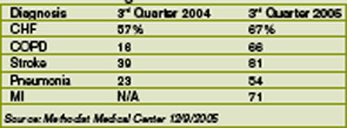
Methodist Medical ranks #1 in Tennessee on its compliance with Medicare core measures. Coletta Manning, RN, MHA, Methodist’s director of outcomes management, provided these data to show how they did it: (Table 1)
Manning cites this caveat in interpreting the data: When a patient is admitted and a diagnosis associated with a core measure is not made until the second day of hospitalization, the case isn’t considered in compliance. An example is abdominal pain that on further examination turns out to be caused by a myocardial infarction. If that were the case, the core measure of giving aspirin immediately would not be met because the diagnosis was not made within the first 24 hours of admission. “We get dinged sometimes,” says Manning.
Internally, the hospitalists measure their performance on compliance with the group’s own care design (critical pathway) utilization. Dr. Frost is pleased with the hospitalists’ growing acceptance of care designs and is working with Team Health on a sophisticated computer system to help the hospitalists use
If a busy doctor is a happy doctor, then the hospitalists in the Methodist Medical Group at Methodist Hospital in Oak Ridge, Tenn., are ecstatic. Chris Frost, MD, the hospital medicine group’s chief hospitalist and medical director, has seen the group’s average daily census climb to 70, then 90, and now 100 patients. Fortunately, the group is growing, too, from nine full-time employees in late October 2005 to 12 before year-end, a projection for 15 by first quarter 2006, and several intensivists to help by March.
The hospital itself has grown as well: It’s in the midst of a $40 million renovation and expansion started in 2004. By August 2006, Methodist Medical will have 230 new private rooms, 12 beds in a new acute care unit, and an emergency department twice the size of its predecessor.
—Chris Frost, MD
How It Started
Methodist Medical Group has been shaped by Oak Ridge’s traditions and demographics. Nestled between the Great Smokey and Cumberland Mountains, Oak Ridge was one of three research and production sites for the Manhattan Project (an effort by the United States, in conjunction with Canada and the United Kingdom to develop nuclear weapons). At its height, Oak Ridge had 75,000 inhabitants. Now its population is 28,000, with a Medicare age group that comprises 42% more of its population than the average Tennessee city. It continues as a magnet for scientists, and its physicians—both office-based and hospitalists—form a close-knit community.
Given its dynamics and demographics, when Oak Ridge’s primary care physicians asked Methodist Hospital in 1993 for help in admitting their patients, the hospital responded affirmatively. Hospital President Jan McNally, BSN, MHA, recalls how things unfolded. “Dr. Richard Dew, a highly respected primary practice physician with a busy office, wanted to change his life. His son had died and he decided to close his office, but he wanted to stay in medicine in Oak Ridge. He agreed to practice inpatient medicine at Methodist,” she explains. “The beauty of it was that he was universally respected. Barriers to the program fell because the admitting doctors and the patients all knew him.”
When Dr. Dew retired in 2002, Anthony Garton, MD, who closed his solo practice to join Dr. Dew as Methodist’s second hospitalist, stayed on. Dr. Garton became a hospitalist because “office procedures just brought me to break even financially. Only the things I did that didn’t have office overhead, such as being medical director at a nursing home and doing physicals for Boeing, made sense financially.” Dr. Garton, who worked with a nephrology group for 13 years, made a smooth transition to a hospitalist career.
From the hospitalist program’s inception, Methodist turned to Team Health, Inc., of Knoxville, an outsourcer of medical personnel, for staffing. Team Health Vice President Kenneth Burns saw a natural fit; his firm already supplied Methodist’s emergency department physicians.
“We understood the problems faced by Methodist’s ED docs,” says Burns. “Patients got stuck there and couldn’t be admitted rapidly to inpatient floors.”
He identified the hospitalist’s necessary skills as an ability to cooperate with community physicians, and an interest in improving processes to boost care quality and decrease costs. Methodist’s hospitalists have been independent contractors since the program’s inception. They receive hourly wages plus incentives based on productivity and metrics negotiated with hospital administrators.
Team Health recently rethought the model as potential recruits balked. In 2006 Methodist’s hospitalists became employees, with health benefits and defined contribution plans. Hospitalist Helen Bidawid, MD, says being employees improves recruiting because many doctors—particularly those just out of residency—find getting loans, buying health insurance, and other business associated with independent contractor status troublesome.
Symbiosis
The relationship of Methodist’s hospitalist program to Oak Ridge’s community physicians has changed over the years. Early on the group hired a hospitalist Dr. Garton describes as “very bright, knew his medicine, and would wow them in academia, but he antagonized the local docs. He left after one year and that was good because our census got low.”
Tact wasn’t that hospitalist’s long suit, and the community doctors who were uncomfortable with him didn’t refer many patients to the hospitalists. With the odd man out, hiring new hospitalists such as Joel Perkerson, MD, put the program back on track.
Dr. Perkerson left an office practice he had been in for 12 years. “I was drowning in paperwork, and it was so frustrating,” he says. “I couldn’t get my homework done or help my son, who was struggling academically.”
Having been both an office- and a hospital-based physician in Oak Ridge, he says it’s too hectic to do both. Being a hospitalist is challenging enough.
“It’s like the movie ‘Field of Dreams.’ Build it, and they will come. Demand keeps growing for our services and we all work a lot of hours,” he says, crediting the increasing load both to Dr. Frost’s leadership, which has made the subspecialists comfortable with the hospitalists’ management of medically complex patients, and to ex-TennCare patients flooding the emergency department. (See “When Politics Collide with Healthcare,” p. 38.)
Under Dr. Frost’s direction (he took over as medical director in 2003 after being recruited as assistant medical director), the hospitalist program has thrived. Dr. Frost has built relationships with Oak Ridge’s subspecialists, particularly pulmonologists, hematologists, oncologists, gastroenterologists, and orthopedists. Typical of Dr. Frost’s leadership is his response to Oak Ridge’s only pulmonary group imploding from six physicians to two. The hospitalists now admit all the group’s patients and Dr. Frost worked with Team Health to recruit intensivists for those very ill patients.
“I’m very pleased with the growth of the hospitalist program,” says Dr. Frost. “We feel empowered that more and more primary care doctors and subspecialists are allowing us to admit and take an active role in managing their patients.”
Tackling one problem that has bedeviled other hospitalist programs, Jan McNally has added a mechanism to ensure cooperation from referring doctors. She expects referring subspecialists to come for consults ASAP when a hospitalist calls.
“We must have that commitment from specialists,” she says. “We have about 15 percent who are laggards, but we will impose disciplinary action if they don’t come when called.”
Dr. Frost favors specialists willing to turn their patients over to the hospitalists in order to improve care quality. He diligently writes care guidelines, focuses on core measures, has deepened discussion of end-of-life care issues, and built such strong esprit de corps that the hospitalists willingly work long and irregular schedules until more physicians arrive. Helen Bidawid, MD, who has been a Methodist hospitalist for about a year, enjoys the hospitalist group, doesn’t mind pitching in to support her colleagues, and says “we function very well together. We watch out for all of our patients, share our responsibilities, and ask each other for help.”
Dr. Bidawid, who was in a non-supportive hospitalist group before her current position, asked herself before she arrived at Methodist: “’Will I be nurtured here or thrown to the wolves?’ Fortunately, I found a very supportive environment.”
With the course set, Dr. Frost still has challenges ahead. There’s growing patient volume, more complex cases to co-manage, carve-outs such as cardiology, neurology, and stroke care, and TennCare disenrollees to contend with.
“Our goal is to add value to Methodist Medical Center,” he says. “As a 24/7 hospitalist program, we help the medical community to be more profitable by enabling them to see more patients in the office and doing more procedures in the hospital. Better communication between physicians, patients, and their families benefits everyone and, we hope, will grow Methodist’s market share.” TH
Marlene Piturro is based in New York.
Quality Counts
Methodist Medical ranks #1 in Tennessee on its compliance with Medicare core measures. Coletta Manning, RN, MHA, Methodist’s director of outcomes management, provided these data to show how they did it: (Table 1)
Manning cites this caveat in interpreting the data: When a patient is admitted and a diagnosis associated with a core measure is not made until the second day of hospitalization, the case isn’t considered in compliance. An example is abdominal pain that on further examination turns out to be caused by a myocardial infarction. If that were the case, the core measure of giving aspirin immediately would not be met because the diagnosis was not made within the first 24 hours of admission. “We get dinged sometimes,” says Manning.
Internally, the hospitalists measure their performance on compliance with the group’s own care design (critical pathway) utilization. Dr. Frost is pleased with the hospitalists’ growing acceptance of care designs and is working with Team Health on a sophisticated computer system to help the hospitalists use