User login
Surveillance Instead of Surgery for Low-Risk DCIS?
At 2 years, investigators on the COMET trial found no clinically meaningful difference in the rates of ipsilateral invasive breast cancer among women randomized to active surveillance vs standard upfront surgery with or without radiation.
The 2-year findings suggest that surveillance is safe in the short term.
“While these results are provocative, I don’t think they’re quite practice-changing yet,” said lead investigator Shelley Hwang, MD, a surgical breast oncologist at Duke University in Durham, North Carolina, who presented the findings at the San Antonio Breast Cancer Symposium (SABCS) 2024.
For one thing, it generally takes longer than 2 years for DCIS to convert to invasive cancer, so it will be important to wait for the planned analyses at 5, 7, and 10 years to make sure there isn’t an excess number of invasive breast cancers in the surveillance arm, Hwang said.
If the results prove durable, however, the findings will likely be “practice-changing” for women who were at least 40 years old and had grade 1 or 2 hormone receptor–positive DCIS at low risk for conversion, Hwang said.
The goal of active surveillance is to prevent unnecessary treatment. During surveillance, lesions are monitored for changes that indicate conversion to more advanced disease, at which point guideline-concordant care begins.
Although DCIS can convert to invasive breast cancer, this doesn’t always happen. As a result, upfront surgery and radiation aren’t necessary for some women.
The COMET trial aimed to determine the short-term safety of an active monitoring approach compared with guideline-concordant care in patients with low-risk DCIS.
The prospective, randomized noninferiority trial included women aged 40 years or older with a new diagnosis of HR–positive grade 1 or grade 2 DCIS without invasive cancer from 100 US Alliance Cancer Cooperative Group clinical trial sites.
In the trial, 484 women with DCIS were randomized to active surveillance — breast mammography and physical exam every 6 months — and 473 were randomized to standard upfront surgery with or without radiation. Overall, 15.7% of participants were Black and 75.0% were White.
Patients in either group could elect to have endocrine therapy, typically over a 5-year period (71% of women in the active monitoring group and 65.5% in the surgery group opted for endocrine therapy).
At 2 years, the cumulative rate of ipsilateral invasive breast cancer was 4.2% in the surveillance group vs 5.9% in the upfront surgery arm.
The study also included a planned per-protocol analysis among 673 patients who strictly followed the study protocol — 246 in the guideline-concordant care group who had received surgery by 6 months and 427 in the surveillance group who initiated the active monitoring protocol at 6 months.
With almost half of patients randomized to surgery declined to have it, which indicates that patients are interested in active monitoring, Hwang said.
At 2 years, the cumulative rate of invasive breast cancer was 3.1% in the active surveillance group vs 8.7% in the upfront surgery arm.
Among patients receiving endocrine therapy, the rate of invasive cancer was 7.15% in the surgery group and 3.21% in the surveillance arm.
Endocrine therapy “may have resulted in a reduced rate of invasive cancer in the active monitoring group,” the study authors noted.
These findings bring up the question of whether endocrine therapy might be just as good as surgery for low-risk DCIS, Hwang added. Given that one third of women undergo mastectomy for DCIS, “I think it’s not an inconsequential question,” Hwang said.
The findings, however, also suggest that surveillance sometimes leaves invasive cancer behind, Hwang explained. Nearly all invasive cancers in the surgery group were found during the initial operation , which may explain the slightly higher rates of invasive cancers in this group. Had the active monitoring group undergone surgery as well, the incidence of invasive cancer may have been the same in both arms, Hwang said.
However, when invasive cancers were removed, there were no significant differences in tumor size, node status, or tumor grade between the two groups, suggesting that there might not be a clinical penalty for delayed intervention with active monitoring, Hwang said.
With more than 10% of patients in the surgery group opting for mastectomy, compared with 1.8% in the active monitoring group, the active monitoring approach may not increase the likelihood of an eventual need for more extensive surgery, the COMET authors explained.
What Strategy Do Patients Prefer?
A companion analysis of patient-reported outcomes in COMET found no meaningful differences in quality of life, symptoms, or anxiety among patients who opted for surveillance over surgery. Results from questionnaires on quality of life, anxiety, depression, and breast cancer concerns were comparable between the two groups, with no evidence of a substantial impact of one approach over the other at 2 years.
“The results of this secondary analysis suggest that the lived experiences of individuals with low-risk DCIS are similar during early follow-up regardless of treatment allocation,” the COMET investigators concluded.
Overall, the findings from COMET provide reassuring short-term data, said Neil Iyengar, MD, a medical breast oncologist at Memorial Sloan Kettering Cancer Center in New York City.
DCIS is not an aggressive cancer, and it’s not going to invade any time soon, so patients have time to consider their options, Iyengar told Medscape Medical News.
The 2-year findings from COMET also help inform patient discussions. “I can tell patients if they decide not to have surgery what the likelihood is that they are going to convert into invasive cancer” after 2 years, he said.
COMET was published in JAMA, and the PRO analysis was published in JAMA Oncology to coincide with the study presentations.
COMET is funded by the Patient-Centered Outcomes Research Institute and others. Hwang is a consultant for Merck and on the advisory board of Clinetic, Exai Bio, and Havah Therapeutics. Iyengar is an advisor and/or researcher for AstraZeneca, Novartis, Pfizer, and other companies.
A version of this article first appeared on Medscape.com.
FROM SABCS 2024
FDA Approves Cosibelimab for Cutaneous SCC
The programmed death ligand-1 (PD-L1)–blocking antibody is the first and only treatment of its kind approved for advanced CSCC, according to a Checkpoint Therapeutics press release. The FDA approval was based on findings from the multicenter, open-label Study CK-301-101 trial of 109 patients.
In that trial, the objective response rate (ORR) was 47% in 78 patients with metastatic CSCC and 48% in 31 patients with locally advanced CSCC. Median duration of response (DOR) in treated patients was not reached in those with metastatic disease and was 17.7 months in those with locally advanced disease, according to the FDA approval notice.
Adverse reactions occurring in at least 10% of patients included fatigue, musculoskeletal pain, rash, diarrhea, hypothyroidism, constipation, nausea, headache, pruritus, edema, localized infection, and urinary tract infection.
The recommended treatment dose, according to the prescribing information, is 1200 mg given as an intravenous infusion over 60 minutes every 3 weeks until disease progression or unacceptable toxicity.
The agent offers “a differentiated treatment option versus available therapies by binding to PD-L1, rather than programmed death receptor-1 (PD-1), to release the inhibitory effects of PD-L1 on the anti-tumor immune response,” Checkpoint Therapeutics president and chief executive officer James Oliviero stated in the company press release.
The agent has also “demonstrated the ability to induce antibody-dependent cell-mediated cytotoxicity, another potential differentiating feature of the drug compared to existing marketing therapies for CSCC,” Oliviero noted.
“CSCC is the second most common form of skin cancer, and those diagnosed with advanced disease that has recurred or metastasized face a poor prognosis,” stated Emily Ruiz, MD, academic director of the Mohs and Dermatologic Surgery Center at Brigham and Women’s Hospital and director of the High-Risk Skin Cancer Clinic at Dana-Farber Brigham Cancer Center.
“With its dual mechanisms of action and compelling safety profile, this promising drug will provide US oncologists with an important new immunotherapy option for the treatment of CSCC,” she added.
A version of this article appeared on Medscape.com.
Does Intensive Follow-Up Testing Improve Survival in CRC?
TOPLINE:
, according to findings from a secondary analysis.
METHODOLOGY:
- After curative surgery for CRC, intensive patient follow-up is common in clinical practice. However, there’s limited evidence to suggest that more frequent testing provides a long-term survival benefit.
- In the COLOFOL trial, patients with stage II or III CRC who had undergone curative resection were randomly assigned to either high-frequency follow-up (CT scans and CEA screening at 6, 12, 18, 24, and 36 months) or low-frequency follow-up (testing at 12 and 36 months) after surgery.
- This secondary analysis of the COLOFOL trial included 2456 patients (median age, 65 years), 1227 of whom received high-frequency follow-up and 1229 of whom received low-frequency follow-up.
- The main outcome of the secondary analysis was 10-year overall mortality and CRC–specific mortality rates.
- The analysis included both intention-to-treat and per-protocol approaches, with outcomes measured through December 2020.
TAKEAWAY:
- In the intention-to-treat analysis, the 10-year overall mortality rates were similar between the high- and low-frequency follow-up groups — 27.1% and 28.4%, respectively (risk difference, 1.3%; P = .46).
- A per-protocol analysis confirmed these findings: The 10-year overall mortality risk was 26.4% in the high-frequency group and 27.8% in the low-frequency group.
- The 10-year CRC–specific mortality rate was also similar between the high-frequency and low-frequency groups — 15.6% and 16.0%, respectively — (risk difference, 0.4%; P = .72). The same pattern was seen in the per-protocol analysis, which found a 10-year CRC–specific mortality risk of 15.6% in the high-frequency group and 15.9% in the low-frequency group.
- Subgroup analyses by cancer stage and location (rectal and colon) also revealed no significant differences in mortality outcomes between the two follow-up groups.
IN PRACTICE:
“This secondary analysis of the COLOFOL randomized clinical trial found that, among patients with stage II or III colorectal cancer, more frequent follow-up testing with CT scan and CEA screening, compared with less frequent follow-up, did not result in a significant rate reduction in 10-year overall mortality or colorectal cancer-specific mortality,” the authors concluded. “The results of this trial should be considered as the evidence base for updating clinical guidelines.”
SOURCE:
The study, led by Henrik Toft Sørensen, MD, PhD, DMSc, DSc, Aarhus University Hospital and Aarhus University, Aarhus, Denmark, was published online in JAMA Network Open.
LIMITATIONS:
The staff turnover at recruitment centers potentially affected protocol adherence. The inability to blind patients and physicians to the follow-up frequency was another limitation. The low-frequency follow-up protocol was less intensive than that recommended in the current guidelines by the National Comprehensive Cancer Network and the American Society of Clinical Oncology, potentially limiting comparisons to current standard practices.
DISCLOSURES:
The initial trial received unrestricted grants from multiple organizations including the Nordic Cancer Union, A.P. Møller Foundation, Beckett Foundation, Danish Cancer Society, and Swedish Cancer Foundation project. The authors reported no relevant conflicts of interest.
This article was created using several editorial tools, including AI, as part of the process. Human editors reviewed this content before publication. A version of this article first appeared on Medscape.com.
Tamoxifen Reduces Risk for Invasive Recurrence of Ductal Carcinoma
These findings come from an exploratory analysis of combined data from two clinical trials. They suggest that, for this select group of patients, the choice to forgo radiation following definitive surgery may be an acceptable option, assuming that they follow a full course of endocrine therapy.
“In the absence of survival impact for adjuvant therapy, the decision to recommend radiation therapy or endocrine therapy should be part of a shared decision process, and I think that this data helps us provide clearer data points to our patients to help them make choices between endocrine therapy and radiation therapy in the setting of good-risk [ductal carcinoma in situ],” said Jean L. Wright, MD, from the University of North Carolina at Chapel Hill.
She presented the findings in an oral abstract session and media briefing at San Antonio Breast Cancer Symposium (SABCS) 2024.
Trial Results Combined
Wright and colleagues looked at pooled data from two clinical trials that enrolled patients with low- or intermediate-grade DCIS with tumor size no larger than 2.5 cm, grade 1 or 2 lesions, and with surgical margins ≥ 3 mm.
The trials included NRG/RTOG 9804, with 317 patients who fit the “good-risk category,” and ECOG-ACRIN E5194, which included a cohort of 561 patients that met the good-risk definition used for the exploratory analysis.
In each trial, tamoxifen use was optional, and choices were tracked. In the NRG/RTOG trial, 66% of patients used tamoxifen and 34% did not. The respective percentages in the ECOG/ACRIN trial were 30% and 70%.
The majority of patients were adherent to the 5-year prescribed course of tamoxifen, Wright said.
Analysis Details
In the combined data, the median age of patients who used tamoxifen vs who did not use tamoxifen was 58 vs 61 years.
In all, 23% of women in both the tamoxifen yes or no groups were premenopausal, with the remainder either postmenopausal or of unknown menopausal status.
After a median follow-up of 14.85 years, the rate of 15-year ipsilateral breast recurrence (IBR) was 19% for patients who did not receive tamoxifen vs 11.4% for those who did. This translated into a hazard ratio for IBR on tamoxifen of 0.52 (P = .001).
Tamoxifen also reduced the risk for invasive recurrence in the same breast, with a 15-year invasive IBR rate of 11.5% in the no tamoxifen group vs 6% in the yes tamoxifen group.
However, as noted before, tamoxifen use was not associated with significant reduction in the risk for noninvasive DCIS recurrence in the same breast, as evidenced by a 15-year DCIS IBR rate of 8.1% without tamoxifen and 5.5% with tamoxifen, a difference that did not reach statistical significance.
A Surprising Result
One finding from the data that seemed to defy clinical wisdom was that tamoxifen use did not appear to significantly reduce the risk for events in the other breast. The 15-year rate of contralateral breast events was 8.8% in the no-tamoxifen group vs 5.6% in the yes tamoxifen group, a difference that was not statically significant.
“It was surprising that there was so little effect on contralateral disease,” commented Elinor Sawyer, MBBS, PhD, the invited discussant.
“But I think it’s really important, this decrease in ipsilateral invasive recurrence [with tamoxifen] because there are studies such as the Sloane study from the United Kingdom that show that if you develop an invasive recurrence after DCIS, you have a worse survival than those who develop a pure DCIS recurrence,” she said.
At the media briefing held prior to Wright’s presentation, moderator Virginia Kaklamani, MD, leader of the breast cancer program at UT Health San Antonio in Texas, also said that she found it surprising that tamoxifen did not reduce risk for contralateral breast cancer “since every study that we’ve done has shown that.”
“I also found that result a little bit surprising,” Wright agreed.
“I think the main feature that we want to focus on is that this was a group of patients with a very clear inclusion criteria of this good-risk DCIS, and even though the definition of good-risk DCIS included patients with [tumors] up to 2.5 cm in DCIS, we saw that, in reality, the patients enrolled had very small DCIS. So I’m wondering if it’s perhaps that it’s related to the fact that patients that were enrolled in these studies had really low-risk features and perhaps just had a lower risk of contralateral breast events as compared to a broader population of patients with DCIS,” she said.
The analysis by Wright and colleagues was supported by grants from the National Cancer Institute. Wright reported receiving honoraria from ASTRO and PER. Sawyer disclosed receiving grants/research support from Pfizer, Seagen, and IQIVIA. Kaklamani disclosed serving as a speaker and/or consultant for AstraZeneca, Celldex Therapeutics, Daiichi Sankyo, Genentech, Gilead, Lilly, Menarini, and Novartis and receiving research support from Eisai.
A version of this article first appeared on Medscape.com.
FROM SABCS 2024
Imlunestrant Shows PFS Benefit in Advanced Breast Cancer
according to recent findings from the EMBER-3 trial.
This progression-free survival benefit with imlunestrant did not extend to the overall population, but a combination of imlunestrant plus abemaciclib did lead to a significant improvement in progression-free survival compared with imlunestrant alone, regardless of patients’ ESR1 mutation status.
Lead author Komal Jhaveri, MD, a breast oncologist at Memorial Sloan Kettering Cancer Center in New York City, called the findings “encouraging.”
The phase 3 results raise the possibility of a second-line all-oral targeted therapy option for patients with ER–positive, HER2–negative advanced breast cancer, said Jhaveri, who presented the findings at the San Antonio Breast Cancer Symposium (SABCS) 2024, which were published simultaneously in The New England Journal of Medicine.
However, outside experts provided a note of caution that the trial design may limit how relevant the findings are to clinical practice.
First-line treatment for advanced ER–positive, HER2–negative breast cancer includes an aromatase inhibitor, such as exemestane, and a CDK4/6 inhibitor, such as abemaciclib. However, an ESR1 mutation may develop, which can undermine the effectiveness of the aromatase inhibitor. These patients may swap in a selective estrogen receptor degrader (SERD) — typically, the injectable fulvestrant — in place of the aromatase inhibitor.
Over the past several years, researchers have searched for a better agent than fulvestrant because this injectable drug has limited efficacy in patients with ESR1 mutations, and the monthly intramuscular shots are painful and inconvenient for patients.
The oral SERD imlunestrant is one such candidate.
The EMBER-3 trial initially randomized 661 patients after progression/recurrence evenly to either imlunestrant monotherapy (400 mg once daily) or a standard treatment arm that included either exemestane or fulvestrant, with 90% of these patients receiving fulvestrant. Investigators added a third combination arm shortly after the trial started, which included 213 patients who received imlunestrant plus abemaciclib.
About 60% of the overall population had received prior CDK4/6 inhibitors, primarily palbociclib and ribociclib. About 37% of the study population had ESR1 mutations.
Among patients with ESR1 mutations, imlunestrant monotherapy led to a significant improvement in median progression-free survival of 5.5 months vs 3.8 months in the standard care arm (P < .001). Among all patients, however, progression-free survival was no different between the two arms — 5.6 months in the imlunestrant group vs 5.5 months in the standard care group.
When comparing the two treatment arms in the overall population, the median progression-free survival was significantly better in patients who received imlunestrant plus abemaciclib — 9.4 months vs 5.5 months in the imlunestrant group (hazard ratio [HR] for progression or death, 0.57; P < .001). The progression-free survival benefit held across most patient subgroups, regardless of ESR1 mutation status, as well as among patients who had received a CDK4/6 inhibitor previously.
Data from other studies presented at SABCS indicate that another SERD, elacestrant, in combination with abemaciclib, may provide a similar progression-free survival benefit in this patient population. Elacestrant was approved by the US Food and Drug Administration (FDA) in January 2023 for second-line treatment of patients with advanced breast cancer and ESR1 mutations.
The EMBER trial also reported early overall survival findings. Although immature, overall survival trends favored imlunestrant over the standard treatment. The estimated overall survival at 18 months was 77% in the imlunestrant group and 58.6% in the standard therapy group among patients with ESR1 mutations (HR, 0.55), and 78.6% in the imlunestrant group vs 71.8% in the standard-therapy group for all patients (HR, 0.69).
Common all-grade adverse events with imlunestrant vs standard therapy included fatigue (22.6% vs 13.3%), diarrhea (21.4% vs 11.7%), and nausea (17% vs 13%). Grade 3 or higher anemia and neutropenia were low and similar in both arms.
All-grade diarrhea (86%) and nausea (49%) were more common with the combination of imlunestrant and abemaciclib.
The incidence of grade 3 or higher events was 17% with imlunestrant monotherapy, 21% for standard treatment, and 49% for imlunestrant plus abemaciclib.
EMBER-3 discussant Harold Burstein, MD, PhD, a breast oncologist at the Dana-Farber Cancer Institute, Boston, said that, overall, oral SERDs are starting to “break out from the ESR1 mutation box,” perhaps reflecting the idea that an agent more active than fulvestrant in combination with a non-cross–resistant CDK4/6 inhibitor like abemaciclib might lead to a better long-term outcome, regardless of ESR1 status.
A major limit of EMBER-3, however, is that it did not compare imlunestrant/abemaciclib with fulvestrant/abemaciclib, which would have been a true standard-of-care control, said Burstein.
Kathy Miller, MD, a breast oncologist at Indiana University, Indianapolis, agreed.
She was also concerned about the use of monotherapy in the standard care arm.
“Monotherapy hormone therapy is not what people would be treated with,” Miller said. Patients would typically get fulvestrant with either a targeted therapy or everolimus.
Without appropriate controls, “the data are impossible to interpret” in the context of current practice, Miller told Medscape Medical News.
Eli Lilly, maker of imlunestrant, funded, designed, and largely conducted the trial. Jhaveri is a consultant and researcher for the company. Burstein and Miller had no disclosures. Miller is an editorial advisor for Medscape Oncology.
A version of this article appeared on Medscape.com.
FROM SABCS 2024
Skin Cancer Screening: Biopsy-Free Technology Advancing
NEW YORK CITY — now in routine use at his own institution.
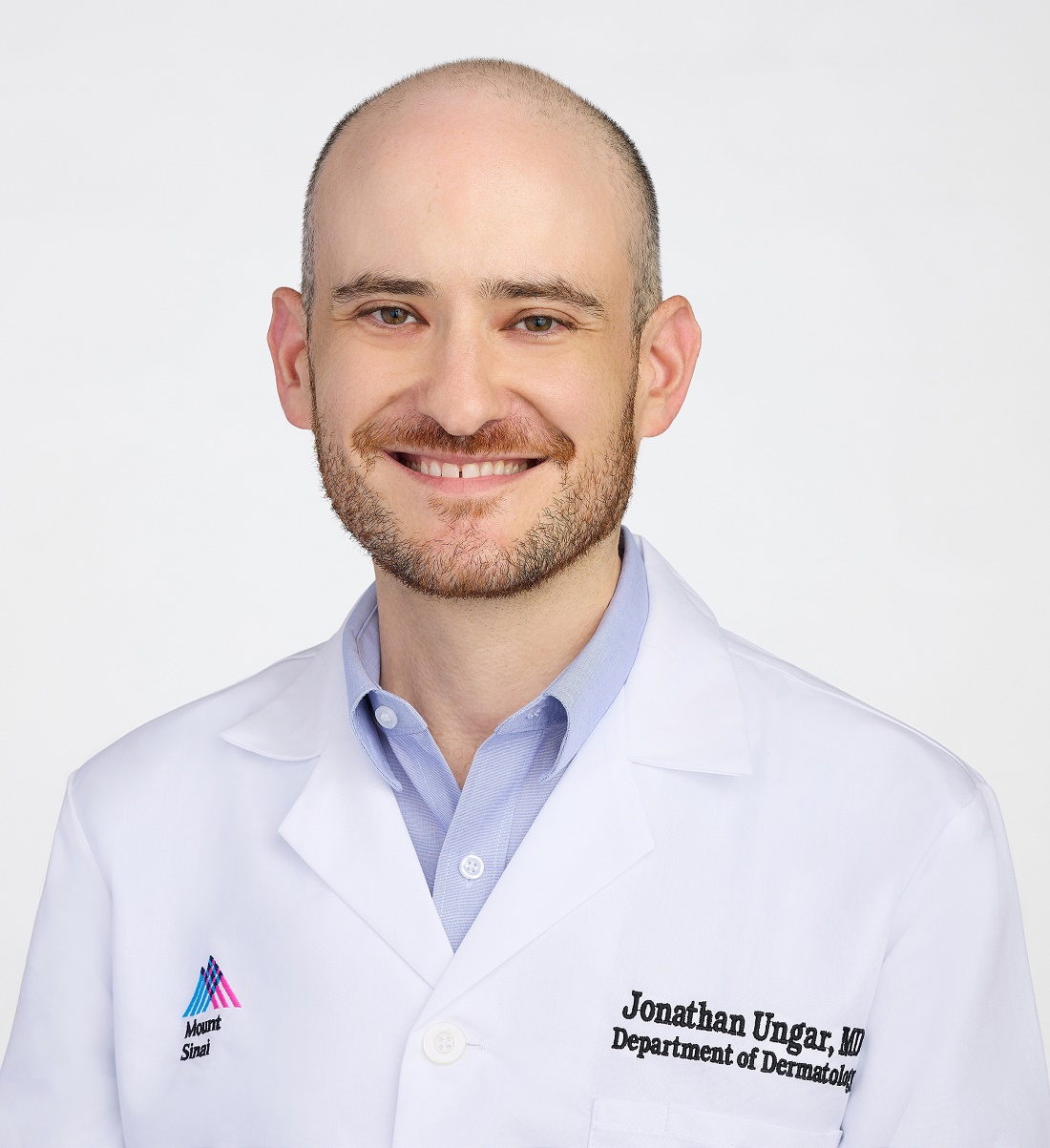
For skin cancer screening, existing and coming technologies represent “the future of dermatology,” but “we can and should be [already] trying to incorporate these into routine practice,” said Jonathan Ungar, MD, assistant professor of dermatology at the Icahn School of Medicine at Mount Sinai, New York City.
Technologies such as electrical impedance spectroscopy (EIS), optical coherence tomography (OCT), and reflectance confocal microscopy (RCM) have immediate utility for improving skin cancer detection with fewer biopsies, but this is just the beginning, according to Ungar, who is also medical director of the Kimberly and Eric J. Waldman Melanoma and Skin Cancer Center at Mount Sinai, New York City.
“There is going to be a day when we are not cutting to make a diagnosis,” he said during a presentation at the 27th Annual Winter Symposium — Advances in Medical and Surgical Dermatology (MSWS) 2024.
Four Noninvasive Tools Are in Routine Use
Each of these technologies, along with total body photography (TBP), is currently in use at Mount Sinai as well as other tertiary centers to improve diagnostic accuracy at the same time they reduce invasive tests. The initial excitement about these technologies was based on their potential to avoid biopsy in cosmetically sensitive areas, but Ungar suggested that wider application is being driven by better rates of detection, less morbidity, and improved patient satisfaction.
Patients are happy to avoid invasive procedures whenever they can, Ungar noted. In addition to concern about pain or discomfort and a small but measurable risk for infection, patients face a wound that requires healing and the potential for an enduring scar whether the histology is positive for a malignancy.
While none of the four technologies Ungar outlined typically provide a yes or no answer regarding the presence of a malignancy, they do improve diagnostic accuracy with a lower rate of biopsy.
Each Noninvasive Tool Reduces Biopsy Rates
In the case of EIS, for example, the impedance of a painless and harmless electrical current directed into the skin with a handheld probe differentiates normal from abnormal skin through an EIS algorithm. Ungar said it does not require training. A result negative for an abnormality has about a 90% predictive value, and it means that a biopsy can be avoided if there are no other reasons for suspicion.
With a price estimated in the thousands of dollars, the device and software are “not so expensive,” particularly when the tool results in fewer biopsies, Ungar noted.
OCT has a similar profile. Again, used as an adjunct to other types of evaluations, including a history and visual inspection, this helps in modulating suspicion of malignancy. In published studies, OCT has proven superior to dermatoscopy for cancer detection. Citing a 14-study meta-analysis, Ungar said that the sensitivity of OCT for melanoma exceeds, and the specificity approaches, 90%. For basal cell cancers, it is even better.
RCM involves directing a laser into the skin to detect abnormal cells that reflect light. It enables visualization of the skin by layers to the papillary dermis in a detail that is comparable with histology, according to Ungar. Imaging performed with the device used at Mount Sinai (VivaScope 1500, Caliber Imaging & Diagnostics) is reimbursed by Medicare.
Once comfortable with the technology, scanning and interpretation take slightly more time than that required of EIS or OCT, but, like the others, it is painless and helpful for determining whether further evaluation is needed, according to Ungar.
“It is extremely useful in reducing the number of biopsies,” whether melanoma or basal cell malignancies, he said.
Total Body Photograph Helps With Serial Screens
While not specifically a diagnostic tool, TBP can also play a role in reducing biopsies through its highly efficient ability to document the evolution of lesions over time.
As its name implies, essentially the entire body surface is captured by multiple cameras mounted in a circle around the patient. Unlike sequential photos that require far more time to take and store and are challenging to organize and retrieve, the device used at Mount Sinai (Vectra Wb180 1360, Canfield Scientific) can complete the photos in about 2 minutes.
Software for organizing and storing the photos, to which dermatoscope images of individual lesions can be attached if helpful, results in efficient retrieval of photos at sequential visits for evaluating change in any specific lesion.
“It is very easy to use,” according to Ungar, who noted that although the underlying idea is not, the technology of taking, storing, and retrieving photographs has been “perhaps perfected” with this approach.
Noninvasive Screening Training Is Appropriate
Year after year, dermatology residents undergo intensive instruction to master the traditional methods of skin examination with the naked eye and the help of a dermatoscope, but Ungar considers the noninvasive tools to be another step forward. They lower miss rates while reducing the need for histopathology.
Adding these new technologies to routine patient care resonates for many experts, even if the protocols of when to use with the tool are not well established.
Angela J. Lamb, MD, an associate professor of dermatology at Mount Sinai, who has been following the work of Ungar with interest, sees merit in his argument. Not surprisingly, she thinks that any approach shown to boost skin cancer detection is something that deserves attention, but she thinks the effort to safely eliminate biopsies with a low likelihood of a positive finding cannot be ignored.
“Patients want to avoid biopsies when they can,” Lamb told this news organization, and she does not think this is limited to biopsies on the face or other cosmetically sensitive areas.
As a result, she said that she does see the rationale for incorporating the newer technologies into routine care and called this an “important” effort to improve the patient experience as well as reduce missed lesions.
Ungar reported financial relationships with AbbVie, Bristol-Myers Squibb, Castle Biosciences, Dermavant, Janssen Pharmaceuticals, Menlo Therapeutics, Mitsubishi Tanabe Pharma America, and UCB. Lamb reported no potential conflicts of interest.
A version of this article first appeared on Medscape.com.
NEW YORK CITY — now in routine use at his own institution.
For skin cancer screening, existing and coming technologies represent “the future of dermatology,” but “we can and should be [already] trying to incorporate these into routine practice,” said Jonathan Ungar, MD, assistant professor of dermatology at the Icahn School of Medicine at Mount Sinai, New York City.
Technologies such as electrical impedance spectroscopy (EIS), optical coherence tomography (OCT), and reflectance confocal microscopy (RCM) have immediate utility for improving skin cancer detection with fewer biopsies, but this is just the beginning, according to Ungar, who is also medical director of the Kimberly and Eric J. Waldman Melanoma and Skin Cancer Center at Mount Sinai, New York City.
“There is going to be a day when we are not cutting to make a diagnosis,” he said during a presentation at the 27th Annual Winter Symposium — Advances in Medical and Surgical Dermatology (MSWS) 2024.
Four Noninvasive Tools Are in Routine Use
Each of these technologies, along with total body photography (TBP), is currently in use at Mount Sinai as well as other tertiary centers to improve diagnostic accuracy at the same time they reduce invasive tests. The initial excitement about these technologies was based on their potential to avoid biopsy in cosmetically sensitive areas, but Ungar suggested that wider application is being driven by better rates of detection, less morbidity, and improved patient satisfaction.
Patients are happy to avoid invasive procedures whenever they can, Ungar noted. In addition to concern about pain or discomfort and a small but measurable risk for infection, patients face a wound that requires healing and the potential for an enduring scar whether the histology is positive for a malignancy.
While none of the four technologies Ungar outlined typically provide a yes or no answer regarding the presence of a malignancy, they do improve diagnostic accuracy with a lower rate of biopsy.
Each Noninvasive Tool Reduces Biopsy Rates
In the case of EIS, for example, the impedance of a painless and harmless electrical current directed into the skin with a handheld probe differentiates normal from abnormal skin through an EIS algorithm. Ungar said it does not require training. A result negative for an abnormality has about a 90% predictive value, and it means that a biopsy can be avoided if there are no other reasons for suspicion.
With a price estimated in the thousands of dollars, the device and software are “not so expensive,” particularly when the tool results in fewer biopsies, Ungar noted.
OCT has a similar profile. Again, used as an adjunct to other types of evaluations, including a history and visual inspection, this helps in modulating suspicion of malignancy. In published studies, OCT has proven superior to dermatoscopy for cancer detection. Citing a 14-study meta-analysis, Ungar said that the sensitivity of OCT for melanoma exceeds, and the specificity approaches, 90%. For basal cell cancers, it is even better.
RCM involves directing a laser into the skin to detect abnormal cells that reflect light. It enables visualization of the skin by layers to the papillary dermis in a detail that is comparable with histology, according to Ungar. Imaging performed with the device used at Mount Sinai (VivaScope 1500, Caliber Imaging & Diagnostics) is reimbursed by Medicare.
Once comfortable with the technology, scanning and interpretation take slightly more time than that required of EIS or OCT, but, like the others, it is painless and helpful for determining whether further evaluation is needed, according to Ungar.
“It is extremely useful in reducing the number of biopsies,” whether melanoma or basal cell malignancies, he said.
Total Body Photograph Helps With Serial Screens
While not specifically a diagnostic tool, TBP can also play a role in reducing biopsies through its highly efficient ability to document the evolution of lesions over time.
As its name implies, essentially the entire body surface is captured by multiple cameras mounted in a circle around the patient. Unlike sequential photos that require far more time to take and store and are challenging to organize and retrieve, the device used at Mount Sinai (Vectra Wb180 1360, Canfield Scientific) can complete the photos in about 2 minutes.
Software for organizing and storing the photos, to which dermatoscope images of individual lesions can be attached if helpful, results in efficient retrieval of photos at sequential visits for evaluating change in any specific lesion.
“It is very easy to use,” according to Ungar, who noted that although the underlying idea is not, the technology of taking, storing, and retrieving photographs has been “perhaps perfected” with this approach.
Noninvasive Screening Training Is Appropriate
Year after year, dermatology residents undergo intensive instruction to master the traditional methods of skin examination with the naked eye and the help of a dermatoscope, but Ungar considers the noninvasive tools to be another step forward. They lower miss rates while reducing the need for histopathology.
Adding these new technologies to routine patient care resonates for many experts, even if the protocols of when to use with the tool are not well established.
Angela J. Lamb, MD, an associate professor of dermatology at Mount Sinai, who has been following the work of Ungar with interest, sees merit in his argument. Not surprisingly, she thinks that any approach shown to boost skin cancer detection is something that deserves attention, but she thinks the effort to safely eliminate biopsies with a low likelihood of a positive finding cannot be ignored.
“Patients want to avoid biopsies when they can,” Lamb told this news organization, and she does not think this is limited to biopsies on the face or other cosmetically sensitive areas.
As a result, she said that she does see the rationale for incorporating the newer technologies into routine care and called this an “important” effort to improve the patient experience as well as reduce missed lesions.
Ungar reported financial relationships with AbbVie, Bristol-Myers Squibb, Castle Biosciences, Dermavant, Janssen Pharmaceuticals, Menlo Therapeutics, Mitsubishi Tanabe Pharma America, and UCB. Lamb reported no potential conflicts of interest.
A version of this article first appeared on Medscape.com.
FROM MSWS 2024
Some Antihypertensives Linked to HCC Risk in Patients With MASLD and Cirrhosis
SAN DIEGO — according to new research.
In particular, the use of calcium channel blockers (CCBs), angiotensin-converting enzyme (ACE) inhibitors, and angiotensin receptor blockers (ARBs) was associated with a higher risk of developing HCC, compared with not using these medications.
About half of patients with MASLD have hypertension, and the use of antihypertensives in these patients is beneficial to reduce the risk for cardiovascular disease and complications related to MASLD, said lead author Ahmed Elhariri, MD, a research fellow at the University of Texas MD Anderson Cancer Center, Houston, who conducted the study as a research assistant in gastroenterology and hepatology at the Baylor College of Medicine, also in Houston.
However, previous studies have suggested a possible link between these medications and cancer development, “especially CCBs and breast and lung cancer,” said Elhariri, who presented the findings at The Liver Meeting 2024: American Association for the Study of Liver Diseases (AASLD).
Analyzing Potential Risks
In a case-control study, Elhariri and colleagues analyzed antihypertensive medication use among patients with MASLD-induced HCC, as defined by histology or radiology based on the Liver Imaging Reporting & Data System, and control patients with MASLD but without HCC.
Between 2020 and 2024, the research team recruited 153 newly diagnosed HCC cases with different etiologies and 170 patients with MASLD but without HCC from Baylor College of Medicine’s outpatient clinics. For this study, they selected 47 age- and sex-matched pairs, all of whom had cirrhosis. Only those with a history of hypertension were included, however. Data on risk factors of metabolic syndrome (including diabetes) and HCC were collected, along with details about medication use such as metformin and statins.
A total of 42 patients with MASLD and HCC and 39 MASLD control individuals had a history of hypertension and were treated with antihypertensive medications. The mean age was 66.5 years for the HCC group and 63.5 years for the control group, and the mean body mass index (BMI) was 31.1 for the HCC group and 31.7 for the control group.
After adjusting for age, sex, BMI, Hispanic ethnicity, and use of other medications, patients taking CCBs had an increased HCC risk (odds ratio [OR], 2.76), compared with those not taking CCBs. Patients taking ACE inhibitors or ARBs also had an increased HCC risk (OR, 2.54), compared with those not taking ACE inhibitors or ARBs.
However, there wasn’t a statistically significant difference in HCC risk among patients taking beta-blockers (OR, 0.87).
“Patients with fatty liver in the presence of metabolic syndrome, especially in the presence of cirrhosis and antihypertensives, need to have stricter surveillance for liver cancer,” Elhariri said.
“We need to carefully review blood pressure medications in patients with MASLD and cirrhosis,” he said. CCBs, ACE inhibitors, and ARBs can be replaced with beta-blockers, “which have been shown to reduce progression of cirrhosis-related complications.”
Considering Clinical Implications
“Although our study showed some association between the use of some commonly used antihypertensives and the risk for HCC in this high-risk population, it is based on data collected retrospectively on a small number of selected patients with advanced liver disease,” Elhariri noted.
The associations and underlying mechanisms should be studied in larger populations and prospective trials, he said. “Until we have more data with a significantly larger sample size, it’s premature to raise the concern in the general population.”
“The cardiovascular benefits of controlling blood pressure far outweigh the risk of liver cancer in patients with metabolic syndrome,” Elhariri added.
In ongoing studies, researchers are investigating ways to improve patient outcomes and reduce the negative effects of cirrhosis-associated complications among patients with MASLD and metabolic dysfunction–associated steatohepatitis (MASH), Muhammad Ali Butt, MD, a hepatology fellow at Beth Israel Lahey Hospital & Medical Center in Burlington, Massachusetts, said in an interview.
Butt, who wasn’t involved with this study, presented separate research on statins in MASH patients with cirrhosis, which indicated statistically significant decreases in portal hypertension, thrombosis, hepatorenal syndrome, hepatic encephalopathy, and mortality.
“We know patients with MASLD- and MASH-associated cirrhosis commonly have other comorbidities, including high cardiovascular risks, diabetes, and hyperlipidemia,” he said. “All of these conditions indicate patients to be on other medications such as antihypertensives or statins. It’s important to know the role these medications play, especially given the high-risk profile of these patients.”
Elhariri and Butt reported no relevant disclosures.
A version of this article first appeared on Medscape.com.
FROM AASLD 2024
Melanoma: Study Addresses Impact of Indoor Tanning on Tumor Mutational Burden
TOPLINE:
in a retrospective cohort study. Higher TMB was linked to older age, head and neck tumors, and a history of nonmelanoma skin cancer (NMSC).
METHODOLOGY:
- Researchers conducted a retrospective cohort study at a tertiary care cancer center between 2013 and 2022.
- A total of 617 patients (median age at diagnosis, 61 years; 62.9% men) with melanoma who had next-generation sequencing data and indoor tanning bed exposure history available were included.
- Analysis involved multivariable modeling to evaluate the association between tanning bed use and TMB.
- Patients’ demographics, pathologic staging, TMB, and dermatologic history, including Fitzpatrick skin type, history of exposure to ultraviolet (UV) light, indoor tanning, NMSC, atypical nevi, and blistering sunburns, were considered for the analysis.
TAKEAWAY:
- About 22% of participants had an indoor tanning history. Indoor tanning exposure showed no association with TMB after adjustment for all possible predictors.
- A significant association was found between TMB and age at diagnosis, primary melanoma site, and history of NMSC (P < .001 for all).
- Patients with a history of atypical nevi demonstrated a significantly lower TMB than those without (log2 TMB, 3.89 vs 4.15; P = .01).
- Tumors of the head and neck exhibited a significantly higher TMB than those occurring in other primary sites, while skin-localized melanomas at diagnosis showed a significantly higher TMB than node-positive or metastatic stage III or IV tumors (log2 TMB, 3.88 vs 3.48; P = .005).
IN PRACTICE:
“Despite the known association between indoor tanning and melanoma risk,” the study did not find an association between indoor tanning and melanoma TMB, which “suggests that cumulative lifetime sun exposure may be a greater primary driver of TMB than intermittent radiation during indoor tanning,” the authors of the study wrote.
SOURCE:
The study was led by Grace B. Hanrahan, BA, of the Center for Melanoma Oncology, Dana-Farber Cancer Institute, Boston, Massachusetts, and was published online on December 11 in JAMA Dermatology.
LIMITATIONS:
The study was conducted at a tertiary referral center, potentially representing a higher-risk subset with more advanced disease than the broader population. Additionally, the retrospective collection of UV exposure history, including indoor tanning and blistering sunburns, may have introduced recall bias.
DISCLOSURES:
The authors did not disclose any funding information. No conflicts of interest were reported.
This article was created using several editorial tools, including artificial intelligence, as part of the process. Human editors reviewed this content before publication. A version of this article first appeared on Medscape.com.
Malpractice in the Age of AI
Instead of sitting behind a laptop during patient visits, the pediatrician directly faces the patient and parent, relying on an ambient artificial intelligence (AI) scribe to capture the conversation for the electronic health record (EHR). A geriatrician doing rounds at the senior living facility plugs each patient’s medications into an AI tool, checking for drug interactions. And a busy hospital radiology department runs all its emergency head CTs through an AI algorithm, triaging potential stroke patients to ensure they receive the highest priority. None of these physicians have been sued for malpractice for AI usage, but they wonder if they’re at risk.
In a recent Medscape report, AI Adoption in Healthcare, 224 physicians responded to the statement: “I want to do more with AI but I worry about malpractice risk if I move too fast.” Seventeen percent said that they strongly agreed while 23% said they agreed — a full 40% were concerned about using the technology for legal reasons.
Malpractice and AI are on many minds in healthcare, especially in large health systems, Deepika Srivastava, chief operating officer at The Doctors Company, told this news organization. “AI is at the forefront of the conversation, and they’re [large health systems] raising questions. Larger systems want to protect themselves.”
The good news is there’s currently no sign of legal action over the clinical use of AI. “We’re not seeing even a few AI-related suits just yet,” but the risk is growing, Srivastava said, “and that’s why we’re talking about it. The legal system will need to adapt to address the role of AI in healthcare.”
How Doctors Are Using AI
Healthcare is incorporating AI in multiple ways based on the type of tool and function needed. Narrow AI is popular in fields like radiology, comparing two large data sets to find differences between them. Narrow AI can help differentiate between normal and abnormal tissue, such as breast or lung tumors. Almost 900 AI health tools have Food and Drug Administration approval as of July 2024, discerning abnormalities and recognizing patterns better than many humans, said Robert Pearl, MD, author of ChatGPT, MD: How AI-Empowered Patients & Doctors Can Take Back Control of American Medicine and former CEO of The Permanente Medical Group.
Narrow AI can improve diagnostic speed and accuracy for other specialties, too, including dermatology and ophthalmology, Pearl said. “It’s less clear to me if it will be very beneficial in primary care, neurology, and psychiatry, areas of medicine that involve a lot of words.” In those specialties, some may use generative AI as a repository of resources. In clinical practice, ambient AI is also used to create health records based on patient/clinician conversations.
In clinical administration, AI is used for scheduling, billing, and submitting insurance claims. On the insurer side, denying claims based on AI algorithms has been at the heart of legal actions, making recent headlines.
Malpractice Risks When Using AI
Accuracy and privacy should be at the top of the list for malpractice concerns with AI. With accuracy, liability could partially be determined by use type. If a diagnostic application makes the wrong diagnosis, “the company has legal accountability because it created and had to test it specific to the application that it’s being recommended for,” Pearl said.
However, keeping a human in the loop is a smart move when using AI diagnostic tools. The physician should still choose the AI-suggested diagnosis or a different one. If it’s the wrong diagnosis, “it’s really hard to currently say where is the source of the error? Was it the physician? Was it the tool?” Srivastava added.
With an incorrect diagnosis by generative AI, liability is more apparent. “You’re taking that accountability,” Pearl said. Generative AI operates in a black box, predicting the correct answer based on information stored in a database. “Generative AI tries to draw a correlation between what it has seen and predicting the next output,” said Alex Shahrestani, managing partner of Promise Legal PLLC, a law firm in Austin, Texas. He serves on the State Bar of Texas’s Taskforce on AI and the Law and has participated in advisory groups related to AI policies with the National Institute of Standards and Technology. “A doctor should know to validate information given back to them by AI,” applying their own medical training and judgment.
Generative AI can provide ideas. Pearl shared a story about a surgeon who was unable to remove a breathing tube that was stuck in a patients’ throat at the end of a procedure. The surgeon checked ChatGPT in the operating room, finding a similar case. Adrenaline in the anesthetic restricted the blood vessels, causing the vocal cords to stick together. Following the AI information, the surgeon allowed more time for the anesthesia to diffuse. As it wore off, the vocal cords separated, easing the removal of the breathing tube. “That is the kind of expertise it can provide,” Pearl said.
Privacy is a common AI concern, but it may be more problematic than it should be. “Many think if you talk to an AI system, you’re surrendering personal information the model can learn from,” said Shahrestani. Platforms offer opt-outs. Even without opting out, the model won’t automatically ingest your interactions. That’s not a privacy feature, but a concern by the developer that the information may not help the model.
“If you do use these opt-out mechanisms, and you have the requisite amount of confidentiality, you can use ChatGPT without too much concern about the patient information being released into the wild,” Shahrestani said. Or use systems with stricter requirements that keep all data on site.
Malpractice Insurance Policies and AI
Currently, malpractice policies do not specify AI coverage. “We don’t ask right now to list all the technology you’re using,” said Srivastava. Many EHR systems already incorporate AI. If a human provider is in the loop, already vetted and insured, “we should be okay when it comes to the risk of malpractice when doctors are using AI because it’s still the risk that we’re ensuring.”
Insurers are paying attention, though. “Traditional medical malpractice law does require re-evaluation because the rapid pace of AI development has outpaced the efforts to integrate it into the legal system,” Srivastava said.
Some, including Pearl, believe AI will actually lower the malpractice risk. Having more data points to consider can make doctors’ jobs faster, easier, and more accurate. “I believe the technology will decrease lawsuits, not increase them,” said Pearl.
Meanwhile, How Can Doctors Protect Themselves From an AI Malpractice Suit?
Know your tool: Providers should understand the tool they’re deploying, what it provides, how it was built and trained (including potential biases), how it was tested, and the guidelines for how to use it or not use it, said Srivastava. Evaluate each tool, use case, and risk separately. “Don’t just say it’s all AI.”
With generative AI, users will have better success requesting information that has been available longer and is more widely accessed. “It’s more likely to come back correctly,” said Shahrestani. If the information sought is fairly new or not widespread, the tool may try to draw problematic conclusions.
Document: “Document, document, document. Just making sure you have good documentation can really help you if litigation comes up and it’s related to the AI tools,” Srivastava said.
Try it out: “I recommend you use [generative AI] a lot so you understand its strengths and shortcomings,” said Shahrestani. “If you wait until things settle, you’ll be further behind.”
Pretend you’re the patient and give the tool the information you’d give a doctor and see the results, said Pearl. It will provide you with an idea of what it can do. “No one would sue you because you went to the library to look up information in the textbooks,” he said — using generative AI is similar. Try the free versions first; if you begin relying on it more, the paid versions have better features and are inexpensive.
A version of this article first appeared on Medscape.com.
Have Your Cake and Eat It, Too: Findings Based on Ingredients in Christmas Desserts From The Great British Bake Off
This transcript has been edited for clarity.
Hello. I’m David Kerr, professor of cancer medicine at University of Oxford. As I become, sadly, older, I’ve become much more interested in the concept of cancer prevention than cancer treatment. Of course, I’m still a practicing cancer physician and researcher. That’s my daily bread and butter. But prevention is important.
There’s a really interesting article in the Christmas edition of The BMJ. This is an opportunity for us to take good science, but lighthearted science, to titillate and amuse our Christmas readers. This is a nice article from the States led by Joshua Wallach. As I say, this brings together good science in a sometimes absurd setting. I’ll read its title: “Association of Health Benefits and Harms of Christmas Dessert Ingredients in Recipes From The Great British Bake Off: Umbrella Review of Umbrella Reviews of Meta-analyses of Observational Studies.”
It’s obviously a very strong statistical underpinning from this group from Yale, predominantly — a half-decent university, as those of us from Oxford would have to admit. They used The Great British Bake Off website, Embase, Medline, and Scopus. They looked at the whole host of umbrella reviews and so on.
They were interested in looking at the relative balance of dangerous and protective ingredients that were recommended in Christmas desserts on this immensely popular television show called The Great British Bake Off. Some of you have watched it and have enjoyed watching the trials and tribulations of the various contestants.
They looked at 48 recipes for Christmas desserts, including cakes, biscuits, pastries, puddings, and conventional desserts. Of all these, there were 178 unique ingredients. Literature research then parsed whether these ingredients were good for you or bad for you.
It was very interesting that, when they put the summary together, the umbrella review of umbrella reviews of meta-analyses compressed together, it was good news for us all. Recipes for Christmas desserts, particularly from The Great British Bake Off — which should be enormously proud of this — tend to use ingredient groups that are associated with reductions rather than increases in the risk for disease. Hurrah!
This means that, clearly, Christmas is a time in which those of us who can, tend to overindulge in food. The granddad falling asleep with a full tummy, sitting with the family in front of a hot fire — all of us can remember and imagine all of that.
Perhaps the most important takeaway point from this observationally, critically important study is that, yes — at Christmas time, enjoy the dessert. You can have your cake and eat it, too. You heard it here. It’s philosophically true and statistically proven: You can have your cake and eat it.
Thanks for listening. I’d be very interested in your own recipes, and whether we think that the American Thanksgiving desserts correlate with British Christmas desserts in some way and are beneficial to your health.
Have a look at this article that is cleverly, wittily written. As always, Medscapers, for the time being, thanks for listening. Over and out.
Dr Kerr, Professor, Nuffield Department of Clinical Laboratory Science, University of Oxford; Professor of Cancer Medicine, Oxford Cancer Centre, Oxford, United Kingdom, has disclosed ties with Celleron Therapeutics, Oxford Cancer Biomarkers (Board of Directors); Afrox (charity; Trustee); GlaxoSmithKline and Bayer HealthCare Pharmaceuticals (Consultant); Genomic Health; Merck Serono, Roche.
A version of this article appeared on Medscape.com.