User login
Hydroxyurea proves comparable to transfusion in SCD

Credit: St Jude Children’s
Research Hospital
Treatment with hydroxyurea is a comparable alternative to blood transfusions for reducing the risk of stroke in children with sickle cell disease (SCD), according to the National Heart Lung and Blood Institute (NHLBI).
Early results of the phase 3 TWiTCH trial showed that daily doses of hydroxyurea lower the transcranial Doppler (TCD) blood velocity in children with SCD to a similar degree as blood transfusions, thereby decreasing the risk of stroke.
Based on these findings, the NHLBI has terminated the trial early, about a year before it was originally scheduled to end.
“Early results indicate that TWiTCH is a success,” said Russell E. Ware, MD, PhD, principal investigator of the study and director of hematology at Cincinnati Children’s Hospital Medical Center.
“A group of outside experts has been reviewing the TWiTCH data every few months to ensure the safety of children in the clinical trial and to monitor the data. This group met recently and, after careful consideration of the interim data results, recommended that the study be stopped since hydroxyurea worked as well as transfusions to lower TCD velocities.”
The NHLBI said it cannot release results from the TWiTCH trial, but some details are available. Researchers enrolled 121 children between the ages of 4 and 16 who had SCD and abnormally elevated TCD velocities.
Half of patients received transfusions, and the other half received daily hydroxyurea, which has not yet been approved for children with SCD.
The clinical data-collection portion of the study was originally scheduled for the 24-month mark, but collection was stopped after only half of the children completed the treatment phase.
At the first interim analysis, a data and safety monitoring board found that hydroxyurea was not inferior to regular blood transfusions in lowering TCD velocities. And the board said the strength of the statistical finding was unlikely to change with the collection of additional data.
After reviewing the board’s recommendation, the NHLBI decided to end the study early, as the primary endpoint had been met.
Study participants have been notified about the trial’s ending, and they will have the opportunity to receive the care they feel is best suited for them.
“No child should ever suffer a stroke, which is why it was so important for the NHLBI to support the TWiTCH trial,” said Gary Gibbons, MD, director of the NHLBI. “This critical research finding opens the door to more treatment options for clinicians trying to prevent strokes in children living with the sickle cell disease.” ![]()
Reintroducing heparin in patients with a history of HIT
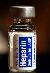
Two tests used to measure antibodies in patients with a history of heparin-induced thrombocytopenia (HIT) can produce radically different results, new research shows.
The anti-PF4/heparin IgG-specific enzyme-immunoassay can show that HIT antibody levels are high, while the functional platelet serotonin-release assay indicates that levels are low.
And researchers said the functional assay’s results may be the better indicator of a patient’s readiness for re-exposure to heparin.
Theodore Warkentin, MD, of McMaster University in Hamilton, Ontario, Canada, and his colleagues expressed this viewpoint and detailed the research supporting it in Blood.
When patients with a history of HIT require urgent heart surgery, physicians must test for the presence of HIT antibodies to determine whether the patient can be re-exposed to heparin during the procedure.
Hematologists use two types of tests to measure HIT antibodies—a functional platelet serotonin-release assay and an anti-PF4/heparin IgG-specific enzyme-immunoassay. If the more widely used immunoassay indicates the presence of HIT antibodies in a patient, surgery is usually delayed or plasma exchange is performed to lower the antibodies.
Practitioners have historically understood the two assays to provide similar conclusions, but a case report suggested otherwise.
A 76-year-old female with kidney cancer and previous HIT required urgent cardiac surgery to remove a tumor that had spread to her heart. After both her initial functional and immunoassays indicated the presence of HIT antibodies, her doctors deemed her ineligible for surgery.
But after repeated plasma exchange, the researchers performed both the functional and immunoassays on the patient again, and, this time, they observed strikingly different results.
“We were surprised to see that levels of HIT antibodies in this patient fell very quickly according to the functional assay, yet the antibodies detected by the immunoassay remained high,” Dr Warkentin said.
“This suggested to us that, while physicians in many situations may be waiting for the immunoassay to indicate lower antibody levels, patients in urgent need of heart surgery may be ready much earlier than the results suggest.”
To better understand the dissociation between the results of the two tests, Dr Warkentin’s team developed a model comparing functional and immunoassay results among 15 HIT blood samples. The samples were sequentially diluted and tested with both assays to mimic the effects of repeated plasma exchange.
The researchers observed that HIT antibody levels as measured by the functional assay decreased rather quickly, while the immunoassay continued to indicate high levels.
This suggests the sensitivity of the immunoassay may provide an overly conservative estimate of HIT antibody levels and their clinical relevance. The analysis illustrates how quickly platelet-activating properties can decline in a patient, either naturally or by using plasma exchange.
The observations also support the use of repeated plasma exchange as a therapeutic strategy prior to planned heparin re-exposure among patients with a recent HIT episode who require urgent cardiac surgery.
“Based on these findings, physicians should consider utilizing both of these tests when preparing a patient with a history of HIT for urgent heart surgery, considering the functional assay result as the stronger indicator of a patient’s readiness,” Dr Warkentin said.
“For these patients, [plasma exchange] can be a useful option to help rapidly reduce their remaining HIT antibody levels, minimize their risk of developing clots, and get them into the operating room sooner.” ![]()
Sickle cell trait linked to increased risk of CKD

while another looks on
Credit: NCI
Sickle cell trait may increase the risk of chronic kidney disease (CKD) and poor kidney function, according to a study published in JAMA.
Researchers evaluated nearly 16,000 African Americans and found that subjects with sickle cell trait had a greater risk of CKD and incident CKD than subjects who did not have the trait.
Trait carriers were also more likely to have albuminuria and a decrease in estimated glomerular filtration rate (eGFR), both characteristics of poor kidney function.
This study was released to coincide with its presentation at the American Society of Nephrology’s Kidney Week Annual Meeting.
Rakhi P. Naik, MD, of Johns Hopkins University in Baltimore, and her colleagues conducted this research to investigate the relationship between sickle cell trait and kidney impairment.
The team looked at data from 5 large, population-based studies. They evaluated 15,975 self-identified African Americans—1248 of whom had sickle cell trait and 14,727 who did not.
The researchers assessed the incidence of CKD, which was defined as an eGFR of <60 mL/min/1.73m2 at baseline or follow-up, and incident CKD. They also assessed the rate of albuminuria, which was defined as a spot urine albumin:creatinine ratio of >30mg/g or albumin excretion rate >30mg/24 hours, and decline in eGFR, which was defined as a decrease of >3 mL/min/1.73m2 per year.
CKD and incident CKD were more common among sickle cell trait carriers than noncarriers. CKD was present in 19.2% (239/1247) of carriers and 13.5% (1994/14,722) of noncarriers. And incident CKD was present in 20.7% (140/675) of carriers and 13.7% (1158/8481) of noncarriers.
Sickle cell trait was associated with a faster decline in eGFR, as 22.6% (150/665) of carriers and 19.0% (1569/8249) of noncarriers met the definition of eGFR decline.
And the trait was associated with a higher incidence of albuminuria, as 31.8% (154/485) of carriers had albuminuria, compared to 19.6% (1168/5947) of noncarriers.
So subjects with sickle cell trait had a greater risk of CKD (odds ratio [OR], 1.57), incident CKD (OR, 1.79), decline in eGFR (OR, 1.32), and albuminuria (OR, 1.86).
The researchers said the associations found in this study may offer an additional genetic explanation for the increased risk of CKD observed among African Americans compared with other racial groups.
They added that the study also highlights the need for further research into the renal complications of sickle cell trait. Because screening for the trait is widely performed, accurate characterization of disease associations with sickle cell trait is needed to inform policy and treatment recommendations. ![]()
Results support transfusing with caution in TTP, HIT
PHILADELPHIA—Results of a large study support the recommendation that patients with platelet consumptive disorders only receive platelet transfusions if they exhibit severe or life-threatening bleeding that is refractory to other therapies.
The research indicated that platelet transfusions may increase the risk of arterial thrombosis and mortality among hospitalized patients with thrombotic thrombocytopenic purpura (TTP) and those with heparin-induced thrombocytopenia (HIT).
Platelet transfusions were also associated with a greater risk of acute myocardial infarction in TTP patients.
However, transfused patients with immune thrombocytopenia (ITP) did not have an increased risk of such complications.
The study did not establish a causal link between transfusions and complications, as it was retrospective and the researchers did not know the exact timing of events.
However, the complications and the transfusions did occur during the same hospital admission, noted Ruchika Goel, MD, of Johns Hopkins University in Baltimore, Maryland. She presented these findings at the AABB Annual Meeting 2014 (abstract S41-030G).
Dr Goel and her colleagues conducted this study to assess current platelet transfusion practices in the US in hospitalized patients with TTP, HIT, and ITP. The team wanted to explore any associations between transfusions and bleeding, venous and arterial thrombotic events, acute myocardial infarction, stroke, and in-hospital mortality in these patients.
“Currently, very little data are available on the risks and benefits associated with platelet transfusions in various platelet consumptive or disruptive disorders,” Dr Goel said. “Thus, evidence-based platelet transfusion guidelines in these disorders are either non-existent or they’re based on consensus statements, with not much supportive data.”
With this in mind, the researchers analyzed data from the Nationwide Inpatient Sample, a stratified probability sample of 20% of all discharges at community hospitals in the US, which covers more than 1100 hospitals across 47 states. The team looked at 5 years of data spanning the period from 2007 through 2011.
They included patients in whom TTP and ITP were the primary admitting diagnoses and patients in whom HIT was 1 of the top 3 diagnoses. Hospitalizations in which patients had a prior history of thrombosis were excluded, as were hospitalizations with any thrombosis/thromboembolism listed as the primary admitting diagnosis (implying that it was already present at admission).
So the analysis included 10,624 patients with TTP, 6332 with HIT, and 79,980 with ITP. The median ages were 47.4, 61.8, and 47.5, respectively. And platelet transfusions were given to 10.1%, 7.1%, and 25.8% of patients, respectively.
When the researchers adjusted their analysis for age and gender, they discovered a significantly increased risk of bleeding among all transfused patients. The odds ratios (ORs) were 2.3 for TTP, 5.5 for HIT, and 5.1 for ITP patients.
“The odds of platelet transfusion were significantly higher in patients who had bleeding, thus implying that . . . that was the indication for the transfusion—an actual bleeding complication,” Dr Goel said.
The results also showed that none of the transfused patients had a significantly increased risk of venous thrombosis or stroke. The ORs for venous thrombosis were 1.1 for TTP, 0.8 for HIT, and 1.3 for ITP patients. And the ORs for stroke were 1.6, 0.5, and 1.3, respectively.
However, both TTP patients and HIT patients had a significantly increased risk of arterial thrombosis. The ORs were 5.8 for TTP, 3.4 for HIT, and 0.3 for ITP patients.
TTP patients also had a significantly increased risk of acute myocardial infarction. The ORs were 2.0 for TTP, 1.9 for HIT, and 1.3 for ITP patients.
And patients with TTP and HIT had a significantly increased risk of in-hospital mortality. The ORs were 2.0 for TTP, 5.2 for HIT, and 1.1 for ITP patients.
Dr Goel noted that this study had several limitations. The temporality of events was not reported, there was no information on platelet thresholds for transfusion or disease severity and the effect on outcomes, and accuracy was limited by the precision of discharge coding.
Therefore, further studies are needed to assess whether platelet transfusions are directly responsible for complications or if they serve as a surrogate marker for the severity of illness.
“We propose that, until such studies or trials are indeed available, which are very hard [to conduct in] these rare disorders, platelets should continue to be considered relatively contraindicated and used only for severe or life-threatening bleeding which is refractory to other therapies,” Dr Goel concluded. ![]()
Transfusions benefit adults with sickle cell disease
PHILADELPHIA—Blood transfusions can provide pain relief in adults with sickle cell disease (SCD) who have failed treatment with hydroxyurea, a pilot study suggests.
Patients had fewer visits to the emergency department (ED) and fewer hospital admissions for pain control after they received chronic transfusions for pain prophylaxis than they did prior to receiving transfusions.
Matthew S. Karafin, MD, of the Blood Center of Wisconsin in Milwaukee, presented these results at the AABB Annual Meeting 2014 (abstract S42-030G).
“Pain in adults with sickle cell disease is probably one of the most important things that we deal with in our clinics,” he began. “It is the leading cause of morbidity in this population.”
Dr Karafin also noted that adults with SCD seem to experience pain differently from children, reporting more of a constant pain, as opposed to the episodic pain observed in kids. And although previous studies have suggested that transfusions do provide pain relief in SCD, most of those studies have focused on children.
So Dr Karafin and his colleagues set out to determine the impact of prophylactic transfusions on the rate of serious pain episodes in adults with SCD. The team retrospectively analyzed a cohort of patients who received chronic transfusions at 3- to 8-week intervals from January 2009 to October 2013.
The researchers defined chronic transfusions as receiving blood—either simple transfusions or red cell exchanges—in an outpatient setting 3 days a week with the goal of controlling hemoglobin (Hb) S percentage, maintaining it at less than 30%.
Patients had to have at least 1 ED or hospital visit for severe pain per month prior to starting transfusions, they were required to have failed hydroxyurea therapy, and they had to have at least 3 months both on and off chronic transfusions. The patients could have no other reason for receiving chronic transfusions (ie, no previous stroke).
So the study included 17 patients, 12 of whom were female. Fifteen (88.1%) had Hb SS disease, and 2 had Hb SC disease. Their median age was 26 (range, 20-54).
“We were able to record 541 total ED admissions over the study period and 404 total hospital admissions,” Dr Karafin said. “The median study evaluation period pre-transfusion was about 3.5 years, and we were able to study [patients for] a median of more than a year for the post-transfusion protocol period.”
Dr Karafin also noted that most of the patients were not transfusion-naïve, but they received significantly more units after being placed on the transfusion protocol.
The median number of red cell units received per 100 days was 1.2 (range, 0-7.2) pre-transfusion and 10.2 (range, 6.7-24.3) post-transfusion (P=0.0003). Nine of the patients received simple transfusions, and 8 received red cell exchanges.
There was a significant difference in the median Hb S pre- and post-transfusion—79% (range, 26.5%-89.6%) and 30.2% (range, 10.9%-57.4%), respectively (P=0.0003).
But there was no significant difference in median ferritin levels—1128.2 ng/mL (range, 65.4-11,130) and 2632.8 ng/mL (range, 16.7-8023.6), respectively (P=0.18). Dr Karafin said this could be explained by the fact that patients were not transfusion-naïve prior to starting the protocol.
Similarly, the median new alloantibody rate per 100 units was 0 both pre- and post-transfusion. This may be due to the fact that all patients received C-, E-, and KEL-matched blood, as well as the freshest available units, Dr Karafin said.
He and his colleagues also found that the median ED admission rate was significantly lower post-transfusion compared to pre-transfusion—0.79 (range, 0-6.6) and 2 (range, 0.4-11) visits every 100 days, respectively (P=0.04).
Thirteen patients (76.5%) had a reduced ED visit rate after chronic transfusion, and there was a 60.5% reduction in the ED visit rate overall.
Likewise, the median hospital admission rate decreased from 1.7 per 100 days (range, 0.05-5.8) pre-transfusion to 1.3 per 100 days (range, 0.2-3.2) post-transfusion (P=0.004).
Fifteen patients (88.2%) had reduced hospital admissions after chronic transfusion, and there was a 20.3% reduction in hospital admissions overall.
Dr Karafin noted that this study had a number of limitations, including a small number of patients, its retrospective nature, and the fact that it was conducted at a comprehensive SCD clinic.
“However, limitations aside, we found significant evidence to support that the findings observed in children seem to be similar in the adult population,” he said.
Namely, chronic transfusions can prevent serious pain episodes in adults with SCD who have failed treatment with hydroxyurea. ![]()
Number of cord blood units doesn’t affect survival

Credit: NHS
Single and double cord blood transplants produce similar outcomes, according to a study of young patients with hematologic disorders.
Researchers found that rates of overall and disease-free survival were not significantly different in patients who received a single unit of cord blood and those who received two units.
Other outcome measures, such as neutrophil recovery, relapse, and transplant-related death, were similar between the two groups as well.
However, patients who received a single cord blood unit showed improved platelet recovery, a lower incidence of grade 3-4 acute graft-vs-host disease (GVHD), and a lower rate of extensive chronic GVHD.
John Wagner, Jr, MD, of the University of Minnesota in Minneapolis, and his colleagues reported these results in NEJM. Dr Wagner previously presented results from this study at ASH 2012.
“Based on promising early studies using two cord blood units in adults for whom one unit is often not sufficient, we designed this study in order to determine if the higher number of blood-forming stem cells in two cord blood units might improve survival,” Dr Wagner said. “What we found, however, was that both treatment arms performed very well, with similar rates of white blood cell recovery and survival.”
The researchers enrolled 224 patients, ages 1 to 21 years, with hematologic disorders, including acute and chronic leukemias as well as myelodysplastic syndromes.
Patients were randomized to receive double-unit (n=111) or single-unit (n=113) cord blood transplants after a uniform myeloablative conditioning regimen and immunoprophylaxis for GVHD.
The researchers matched the treatment arms for age, sex, self-reported race, performance status, degree of donor-recipient HLA matching, disease type, and disease status at transplant.
Survival and relapse
The study’s primary endpoint was 1-year survival, which was 65% in the double-unit arm and 73% in the single-unit arm (P=0.17). In a multivariate analysis, the risk of death did not differ significantly between the arms (hazard ratio=1.34, P=0.20).
Similarly, there was no significant difference in 1-year disease-free survival between the double- and single-unit arms—64% and 70%, respectively (P=0.11). In a multivariate analysis, the risk of relapse or death did not differ significantly between arms (hazard ratio=1.48, P=0.08).
It therefore follows that rates of relapse and transplant-related death were similar at 1 year as well. The incidence of relapse was 14% in the double-unit arm and 12% in the single-unit arm (P=0.12). And rates of transplant-related death were 22% and 19%, respectively (P=0.43).
Recovery and GVHD
The incidence of neutrophil recovery was similar between treatment arms—88% in the double-unit arm and 89% in the single-unit arm (P=0.29) at a median of 23 days (range, 11 to 133) and 21 days (range, 11 to 62) after transplant, respectively.
However, the rate of platelet recovery was significantly higher in the single-unit arm—76% vs 65% (P=0.04). Furthermore, the median time to platelet recovery was 58 days (range, 28 to 295) in the single-unit arm and 84 days (range, 22 to 716) in the double-unit arm.
The rate of grade 2-4 acute GVHD was similar between the treatment arms (P=0.78), but patients in the double-unit arm had a higher incidence of grade 3-4 acute GVHD—23% vs 13% (P=0.02).
There was no difference in the incidence of any chronic GVHD at 1 year after transplant—32% in the double-unit arm and 30% in the single-unit arm (P=0.51). But there was a higher incidence of extensive chronic GVHD after double-unit transplant—15% vs 9% (P=0.05).
“This is helpful news for physicians considering the best treatment options for their patients,” said Joanne Kurtzberg, MD, of Duke University Medical Center in Durham, North Carolina.
“We found children who have a cord blood unit with an adequate number of cells do not benefit from receiving two units. This reduces the cost of a cord blood transplant for the majority of pediatric patients needing the procedure. However, for larger children without an adequately dosed single cord blood unit, using two units will provide access to a potentially life-saving transplant.” ![]()
Drug gets orphan status for PNH in US

The US Food and Drug Administration (FDA) has granted the complement inhibitor AMY-101 orphan status as a treatment for paroxysmal nocturnal
hemoglobinuria (PNH).
Roughly 2 months ago, the European Medicines Agency (EMA) did the same.
Orphan designation will allow Amyndas Pharmaceuticals, the company developing AMY-101, to proceed with expedited clinical development. The company is
planning to move the drug into clinical trials in 2015.
If AMY-101 is approved by the FDA, orphan status will allow for a 7-year period of market exclusivity from product launch in the US. It will also allow Amyndas to apply for research funding, tax credits for certain research expenses, and assistance for clinical research study design. It provides a waiver from the FDA’s Prescription Drug User Fee as well.
“Receiving the orphan drug designation from both the FDA and the EMA is an important achievement and a key milestone in the development pathway of AMY-101, and we are optimistic regarding the long-term potential of this potent complement inhibitor,” said John Lambris, PhD, of the University of Pennsylvania.
Dr Lambris developed AMY-101 at the University of Pennsylvania, and the university licensed the drug to Amyndas Pharmaceuticals. Dr Lambris is a founder and equity holder of Amyndas Pharmaceuticals.
About AMY-101 and PNH
PNH is caused by the defective expression of regulatory proteins on the surface of blood cells, which leaves them vulnerable to complement attack. This can lead to hemolysis, which results in severe anemia and contributes to a high risk of thrombosis.
The monoclonal antibody eculizumab is often successful in treating PNH, but roughly a third of patients do not respond well to the drug and still require blood transfusions to manage their anemia.
Research has suggested this lack of response is due to fragments of complement C3 proteins on the surface of the patients’ red blood cells, which are eventually attacked by immune cells.
In an attempt to overcome this problem, Dr Lambris and his colleagues developed AMY-101. The drug is designed to inhibit C3, thereby preventing hemolysis and immune cell recognition.
The researchers have investigated the effects of AMY-101 on self-attack and the resulting hemolysis in human PNH cells and found the drug to be active.
These results have not been published, but the group has published results with a C3 inhibitor known as Cp40, and AMY-101 is based on Cp40.
The researchers reported in Blood that Cp40 and its long-acting form, PEG-Cp40, effectively inhibited hemolysis and efficiently prevented the deposition of C3 fragments on red blood cells from patients with PNH. ![]()
The US Food and Drug Administration (FDA) has granted the complement inhibitor AMY-101 orphan status as a treatment for paroxysmal nocturnal
hemoglobinuria (PNH).
Roughly 2 months ago, the European Medicines Agency (EMA) did the same.
Orphan designation will allow Amyndas Pharmaceuticals, the company developing AMY-101, to proceed with expedited clinical development. The company is
planning to move the drug into clinical trials in 2015.
If AMY-101 is approved by the FDA, orphan status will allow for a 7-year period of market exclusivity from product launch in the US. It will also allow Amyndas to apply for research funding, tax credits for certain research expenses, and assistance for clinical research study design. It provides a waiver from the FDA’s Prescription Drug User Fee as well.
“Receiving the orphan drug designation from both the FDA and the EMA is an important achievement and a key milestone in the development pathway of AMY-101, and we are optimistic regarding the long-term potential of this potent complement inhibitor,” said John Lambris, PhD, of the University of Pennsylvania.
Dr Lambris developed AMY-101 at the University of Pennsylvania, and the university licensed the drug to Amyndas Pharmaceuticals. Dr Lambris is a founder and equity holder of Amyndas Pharmaceuticals.
About AMY-101 and PNH
PNH is caused by the defective expression of regulatory proteins on the surface of blood cells, which leaves them vulnerable to complement attack. This can lead to hemolysis, which results in severe anemia and contributes to a high risk of thrombosis.
The monoclonal antibody eculizumab is often successful in treating PNH, but roughly a third of patients do not respond well to the drug and still require blood transfusions to manage their anemia.
Research has suggested this lack of response is due to fragments of complement C3 proteins on the surface of the patients’ red blood cells, which are eventually attacked by immune cells.
In an attempt to overcome this problem, Dr Lambris and his colleagues developed AMY-101. The drug is designed to inhibit C3, thereby preventing hemolysis and immune cell recognition.
The researchers have investigated the effects of AMY-101 on self-attack and the resulting hemolysis in human PNH cells and found the drug to be active.
These results have not been published, but the group has published results with a C3 inhibitor known as Cp40, and AMY-101 is based on Cp40.
The researchers reported in Blood that Cp40 and its long-acting form, PEG-Cp40, effectively inhibited hemolysis and efficiently prevented the deposition of C3 fragments on red blood cells from patients with PNH.
Megakaryocytes can control HSCs, team finds

to a megakaryocyte (red)
Credit: Meng Zhao
For the first time, researchers have shown that hematopoietic stem cells (HSCs) can be directly controlled by their own progeny, megakaryocytes.
Preclinical experiments revealed that megakaryocytes maintain HSC quiescence during homeostasis and promote HSC regeneration after chemotherapeutic stress.
The discovery suggests megakaryocytes might be used to treat patients with low blood cell counts and to expand HSCs for transplant.
The researchers described these findings in Nature Medicine.
The team examined the relationship between megakaryocytes and HSCs in mouse bone marrow. And they discovered that, as a terminally differentiated progeny, megakaryocytes regulate HSCs by performing two previously unknown functions.
“Megakaryocytes can directly regulate the amount of hematopoietic stem cells by telling the cells when they need to keep in the quiescent stage and when they need to start proliferating to meet increased demand,” said study author Meng Zhao, PhD, of the Stowers Institute for Medical Research in Kansas City, Missouri.
The researchers found that the protein transforming growth factor B1 (TGF-B1), contained in megakaryocytes, signaled quiescence.
And, when under stress from chemotherapy, megakaryocytes signaled fibroblast growth factor 1 (FGF1), to stimulate HSC proliferation.
“Our findings suggest that megakaryocytes are required for the recovery of hematopoietic stem cells post-chemotherapy,” said Linheng Li, PhD, also of the Stowers Institute.
The discovery could provide insight for using megakaryocyte-derived factors, such as TGF-B1 and FGF1, clinically to facilitate the regeneration of HSCs, he added.
Engineering a megakaryocyte niche that supports the growth of HSCs in culture is the next step for the researchers. They are also investigating whether a megakaryocyte niche can be used to help expand human HSCs in vitro for transplant.
These findings are supported by similar research also reported in Nature Medicine.
Exome sequencing shows potential as diagnostic tool

Credit: Graham Colm
In a large study, whole-exome sequencing provided 25% of patients with a diagnosis related to a known genetic disease, giving young
patients and their parents some long-sought answers.
The technology was able to detect a number of rare genetic events and new mutations contributing to disease.
Among the medically actionable findings were mutations related to Fanconi anemia, erythrocytosis, hemolytic anemia, and von Willebrand disease.
“The findings in this report, I believe, will forever change the future practice of pediatrics and medicine as a whole,” said study author James R. Lupski, MD, PhD, of the Baylor College of Medicine in Houston.
“It is just a matter of time before genomics moves up on the physician’s list of things to do and is ordered before formulating a differential diagnosis. It will be the new ‘family history’ that, better yet, gets you both the important variants inherited from each parent and the new mutations that contribute to disease susceptibility.”
This research was published in JAMA and is set to be presented on October 21 at the American Society of Human Genetics Annual Meeting in San Diego.
The researchers had previously conducted a pilot study of whole-exome sequencing that included 250 patients and revealed a 25% molecular diagnostic rate.
This current study included 2000 patients (88% pediatric) with clinical whole-exome sequencing analyzed between June 2012 and August 2014. The majority of patients—87.8%—had neurological disorders or a developmental delay, and 12.2% had non-neurological disorders.
The researchers collected peripheral blood, tissue, or extracted DNA samples from patients and their parents. The team sequenced patients’ DNA and compared those results to the normal reference. Any disease-associated mutations were then compared with the parent’s DNA to determine if the child inherited it from one or both parents.
In all, 504 patients (25.2%) received a molecular diagnosis, and 58% of the diagnostic mutations had not previously been reported. Two hundred and eighty patients had a single mutation that caused disease, 181 were autosomal recessive, 65 were X-linked, and 1 was presumed inherited through the mitochondria.
In 5 cases, the patient inherited 2 copies of the mutated gene from the same parent. Of the dominant mutations, 208 were de novo mutations not inherited from either parent, 32 were inherited, and 40 were not determined because parental samples were not available.
Among the de novo mutations, 5 demonstrated mosaicism, which suggested the mutation occurred after fertilization.
The researchers found 708 presumptive causative variant alleles in the 504 cases. Almost 30% of the diagnoses occurred in disease genes only identified by researchers in the last 3 years. In 65 cases, there was no available genetic test other than whole-exome sequencing to find the mutated gene at the time the test was ordered.
Twenty-three patients (about 5%) had mutations in 2 different genes, which could account for various aspects of the patient’s medical condition.
“Doctors generally try to find one diagnosis that explains all the issues a patient may have,” said study author Christine Eng, MD, of the Baylor College of Medicine.
“We have found that, in some cases, a patient may have a blended phenotype of 2 different conditions. That patients may have 2 different rare genetic diseases to explain their condition was an unexpected finding prior to the use of whole-exome sequencing.”
In the 2000 cases, incidental findings of medically actionable results that could result in early diagnosis, screening, or treatment were found in 92 patients. Three patients had more than 1 finding.
“Clinical exome sequencing can assist diagnosis in a wide range of disorders that are diagnostic dilemmas,” Dr Lupski said. “Rare variants and Mendelian disease are important contributors to disease populations. This is in sharp contrast to the thinking of population geneticists, who investigate how common variants contribute to disease susceptibility.”
“We find ‘rare variants’ in aggregate actually contribute to disease susceptibility in a big way. The individual diseases may be rare, but there are thousands of such diseases and many more being defined through genomics.”
Study points to potential anemia treatment

Credit: Georges Seguin
Acetate, the major component of vinegar, can stimulate erythropoiesis in anemic mice, according to research published in Nature Medicine.
The study suggests acetate supplements could eventually be a suitable addition or even an alternative to erythropoietin (EPO) therapy.
“[W]e may be able to treat acutely or chronically anemic patients with acetate supplements and thereby reduce the need for blood transfusions or erythropoietin therapy,” said Joseph Garcia, MD, PhD, of the UT Southwestern Medical Center in Dallas, Texas.
He and his colleagues began this study by identifying a pathway that controls erythropoiesis in conditions of stress, such as hypoxia.
Studying genetically modified mice, the researchers discovered that hypoxia stimulates the production of acetate. Acetate, in turn, activates a molecular pathway that ultimately results in erythropoiesis by triggering EPO production.
EPO is regulated by the stress-responsive transcription factor hypoxia-inducible factor-2 (HIF-2). And the researchers had previously shown that the lysine acetyltransferase CREB-binding protein (CBP) is required for HIF-2a acetylation and efficient HIF-2-dependent EPO induction during hypoxia.
With this study, the team found that these processes require acetate-dependent acetyl CoA synthetase 2 (ACSS2) as well.
Experiments showed that ACSS2 is required for HIF-2a acetylation, CBP-HIF-2a complex formation, CBP-HIF-2a recruitment to the EPO enhancer, and efficient induction of EPO gene expression.
The researchers administered acetate to acutely anemic mice and found the treatment augments stress erythropoiesis in an ACSS2-dependent manner.
When they administered acetate to mice with acquired and inherited chronic anemia, the team observed increases in EPO expression and resting hematocrit.
“Our study shows that acetate functions as a biochemical ‘flare,’” Dr Garcia said, “linking changes in cell metabolism that occur during hypoxia with the activation of a selective stress signaling pathway.”