User login
Are doctors savers or spenders?
In a poll that ran from August 30 to Sept. 21, conducted by Medscape, physicians were asked if they lived within their means. They were asked whether they pay their bills on time, save at least 20% of their monthly income toward retirement, pay down student loan debt, and contribute to their kids’ college savings or a rainy-day emergency fund.
Medscape polled 468 U.S. physicians and 159 living outside of the United States. Eighty-nine percent of U.S. respondents report living within their means, while only 11% said they don’t.
Medscape’s Physician Wealth & Debt Report 2022 similarly reported that of 13,000 physicians in more than 29 specialties, 94% said they live at or below their means.
For example, over half of physicians have a net worth above $1 million. In contrast, according to Credit Suisse’s Global Wealth Report, less than 7% of the general population has a seven-figure net worth.
So just how do physicians stack up financially?
Habits of physician super savers
Physicians who consider themselves savers likely have money habits that correlate. They buy things on sale, are DIYers for home projects and maintenance, and wait to buy luxury or large expenses when the timing is right, an item is on sale, or they’ve saved for it.
For example, when it comes to life’s luxuries like buying a new car or dining out, overall, physicians seem to be more frugal, as 43% of those who buy cars said they only buy a new car every 10 years; 30% said they buy a new vehicle every 6-7 years, and 22% said every 4-5 years.
When asked about weekly dine-out or delivery habits, 82% of those polled who said they dine out, or order takeout, do so a nominal 1-2 times per week. That’s on par with the Centers for Disease Control, which reports that 3 in 5 Americans eat out once weekly. Another 14% of polled physicians said they dine out 3-5 nights per week. Only 4% revealed they eat out or grab to-go food more than 5 nights a week.
When hiring for essential home maintenance, like house cleaning and pool or lawn service, almost a third of physicians we polled who require such maintenance employ a service for these tasks, and 23% hire out often while 21% hire out only sometimes. However, 14% say they rarely hire out for home maintenance, and 11% never do.
Since physicians are typically tight on time, they tend to favor outsourcing things like housecleaning, lawn service, landscaping, maintenance, and even cooking. So, the fact that a quarter of physicians polled rarely or never hire out for household help is somewhat surprising.
Most physicians also prioritize saving. When asked how important it is to save money consistently, 93% think it’s either extremely or very important, while only 6% think it’s somewhat important.
Barriers to wealth
When asked what barriers prevent them from saving at least 20% of their monthly income, physician respondents who said they live within their means and encountered barriers reported that family necessities (35%), student loan debt (19%), and mortgage sizes (18%) were the top reasons. The average doctor earns five times as much as the average American, according to the Global Wealth Report.
“What prevents me from saving is holding too much debt, responsibilities at home, bills, being unprepared for what is coming, and making excuses to spend even when it’s not necessary,” says Sean Ormond, MD, a dual board-certified physician in Anesthesiology and Pain Management in Phoenix.
When physician respondents who said they didn’t live within their means were asked about the barriers preventing them from saving at least 20% of their monthly income, they cited the cost of family necessities (49%), the size of their mortgage (47%), credit card debt (30%), student loan debt (21%), other loans (15%), and car lease/loan (13%).
“My most significant financial splurge is vacation, since I always choose the best, and the best comes at an extra cost,” says Dr. Ormond.
What’s your financial grade?
Finally, physicians were asked who they considered better at saving money, themselves or their spouse/domestic partner. Forty-four percent think they are the better saver, whereas 41% said that both they and their partner were equally good at saving. Thirteen percent credited their partner with better saving habits, and 2% said neither themselves nor their partner were good at saving money.
More than half (63%) of physicians polled pay off their credit card balance monthly, but 18% carry a $1,000-$5,000 balance, 10% have $5,000-$10,000 in credit card debt, and 6% hold more than $10,000 of credit card debt.
“I would grade myself with a B, because however much I love having the best, I still have a budget, and I always ensure that I follow it to the dot,” says Dr. Ormond.
A version of this article first appeared on Medscape.com.
In a poll that ran from August 30 to Sept. 21, conducted by Medscape, physicians were asked if they lived within their means. They were asked whether they pay their bills on time, save at least 20% of their monthly income toward retirement, pay down student loan debt, and contribute to their kids’ college savings or a rainy-day emergency fund.
Medscape polled 468 U.S. physicians and 159 living outside of the United States. Eighty-nine percent of U.S. respondents report living within their means, while only 11% said they don’t.
Medscape’s Physician Wealth & Debt Report 2022 similarly reported that of 13,000 physicians in more than 29 specialties, 94% said they live at or below their means.
For example, over half of physicians have a net worth above $1 million. In contrast, according to Credit Suisse’s Global Wealth Report, less than 7% of the general population has a seven-figure net worth.
So just how do physicians stack up financially?
Habits of physician super savers
Physicians who consider themselves savers likely have money habits that correlate. They buy things on sale, are DIYers for home projects and maintenance, and wait to buy luxury or large expenses when the timing is right, an item is on sale, or they’ve saved for it.
For example, when it comes to life’s luxuries like buying a new car or dining out, overall, physicians seem to be more frugal, as 43% of those who buy cars said they only buy a new car every 10 years; 30% said they buy a new vehicle every 6-7 years, and 22% said every 4-5 years.
When asked about weekly dine-out or delivery habits, 82% of those polled who said they dine out, or order takeout, do so a nominal 1-2 times per week. That’s on par with the Centers for Disease Control, which reports that 3 in 5 Americans eat out once weekly. Another 14% of polled physicians said they dine out 3-5 nights per week. Only 4% revealed they eat out or grab to-go food more than 5 nights a week.
When hiring for essential home maintenance, like house cleaning and pool or lawn service, almost a third of physicians we polled who require such maintenance employ a service for these tasks, and 23% hire out often while 21% hire out only sometimes. However, 14% say they rarely hire out for home maintenance, and 11% never do.
Since physicians are typically tight on time, they tend to favor outsourcing things like housecleaning, lawn service, landscaping, maintenance, and even cooking. So, the fact that a quarter of physicians polled rarely or never hire out for household help is somewhat surprising.
Most physicians also prioritize saving. When asked how important it is to save money consistently, 93% think it’s either extremely or very important, while only 6% think it’s somewhat important.
Barriers to wealth
When asked what barriers prevent them from saving at least 20% of their monthly income, physician respondents who said they live within their means and encountered barriers reported that family necessities (35%), student loan debt (19%), and mortgage sizes (18%) were the top reasons. The average doctor earns five times as much as the average American, according to the Global Wealth Report.
“What prevents me from saving is holding too much debt, responsibilities at home, bills, being unprepared for what is coming, and making excuses to spend even when it’s not necessary,” says Sean Ormond, MD, a dual board-certified physician in Anesthesiology and Pain Management in Phoenix.
When physician respondents who said they didn’t live within their means were asked about the barriers preventing them from saving at least 20% of their monthly income, they cited the cost of family necessities (49%), the size of their mortgage (47%), credit card debt (30%), student loan debt (21%), other loans (15%), and car lease/loan (13%).
“My most significant financial splurge is vacation, since I always choose the best, and the best comes at an extra cost,” says Dr. Ormond.
What’s your financial grade?
Finally, physicians were asked who they considered better at saving money, themselves or their spouse/domestic partner. Forty-four percent think they are the better saver, whereas 41% said that both they and their partner were equally good at saving. Thirteen percent credited their partner with better saving habits, and 2% said neither themselves nor their partner were good at saving money.
More than half (63%) of physicians polled pay off their credit card balance monthly, but 18% carry a $1,000-$5,000 balance, 10% have $5,000-$10,000 in credit card debt, and 6% hold more than $10,000 of credit card debt.
“I would grade myself with a B, because however much I love having the best, I still have a budget, and I always ensure that I follow it to the dot,” says Dr. Ormond.
A version of this article first appeared on Medscape.com.
In a poll that ran from August 30 to Sept. 21, conducted by Medscape, physicians were asked if they lived within their means. They were asked whether they pay their bills on time, save at least 20% of their monthly income toward retirement, pay down student loan debt, and contribute to their kids’ college savings or a rainy-day emergency fund.
Medscape polled 468 U.S. physicians and 159 living outside of the United States. Eighty-nine percent of U.S. respondents report living within their means, while only 11% said they don’t.
Medscape’s Physician Wealth & Debt Report 2022 similarly reported that of 13,000 physicians in more than 29 specialties, 94% said they live at or below their means.
For example, over half of physicians have a net worth above $1 million. In contrast, according to Credit Suisse’s Global Wealth Report, less than 7% of the general population has a seven-figure net worth.
So just how do physicians stack up financially?
Habits of physician super savers
Physicians who consider themselves savers likely have money habits that correlate. They buy things on sale, are DIYers for home projects and maintenance, and wait to buy luxury or large expenses when the timing is right, an item is on sale, or they’ve saved for it.
For example, when it comes to life’s luxuries like buying a new car or dining out, overall, physicians seem to be more frugal, as 43% of those who buy cars said they only buy a new car every 10 years; 30% said they buy a new vehicle every 6-7 years, and 22% said every 4-5 years.
When asked about weekly dine-out or delivery habits, 82% of those polled who said they dine out, or order takeout, do so a nominal 1-2 times per week. That’s on par with the Centers for Disease Control, which reports that 3 in 5 Americans eat out once weekly. Another 14% of polled physicians said they dine out 3-5 nights per week. Only 4% revealed they eat out or grab to-go food more than 5 nights a week.
When hiring for essential home maintenance, like house cleaning and pool or lawn service, almost a third of physicians we polled who require such maintenance employ a service for these tasks, and 23% hire out often while 21% hire out only sometimes. However, 14% say they rarely hire out for home maintenance, and 11% never do.
Since physicians are typically tight on time, they tend to favor outsourcing things like housecleaning, lawn service, landscaping, maintenance, and even cooking. So, the fact that a quarter of physicians polled rarely or never hire out for household help is somewhat surprising.
Most physicians also prioritize saving. When asked how important it is to save money consistently, 93% think it’s either extremely or very important, while only 6% think it’s somewhat important.
Barriers to wealth
When asked what barriers prevent them from saving at least 20% of their monthly income, physician respondents who said they live within their means and encountered barriers reported that family necessities (35%), student loan debt (19%), and mortgage sizes (18%) were the top reasons. The average doctor earns five times as much as the average American, according to the Global Wealth Report.
“What prevents me from saving is holding too much debt, responsibilities at home, bills, being unprepared for what is coming, and making excuses to spend even when it’s not necessary,” says Sean Ormond, MD, a dual board-certified physician in Anesthesiology and Pain Management in Phoenix.
When physician respondents who said they didn’t live within their means were asked about the barriers preventing them from saving at least 20% of their monthly income, they cited the cost of family necessities (49%), the size of their mortgage (47%), credit card debt (30%), student loan debt (21%), other loans (15%), and car lease/loan (13%).
“My most significant financial splurge is vacation, since I always choose the best, and the best comes at an extra cost,” says Dr. Ormond.
What’s your financial grade?
Finally, physicians were asked who they considered better at saving money, themselves or their spouse/domestic partner. Forty-four percent think they are the better saver, whereas 41% said that both they and their partner were equally good at saving. Thirteen percent credited their partner with better saving habits, and 2% said neither themselves nor their partner were good at saving money.
More than half (63%) of physicians polled pay off their credit card balance monthly, but 18% carry a $1,000-$5,000 balance, 10% have $5,000-$10,000 in credit card debt, and 6% hold more than $10,000 of credit card debt.
“I would grade myself with a B, because however much I love having the best, I still have a budget, and I always ensure that I follow it to the dot,” says Dr. Ormond.
A version of this article first appeared on Medscape.com.
New COVID variant gaining traction in U.S.
, according to the CDC’s latest data.
Just 1 month ago, the variant accounted for less than 1% of cases.
“When you get variants like that, you look at what their rate of increase is as a relative proportion of the variants, and this has a pretty troublesome doubling time,” Anthony Fauci, MD, said in an interview with CBS News. Dr. Fauci is the director of the National Institute of Allergy and Infectious Diseases and also the chief medical adviser to President Joe Biden.
There are also concerning features of the BQ.1 variant, which include mutations that could potentially escape vaccines and treatments for COVID-19.
Currently, the most widespread variant in the U.S. is the Omicron subvariant known as BA.5, which accounts for 68% of all infections. One of the go-to treatments for BA.5 infections is monoclonal antibodies, which may not be as effective when fighting the up-and-coming strains of BQ.1 and its descendant BQ.1.1, according to experts.
“That’s the reason why people are concerned about BQ.1.1, for the double reason of its doubling time and the fact that it seems to elude important monoclonal antibodies,” Dr. Fauci told CBS News.
Currently, BQ.1 and BQ.1.1 appear most widespread in the New York and New Jersey region, accounting for nearly 20% of infections there, according to the CDC.
But because the new variant is a descendant of Omicron, Dr. Fauci said the currently available booster shots are still the best first line of protection against this up-and-coming threat.
“The bad news is that there’s a new variant that’s emerging and that has qualities or characteristics that could evade some of the interventions we have. But, the somewhat encouraging news is that it’s a BA.5 sub-lineage, so there is almost certainly going to be some cross-protection that you can boost up,” he said.
A version of this article first appeared on WebMD.com.
, according to the CDC’s latest data.
Just 1 month ago, the variant accounted for less than 1% of cases.
“When you get variants like that, you look at what their rate of increase is as a relative proportion of the variants, and this has a pretty troublesome doubling time,” Anthony Fauci, MD, said in an interview with CBS News. Dr. Fauci is the director of the National Institute of Allergy and Infectious Diseases and also the chief medical adviser to President Joe Biden.
There are also concerning features of the BQ.1 variant, which include mutations that could potentially escape vaccines and treatments for COVID-19.
Currently, the most widespread variant in the U.S. is the Omicron subvariant known as BA.5, which accounts for 68% of all infections. One of the go-to treatments for BA.5 infections is monoclonal antibodies, which may not be as effective when fighting the up-and-coming strains of BQ.1 and its descendant BQ.1.1, according to experts.
“That’s the reason why people are concerned about BQ.1.1, for the double reason of its doubling time and the fact that it seems to elude important monoclonal antibodies,” Dr. Fauci told CBS News.
Currently, BQ.1 and BQ.1.1 appear most widespread in the New York and New Jersey region, accounting for nearly 20% of infections there, according to the CDC.
But because the new variant is a descendant of Omicron, Dr. Fauci said the currently available booster shots are still the best first line of protection against this up-and-coming threat.
“The bad news is that there’s a new variant that’s emerging and that has qualities or characteristics that could evade some of the interventions we have. But, the somewhat encouraging news is that it’s a BA.5 sub-lineage, so there is almost certainly going to be some cross-protection that you can boost up,” he said.
A version of this article first appeared on WebMD.com.
, according to the CDC’s latest data.
Just 1 month ago, the variant accounted for less than 1% of cases.
“When you get variants like that, you look at what their rate of increase is as a relative proportion of the variants, and this has a pretty troublesome doubling time,” Anthony Fauci, MD, said in an interview with CBS News. Dr. Fauci is the director of the National Institute of Allergy and Infectious Diseases and also the chief medical adviser to President Joe Biden.
There are also concerning features of the BQ.1 variant, which include mutations that could potentially escape vaccines and treatments for COVID-19.
Currently, the most widespread variant in the U.S. is the Omicron subvariant known as BA.5, which accounts for 68% of all infections. One of the go-to treatments for BA.5 infections is monoclonal antibodies, which may not be as effective when fighting the up-and-coming strains of BQ.1 and its descendant BQ.1.1, according to experts.
“That’s the reason why people are concerned about BQ.1.1, for the double reason of its doubling time and the fact that it seems to elude important monoclonal antibodies,” Dr. Fauci told CBS News.
Currently, BQ.1 and BQ.1.1 appear most widespread in the New York and New Jersey region, accounting for nearly 20% of infections there, according to the CDC.
But because the new variant is a descendant of Omicron, Dr. Fauci said the currently available booster shots are still the best first line of protection against this up-and-coming threat.
“The bad news is that there’s a new variant that’s emerging and that has qualities or characteristics that could evade some of the interventions we have. But, the somewhat encouraging news is that it’s a BA.5 sub-lineage, so there is almost certainly going to be some cross-protection that you can boost up,” he said.
A version of this article first appeared on WebMD.com.
Since when does providing pediatric care require courage?
I have been noticing “jokes” lately about doctors. Magazine cartoons depict doctors as conveying a bad prognosis in abrupt, indirect, vague ways. I remember, from medical school, the joke about pediatricians being doctors for patients “from the waist up” – wimps about tough topics such as sexuality. As an inherently shy person, I have appreciated the structure of the contractual relationship with families that both gives me permission and requires me to be direct about topics that would not be socially acceptable to discuss in other relationships.
Examples include our asking about bowel movements, genital symptoms, marital conflict, past abortions, food insecurity, adherence to medication, history of trauma or discrimination, substance use, illegal conduct, and suicidal ideation, among others. By bringing up these topics nonjudgmentally and with skill, we are demonstrating openness and making it safe for the patient/parent to ask questions about their own concerns.
Since these may be topics for which we lack knowledge or have very little power to help, it is far easier to not bring them up. Yet failing to have the courage to elicit sensitive information may delay the correct diagnosis, result in inappropriate tests or treatments, or miss factors critical in either the cause or solution for the patient’s problems.
Historically, being a physician has conveyed a promise of confidentiality and always trying to do what is best for the patient. The fact that we had to swear an oath to do so may also indicate that these things are not easy to do.
Yes, we need a lot of knowledge to know what is truly in the patient’s best interest. But we need to take personal risks to do it as well. There have been times when a parent has shouted “That is none of your business” at me or stormed out. Although only one patient has ever connected when striking out at me, other clinicians have not been as lucky (or had such small patients); some have even been killed.
For those of us in private practice, upsetting a patient with our well-intentioned words may mean losing them from our income stream or having them post negative comments online, which may affect our reputation in the community. Patients may not return for needed follow-up if a conversation was too uncomfortable for them. Current political divisions make this even trickier.
These days anxiety about getting behind in seeing the next patient may be a covert reason for avoiding difficult conversations as tears or anger take extra time. Certainly, fears of these outcomes can make us hold back from talking about important but potentially upsetting topics.
Of course, courage does not just mean being direct with questions, stating your observations, or giving advice. Courage requires thoughtfulness about possible adverse outcomes and their effect on others. It is not just “stupid bravery,” to proceed even when sensing danger. Courage is thus best paired with skill. It is:
- Setting up potentially difficult discussions with privacy (from the child or parent), seating, and enough time to listen.
- Normalizing questions by saying “I ask all my patients about ...” so patients do not feel singled out.
- Asking the patient or family first what they think is going on and how their own culture might regard the issue.
- Using simple language and arranging a translator when needed.
- Not just stating facts but checking “to be sure I explained well enough” rather than setting a patient up to appear ignorant for not understanding.
- Offering to contact the patient or other family member/support soon to review what you said and answer more questions.
- Offering a second opinion option.
- Promising to get more information when you do not know.
- Always leaving room for hope and sharing in that hope with them.
And it is crucial to have a way to keep notes about past trauma or difficult topics for a patient so neither you nor subsequent clinicians unnecessarily ask about sensitive topics.
Courage includes facing difficult situations without undue delay. Making that call about an abnormal test result right away, even when you are tired and upset, takes courage. Each time you overcome your own reluctance it takes moral strength but tends to make future courageous acts easier. Speaking up to a specialist on rounds when you think he or she is incorrect takes courage to serve the patient’s best interest. Being willing to try a new workflow in your office takes the courage to risk looking awkward or being judged by your team, but can be essential to progress. Asking for help or an opinion, sometimes from a medical assistant or student, can take courage but may reduce status barriers and improve relationships. Standing up for your values when they are not popular may take courage in some organizations. It takes courage to admit a mistake, even when your mistake may not otherwise be noticed.
How can we grow in courage? T. Berry Brazelton, MD, was a model of courage for me during my training – able and willing to tell about a child’s delays or ask about a parent’s well-being with empathy and by giving hope. Dr. Brazelton, pediatrician, developer of the Neonatal Behavioral Assessment Scale, professor at Harvard Medical School and founder of its Child Development Unit, was a world-renowned educator about the development of children and founder of the Touchpoints program. Our goal should be to promise to partner with the family in dealing with the problem, no matter how difficult or tender. I hope you had role models who not only said what to do but also demonstrated it with patients.
We had the privilege of hearing stories of difficult situations from hundreds of pediatricians in group sessions over the years in the Collaborative Office Rounds program. What group members often said that they valued most from these sessions was hearing examples of words they might say in these cases, either modeled by the coleaders or suggested by their pediatrician peers. Opportunities to share the tough times with trusted empathic peers is an important resource rarer and thus even more worth securing for yourself.
Being courageous may not be natural part of your personality but Aristotle said, “We become what we repeatedly do.” Even if you do not consider yourself so now, with practice you can become courageous and reap its benefits for your patients and yourself.
Dr. Howard is assistant professor of pediatrics at Johns Hopkins University, Baltimore, and creator of CHADIS. She had no other relevant disclosures. Dr. Howard’s contribution to this publication was as a paid expert to MDedge News. E-mail her at [email protected].
I have been noticing “jokes” lately about doctors. Magazine cartoons depict doctors as conveying a bad prognosis in abrupt, indirect, vague ways. I remember, from medical school, the joke about pediatricians being doctors for patients “from the waist up” – wimps about tough topics such as sexuality. As an inherently shy person, I have appreciated the structure of the contractual relationship with families that both gives me permission and requires me to be direct about topics that would not be socially acceptable to discuss in other relationships.
Examples include our asking about bowel movements, genital symptoms, marital conflict, past abortions, food insecurity, adherence to medication, history of trauma or discrimination, substance use, illegal conduct, and suicidal ideation, among others. By bringing up these topics nonjudgmentally and with skill, we are demonstrating openness and making it safe for the patient/parent to ask questions about their own concerns.
Since these may be topics for which we lack knowledge or have very little power to help, it is far easier to not bring them up. Yet failing to have the courage to elicit sensitive information may delay the correct diagnosis, result in inappropriate tests or treatments, or miss factors critical in either the cause or solution for the patient’s problems.
Historically, being a physician has conveyed a promise of confidentiality and always trying to do what is best for the patient. The fact that we had to swear an oath to do so may also indicate that these things are not easy to do.
Yes, we need a lot of knowledge to know what is truly in the patient’s best interest. But we need to take personal risks to do it as well. There have been times when a parent has shouted “That is none of your business” at me or stormed out. Although only one patient has ever connected when striking out at me, other clinicians have not been as lucky (or had such small patients); some have even been killed.
For those of us in private practice, upsetting a patient with our well-intentioned words may mean losing them from our income stream or having them post negative comments online, which may affect our reputation in the community. Patients may not return for needed follow-up if a conversation was too uncomfortable for them. Current political divisions make this even trickier.
These days anxiety about getting behind in seeing the next patient may be a covert reason for avoiding difficult conversations as tears or anger take extra time. Certainly, fears of these outcomes can make us hold back from talking about important but potentially upsetting topics.
Of course, courage does not just mean being direct with questions, stating your observations, or giving advice. Courage requires thoughtfulness about possible adverse outcomes and their effect on others. It is not just “stupid bravery,” to proceed even when sensing danger. Courage is thus best paired with skill. It is:
- Setting up potentially difficult discussions with privacy (from the child or parent), seating, and enough time to listen.
- Normalizing questions by saying “I ask all my patients about ...” so patients do not feel singled out.
- Asking the patient or family first what they think is going on and how their own culture might regard the issue.
- Using simple language and arranging a translator when needed.
- Not just stating facts but checking “to be sure I explained well enough” rather than setting a patient up to appear ignorant for not understanding.
- Offering to contact the patient or other family member/support soon to review what you said and answer more questions.
- Offering a second opinion option.
- Promising to get more information when you do not know.
- Always leaving room for hope and sharing in that hope with them.
And it is crucial to have a way to keep notes about past trauma or difficult topics for a patient so neither you nor subsequent clinicians unnecessarily ask about sensitive topics.
Courage includes facing difficult situations without undue delay. Making that call about an abnormal test result right away, even when you are tired and upset, takes courage. Each time you overcome your own reluctance it takes moral strength but tends to make future courageous acts easier. Speaking up to a specialist on rounds when you think he or she is incorrect takes courage to serve the patient’s best interest. Being willing to try a new workflow in your office takes the courage to risk looking awkward or being judged by your team, but can be essential to progress. Asking for help or an opinion, sometimes from a medical assistant or student, can take courage but may reduce status barriers and improve relationships. Standing up for your values when they are not popular may take courage in some organizations. It takes courage to admit a mistake, even when your mistake may not otherwise be noticed.
How can we grow in courage? T. Berry Brazelton, MD, was a model of courage for me during my training – able and willing to tell about a child’s delays or ask about a parent’s well-being with empathy and by giving hope. Dr. Brazelton, pediatrician, developer of the Neonatal Behavioral Assessment Scale, professor at Harvard Medical School and founder of its Child Development Unit, was a world-renowned educator about the development of children and founder of the Touchpoints program. Our goal should be to promise to partner with the family in dealing with the problem, no matter how difficult or tender. I hope you had role models who not only said what to do but also demonstrated it with patients.
We had the privilege of hearing stories of difficult situations from hundreds of pediatricians in group sessions over the years in the Collaborative Office Rounds program. What group members often said that they valued most from these sessions was hearing examples of words they might say in these cases, either modeled by the coleaders or suggested by their pediatrician peers. Opportunities to share the tough times with trusted empathic peers is an important resource rarer and thus even more worth securing for yourself.
Being courageous may not be natural part of your personality but Aristotle said, “We become what we repeatedly do.” Even if you do not consider yourself so now, with practice you can become courageous and reap its benefits for your patients and yourself.
Dr. Howard is assistant professor of pediatrics at Johns Hopkins University, Baltimore, and creator of CHADIS. She had no other relevant disclosures. Dr. Howard’s contribution to this publication was as a paid expert to MDedge News. E-mail her at [email protected].
I have been noticing “jokes” lately about doctors. Magazine cartoons depict doctors as conveying a bad prognosis in abrupt, indirect, vague ways. I remember, from medical school, the joke about pediatricians being doctors for patients “from the waist up” – wimps about tough topics such as sexuality. As an inherently shy person, I have appreciated the structure of the contractual relationship with families that both gives me permission and requires me to be direct about topics that would not be socially acceptable to discuss in other relationships.
Examples include our asking about bowel movements, genital symptoms, marital conflict, past abortions, food insecurity, adherence to medication, history of trauma or discrimination, substance use, illegal conduct, and suicidal ideation, among others. By bringing up these topics nonjudgmentally and with skill, we are demonstrating openness and making it safe for the patient/parent to ask questions about their own concerns.
Since these may be topics for which we lack knowledge or have very little power to help, it is far easier to not bring them up. Yet failing to have the courage to elicit sensitive information may delay the correct diagnosis, result in inappropriate tests or treatments, or miss factors critical in either the cause or solution for the patient’s problems.
Historically, being a physician has conveyed a promise of confidentiality and always trying to do what is best for the patient. The fact that we had to swear an oath to do so may also indicate that these things are not easy to do.
Yes, we need a lot of knowledge to know what is truly in the patient’s best interest. But we need to take personal risks to do it as well. There have been times when a parent has shouted “That is none of your business” at me or stormed out. Although only one patient has ever connected when striking out at me, other clinicians have not been as lucky (or had such small patients); some have even been killed.
For those of us in private practice, upsetting a patient with our well-intentioned words may mean losing them from our income stream or having them post negative comments online, which may affect our reputation in the community. Patients may not return for needed follow-up if a conversation was too uncomfortable for them. Current political divisions make this even trickier.
These days anxiety about getting behind in seeing the next patient may be a covert reason for avoiding difficult conversations as tears or anger take extra time. Certainly, fears of these outcomes can make us hold back from talking about important but potentially upsetting topics.
Of course, courage does not just mean being direct with questions, stating your observations, or giving advice. Courage requires thoughtfulness about possible adverse outcomes and their effect on others. It is not just “stupid bravery,” to proceed even when sensing danger. Courage is thus best paired with skill. It is:
- Setting up potentially difficult discussions with privacy (from the child or parent), seating, and enough time to listen.
- Normalizing questions by saying “I ask all my patients about ...” so patients do not feel singled out.
- Asking the patient or family first what they think is going on and how their own culture might regard the issue.
- Using simple language and arranging a translator when needed.
- Not just stating facts but checking “to be sure I explained well enough” rather than setting a patient up to appear ignorant for not understanding.
- Offering to contact the patient or other family member/support soon to review what you said and answer more questions.
- Offering a second opinion option.
- Promising to get more information when you do not know.
- Always leaving room for hope and sharing in that hope with them.
And it is crucial to have a way to keep notes about past trauma or difficult topics for a patient so neither you nor subsequent clinicians unnecessarily ask about sensitive topics.
Courage includes facing difficult situations without undue delay. Making that call about an abnormal test result right away, even when you are tired and upset, takes courage. Each time you overcome your own reluctance it takes moral strength but tends to make future courageous acts easier. Speaking up to a specialist on rounds when you think he or she is incorrect takes courage to serve the patient’s best interest. Being willing to try a new workflow in your office takes the courage to risk looking awkward or being judged by your team, but can be essential to progress. Asking for help or an opinion, sometimes from a medical assistant or student, can take courage but may reduce status barriers and improve relationships. Standing up for your values when they are not popular may take courage in some organizations. It takes courage to admit a mistake, even when your mistake may not otherwise be noticed.
How can we grow in courage? T. Berry Brazelton, MD, was a model of courage for me during my training – able and willing to tell about a child’s delays or ask about a parent’s well-being with empathy and by giving hope. Dr. Brazelton, pediatrician, developer of the Neonatal Behavioral Assessment Scale, professor at Harvard Medical School and founder of its Child Development Unit, was a world-renowned educator about the development of children and founder of the Touchpoints program. Our goal should be to promise to partner with the family in dealing with the problem, no matter how difficult or tender. I hope you had role models who not only said what to do but also demonstrated it with patients.
We had the privilege of hearing stories of difficult situations from hundreds of pediatricians in group sessions over the years in the Collaborative Office Rounds program. What group members often said that they valued most from these sessions was hearing examples of words they might say in these cases, either modeled by the coleaders or suggested by their pediatrician peers. Opportunities to share the tough times with trusted empathic peers is an important resource rarer and thus even more worth securing for yourself.
Being courageous may not be natural part of your personality but Aristotle said, “We become what we repeatedly do.” Even if you do not consider yourself so now, with practice you can become courageous and reap its benefits for your patients and yourself.
Dr. Howard is assistant professor of pediatrics at Johns Hopkins University, Baltimore, and creator of CHADIS. She had no other relevant disclosures. Dr. Howard’s contribution to this publication was as a paid expert to MDedge News. E-mail her at [email protected].
Drug abusers will find a way
Recently, when I logged on to see what medication refills had come in, I was greeted with a notice that Walgreens would no longer carry promethazine/codeine cough syrup. It wouldn’t surprise me if other pharmacies follow.
This doesn’t affect me much. As a neurologist I’ve never prescribed it, and as a patient I’ve never used it.

The unwritten reason was likely because of its popularity for abuse. It is often mixed with various beverages and called “purple drank.” It has both social, and legal, consequences that can come back and bite the pharmacy.
A friend of mine commented that if everything that can be abused gets banned, all we’ll be left with are Tylenol and Preparation H. Another friend made the comment that it’s a shame, because codeine is a remarkably effective antitussive.
I agree with both of them, but Walgreens is pulling only the combo preparation off the shelves. Codeine and promethazine are still available. The former is on WHO’s list of essential medications.
They’d still find a way to get the components and whip up some equivalent. Human innovation can be remarkable. All of us who trained in the inner city (which is pretty much all of us at some point) have seen people who drank mouthwash, hairspray, and who knows what else in desperation.
No one believes it’s going to stop drug abuse, but it will make it harder to have purple drank, which is often passed around as a low-level drug at parties. Putting Sudafed behind the counter has reduced, though not stopped, meth. Walter White can tell you that.
A patient of mine who’s a pharmacist also was talking about this. He’s in favor of it, as he’s tired of dealing with people trying to get it through faked prescriptions and bogus visits to urgent care pretending to have a cough, not to mention the additional paperwork and reporting requirements that a controlled drug carries.
I agree with it, mostly, but there are those who truly do need it at times, and who now will have to take it as individual components, or find a pharmacy that does carry it. The issue here becomes that, by punishing the abusers, you’re also punishing the responsible.
The vast majority of alcohol users are responsible drinkers. I have the occasional beer myself. Unfortunately, there are a comparative few who aren’t, and their actions can bring tremendous grief to many others. So we have tougher laws all around that we all have to follow.
I agree with Walgreens actions on this, but still find myself wondering how much of a difference it will make.
Probably not as much as I hope.
Dr. Block has a solo neurology practice in Scottsdale, Ariz.
Recently, when I logged on to see what medication refills had come in, I was greeted with a notice that Walgreens would no longer carry promethazine/codeine cough syrup. It wouldn’t surprise me if other pharmacies follow.
This doesn’t affect me much. As a neurologist I’ve never prescribed it, and as a patient I’ve never used it.
The unwritten reason was likely because of its popularity for abuse. It is often mixed with various beverages and called “purple drank.” It has both social, and legal, consequences that can come back and bite the pharmacy.
A friend of mine commented that if everything that can be abused gets banned, all we’ll be left with are Tylenol and Preparation H. Another friend made the comment that it’s a shame, because codeine is a remarkably effective antitussive.
I agree with both of them, but Walgreens is pulling only the combo preparation off the shelves. Codeine and promethazine are still available. The former is on WHO’s list of essential medications.
They’d still find a way to get the components and whip up some equivalent. Human innovation can be remarkable. All of us who trained in the inner city (which is pretty much all of us at some point) have seen people who drank mouthwash, hairspray, and who knows what else in desperation.
No one believes it’s going to stop drug abuse, but it will make it harder to have purple drank, which is often passed around as a low-level drug at parties. Putting Sudafed behind the counter has reduced, though not stopped, meth. Walter White can tell you that.
A patient of mine who’s a pharmacist also was talking about this. He’s in favor of it, as he’s tired of dealing with people trying to get it through faked prescriptions and bogus visits to urgent care pretending to have a cough, not to mention the additional paperwork and reporting requirements that a controlled drug carries.
I agree with it, mostly, but there are those who truly do need it at times, and who now will have to take it as individual components, or find a pharmacy that does carry it. The issue here becomes that, by punishing the abusers, you’re also punishing the responsible.
The vast majority of alcohol users are responsible drinkers. I have the occasional beer myself. Unfortunately, there are a comparative few who aren’t, and their actions can bring tremendous grief to many others. So we have tougher laws all around that we all have to follow.
I agree with Walgreens actions on this, but still find myself wondering how much of a difference it will make.
Probably not as much as I hope.
Dr. Block has a solo neurology practice in Scottsdale, Ariz.
Recently, when I logged on to see what medication refills had come in, I was greeted with a notice that Walgreens would no longer carry promethazine/codeine cough syrup. It wouldn’t surprise me if other pharmacies follow.
This doesn’t affect me much. As a neurologist I’ve never prescribed it, and as a patient I’ve never used it.
The unwritten reason was likely because of its popularity for abuse. It is often mixed with various beverages and called “purple drank.” It has both social, and legal, consequences that can come back and bite the pharmacy.
A friend of mine commented that if everything that can be abused gets banned, all we’ll be left with are Tylenol and Preparation H. Another friend made the comment that it’s a shame, because codeine is a remarkably effective antitussive.
I agree with both of them, but Walgreens is pulling only the combo preparation off the shelves. Codeine and promethazine are still available. The former is on WHO’s list of essential medications.
They’d still find a way to get the components and whip up some equivalent. Human innovation can be remarkable. All of us who trained in the inner city (which is pretty much all of us at some point) have seen people who drank mouthwash, hairspray, and who knows what else in desperation.
No one believes it’s going to stop drug abuse, but it will make it harder to have purple drank, which is often passed around as a low-level drug at parties. Putting Sudafed behind the counter has reduced, though not stopped, meth. Walter White can tell you that.
A patient of mine who’s a pharmacist also was talking about this. He’s in favor of it, as he’s tired of dealing with people trying to get it through faked prescriptions and bogus visits to urgent care pretending to have a cough, not to mention the additional paperwork and reporting requirements that a controlled drug carries.
I agree with it, mostly, but there are those who truly do need it at times, and who now will have to take it as individual components, or find a pharmacy that does carry it. The issue here becomes that, by punishing the abusers, you’re also punishing the responsible.
The vast majority of alcohol users are responsible drinkers. I have the occasional beer myself. Unfortunately, there are a comparative few who aren’t, and their actions can bring tremendous grief to many others. So we have tougher laws all around that we all have to follow.
I agree with Walgreens actions on this, but still find myself wondering how much of a difference it will make.
Probably not as much as I hope.
Dr. Block has a solo neurology practice in Scottsdale, Ariz.

Sacral blistering
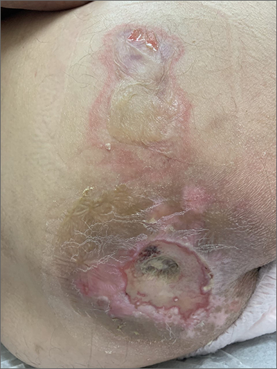
This patient had sustained multiple pressure injuries. The superior aspect of this image shows bullous change with intact dermis, which would classify that area of injury as a Stage 2 pressure injury.1 An older injury in the coccygeal area was through the dermis (Stage 3), with some eschar seen at the base, making that area unstageable.1 (There may have been deeper injury under the eschar.)
This patient was at heightened risk for pressure injury because of his paraplegia.2 Fortunately, he had some preserved sensation. However, his rotator cuff surgery made it harder for him to smoothly transfer to and from the wheelchair, leading to sheer forces against his skin. Social determinates of health care pose an additional risk for pressure injuries. Without a properly fitted wheelchair and cushion, there is an increased risk of localized pressure over both bony prominences and parts of the body that come into contact with mechanical elements of the wheelchair.
Treatment for all pressure injuries includes relief of pressure on the affected area. In this patient’s case, he had to stay in bed (and out of the wheelchair) so that he could heal.
This patient was very knowledgeable about his condition and pressure injuries. He had already arranged for a wheelchair fitting and a visit with the wound care team. His Stage 2 injury had a bullous change instead of absent epithelium, so rather than an adherent hydrocolloid dressing (which would likely remove the loosened epithelium), he was provided with a nonadherent dressing to the area, then an absorbent foam overdressing.
An image of the patient’s deeper sacral injury was shared with the wound care team, which recommended filling the area with a silver rope dressing for its absorptive filler and antibacterial properties. The area was then covered with an absorbent foam. The patient planned to follow up with the Wound Care Clinic for reevaluation and ongoing treatment.
Images and text courtesy of Daniel Stulberg, MD, FAAFP, Professor and Chair, Department of Family and Community Medicine, Western Michigan University Homer Stryker, MD School of Medicine Kalamazoo.
1. 2019 Guideline. The National Pressure Injury Advisory Panel. Accessed October 9, 2022. https://npiap.com/page/2019Guideline
2. Ricci JA, Bayer LR, Orgill DP. Evidence-based medicine: the evaluation and treatment of pressure injuries. Plast Reconstr Surg. 2017;139:275e-286e. doi: 10.1097/PRS.0000000000002850
This patient had sustained multiple pressure injuries. The superior aspect of this image shows bullous change with intact dermis, which would classify that area of injury as a Stage 2 pressure injury.1 An older injury in the coccygeal area was through the dermis (Stage 3), with some eschar seen at the base, making that area unstageable.1 (There may have been deeper injury under the eschar.)
This patient was at heightened risk for pressure injury because of his paraplegia.2 Fortunately, he had some preserved sensation. However, his rotator cuff surgery made it harder for him to smoothly transfer to and from the wheelchair, leading to sheer forces against his skin. Social determinates of health care pose an additional risk for pressure injuries. Without a properly fitted wheelchair and cushion, there is an increased risk of localized pressure over both bony prominences and parts of the body that come into contact with mechanical elements of the wheelchair.
Treatment for all pressure injuries includes relief of pressure on the affected area. In this patient’s case, he had to stay in bed (and out of the wheelchair) so that he could heal.
This patient was very knowledgeable about his condition and pressure injuries. He had already arranged for a wheelchair fitting and a visit with the wound care team. His Stage 2 injury had a bullous change instead of absent epithelium, so rather than an adherent hydrocolloid dressing (which would likely remove the loosened epithelium), he was provided with a nonadherent dressing to the area, then an absorbent foam overdressing.
An image of the patient’s deeper sacral injury was shared with the wound care team, which recommended filling the area with a silver rope dressing for its absorptive filler and antibacterial properties. The area was then covered with an absorbent foam. The patient planned to follow up with the Wound Care Clinic for reevaluation and ongoing treatment.
Images and text courtesy of Daniel Stulberg, MD, FAAFP, Professor and Chair, Department of Family and Community Medicine, Western Michigan University Homer Stryker, MD School of Medicine Kalamazoo.
This patient had sustained multiple pressure injuries. The superior aspect of this image shows bullous change with intact dermis, which would classify that area of injury as a Stage 2 pressure injury.1 An older injury in the coccygeal area was through the dermis (Stage 3), with some eschar seen at the base, making that area unstageable.1 (There may have been deeper injury under the eschar.)
This patient was at heightened risk for pressure injury because of his paraplegia.2 Fortunately, he had some preserved sensation. However, his rotator cuff surgery made it harder for him to smoothly transfer to and from the wheelchair, leading to sheer forces against his skin. Social determinates of health care pose an additional risk for pressure injuries. Without a properly fitted wheelchair and cushion, there is an increased risk of localized pressure over both bony prominences and parts of the body that come into contact with mechanical elements of the wheelchair.
Treatment for all pressure injuries includes relief of pressure on the affected area. In this patient’s case, he had to stay in bed (and out of the wheelchair) so that he could heal.
This patient was very knowledgeable about his condition and pressure injuries. He had already arranged for a wheelchair fitting and a visit with the wound care team. His Stage 2 injury had a bullous change instead of absent epithelium, so rather than an adherent hydrocolloid dressing (which would likely remove the loosened epithelium), he was provided with a nonadherent dressing to the area, then an absorbent foam overdressing.
An image of the patient’s deeper sacral injury was shared with the wound care team, which recommended filling the area with a silver rope dressing for its absorptive filler and antibacterial properties. The area was then covered with an absorbent foam. The patient planned to follow up with the Wound Care Clinic for reevaluation and ongoing treatment.
Images and text courtesy of Daniel Stulberg, MD, FAAFP, Professor and Chair, Department of Family and Community Medicine, Western Michigan University Homer Stryker, MD School of Medicine Kalamazoo.
1. 2019 Guideline. The National Pressure Injury Advisory Panel. Accessed October 9, 2022. https://npiap.com/page/2019Guideline
2. Ricci JA, Bayer LR, Orgill DP. Evidence-based medicine: the evaluation and treatment of pressure injuries. Plast Reconstr Surg. 2017;139:275e-286e. doi: 10.1097/PRS.0000000000002850
1. 2019 Guideline. The National Pressure Injury Advisory Panel. Accessed October 9, 2022. https://npiap.com/page/2019Guideline
2. Ricci JA, Bayer LR, Orgill DP. Evidence-based medicine: the evaluation and treatment of pressure injuries. Plast Reconstr Surg. 2017;139:275e-286e. doi: 10.1097/PRS.0000000000002850
Cerebral palsy: Video clues suggest dystonia
CINCINNATI – Dystonia is a frequent complication seen in cerebral palsy, but it often goes undiagnosed. Using a unique video analysis, researchers have identified some movement features that have the potential to simplify diagnosis.
“[We have] previously demonstrated that by the age of 5 years, only 30% of children seen in a clinical setting have had their predominant motor phenotype identified, including dystonia. This helps demonstrate a broad diagnostic gap and the need for novel solutions,” said Laura Gilbert, DO, during her presentation of the results at the 2022 annual meeting of the Child Neurology Society.
Diagnosis of dystonia is challenging because of its clinical variability, and diagnostic tools often require a trained physician, which limits access to diagnoses. Expert clinician consensus therefore remains the gold standard for diagnosis of dystonia.
Another clinical need is that specific features of dystonia have not been well described in the upper extremities, and the research suggests there could be differences in brain injuries contributing to dystonia in the two domains.
The researchers set out to discover expert-identified features of patient videos that could be used to allow nonexperts to make a diagnosis of dystonia.
The researchers analyzed 26 videos with upper extremity exam maneuvers performed on children with periventricular leukomalacia at St. Louis Children’s Hospital Cerebral Palsy Center from 2005 to 2018. Among the study cohort, 65% of patients were male, 77% were White, and 11% were Black; 24% of patients were Gross Motor Function Classification Scale I, 24% were GMFCS II, 24% were GMFCS III, 16% were GMFCS IV, and 12% were GMFCS V. A total of 12% of patients were older than 20, 11% were aged 15-20, 38% were aged 10-15, 31% were aged 5-10, and 8% were age 5 or younger.
Video clues aid diagnosis
Three pediatric movement disorder specialists independently reviewed each video and assessed severity of dystonia. They then met over Zoom to reach a diagnostic consensus for each case.
The research team performed a content analysis of the experts’ discussions and identified specific statement fragments. The frequency of these fragments was then linked to severity of dystonia.
A total of 45% of the statement fragments referenced movement codes, which in turn comprised five content areas: 33% referenced a body part, 24% focused on laterality, 22% described movement features, 18% an action, and 3% described exam maneuvers. Examples included shoulder as a body part, flexion as an action descriptor, brisk as a movement feature, unilateral, and finger-nose-finger for exam maneuver.
With increasing dystonia severity, the shoulder was more often cited and hand was cited less often. Mirror movements, defined as involuntary, contralateral movements that are similar to the voluntary action, occurred more often in patients with no dystonia or only mild dystonia. Variability of movement over time, which is a distinguishing feature found in lower extremities, was not significantly associated with dystonia severity.
Within the category of exam maneuver, hand opening and closing was the most commonly cited, and it was cited more frequently among individuals with mild dystonia (70% vs. about 10% for both no dystonia and moderate to severe dystonia; P < .005).
“So how can we adopt this clinically? First, we can add in a very brief exam maneuver of hand opening and closing that can help assess for mild dystonia. Shoulder involvement may suggest more severe dystonia, and we must recognize the dystonia features seem to differ by body region and the triggering task. Overall, to help improve dystonia diagnosis, we must continue to work towards understanding these salient features to fully grasp the breadth of dystonia manifestations in people with [cerebral palsy],” said Dr. Gilbert, who is a pediatric movements disorder fellow at Washington University in St. Louis.
Key features help determine dystonia severity
The study is particularly interesting for its different findings in upper extremities versus lower extremities, according to Keith Coffman, MD, who comoderated the session where the study was presented. “That same group showed that there are very clear differences in lower-extremity function, but when they looked at upper extremity, there really weren’t robust differences. What it may show is that the features of cerebral palsy regarding dystonia may be very dependent on what type of injury you have to your brain. Because when you think about where the motor fibers that provide leg function, they live along the medial walls of the brain right along the midline, whereas the representation of the hand and arm are more out on the lateral side of the brain. So it may be that those regional anatomy differences and where the injury occurred could be at the baseline of why they had such differences in motor function,” said Dr. Coffman, who is a professor of pediatrics at University of Missouri–Kansas City and director of the movement disorders program at Children’s Mercy Hospital, also in Kansas City, Mo.
He suggested that the researchers might also do kinematic analysis of the videos to make predictions using quantitative differences in movement.
The research has the potential to improve dystonia diagnosis, according to comoderator Marc Patterson, MD, professor of neurology, pediatrics, and medical genetics at Mayo Clinic in Rochester, Minn. “I think they really pointed to some key features that can help clinicians distinguish [dystonia severity]. Something like the speed of opening and closing the hands [is a] fairly simple thing. That was to me the chief value of that study,” Dr. Patterson said.
Dr. Gilbert reported no relevant disclosures.
CINCINNATI – Dystonia is a frequent complication seen in cerebral palsy, but it often goes undiagnosed. Using a unique video analysis, researchers have identified some movement features that have the potential to simplify diagnosis.
“[We have] previously demonstrated that by the age of 5 years, only 30% of children seen in a clinical setting have had their predominant motor phenotype identified, including dystonia. This helps demonstrate a broad diagnostic gap and the need for novel solutions,” said Laura Gilbert, DO, during her presentation of the results at the 2022 annual meeting of the Child Neurology Society.
Diagnosis of dystonia is challenging because of its clinical variability, and diagnostic tools often require a trained physician, which limits access to diagnoses. Expert clinician consensus therefore remains the gold standard for diagnosis of dystonia.
Another clinical need is that specific features of dystonia have not been well described in the upper extremities, and the research suggests there could be differences in brain injuries contributing to dystonia in the two domains.
The researchers set out to discover expert-identified features of patient videos that could be used to allow nonexperts to make a diagnosis of dystonia.
The researchers analyzed 26 videos with upper extremity exam maneuvers performed on children with periventricular leukomalacia at St. Louis Children’s Hospital Cerebral Palsy Center from 2005 to 2018. Among the study cohort, 65% of patients were male, 77% were White, and 11% were Black; 24% of patients were Gross Motor Function Classification Scale I, 24% were GMFCS II, 24% were GMFCS III, 16% were GMFCS IV, and 12% were GMFCS V. A total of 12% of patients were older than 20, 11% were aged 15-20, 38% were aged 10-15, 31% were aged 5-10, and 8% were age 5 or younger.
Video clues aid diagnosis
Three pediatric movement disorder specialists independently reviewed each video and assessed severity of dystonia. They then met over Zoom to reach a diagnostic consensus for each case.
The research team performed a content analysis of the experts’ discussions and identified specific statement fragments. The frequency of these fragments was then linked to severity of dystonia.
A total of 45% of the statement fragments referenced movement codes, which in turn comprised five content areas: 33% referenced a body part, 24% focused on laterality, 22% described movement features, 18% an action, and 3% described exam maneuvers. Examples included shoulder as a body part, flexion as an action descriptor, brisk as a movement feature, unilateral, and finger-nose-finger for exam maneuver.
With increasing dystonia severity, the shoulder was more often cited and hand was cited less often. Mirror movements, defined as involuntary, contralateral movements that are similar to the voluntary action, occurred more often in patients with no dystonia or only mild dystonia. Variability of movement over time, which is a distinguishing feature found in lower extremities, was not significantly associated with dystonia severity.
Within the category of exam maneuver, hand opening and closing was the most commonly cited, and it was cited more frequently among individuals with mild dystonia (70% vs. about 10% for both no dystonia and moderate to severe dystonia; P < .005).
“So how can we adopt this clinically? First, we can add in a very brief exam maneuver of hand opening and closing that can help assess for mild dystonia. Shoulder involvement may suggest more severe dystonia, and we must recognize the dystonia features seem to differ by body region and the triggering task. Overall, to help improve dystonia diagnosis, we must continue to work towards understanding these salient features to fully grasp the breadth of dystonia manifestations in people with [cerebral palsy],” said Dr. Gilbert, who is a pediatric movements disorder fellow at Washington University in St. Louis.
Key features help determine dystonia severity
The study is particularly interesting for its different findings in upper extremities versus lower extremities, according to Keith Coffman, MD, who comoderated the session where the study was presented. “That same group showed that there are very clear differences in lower-extremity function, but when they looked at upper extremity, there really weren’t robust differences. What it may show is that the features of cerebral palsy regarding dystonia may be very dependent on what type of injury you have to your brain. Because when you think about where the motor fibers that provide leg function, they live along the medial walls of the brain right along the midline, whereas the representation of the hand and arm are more out on the lateral side of the brain. So it may be that those regional anatomy differences and where the injury occurred could be at the baseline of why they had such differences in motor function,” said Dr. Coffman, who is a professor of pediatrics at University of Missouri–Kansas City and director of the movement disorders program at Children’s Mercy Hospital, also in Kansas City, Mo.
He suggested that the researchers might also do kinematic analysis of the videos to make predictions using quantitative differences in movement.
The research has the potential to improve dystonia diagnosis, according to comoderator Marc Patterson, MD, professor of neurology, pediatrics, and medical genetics at Mayo Clinic in Rochester, Minn. “I think they really pointed to some key features that can help clinicians distinguish [dystonia severity]. Something like the speed of opening and closing the hands [is a] fairly simple thing. That was to me the chief value of that study,” Dr. Patterson said.
Dr. Gilbert reported no relevant disclosures.
CINCINNATI – Dystonia is a frequent complication seen in cerebral palsy, but it often goes undiagnosed. Using a unique video analysis, researchers have identified some movement features that have the potential to simplify diagnosis.
“[We have] previously demonstrated that by the age of 5 years, only 30% of children seen in a clinical setting have had their predominant motor phenotype identified, including dystonia. This helps demonstrate a broad diagnostic gap and the need for novel solutions,” said Laura Gilbert, DO, during her presentation of the results at the 2022 annual meeting of the Child Neurology Society.
Diagnosis of dystonia is challenging because of its clinical variability, and diagnostic tools often require a trained physician, which limits access to diagnoses. Expert clinician consensus therefore remains the gold standard for diagnosis of dystonia.
Another clinical need is that specific features of dystonia have not been well described in the upper extremities, and the research suggests there could be differences in brain injuries contributing to dystonia in the two domains.
The researchers set out to discover expert-identified features of patient videos that could be used to allow nonexperts to make a diagnosis of dystonia.
The researchers analyzed 26 videos with upper extremity exam maneuvers performed on children with periventricular leukomalacia at St. Louis Children’s Hospital Cerebral Palsy Center from 2005 to 2018. Among the study cohort, 65% of patients were male, 77% were White, and 11% were Black; 24% of patients were Gross Motor Function Classification Scale I, 24% were GMFCS II, 24% were GMFCS III, 16% were GMFCS IV, and 12% were GMFCS V. A total of 12% of patients were older than 20, 11% were aged 15-20, 38% were aged 10-15, 31% were aged 5-10, and 8% were age 5 or younger.
Video clues aid diagnosis
Three pediatric movement disorder specialists independently reviewed each video and assessed severity of dystonia. They then met over Zoom to reach a diagnostic consensus for each case.
The research team performed a content analysis of the experts’ discussions and identified specific statement fragments. The frequency of these fragments was then linked to severity of dystonia.
A total of 45% of the statement fragments referenced movement codes, which in turn comprised five content areas: 33% referenced a body part, 24% focused on laterality, 22% described movement features, 18% an action, and 3% described exam maneuvers. Examples included shoulder as a body part, flexion as an action descriptor, brisk as a movement feature, unilateral, and finger-nose-finger for exam maneuver.
With increasing dystonia severity, the shoulder was more often cited and hand was cited less often. Mirror movements, defined as involuntary, contralateral movements that are similar to the voluntary action, occurred more often in patients with no dystonia or only mild dystonia. Variability of movement over time, which is a distinguishing feature found in lower extremities, was not significantly associated with dystonia severity.
Within the category of exam maneuver, hand opening and closing was the most commonly cited, and it was cited more frequently among individuals with mild dystonia (70% vs. about 10% for both no dystonia and moderate to severe dystonia; P < .005).
“So how can we adopt this clinically? First, we can add in a very brief exam maneuver of hand opening and closing that can help assess for mild dystonia. Shoulder involvement may suggest more severe dystonia, and we must recognize the dystonia features seem to differ by body region and the triggering task. Overall, to help improve dystonia diagnosis, we must continue to work towards understanding these salient features to fully grasp the breadth of dystonia manifestations in people with [cerebral palsy],” said Dr. Gilbert, who is a pediatric movements disorder fellow at Washington University in St. Louis.
Key features help determine dystonia severity
The study is particularly interesting for its different findings in upper extremities versus lower extremities, according to Keith Coffman, MD, who comoderated the session where the study was presented. “That same group showed that there are very clear differences in lower-extremity function, but when they looked at upper extremity, there really weren’t robust differences. What it may show is that the features of cerebral palsy regarding dystonia may be very dependent on what type of injury you have to your brain. Because when you think about where the motor fibers that provide leg function, they live along the medial walls of the brain right along the midline, whereas the representation of the hand and arm are more out on the lateral side of the brain. So it may be that those regional anatomy differences and where the injury occurred could be at the baseline of why they had such differences in motor function,” said Dr. Coffman, who is a professor of pediatrics at University of Missouri–Kansas City and director of the movement disorders program at Children’s Mercy Hospital, also in Kansas City, Mo.
He suggested that the researchers might also do kinematic analysis of the videos to make predictions using quantitative differences in movement.
The research has the potential to improve dystonia diagnosis, according to comoderator Marc Patterson, MD, professor of neurology, pediatrics, and medical genetics at Mayo Clinic in Rochester, Minn. “I think they really pointed to some key features that can help clinicians distinguish [dystonia severity]. Something like the speed of opening and closing the hands [is a] fairly simple thing. That was to me the chief value of that study,” Dr. Patterson said.
Dr. Gilbert reported no relevant disclosures.
AT CNS 2022
Newborn with white oral lesions
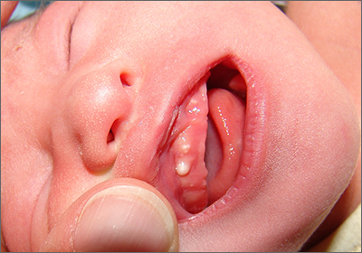
These lesions, called Bohn nodules, manifest on the buccal or lingual portion of the maxillary alveolar ridge and, less frequently, on the mandibular alveolar ridge. Because Bohn nodules are white and have a firm consistency, they are often confused with teeth. They can be differentiated by location, as teeth usually erupt from the distal aspect of the alveolar ridge.
Bohn nodules are epithelial cysts that are filled with keratin, which gives them their white color. They are caused by portions of epithelium that get trapped under surrounding epithelial cells. Bohn nodules usually resolve when the overlying epithelium ruptures and releases the keratinaceous material (usually by the time the child is 3 months of age).1
These nodules can be confused with neonatal or supernumerary teeth. Neonatal teeth can be abnormally small and pointed; they are true deciduous teeth that have erupted early. If they are removed, the child will not replace them until the time of their adult tooth eruption. Additionally, there are supernumerary teeth, which are extra teeth that are often abnormally shaped and loosely adherent. These abnormal teeth warrant extraction to avoid trauma to the tongue or aspiration.2
In this case, the family was advised regarding the benign nature of the nodules and the expectation that they would spontaneously resolve.
Images and text courtesy of Daniel Stulberg, MD, FAAFP, Professor and Chair, Department of Family and Community Medicine, Western Michigan University Homer Stryker, MD School of Medicine Kalamazoo.
1. Gupta N, Ramji S. Bohn's nodules: an under-recognised entity. Arch Dis Child Fetal Neonatal Ed. 2013;98:F464. doi: 10.1136/archdischild-2012-302922
2. DeSeta M, Holden E, Siddik D, et al. Natal and neonatal teeth: a review and case series. Br Dent J. 2022;232:449-453. doi: 10.1038/s41415-022-4091-3
These lesions, called Bohn nodules, manifest on the buccal or lingual portion of the maxillary alveolar ridge and, less frequently, on the mandibular alveolar ridge. Because Bohn nodules are white and have a firm consistency, they are often confused with teeth. They can be differentiated by location, as teeth usually erupt from the distal aspect of the alveolar ridge.
Bohn nodules are epithelial cysts that are filled with keratin, which gives them their white color. They are caused by portions of epithelium that get trapped under surrounding epithelial cells. Bohn nodules usually resolve when the overlying epithelium ruptures and releases the keratinaceous material (usually by the time the child is 3 months of age).1
These nodules can be confused with neonatal or supernumerary teeth. Neonatal teeth can be abnormally small and pointed; they are true deciduous teeth that have erupted early. If they are removed, the child will not replace them until the time of their adult tooth eruption. Additionally, there are supernumerary teeth, which are extra teeth that are often abnormally shaped and loosely adherent. These abnormal teeth warrant extraction to avoid trauma to the tongue or aspiration.2
In this case, the family was advised regarding the benign nature of the nodules and the expectation that they would spontaneously resolve.
Images and text courtesy of Daniel Stulberg, MD, FAAFP, Professor and Chair, Department of Family and Community Medicine, Western Michigan University Homer Stryker, MD School of Medicine Kalamazoo.
These lesions, called Bohn nodules, manifest on the buccal or lingual portion of the maxillary alveolar ridge and, less frequently, on the mandibular alveolar ridge. Because Bohn nodules are white and have a firm consistency, they are often confused with teeth. They can be differentiated by location, as teeth usually erupt from the distal aspect of the alveolar ridge.
Bohn nodules are epithelial cysts that are filled with keratin, which gives them their white color. They are caused by portions of epithelium that get trapped under surrounding epithelial cells. Bohn nodules usually resolve when the overlying epithelium ruptures and releases the keratinaceous material (usually by the time the child is 3 months of age).1
These nodules can be confused with neonatal or supernumerary teeth. Neonatal teeth can be abnormally small and pointed; they are true deciduous teeth that have erupted early. If they are removed, the child will not replace them until the time of their adult tooth eruption. Additionally, there are supernumerary teeth, which are extra teeth that are often abnormally shaped and loosely adherent. These abnormal teeth warrant extraction to avoid trauma to the tongue or aspiration.2
In this case, the family was advised regarding the benign nature of the nodules and the expectation that they would spontaneously resolve.
Images and text courtesy of Daniel Stulberg, MD, FAAFP, Professor and Chair, Department of Family and Community Medicine, Western Michigan University Homer Stryker, MD School of Medicine Kalamazoo.
1. Gupta N, Ramji S. Bohn's nodules: an under-recognised entity. Arch Dis Child Fetal Neonatal Ed. 2013;98:F464. doi: 10.1136/archdischild-2012-302922
2. DeSeta M, Holden E, Siddik D, et al. Natal and neonatal teeth: a review and case series. Br Dent J. 2022;232:449-453. doi: 10.1038/s41415-022-4091-3
1. Gupta N, Ramji S. Bohn's nodules: an under-recognised entity. Arch Dis Child Fetal Neonatal Ed. 2013;98:F464. doi: 10.1136/archdischild-2012-302922
2. DeSeta M, Holden E, Siddik D, et al. Natal and neonatal teeth: a review and case series. Br Dent J. 2022;232:449-453. doi: 10.1038/s41415-022-4091-3
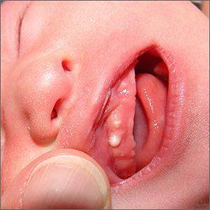
Self-worth training boosts ketamine’s effects in severe depression

The double-blind, randomized clinical trial is the first to assess combining ketamine with a low-cost protective learning program, researchers note.
They add that the findings are an important step toward long-lasting depression treatment for millions of patients whose depression does not improve following first-line therapies.
“One of the biggest challenges in psychiatry and psychology is seeing evidence of longer-term benefits and longer-term compliance,” lead investigator Rebecca B. Price, PhD, associate professor of psychiatry and psychology, University of Pittsburgh, told this news organization.
“Anything that can get somebody well quickly and keep them well for some length of time is really exciting – and a whole paradigm shift for how things have been done up to now,” Dr. Price said.
The findings were published online in the American Journal of Psychiatry.
Promoting self-worth
About one-third of patients with depression remain treatment-resistant even after trying different medications at different doses and at different combinations, the investigators note.
Ketamine and esketamine, a nasal spray formulation of the drug, have been shown previously to improve symptoms in patients with TRD. While the benefits are evident within a few hours of treatment, the effects often wane after just a few weeks.
Ketamine and esketamine must be administered in a clinical setting and patients must be monitored for at least 2 hours after treatment. Repeat dosing is costly, both in time and expense, so clinical researchers have been studying ways to extend the drug’s effects without additional treatments.
The new study combined ketamine treatment with a computer-based active automated self-association training (ASAT) program that the researchers developed. It uses positive words and imagery to promote positive self-image and self-worth.
The trial included 154 adults with treatment-resistant unipolar depression whose symptoms persisted after therapy with at least two medications. Participants received an IV infusion of ketamine 0.5 mg/kg plus active ASAT (n = 53), saline plus active ASAT (n = 51), or ketamine plus sham ASAT (n = 50).
The active program used words like “sweet,” “lovable,” and “worthy” that appeared on the screen interspersed with images of people smiling and the patient’s own photo. Participants were also asked to complete certain mouse-tracking tasks during the session.
The sham ASAT was similar but included neutral words and images. ASAT and sham ASAT were delivered twice daily over 4 consecutive days for 20 minutes.
Clear benefit
Results showed that ketamine rapidly and significantly reduced depression scores within 24 hours of treatment (group-by-time interaction: standardized beta, –1.30; 95% confidence interval, –1.89 to –0.70).
Depression scores in the ketamine-plus-ASAT group remained low and stable over a 30-day period, compared with the saline-plus-ASAT group (standardized beta, –0.61; 95% CI, –0.95 to –0.28).
Participants who received ketamine plus sham ASAT saw initial improvement in symptoms immediately following infusion, but depression symptoms returned after a few weeks.
While researchers hoped to see positive effects from ASAT, “I certainly did not expect to see something so clear to jump right out,” Dr. Price said.
The investigators are now examining whether the computer program can be administered effectively remotely and whether its effects are equally beneficial following treatment with esketamine.
Greatest unmet need
Gerard Sanacora, MD, PhD, professor of psychiatry, and director, Yale Depression Research Program, Yale University, New Haven, Conn., said that extending the effects of ketamine or esketamine without additional dosing is “probably the greatest unmet need in relation to treatments with ketamine and esketamine.”
He added that there are large economic, time, and access burdens associated with the treatment of ketamine.
“Anything we can do to reduce the number of treatments needed or increase the sustainability or the duration of effect would be a tremendous benefit,” said Dr. Sanacora, who was not involved with the research.
Adding an easily accessible, nonpharmacological therapeutic approach to ketamine treatment could be valuable, he said – but more research is needed.
“I’m not sure that this specific associated positive thinking is really the critical component. I think we still have some work to do there. But it does demonstrate that we can use ancillary or augmenting nonpharmacological treatments to extend the effect,” Dr. Sanacora said.
The study was funded by the National Institute of Mental Health and Clinical and Translational Science Institute at the University of Pittsburgh. Dr. Price is the named inventor on a University of Pittsburgh–owned provisional patent filing related to the combination intervention described in this report. Dr. Sanacora reports no relevant financial relationships.
A version of this article first appeared on Medscape.com.
The double-blind, randomized clinical trial is the first to assess combining ketamine with a low-cost protective learning program, researchers note.
They add that the findings are an important step toward long-lasting depression treatment for millions of patients whose depression does not improve following first-line therapies.
“One of the biggest challenges in psychiatry and psychology is seeing evidence of longer-term benefits and longer-term compliance,” lead investigator Rebecca B. Price, PhD, associate professor of psychiatry and psychology, University of Pittsburgh, told this news organization.
“Anything that can get somebody well quickly and keep them well for some length of time is really exciting – and a whole paradigm shift for how things have been done up to now,” Dr. Price said.
The findings were published online in the American Journal of Psychiatry.
Promoting self-worth
About one-third of patients with depression remain treatment-resistant even after trying different medications at different doses and at different combinations, the investigators note.
Ketamine and esketamine, a nasal spray formulation of the drug, have been shown previously to improve symptoms in patients with TRD. While the benefits are evident within a few hours of treatment, the effects often wane after just a few weeks.
Ketamine and esketamine must be administered in a clinical setting and patients must be monitored for at least 2 hours after treatment. Repeat dosing is costly, both in time and expense, so clinical researchers have been studying ways to extend the drug’s effects without additional treatments.
The new study combined ketamine treatment with a computer-based active automated self-association training (ASAT) program that the researchers developed. It uses positive words and imagery to promote positive self-image and self-worth.
The trial included 154 adults with treatment-resistant unipolar depression whose symptoms persisted after therapy with at least two medications. Participants received an IV infusion of ketamine 0.5 mg/kg plus active ASAT (n = 53), saline plus active ASAT (n = 51), or ketamine plus sham ASAT (n = 50).
The active program used words like “sweet,” “lovable,” and “worthy” that appeared on the screen interspersed with images of people smiling and the patient’s own photo. Participants were also asked to complete certain mouse-tracking tasks during the session.
The sham ASAT was similar but included neutral words and images. ASAT and sham ASAT were delivered twice daily over 4 consecutive days for 20 minutes.
Clear benefit
Results showed that ketamine rapidly and significantly reduced depression scores within 24 hours of treatment (group-by-time interaction: standardized beta, –1.30; 95% confidence interval, –1.89 to –0.70).
Depression scores in the ketamine-plus-ASAT group remained low and stable over a 30-day period, compared with the saline-plus-ASAT group (standardized beta, –0.61; 95% CI, –0.95 to –0.28).
Participants who received ketamine plus sham ASAT saw initial improvement in symptoms immediately following infusion, but depression symptoms returned after a few weeks.
While researchers hoped to see positive effects from ASAT, “I certainly did not expect to see something so clear to jump right out,” Dr. Price said.
The investigators are now examining whether the computer program can be administered effectively remotely and whether its effects are equally beneficial following treatment with esketamine.
Greatest unmet need
Gerard Sanacora, MD, PhD, professor of psychiatry, and director, Yale Depression Research Program, Yale University, New Haven, Conn., said that extending the effects of ketamine or esketamine without additional dosing is “probably the greatest unmet need in relation to treatments with ketamine and esketamine.”
He added that there are large economic, time, and access burdens associated with the treatment of ketamine.
“Anything we can do to reduce the number of treatments needed or increase the sustainability or the duration of effect would be a tremendous benefit,” said Dr. Sanacora, who was not involved with the research.
Adding an easily accessible, nonpharmacological therapeutic approach to ketamine treatment could be valuable, he said – but more research is needed.
“I’m not sure that this specific associated positive thinking is really the critical component. I think we still have some work to do there. But it does demonstrate that we can use ancillary or augmenting nonpharmacological treatments to extend the effect,” Dr. Sanacora said.
The study was funded by the National Institute of Mental Health and Clinical and Translational Science Institute at the University of Pittsburgh. Dr. Price is the named inventor on a University of Pittsburgh–owned provisional patent filing related to the combination intervention described in this report. Dr. Sanacora reports no relevant financial relationships.
A version of this article first appeared on Medscape.com.
The double-blind, randomized clinical trial is the first to assess combining ketamine with a low-cost protective learning program, researchers note.
They add that the findings are an important step toward long-lasting depression treatment for millions of patients whose depression does not improve following first-line therapies.
“One of the biggest challenges in psychiatry and psychology is seeing evidence of longer-term benefits and longer-term compliance,” lead investigator Rebecca B. Price, PhD, associate professor of psychiatry and psychology, University of Pittsburgh, told this news organization.
“Anything that can get somebody well quickly and keep them well for some length of time is really exciting – and a whole paradigm shift for how things have been done up to now,” Dr. Price said.
The findings were published online in the American Journal of Psychiatry.
Promoting self-worth
About one-third of patients with depression remain treatment-resistant even after trying different medications at different doses and at different combinations, the investigators note.
Ketamine and esketamine, a nasal spray formulation of the drug, have been shown previously to improve symptoms in patients with TRD. While the benefits are evident within a few hours of treatment, the effects often wane after just a few weeks.
Ketamine and esketamine must be administered in a clinical setting and patients must be monitored for at least 2 hours after treatment. Repeat dosing is costly, both in time and expense, so clinical researchers have been studying ways to extend the drug’s effects without additional treatments.
The new study combined ketamine treatment with a computer-based active automated self-association training (ASAT) program that the researchers developed. It uses positive words and imagery to promote positive self-image and self-worth.
The trial included 154 adults with treatment-resistant unipolar depression whose symptoms persisted after therapy with at least two medications. Participants received an IV infusion of ketamine 0.5 mg/kg plus active ASAT (n = 53), saline plus active ASAT (n = 51), or ketamine plus sham ASAT (n = 50).
The active program used words like “sweet,” “lovable,” and “worthy” that appeared on the screen interspersed with images of people smiling and the patient’s own photo. Participants were also asked to complete certain mouse-tracking tasks during the session.
The sham ASAT was similar but included neutral words and images. ASAT and sham ASAT were delivered twice daily over 4 consecutive days for 20 minutes.
Clear benefit
Results showed that ketamine rapidly and significantly reduced depression scores within 24 hours of treatment (group-by-time interaction: standardized beta, –1.30; 95% confidence interval, –1.89 to –0.70).
Depression scores in the ketamine-plus-ASAT group remained low and stable over a 30-day period, compared with the saline-plus-ASAT group (standardized beta, –0.61; 95% CI, –0.95 to –0.28).
Participants who received ketamine plus sham ASAT saw initial improvement in symptoms immediately following infusion, but depression symptoms returned after a few weeks.
While researchers hoped to see positive effects from ASAT, “I certainly did not expect to see something so clear to jump right out,” Dr. Price said.
The investigators are now examining whether the computer program can be administered effectively remotely and whether its effects are equally beneficial following treatment with esketamine.
Greatest unmet need
Gerard Sanacora, MD, PhD, professor of psychiatry, and director, Yale Depression Research Program, Yale University, New Haven, Conn., said that extending the effects of ketamine or esketamine without additional dosing is “probably the greatest unmet need in relation to treatments with ketamine and esketamine.”
He added that there are large economic, time, and access burdens associated with the treatment of ketamine.
“Anything we can do to reduce the number of treatments needed or increase the sustainability or the duration of effect would be a tremendous benefit,” said Dr. Sanacora, who was not involved with the research.
Adding an easily accessible, nonpharmacological therapeutic approach to ketamine treatment could be valuable, he said – but more research is needed.
“I’m not sure that this specific associated positive thinking is really the critical component. I think we still have some work to do there. But it does demonstrate that we can use ancillary or augmenting nonpharmacological treatments to extend the effect,” Dr. Sanacora said.
The study was funded by the National Institute of Mental Health and Clinical and Translational Science Institute at the University of Pittsburgh. Dr. Price is the named inventor on a University of Pittsburgh–owned provisional patent filing related to the combination intervention described in this report. Dr. Sanacora reports no relevant financial relationships.
A version of this article first appeared on Medscape.com.
FROM AMERICAN JOURNAL OF PSYCHIATRY

‘Disturbing’ lack of follow-up care after psychiatric crises
There is a concerning lack of follow-up care for young people who experience a mental health crisis, new research suggests.
The follow-up rate was less than 30% for those who had visited an ED.
The strongest predictor of follow-up was having received both primary and mental health care during the 6 months prior to using the acute service.
“For people discharging folks after a psychiatric crisis, whether it be in a hospital or emergency room setting, connecting them with their outpatient provider to ensure the transfer of care and continuity of care is vitally important to reduce risks for this population,” coinvestigator Brian Skehan, MD, PhD, assistant professor and psychiatrist, University of Massachusetts, Worcester, said during a press briefing.
If these discharged patients do not have a provider, “make sure they get one,” Lisa Dixon, MD, editor-in-chief of Psychiatric Services, added during the same briefing. “That’s the gift of life potentially for these young people.”
The findings were published online in Psychiatric Services.
Alarming trends
The alarming suicide trends among youths were exacerbated by the COVID-19 pandemic, Dr. Skehan noted.
He cited a 2021 study that showed more than 44% of high school students experienced persistent sadness or hopelessness over the previous year, 1 in 5 seriously considered suicide, and almost 1 in 10 actually attempted suicide.
“When we look at the number of young adults and adolescents struggling with behavioral health issues, the data trend is disturbing nationwide,” Dr. Skehan said.
The current study included participants aged 12-27 years who had private insurance. Many youth in this age category are experiencing significant changes, such as moving from high school to college and from pediatric providers to adult providers – and some “get lost in this transition,” said Dr. Skehan.
He noted many inpatient psychiatric units are not geared to young adults. “They may miss out on some aspects of inpatient care because it’s not geared to their developmental stage,” he said.
Assessing U.S. patient data in the IBM MarketScan commercial database (2013-2018), the researchers created two study samples: 95,153 inpatients and 108,576 patients who used the ED. All had an acute event stemming from a mental health condition.
The investigators explored the role of “established” outpatient care, defined as having had at least one visit with a provider of primary or mental health care in the 6 months prior to the acute psychiatric event.
Covariates included age at time of service (aged 12-17 years or 18-27 years), gender, health care plan type, psychiatric diagnosis, whether the acute event was self-harm or suicide related, and medical complexity.
Low follow-up rates
In the inpatient group, the average age was 18.9 years, the most common length of hospital stay was 4-6 days, and 1.5% left against medical advice. The most common primary diagnosis was major depression (53.7%), followed by bipolar disorder (22.3%). The least common disorders were PTSD, comorbid eating disorders, and disruptive disorders.
About one-third of participants had used both primary and mental health care during the 6 months before hospitalization, whereas 22.8% had no established outpatient care. Established care was most common among those with comorbid eating disorders and least common among those with psychotic disorders.
Results showed 42.7% of the hospitalized patients received follow up within 7 days and 67.4% received follow up within 30 days.
The strongest predictor of mental health follow-up care was established outpatient care. Compared with those who had no such care, those who had received both primary care and mental health care before the acute event had the highest odds of receiving follow-up (within 7 days, adjusted odds ratio, 2.81; 95% confidence interval, 2.68-2.94).
Older age and leaving against medical advice were associated with decreased likelihood of follow-up. Female sex, hospitalizations related to self-harm or suicidality, and longer length of stay were associated with increased likelihood of mental health follow-up care.
Compared with those hospitalized for major depression, those hospitalized for schizophrenia, bipolar disorder, PTSD, disruptive disorders, or comorbid substance use disorder were less likely to receive mental health follow-up. For example, only 23.7% of youth with comorbid substance use discharged from the hospital had follow-up within 7 days.
Similar patterns were observed for 30-day follow-up care.
‘Accessible and appealing’ options needed
In the ED-visit group, the average age was 19.5 years (58% female). Most (70.4%) had no chronic health conditions other than a psychiatric disorder. The primary diagnoses were anxiety disorders or phobias (44.1%) and major depression (23%).
One in four visits included a code for self-harm, suicidal ideation, or suicide attempt. And almost one third lacked established outpatient care before the ED visit.
Results showed 28.6% of the ED group received mental health care follow-up within 7 days and 46.4% received it within 30 days.
Again, the strongest predictor of mental health follow-up was prior outpatient care. For example, compared with participants with no established outpatient care, those with both primary care and mental health care were the most likely to receive follow-up within 7 days (aOR, 4.06; 95% CI, 3.72-4.42).
These numbers “are far from the goal of making sure everybody is getting follow-up care within 7 days of an acute psychiatric event,” Dr. Skehan said.
He stressed the need for “accessible and appealing options for youth.” These could include telehealth services, improved communication among health care providers in the ED, and reducing barriers to access follow-up care.
“This probably highlights the need to have more case management and referral services, and maybe make sure patients have a follow-up appointment before they leave the emergency room,” said Dr. Skehan. “This doesn’t necessarily guarantee they’ll get there but hopefully it makes it more likely they will have that access should they need it.”
The study was funded by grants from the National Institute of General Medical Sciences and the National Center for Advancing Translational Sciences, from the National Institutes of Health. The investigators reported no relevant financial relationships.
A version of this article first appeared on Medscape.com.
There is a concerning lack of follow-up care for young people who experience a mental health crisis, new research suggests.
The follow-up rate was less than 30% for those who had visited an ED.
The strongest predictor of follow-up was having received both primary and mental health care during the 6 months prior to using the acute service.
“For people discharging folks after a psychiatric crisis, whether it be in a hospital or emergency room setting, connecting them with their outpatient provider to ensure the transfer of care and continuity of care is vitally important to reduce risks for this population,” coinvestigator Brian Skehan, MD, PhD, assistant professor and psychiatrist, University of Massachusetts, Worcester, said during a press briefing.
If these discharged patients do not have a provider, “make sure they get one,” Lisa Dixon, MD, editor-in-chief of Psychiatric Services, added during the same briefing. “That’s the gift of life potentially for these young people.”
The findings were published online in Psychiatric Services.
Alarming trends
The alarming suicide trends among youths were exacerbated by the COVID-19 pandemic, Dr. Skehan noted.
He cited a 2021 study that showed more than 44% of high school students experienced persistent sadness or hopelessness over the previous year, 1 in 5 seriously considered suicide, and almost 1 in 10 actually attempted suicide.
“When we look at the number of young adults and adolescents struggling with behavioral health issues, the data trend is disturbing nationwide,” Dr. Skehan said.
The current study included participants aged 12-27 years who had private insurance. Many youth in this age category are experiencing significant changes, such as moving from high school to college and from pediatric providers to adult providers – and some “get lost in this transition,” said Dr. Skehan.
He noted many inpatient psychiatric units are not geared to young adults. “They may miss out on some aspects of inpatient care because it’s not geared to their developmental stage,” he said.
Assessing U.S. patient data in the IBM MarketScan commercial database (2013-2018), the researchers created two study samples: 95,153 inpatients and 108,576 patients who used the ED. All had an acute event stemming from a mental health condition.
The investigators explored the role of “established” outpatient care, defined as having had at least one visit with a provider of primary or mental health care in the 6 months prior to the acute psychiatric event.
Covariates included age at time of service (aged 12-17 years or 18-27 years), gender, health care plan type, psychiatric diagnosis, whether the acute event was self-harm or suicide related, and medical complexity.
Low follow-up rates
In the inpatient group, the average age was 18.9 years, the most common length of hospital stay was 4-6 days, and 1.5% left against medical advice. The most common primary diagnosis was major depression (53.7%), followed by bipolar disorder (22.3%). The least common disorders were PTSD, comorbid eating disorders, and disruptive disorders.
About one-third of participants had used both primary and mental health care during the 6 months before hospitalization, whereas 22.8% had no established outpatient care. Established care was most common among those with comorbid eating disorders and least common among those with psychotic disorders.
Results showed 42.7% of the hospitalized patients received follow up within 7 days and 67.4% received follow up within 30 days.
The strongest predictor of mental health follow-up care was established outpatient care. Compared with those who had no such care, those who had received both primary care and mental health care before the acute event had the highest odds of receiving follow-up (within 7 days, adjusted odds ratio, 2.81; 95% confidence interval, 2.68-2.94).
Older age and leaving against medical advice were associated with decreased likelihood of follow-up. Female sex, hospitalizations related to self-harm or suicidality, and longer length of stay were associated with increased likelihood of mental health follow-up care.
Compared with those hospitalized for major depression, those hospitalized for schizophrenia, bipolar disorder, PTSD, disruptive disorders, or comorbid substance use disorder were less likely to receive mental health follow-up. For example, only 23.7% of youth with comorbid substance use discharged from the hospital had follow-up within 7 days.
Similar patterns were observed for 30-day follow-up care.
‘Accessible and appealing’ options needed
In the ED-visit group, the average age was 19.5 years (58% female). Most (70.4%) had no chronic health conditions other than a psychiatric disorder. The primary diagnoses were anxiety disorders or phobias (44.1%) and major depression (23%).
One in four visits included a code for self-harm, suicidal ideation, or suicide attempt. And almost one third lacked established outpatient care before the ED visit.
Results showed 28.6% of the ED group received mental health care follow-up within 7 days and 46.4% received it within 30 days.
Again, the strongest predictor of mental health follow-up was prior outpatient care. For example, compared with participants with no established outpatient care, those with both primary care and mental health care were the most likely to receive follow-up within 7 days (aOR, 4.06; 95% CI, 3.72-4.42).
These numbers “are far from the goal of making sure everybody is getting follow-up care within 7 days of an acute psychiatric event,” Dr. Skehan said.
He stressed the need for “accessible and appealing options for youth.” These could include telehealth services, improved communication among health care providers in the ED, and reducing barriers to access follow-up care.
“This probably highlights the need to have more case management and referral services, and maybe make sure patients have a follow-up appointment before they leave the emergency room,” said Dr. Skehan. “This doesn’t necessarily guarantee they’ll get there but hopefully it makes it more likely they will have that access should they need it.”
The study was funded by grants from the National Institute of General Medical Sciences and the National Center for Advancing Translational Sciences, from the National Institutes of Health. The investigators reported no relevant financial relationships.
A version of this article first appeared on Medscape.com.
There is a concerning lack of follow-up care for young people who experience a mental health crisis, new research suggests.
The follow-up rate was less than 30% for those who had visited an ED.
The strongest predictor of follow-up was having received both primary and mental health care during the 6 months prior to using the acute service.
“For people discharging folks after a psychiatric crisis, whether it be in a hospital or emergency room setting, connecting them with their outpatient provider to ensure the transfer of care and continuity of care is vitally important to reduce risks for this population,” coinvestigator Brian Skehan, MD, PhD, assistant professor and psychiatrist, University of Massachusetts, Worcester, said during a press briefing.
If these discharged patients do not have a provider, “make sure they get one,” Lisa Dixon, MD, editor-in-chief of Psychiatric Services, added during the same briefing. “That’s the gift of life potentially for these young people.”
The findings were published online in Psychiatric Services.
Alarming trends
The alarming suicide trends among youths were exacerbated by the COVID-19 pandemic, Dr. Skehan noted.
He cited a 2021 study that showed more than 44% of high school students experienced persistent sadness or hopelessness over the previous year, 1 in 5 seriously considered suicide, and almost 1 in 10 actually attempted suicide.
“When we look at the number of young adults and adolescents struggling with behavioral health issues, the data trend is disturbing nationwide,” Dr. Skehan said.
The current study included participants aged 12-27 years who had private insurance. Many youth in this age category are experiencing significant changes, such as moving from high school to college and from pediatric providers to adult providers – and some “get lost in this transition,” said Dr. Skehan.
He noted many inpatient psychiatric units are not geared to young adults. “They may miss out on some aspects of inpatient care because it’s not geared to their developmental stage,” he said.
Assessing U.S. patient data in the IBM MarketScan commercial database (2013-2018), the researchers created two study samples: 95,153 inpatients and 108,576 patients who used the ED. All had an acute event stemming from a mental health condition.
The investigators explored the role of “established” outpatient care, defined as having had at least one visit with a provider of primary or mental health care in the 6 months prior to the acute psychiatric event.
Covariates included age at time of service (aged 12-17 years or 18-27 years), gender, health care plan type, psychiatric diagnosis, whether the acute event was self-harm or suicide related, and medical complexity.
Low follow-up rates
In the inpatient group, the average age was 18.9 years, the most common length of hospital stay was 4-6 days, and 1.5% left against medical advice. The most common primary diagnosis was major depression (53.7%), followed by bipolar disorder (22.3%). The least common disorders were PTSD, comorbid eating disorders, and disruptive disorders.
About one-third of participants had used both primary and mental health care during the 6 months before hospitalization, whereas 22.8% had no established outpatient care. Established care was most common among those with comorbid eating disorders and least common among those with psychotic disorders.
Results showed 42.7% of the hospitalized patients received follow up within 7 days and 67.4% received follow up within 30 days.
The strongest predictor of mental health follow-up care was established outpatient care. Compared with those who had no such care, those who had received both primary care and mental health care before the acute event had the highest odds of receiving follow-up (within 7 days, adjusted odds ratio, 2.81; 95% confidence interval, 2.68-2.94).
Older age and leaving against medical advice were associated with decreased likelihood of follow-up. Female sex, hospitalizations related to self-harm or suicidality, and longer length of stay were associated with increased likelihood of mental health follow-up care.
Compared with those hospitalized for major depression, those hospitalized for schizophrenia, bipolar disorder, PTSD, disruptive disorders, or comorbid substance use disorder were less likely to receive mental health follow-up. For example, only 23.7% of youth with comorbid substance use discharged from the hospital had follow-up within 7 days.
Similar patterns were observed for 30-day follow-up care.
‘Accessible and appealing’ options needed
In the ED-visit group, the average age was 19.5 years (58% female). Most (70.4%) had no chronic health conditions other than a psychiatric disorder. The primary diagnoses were anxiety disorders or phobias (44.1%) and major depression (23%).
One in four visits included a code for self-harm, suicidal ideation, or suicide attempt. And almost one third lacked established outpatient care before the ED visit.
Results showed 28.6% of the ED group received mental health care follow-up within 7 days and 46.4% received it within 30 days.
Again, the strongest predictor of mental health follow-up was prior outpatient care. For example, compared with participants with no established outpatient care, those with both primary care and mental health care were the most likely to receive follow-up within 7 days (aOR, 4.06; 95% CI, 3.72-4.42).
These numbers “are far from the goal of making sure everybody is getting follow-up care within 7 days of an acute psychiatric event,” Dr. Skehan said.
He stressed the need for “accessible and appealing options for youth.” These could include telehealth services, improved communication among health care providers in the ED, and reducing barriers to access follow-up care.
“This probably highlights the need to have more case management and referral services, and maybe make sure patients have a follow-up appointment before they leave the emergency room,” said Dr. Skehan. “This doesn’t necessarily guarantee they’ll get there but hopefully it makes it more likely they will have that access should they need it.”
The study was funded by grants from the National Institute of General Medical Sciences and the National Center for Advancing Translational Sciences, from the National Institutes of Health. The investigators reported no relevant financial relationships.
A version of this article first appeared on Medscape.com.
FROM PSYCHIATRIC SERVICES