User login
Physiology, not mechanics, explains benefit of bariatric procedures
boston – Rather than being a better strategy to block absorption of ingested calories, the future of bariatric surgery depends on treatment combinations that promote weight control through healthy physiology, according to three experts participating in a panel on this topic at the 2018 AGA Tech Summit, sponsored by the AGA Center for GI Innovation and Technology.
“When we think about the mechanisms of surgery, the mechanical model is dead. There is no good supporting evidence for the mechanical model. The current model is all physiological, involving changes in signaling from the gut to the rest of the body,” asserted Lee Kaplan, MD, PhD, AGAF, director of the Weight Center at Massachusetts General Hospital, Boston.
The list of evidence suggesting that change in physiologic function is a far more important explanation for weight loss from bariatric interventions is long, according to Dr. Kaplan. Of his many examples, he noted that pregnant women gain weight normally after bariatric surgery.
“Now, if you cannot absorb food normally after bariatric surgery, how do you gain weight normally when pregnant?” Dr. Kaplan asked. The answer to this and other examples of a disconnect between a simple food-blocking mechanism and what is observed is that bariatric procedures favorably alter signals that control hunger, satiety, and metabolism.
The two other experts on the panel largely agreed. In discussing advances in small-bowel devices for the treatment of type 2 diabetes mellitus, Christopher Thompson, MD, AGAF, director of therapeutic endoscopy at Brigham and Women’s Hospital, Boston, also looked to physiologic effects of bariatric surgery. He placed particular emphasis on the foregut and hindgut hypotheses. These hypotheses are “not yet written in stone,” but they provide a conceptual basis for understanding metabolic changes observed after bariatric procedures.
“One way that gastric bypass might work is that it alters the incretins that drive insulin secretion and sensitivity,” Dr. Thompson said. The same principle has been proposed for a novel incisionless magnetic device developed by Dr. Thompson that is now in clinical trials. The device, which creates an anastomosis and a partial jejunal diversion, achieved a 40% excess weight loss and a significant reduction in hemoglobin A1c levels among patients with type 2 diabetes mellitus in an initial study. Dr. Thompson contended that this effect cannot be explained by a change in nutrient absorption.
A surgeon serving on the panel, Marina Kurian, MD, of New York University’s Langone Medical Center, New York, also referenced the evidence for physiologic effects when speaking about gastric bypass and sleeve gastrectomy. Although both involve a blocking function for food absorption, she agreed that there are several reasons why this may not account for benefits.
“Certainly with gastric bypass, we talk about foregut and hindgut theory in terms of incretin effect,” Dr. Kurian said. She also noted that even the procedures that produce the greatest restriction on food absorption are not typically effective as a single therapeutic approach. Rather, her major point was that no approach, whether surgical, endoscopic, or lifestyle, is generally sufficient to achieve and maintain weight loss indefinitely. In her own practice, she has been moving to a “one-stop shopping” approach to coordinate multiple options.
“Those of us working in obesity are very aware of its chronicity and how one intervention is not enough,” Dr. Kurian said. She suggested that coordinated care among surgeons, gastroenterologists, dietitians, behavioral therapists, and others will provide the road forward even if the next set of surgical procedures or endoscopic devices are incrementally more effective than current options for weight loss.
One reason that a single intervention may not be enough is that obesity is not a single disease but the product of multiple different pathological processes, according to Dr. Kaplan. This is supported by the varied response to current therapies. Producing a variety of examples, he showed that, although there are large weight reductions with the most successful therapies, some patients are exceptional responders, while a proportion of patients lose little or no weight and others actually gain weight. He expressed doubt that there will be a single solution applicable to all patients.
“Patients who respond to one therapy may not respond to another and vice versa, and so the goal is to match each patient with the therapy that is most appropriate and protective for them,” Dr. Kaplan said.
GIs are uniquely positioned to lead a care team to help patients with obesity achieve a healthy weight. The POWER (Practice Guide on Obesity and Weight Management, Education and Resources) white paper provides physicians with a comprehensive, multidisciplinary process to guide and personalize innovative obesity care for safe and effective weight management.
Learn more at http://www.cghjournal.org/article/S1542-3565(16)309880/fulltext.
EXPERT ANALYSIS FROM 2018 AGA TECH SUMMIT
Meta-analysis: Thin struts equal better outcomes for drug-eluting stents
WASHINGTON – Consistent with experimental and recent clinical studies, a meta-analysis of 69 randomized controlled trials found that increasing strut thickness correlated with increasing risk of stent thrombosis as well as risk of myocardial infarction.
The report was presented at CRT 2018, sponsored by the Cardiovascular Research Institute at Medstar Washington (D.C.) Hospital Center.
In the meta-analysis, which compared four categories of strut thickness, the relationship between strut thickness and rates of stent thrombosis was almost linear, according to Micaela Iantorno, MD, a clinical fellow in interventional cardiology at the hospital center.
Thirty-six of the studies compared devices with thin struts to those with thick struts, 15 studies compared devices with ultrathin struts to devices with thin struts, and 11 compared devices with thin struts to devices with struts in the intermediate category. The remaining seven studies compared other strut thicknesses, such as ultrathin to intermediate.
When compared to devices with the thickest struts, there was a stepwise reduction in risk of strut thrombosis for each grade reduction in thickness. Expressed as an odds ratio, devices with intermediate struts were associated with 33% risk reduction, devices with thin struts were associated with a 42% risk reduction, and devices with ultrathin struts were associated with a 57% risk reduction. Each was statistically significant based on the 95% confidence interval, although P values were not reported.
When devices with ultrathin struts were compared to those with thin struts or to those with intermediate thickness struts, the differences in stent thrombosis were not statistically significant, but there were trends favoring the devices with thinner struts. However, the lower risk of stent thrombosis for devices with thin struts relative to those with intermediate thickness was modest and did not approach significance.
For risk of MI, the same type of gradient was observed. Relative to devices with thick struts, devices with ultrathin struts were associated with a 27% reduction, devices with thin struts were associated with a 21% reduction, and devices with struts of intermediate thickness were associated with a 15% reduction. Only the difference for the intermediate-thickness devices fell short of statistical significance.
There were 22 different stent devices represented in this analysis. Bare metal stents and stents with bioresorbable scaffolds were excluded, but devices from all three generations of drug-eluting stents, including devices with bioabsorbable polymers, were included.
Other outcomes, including mortality, cardiovascular mortality, and major adverse cardiovascular events, were evaluated, but a gradient relationship between strut thickness and these outcomes was less apparent. For example, when devices with thick struts were compared to devices with thinner struts, only the ultrathin devices achieved a significant, 15%, reduction in cardiovascular mortality. The 10% reduction in all-cause mortality fell short of statistical significance.
Emphasizing that other factors, such as stent geometry, polymer type, and type of eluting drug, were not considered in this analysis, Dr. Iantorno acknowledged that there are important limitations of this study, but the data are consistent with the hypothesis that “reducing strut thickness might be the key to improving the efficacy and safety profile of coronary stents.”
Sachin Kumar, MD, an interventional cardiologist at the University of Texas Health Science Center at Houston, cautioned that strut thickness “is just one side of the story.” Moderator of the session in which these data were presented, Dr. Kumar said that other factors Dr. Iantorno listed as potentially important, including polymer type and eluting drug, should not be discounted. However, he conceded that in the context of other recent evidence that strut thickness may be important to outcomes, particularly risk of stent thrombosis, this variable has become a focus of design improvements.
SOURCE: Iantorno M. Abstract CRT-100.87.
REPORTING FROM CRT 2018
Key clinical point: The thickness of struts in drug-eluting stents is inversely related to the risk of stent thrombosis.
Major finding: The risk of stent thrombosis is 57% lower for DES devices with ultrathin (less than 81 mcg) rather than thick (at least 121 mcg) struts.
Data source: Meta-analysis of randomized trials.
Disclosures: Dr. Iantorno reports no financial relationships relevant to this study.
Source: Iantorno M. Abstract CRT-100.87.
Clopidogrel flunks platelet reactivity control test in TAVI
WASHINGTON – For antithrombotic therapy after transcatheter aortic valve implantation (TAVI), ticagrelor plus aspirin may be a better strategy than clopidogrel plus aspirin even though the latter combination is guideline recommended, according to a late-breaking, randomized study presented at CRT 2018 sponsored by the Cardiovascular Research Institute at Washington Hospital Center.
Unlike the ticagrelor regimen, which did deliver the goal antiplatelet effect for all 3 months of the study, “clopidogrel did not achieve adequate platelet inhibition before or after TAVI in most patients,” reported Victor A. Jimenez Diaz, MD, a cardiologist at University Hospital, Vigo, Spain.
The current American Heart Association/American College of Cardiology guidelines label the clopidogrel/aspirin combination for the first 6 months after TAVI “reasonable,” but Dr. Jimenez Diaz said that the value of this combination over other antiplatelet strategies has not been supported by a randomized clinical trial. The known variability in response to clopidogrel is among the reasons such data are needed.
Thrombotic and hemorrhagic complications are frequent after TAVI, making choice of antithrombotic treatment an important consideration for improving outcomes, according to Dr. Jimenez Diaz. The aim of the REAC TAVI trial was to evaluate whether ticagrelor provides a more consistent antiplatelet effect than clopidogrel for TAVI patients, which was undertaken at seven participating centers in Spain.
A total of 65 candidates for TAVI were enrolled in this study. The key exclusion criterion was chronic oral anticoagulation therapy. In a baseline assessment, patients in the study, all of whom were on 75 mg clopidogrel plus aspirin, were evaluated for high on-treatment platelet reactivity (HTPR), defined as a score of at least 208 platelet reaction units (PRU) on a standard assay.
The 46 (71%) patients found to have HTPR were randomized to 90 mg ticagrelor twice daily plus aspirin or to remain on the clopidogrel/aspirin combination. Unlike those with HTPR, in whom the mean PRU was 274 units, all of the patients without HTPR, who had a mean PRU of 134 units, remained on the baseline dual antiplatelet therapy. The study was open label.
The primary endpoint was adequate platelet antiaggregation, defined as absence of HTPR (less than 208 PRU), which was greater in the ticagrelor-treated group than the clopidogrel-treated group at 6 hours (91% vs. 4%), 5 days (100% vs. 10%), and 3 months (100% vs. 21%). In the patients without HTPR, the proportion with adequate platelet antiaggregation at these three time points were 73%, 64%, and 78%, respectively.
“The net difference in the randomized arms over the course of the study was 79%,” reported Dr. Jimenez Diaz, emphasizing that the study verified the hypothesis that ticagrelor would provide a more consistent antiplatelet effect than clopidogrel.
Although in-hospital bleeding complications were numerically higher in the clopidogrel-treated group (25% vs. 4%), this difference did not reach significance, and there were no significant differences in bleeding complications at any other time points or overall. There were two deaths in the clopidogrel-treated group, two deaths in the group without baseline HTPR, but no deaths in the ticagrelor-treated group.
While acknowledging that this study was small and not powered to show a difference in clinical events, Dr. Jimenez Diaz said it is important to emphasize that two-thirds of patients had HTPR at baseline. The high rate of HTPR among TAVI patients on clopidogrel and aspirin at baseline was identified as an important message from this study. However, a study is now needed to determine whether a ticagrelor strategy improves clinical outcomes when compared with a clopidogrel strategy.
A panel of experts at the CRT late-breaker session where these results were presented offered mixed reactions. While Jeffrey Popma, MD, director of interventional cardiology at Beth Israel Deaconess Hospital, Boston, called the results both “intriguing” and “provocative,” Ron Waksman, MD, associate director of the division of cardiology at the Medstar Health Institute, Washington, offered a note of caution, commenting that this application of ticagrelor “is off label, and then you would have to be concerned about the bleeding risk.”
However, all agreed that the optimal antithrombotic therapy for TAVI remains poorly defined and that randomized trials are needed to explore this issue.
This investigator-initiated study had no commercial sponsor. Dr. Jimenez Diaz reported no relevant financial relationships.
SOURCE: Jimenez Diaz VA. CRT 2018, Abstract LBT-10.
REPORTING FROM CRT 2018
Key clinical point: For platelet reactivity after transcatheter aortic valve implantation (TAVI), ticagrelor is more effective than clopidogrel.
Major finding:
Study details: A multicenter, randomized trial with 65 patients.
Disclosures: This investigator-initiated study had no commercial sponsor. Dr. Jimenez Diaz reported no relevant financial relationships.
Source: Jimenez Diaz VA. CRT 2018, Abstract LBT-10.
Transcatheter valves underperform for native aortic regurgitation
WASHINGTON – Transcatheter heart valves (THV) developed for the treatment of symptomatic aortic stenosis have been used off label for the treatment of native aortic valve regurgitation (NAVR), but registry data suggest that outcomes have been disappointing, according to a presentation at CRT 2018 sponsored by the Cardiovascular Research Institute at Washington Hospital Center.
“Although significant improvement was seen with newer-generation THV devices, TAVR [transcatheter aortic valve replacement] for NAVR is a challenging approach associated with limited procedural efficacy,” reported Danny Dvir, MD, a prosthetic heart valve specialist and assistant professor of cardiology at the University of Washington, Seattle.
Results improved substantially with second-generation devices. For example, Dr. Dvir reported that the rate of device success climbed from 47% to 82% while correct positioning climbed from 67% to 91%. The proportion of patients without moderate or severe aortic regurgitation after placement of the THV climbed from 69% to 96%.
These improvements were reflected in clinical outcomes at 30 days. When second-generation devices were compared with first-generation devices, there was a reduction in all cause mortality (8% vs. 17%) and cardiac mortality (7% vs. 12%). There were also reductions from first- to second-generation devices in noncardiac mortality (1% vs. 5%), valve-related dysfunction (10% vs. 29%), and proportion of patients in New York Heart Association class III or IV (13% vs. 18%).
The improvement in outcomes from first- to second-generation devices is encouraging, but Dr. Dvir indicated that the main message is that TAVR for NAVR is producing success rates that “are suboptimal” and “not comparable to those being achieved when the indication is aortic stenosis.” The reasons cannot be derived from these data, but he suggested that optimal sizing of the device for NAVR might be different than it is for aortic stenosis.
“I wonder if we should have better devices designed specifically for aortic regurgitation,” Dr. Dvir said.
Despite the improved results with second-generation THV, receipt of a first-generation device was not a significant predictor of mortality at 1 year. Rather, in an analysis of predictors, mortality was significantly increased in those with moderate or worse aortic regurgitation, Society of Thoracic Surgeons risk score of 8% or greater, and acute kidney injury of grade 2 or higher. There was also a trend for increased mortality in those with pulmonary hypertension.
Most of the devices (76%) were placed with a transfemoral approach. No difference in mortality was observed when a transfemoral approach was compared with a nontransfemoral approach.
According to the registry data, a 10%-20% oversizing of the THV was associated with a reduced risk of malpositioning, relative to devices with less than 10% oversizing or greater than 20% oversizing, reinforcing Dr. Dvir’s hypothesis that sizing is a variable affecting outcome in NAVR.
Although Dr. Dvir contended that these data raise issues about the suitability of current THV designs for use in the treatment of NAVR, not all experts were convinced by these data. Jeffrey Popma, MD, director of the interventional cardiology clinical service at Beth Israel Deaconess Hospital, Boston, questioned whether more experience is placing these devices for NAVR might lead to greater success.
“There are two variables to consider,” Dr. Popma said. “One is the valve and one is how much we’ve evolved our procedure over the past couple of years.” He indicated that these data do not preclude advances that would improve results in NAVR even without developing new valves specific for this indication.
Dr. Dvir reported financial relationships with Edwards Lifesciences, Medtronic, Abbott, and Jena.
SOURCE: Dvir D. CRT 2018.
AT THE 2018 CRT MEETING
Key clinical point:
Major finding: With newer-generation THV, rates of incorrect positioning (9%), persistent regurgitation (4%), and 30-day mortality (8%) remain unacceptably high.
Data source: A registry data analysis.
Disclosures: Dr. Dvir reported financial relationships with Edwards Lifesciences, Medtronic, Abbott, and Jena.
Source: Dvir D. CRT 2018.
April 2018: Click for Credit
Here are 4 articles in the April issue of Journal of Clinical Outcomes Management.
1. Young Adult HIV Patients May Be At Increased Risk of Hypertension
2. Written Exposure Therapy Rivals Cognitive Processing Therapy For PTSD
3. Large Database Analysis Suggests Safety of Bariatric Surgery in Seniors
4. Who Fares Best After Successful ECT?
Interventionalists eager for better bioresorbable stents
WASHINGTON – The published results with bioresorbable stents have been disappointing, but most interventionalists think such devices have the potential to prevail over drug-eluting stents by addressing unmet needs, judging from a debate on this topic that took place at CRT 2018 sponsored by the Cardiovascular Research Institute at Washington Hospital Center..
“There is a clinical need for bioresorbable stents [BRS] unless drug-eluting stents [DES] are perfect, and I think we agree that they are not perfect,” argued Gregg Stone, MD, director of cardiovascular research and education at Columbia University Medical Center, New York.

“It does not matter what device you evaluate,” Dr. Stone said. Whether caused by late polymer sensitivity reactions, late strut fractures, or neoatherosclerosis formation, failure can occur suddenly after a decade or more in DES that were performing well up until that point, according to data cited by Dr. Stone. This is one of the key risks that BRS technology was designed to address.
“We now know that the bioresorbable scaffold is replaced by normal tissue at 3 years,” Dr. Stone said. He reported that he is encouraged by the absence of any scaffold thrombosis after 3 years in a recent series of 501 patients with a BRS that have been followed at least 4 years. “This is the first time that we have seen that,” he asserted, adding, “The concept is there.”
His debate opponent, Stephan Windecker, MD, director of invasive cardiology at the Swiss Cardiovascular Center in Bern, cited numerous studies to conclude that “there is clear inferiority” of BRS relative to DES for most important clinical outcomes, including TLF. Although he acknowledged late TLF does occur with DES devices, he contended that new design changes are at least as likely to mitigate risk in DES as improvements in BRS.

However, Dr. Stone suggested that reductions in strut thickness might be the answer for reduced TLF for both BRS and DES devices. If so, there are several reasons to believe that this would ultimately favor BRS as the dominant technology.
“We did not get there with the 150-mcm strut thickness scaffold, but now we have struts as thin as 80 mcm. I’m pretty confident that with smaller struts we will see great results,” countered Dr. Stone.
Returning to the theme that BRS devices address an unmet need, Dr. Stone contended that BRS is the preferred strategy if advances render BRS and drug-eluting stents equally safe and effective. Not least of the reasons is patient preference. “It is an undeniable fact that based on cultural, religious, or personal beliefs, many patients prefer not to live their lives with a permanently implanted device,” he said.
The audience agreed. Asked for a show of hands regarding whether bioresorbable stents technology has the potential to address an unmet need, the majority vote was overwhelmingly in favor of Dr. Stone’s position. Based on a visual vote count that seemed to clearly affirm that BRS addresses and unmet need, Dr. Stone contended that a statistical analysis would have had a P value “with about eight zeros after the decimal point.”
Dr. Stone reports a financial relationship with Reva Medical. Dr. Windecker reports financial relationships with Abbott, Boston Scientific, Biotronik, Edwards Lifesciences, Medtronic, and St. Jude.
WASHINGTON – The published results with bioresorbable stents have been disappointing, but most interventionalists think such devices have the potential to prevail over drug-eluting stents by addressing unmet needs, judging from a debate on this topic that took place at CRT 2018 sponsored by the Cardiovascular Research Institute at Washington Hospital Center..
“There is a clinical need for bioresorbable stents [BRS] unless drug-eluting stents [DES] are perfect, and I think we agree that they are not perfect,” argued Gregg Stone, MD, director of cardiovascular research and education at Columbia University Medical Center, New York.
“It does not matter what device you evaluate,” Dr. Stone said. Whether caused by late polymer sensitivity reactions, late strut fractures, or neoatherosclerosis formation, failure can occur suddenly after a decade or more in DES that were performing well up until that point, according to data cited by Dr. Stone. This is one of the key risks that BRS technology was designed to address.
“We now know that the bioresorbable scaffold is replaced by normal tissue at 3 years,” Dr. Stone said. He reported that he is encouraged by the absence of any scaffold thrombosis after 3 years in a recent series of 501 patients with a BRS that have been followed at least 4 years. “This is the first time that we have seen that,” he asserted, adding, “The concept is there.”
His debate opponent, Stephan Windecker, MD, director of invasive cardiology at the Swiss Cardiovascular Center in Bern, cited numerous studies to conclude that “there is clear inferiority” of BRS relative to DES for most important clinical outcomes, including TLF. Although he acknowledged late TLF does occur with DES devices, he contended that new design changes are at least as likely to mitigate risk in DES as improvements in BRS.
However, Dr. Stone suggested that reductions in strut thickness might be the answer for reduced TLF for both BRS and DES devices. If so, there are several reasons to believe that this would ultimately favor BRS as the dominant technology.
“We did not get there with the 150-mcm strut thickness scaffold, but now we have struts as thin as 80 mcm. I’m pretty confident that with smaller struts we will see great results,” countered Dr. Stone.
Returning to the theme that BRS devices address an unmet need, Dr. Stone contended that BRS is the preferred strategy if advances render BRS and drug-eluting stents equally safe and effective. Not least of the reasons is patient preference. “It is an undeniable fact that based on cultural, religious, or personal beliefs, many patients prefer not to live their lives with a permanently implanted device,” he said.
The audience agreed. Asked for a show of hands regarding whether bioresorbable stents technology has the potential to address an unmet need, the majority vote was overwhelmingly in favor of Dr. Stone’s position. Based on a visual vote count that seemed to clearly affirm that BRS addresses and unmet need, Dr. Stone contended that a statistical analysis would have had a P value “with about eight zeros after the decimal point.”
Dr. Stone reports a financial relationship with Reva Medical. Dr. Windecker reports financial relationships with Abbott, Boston Scientific, Biotronik, Edwards Lifesciences, Medtronic, and St. Jude.
EXPERT ANALYSIS FROM CRT 2018
Balloon pulmonary angioplasty for CTEPH improves heart failure symptoms
WASHINGTON – Balloon pulmonary angioplasty provides meaningful improvements in functional capacity for patients with chronic thromboembolic pulmonary hypertension (CTEPH) who are not candidates for surgical pulmonary thromboendarterectomy, according to a single-center experience with 15 consecutive patients that was presented at CRT 2018 sponsored by the Cardiovascular Research Institute at Washington Hospital Center.
The treatment of choice for CTEPH is pulmonary thromboendarterectomy (PTE), but “a huge percentage of the population with CTEPH” is not eligible or does not undergo surgical treatment, which was the impetus to initiate this intervention, reported Riyaz Bashir, MD, director of vascular and endovascular medicine at Temple University Hospital and professor of medicine at Temple University, Philadelphia.
There have now been 15 CTEPH patients treated with BPA by Dr. Bashir and his team at Temple University. He reported 6-month outcome data on the first 13 patients, all of whom had a history of pulmonary embolism. Three of the patients had a prior PTE.
The primary outcome of interest in this series was functional improvement. Unlike PTE, immediate improvement in hemodynamics is not typically observed immediately after the procedure, but these measures do improve incrementally over time, Dr. Bashir reported. This is reflected in progressive improvements in the 6-minute walk test and in New York Heart Association (NYHA) functional class.
Of the 13 patients treated so far, 6 (46%) were in NYHA class IV and only 2 (15%) were in NYHA class II prior to BPA. Six months after BPA, the proportions had reversed. At that point, seven patients (54%) were in class II and two (15%) in class IV. The remaining patients at both time points were in NYHA class III. Similar improvements were seen in the 6-minute walk test, which typically tracks with NYHA class.
Describing the first case, performed about 2 years ago, Dr. Bashir explained that a tight stenosis in the right lower pulmonary artery of a 44-year-old woman was reached with a multipurpose guiding catheter through femoral access. A 5-mm balloon was used to dilate the stenosis and create a pulsatile flow.
“The goal is not to raise the mean arterial pressure above 35 mm Hg, because this has been associated with significant peripheral edema,” Dr. Bashir explained.
In this patient, progressive improvements in pulmonary pressure, cardiac index, and other hemodynamics were associated with progressive shrinking of the right ventricle over 6 months of follow-up. The walk test improved from 216 m prior to BPA to 421 m at her most recent evaluation.
The average age in the 13 patients treated so far was 55 years, and 75% are males. The mean left ventricular ejection fraction (LVEF) was 55%. According to Dr. Bashir, most patients required at least two treatment sessions and some have required up to four.
There have been two complications – one patient developed hemoptysis that required a brief intubation and the other involved perfusion edema – and no deaths in this series so far, he said.
The outcomes so far, which Dr. Bashir characterized as “an early experience,” provide evidence that BPA is “safe and feasible” for patients with CTEPH who are not surgical candidates. At Dr. Bashir’s institution, where PTE is commonly performed in patients with CTEPH (Dr. Bashir reported that 134 cases were performed over the period of time in which these 15 cases of BPA were performed), there is a plan to compare functional outcomes in CTEPH patients managed with these two different approaches.
Dr. Bashir reported no financial relationships relevant to this study.
SOURCE: Bashir R. CRT 2018
REPORTING FROM CRT 2018
Key clinical point: Balloon pulmonary angioplasty has clinical benefits in chronic thromboembolic pulmonary hypertension (CTEPH) patients not candidates for surgery.
Major finding: At baseline, only 30% of CTEPH patients were at or below NYHA class II, rising to 65% 6 months after balloon pulmonary angioplasty.
Data source: Single-center review of 15 patients.
Disclosures: Dr. Bashir reports no financial relationships relevant to this study.
Source: Bashir R. CRT 2018
Drug coated balloons may match stents for small coronary lesions
WASHINGTON – For treating de novo coronary lesions in vessels smaller than 2.75 mm, drug coated balloon angioplasty is as safe and may be as effective as drug-eluting stents, according to a multicenter randomized trial presented as a late-breaker at CRT 2018 sponsored by the Cardiovascular Research Institute at Washington Hospital Center.
“The aim of PCI [percutaneous coronary intervention] without leaving any metal behind seems to be feasible and safe with drug coated balloons,” reported Victor A. Jimenez Diaz, MD, of the department of cardiology at University Hospital, Vigo, Spain.
In this study, 94 patients with de novo coronary lesions in small diameter vessels were randomized to treatment with a paclitaxel drug coated balloon (DCB) (IN.PACT Falcon, Medtronic) or a zotarolimus drug-eluting stent (DES) (Resolute Integrity, Medtronic). Lesions in vessels between 2.0 and 2.75 mm in diameter were eligible; 137 lesions were treated in a study with seven participating centers in Spain.
For entry, target lesions had to have a stenosis of at least 70% by visual estimation or at least 50% by quantitative coronary angiography. Lesion length was limited to less than 25 mm, and severely calcified lesions were excluded.
The primary endpoint was a composite major adverse coronary event (MACE) endpoint of cardiac death, myocardial infarction, or revascularization at 12 months of follow-up. Crossovers by discretion of the interventional team were permitted.
At 12 months, MACE had occurred in 4.4% of those in the DCB group and 11.1% of those in the DES group. All primary endpoint events in both groups involved revascularizations. The two events in the DCB group were clinically driven target vessel revascularizations. Only one of the five events in the DES group was a clinically driven target lesion revascularization; the remaining four were revascularizations performed for nontarget vessels.
Four patients (8%) in the DCB group crossed over, each because of a dissection and managed with a bare-metal stent. There were four other dissections in the DCB group; one type F dissection resulted in a bailout and three dissections were managed conservatively. There were no crossovers in the DES group, and of the two dissections in the group, both were managed with the originally assigned stenting strategy.
Calling DCB a safe strategy for small de novo coronary stenosis, Dr. Jimenez Diaz said, “The procedural success rates were comparable.” However, he acknowledged that because of low enrollment, the study was “underpowered for clinical events.” The original power calculation called for a study of 200 patients.
“We can say results are encouraging,” Dr. Jimenez Diaz said.
Several discussants at the late-breaker abstracts agreed that DCB is an intriguing option for a difficult problem. They also agreed that restoring blood flow without leaving a permanent device is an attractive concept. However, they emphasized that a larger study is needed to declare that DCB and DES are equivalent strategies in regard to risk of MACE.
While agreeing that more data powered for events are needed, Cindy Grines, MD, chief of cardiology, Hofstra University, Hempstead, N.Y., was among those who suggested that this approach might be “worth a try.” She indicated that this study, which involved interventionalists at multiple centers, does provide support for the safety of DCB.
SOURCE: Jimenez Diaz VA. CRT 2018.
REPORTING FROM CRT 2018
Key clinical point: Drug-coated balloon stents were as safe and effective as drug-eluting stents for treatment of small de novo coronary lesions in a randomized trial.
Major finding: (4.4% vs. 11%).
Study details: A multicenter randomized trial.
Disclosures: Dr. Jimenez Diaz reports no potential conflicts of interest.
Source: Jimenez Diaz VA. CRT 2018.
Debris left after TAVR varies by device
WASHINGTON – All transcatheter aortic valve replacement devices generate debris into the bloodstream, including debris larger than 1 mm, but the amount of the debris differs between devices, according to a study based on collections from a cerebral embolic protection system.
“The quantity of the debris measured by different methods and techniques revealed more debris in patients receiving the Evolut R or Lotus TAVR [transcatheter aortic valve replacement] devices than in patients receiving a Sapien 3 or Sapien XT prosthesis,” reported Tobias Schmidt, MD, a cardiologist at Asklepios Klinik St. Georg, Hamburg, Germany.
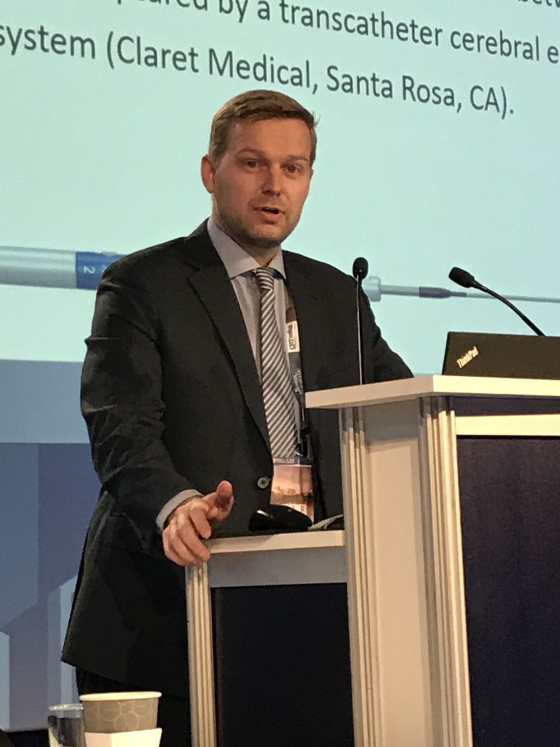
Of the 246 patients, 145 (59%) received a Sapien 3 device, 40 (16%) received an Evolut R, 36 (14%) received a Lotus, and 25 (10%) received a Sapien XT. Particle features based on histopathological and histomorphological analyses were compared. A multivariate analysis was conducted to test for interaction with the amount of debris according to patient demographics and procedural characteristics, which differed for the devices evaluated.
Debris was captured in essentially all patients (99%) and with every TAVR device. On automated histomorphometry, the Lotus device had significantly higher median particle counts relative to the Evolut R (P = .004), the Sapien S3 (P less than .001), and the Sapien XT (P = .02) devices. When stratified by particle size, the Evolut R generated significantly more particles greater than 500 mcm than either the Sapien S3 or the Sapien XT (both P less than .05).
For particle sizes greater than 1 mm, there were significantly higher counts for both the Lotus and Evolut R (both P less than .05) relative to the Sapien S3, but Dr. Schmidt emphasized all devices generated at least some debris this size and that particles greater than 1 mm were retrieved from 53% of all patients. Similarly, when median total particle area was calculated by histomorphometry, the Lotus (P = .0007) and Evolut R (P = .0009) devices were more likely to have particles greater than 1 mm than the Sapien devices, but again, debris with a large total particle area was retrieved from at least some patients in all device groups.
There were significant differences in patient demographics and procedural characteristics by device. For example, patients receiving an Evolut R were significantly more likely than those receiving the Lotus or Sapien S3 valves to have undergone dilation prior to TAVR. Patients receiving the Sapien XT were significantly older than patients receiving any of the other valve types.
In an effort to control for these differences, “multivariate analysis was performed to text for interaction with the amount of debris captured,” Dr. Schmidt explained. “This analysis showed that transcatheter valve type was the only significant predictor for the morphometric findings.”
In this study, most of the debris was found to come from the vascular bed, but foreign body debris and debris from myocardium was also identified. One hypothesis for the greater debris from the Lotus and Evolut R devices is that these are positionable, raising the possibility that manipulation of the devices was the source of the debris, according to Dr. Schmidt. However, this hypothesis needs verification.
One message from this study is that “capture of debris is universal across different transcatheter valve types,” Dr. Schmidt said, adding that this “supports the benefit of using a CPS device in all TAVR procedures.”
Several commentators, including Jeffrey Popma, MD, director of the Interventional Cardiology Clinical Service at Beth Israel Deaconess Hospital, Boston, observed that it would be inappropriate to conclude that the greater debris observed in this study would necessarily translate into a difference in risk of embolic events for the devices evaluated with or without a CPS device. He and others emphasized no relationship to clinical risk was demonstrated in this study.
Dr. Schmidt reports no potential conflicts of interest.
SOURCE: Schmidt T et al. CROI 2018.
WASHINGTON – All transcatheter aortic valve replacement devices generate debris into the bloodstream, including debris larger than 1 mm, but the amount of the debris differs between devices, according to a study based on collections from a cerebral embolic protection system.
“The quantity of the debris measured by different methods and techniques revealed more debris in patients receiving the Evolut R or Lotus TAVR [transcatheter aortic valve replacement] devices than in patients receiving a Sapien 3 or Sapien XT prosthesis,” reported Tobias Schmidt, MD, a cardiologist at Asklepios Klinik St. Georg, Hamburg, Germany.
Of the 246 patients, 145 (59%) received a Sapien 3 device, 40 (16%) received an Evolut R, 36 (14%) received a Lotus, and 25 (10%) received a Sapien XT. Particle features based on histopathological and histomorphological analyses were compared. A multivariate analysis was conducted to test for interaction with the amount of debris according to patient demographics and procedural characteristics, which differed for the devices evaluated.
Debris was captured in essentially all patients (99%) and with every TAVR device. On automated histomorphometry, the Lotus device had significantly higher median particle counts relative to the Evolut R (P = .004), the Sapien S3 (P less than .001), and the Sapien XT (P = .02) devices. When stratified by particle size, the Evolut R generated significantly more particles greater than 500 mcm than either the Sapien S3 or the Sapien XT (both P less than .05).
For particle sizes greater than 1 mm, there were significantly higher counts for both the Lotus and Evolut R (both P less than .05) relative to the Sapien S3, but Dr. Schmidt emphasized all devices generated at least some debris this size and that particles greater than 1 mm were retrieved from 53% of all patients. Similarly, when median total particle area was calculated by histomorphometry, the Lotus (P = .0007) and Evolut R (P = .0009) devices were more likely to have particles greater than 1 mm than the Sapien devices, but again, debris with a large total particle area was retrieved from at least some patients in all device groups.
There were significant differences in patient demographics and procedural characteristics by device. For example, patients receiving an Evolut R were significantly more likely than those receiving the Lotus or Sapien S3 valves to have undergone dilation prior to TAVR. Patients receiving the Sapien XT were significantly older than patients receiving any of the other valve types.
In an effort to control for these differences, “multivariate analysis was performed to text for interaction with the amount of debris captured,” Dr. Schmidt explained. “This analysis showed that transcatheter valve type was the only significant predictor for the morphometric findings.”
In this study, most of the debris was found to come from the vascular bed, but foreign body debris and debris from myocardium was also identified. One hypothesis for the greater debris from the Lotus and Evolut R devices is that these are positionable, raising the possibility that manipulation of the devices was the source of the debris, according to Dr. Schmidt. However, this hypothesis needs verification.
One message from this study is that “capture of debris is universal across different transcatheter valve types,” Dr. Schmidt said, adding that this “supports the benefit of using a CPS device in all TAVR procedures.”
Several commentators, including Jeffrey Popma, MD, director of the Interventional Cardiology Clinical Service at Beth Israel Deaconess Hospital, Boston, observed that it would be inappropriate to conclude that the greater debris observed in this study would necessarily translate into a difference in risk of embolic events for the devices evaluated with or without a CPS device. He and others emphasized no relationship to clinical risk was demonstrated in this study.
Dr. Schmidt reports no potential conflicts of interest.
SOURCE: Schmidt T et al. CROI 2018.
AT CRT 2018
Key clinical point: Based on a study with a cerebral embolic collection system, transcatheter aortic valve replacement devices generate unequal debris.
Major finding: .
Data source: Data analysis from two randomized trials.
Disclosures: Dr. Schmidt reports no potential conflicts of interest.
Source: Schmidt T et al. CROI 2018.
Infection risk from reprocessed duodenoscopes is low, but the goal is zero
BOSTON – Once attributed to human error, the outbreaks of infection from persistent contamination of reprocessed duodenoscopes have eluded an easy fix, according to three experts addressing the problem at the 2018 AGA Tech Summit, sponsored by the AGA Center for GI Innovation and Technology. This session is part of the AGA Center for GI Innovation and Technology’s ongoing efforts to convene stakeholders, including endoscope manufacturers and the Food and Drug Administration, and to collaborate on a solution to ensure zero device-transmitted infections.
Since the problem first was recognized, changes in device design and cleaning protocols have produced a fivefold reduction in the risk of infection.
“The risk is now very low and well beneath the benefits provided by endoscopy, but we will not settle for anything less than complete resolution of the problem,” said David R. Lichtenstein, MD, the director of the endoscopy program at Boston University. Leading off a program that outlined the problem and possible solutions, Dr. Lichtenstein said, “We need to make this a historical issue.”
After scrutiny by the many stakeholders, including the manufacturers, gastroenterologists, and regulatory agencies, the elevator mechanism has remained one focus of concern. The difficulty of cleaning this mechanism was recognized in the earliest devices, but redesigns to permit these channels to be flushed did not resolve the problem completely. In the AGA Tech Summit session, there was general consensus that even dedicated reprocessing technicians fully adherent to current protocols cannot reliably completely clean the currently available duodenoscopes in every case.
“In most instances, it is not the technician at fault and it is extremely difficult to get a flexible scope clean after a messy procedure,” explained Cori Ofstead, MSPH, an epidemiologist and president of Ofstead & Associates. In one study she conducted in endoscopes reprocessed using best practice recommendations, microbial presence could still be detected.
Although risk of contamination increases with repeated use of scopes, with damaged scopes, and after scopes have been used in procedures generating relatively high amounts of debris, Ms. Ofstead said, “If you look, you will find contamination.” While she emphasized the importance of developing incentives that reward quality over efficiency when attempting to reduce human error in duodenoscope reprocessing, she, like Dr. Lichtenstein, believes new strategies are needed to achieve zero tolerance for infection risk.
“Disinfection simply may not be enough,” Ms. Ofstead said. “The solution is likely to be something else, such as single-use scopes or sterilization.” Explaining how processes of sterilization and disinfection differ, David Weber, MD, MPH, professor of epidemiology, University of North Carolina School of Medicine, Chapel Hill, outlined the relative advantages and disadvantages of current options. Dr. Weber, who has been involved in numerous studies regarding duodenoscope decontamination, emphasized that that rigorous cleaning of biological debris is a central tenet of any solution, but he outlined evidence that the current standard of high level disinfection (HLD) has not been sufficient to bring the infection risk to zero.
“There is no simple answer, because every solution so far has introduced some challenges,” Dr. Lichtenstein observed. Listing some examples, he noted that autoclaving results in damage to endoscopes since they are heat labile; that some proposed sterilization techniques may be limited by sterilants that can’t penetrate biofilm formation into deeper layers that harbor viable bacteria and are associated with a risk of toxicity to technicians or patients; and that disposable endoscopes or elevator mechanisms may involve unacceptable costs or operating characteristics. However, he is convinced there is a solution.
“The problem is being addressed from different angles, and I think we will find the solution within several years. This may involve a series of additional incremental improvements, but I think no one with a stake in this issue will sit comfortably until it is no longer a problem,” Dr. Lichtenstein said.
In the discussion that followed the formal presentations, it was generally acknowledged that the ultimate solution remains unclear even though incremental progress is being made. In fact, there have been no major outbreaks in the United States in the last year following the attention given to this issue, which is likely to have promoted more diligence in disinfection, according to Dr. Lichtenstein.
“Disposable endoscopes or disposable parts on endoscopes that facilitate cleaning may be coming, but I think the best current step is a standardized method of testing scopes after cleaning to verify that disinfection has been achieved,” Dr. Lichtenstein said.
Other solutions, including new sterilization techniques may also emerge, but there was general consensus that the human factor will always be important even if the best solution proves to be a disposable endoscope. Dr. Weber said, “You can still place a disposable scope on a contaminated surface. The human factor is always important.”
Michael L. Kochman, MD, the Wilmott Professor of Medicine and director of the Center for Endoscopic Innovation, Research and Training at the University of Pennsylvania, Philadelphia, and past chair of the AGA Center for GI Innovation and Technology, added, “AGA will build on the relationships that we have with the manufacturers of the endoscopes along with companies developing other novel approaches to reprocessing, as well as the FDA and CDC, to further the discussion. Ultimately, we hope to see the availability of devices that are effective and reliably clean so that we do not have to be concerned about patient-to-patient transmission of infection.”
EXPERT ANALYSIS FROM 2018 AGA TECH SUMMIT