User login
CHMP recommends expanded approval for rivaroxaban

The European Medicines Agency’s Committee for Medicinal Products for Human Use (CHMP) has recommended a new indication for rivaroxaban (Xarelto) 2.5 mg tablets.
The recommendation is for rivaroxaban co-administered with acetylsalicylic acid (ASA) for the prevention of atherothrombotic events in adults with coronary artery disease or symptomatic peripheral artery disease at high risk of ischemic events.
The CHMP’s recommendation will be reviewed by the European Commission (EC), which has the authority to approve medicines for use in the European Union, Norway, Iceland, and Liechtenstein.
The EC usually makes a decision within 67 days of the CHMP’s recommendation.
Rivaroxaban is already EC-approved for use in combination with ASA alone or ASA plus clopidogrel or ticlopidine for the prevention of atherothrombotic events in adults with acute coronary syndrome and elevated cardiac biomarkers.
The CHMP’s positive opinion for rivaroxaban in coronary artery disease and symptomatic peripheral artery disease is based on data from the COMPASS study. Results from this study were presented at ESC Congress 2017 and published simultaneously in NEJM.
COMPASS enrolled 27,395 patients with stable atherosclerotic vascular disease. They were randomized to receive rivaroxaban (2.5 mg twice daily) plus aspirin (100 mg once daily), rivaroxaban alone (5 mg twice daily), or aspirin alone (100 mg once daily).
The study’s primary outcome was a composite of cardiovascular death, stroke, or myocardial infarction. This outcome occurred in 4.1% of patients in the rivaroxaban-aspirin arm, 4.9% of those in the rivaroxaban-alone arm, and 5.4% of those in the aspirin-alone arm.
There was a significant difference in the primary outcome between the rivaroxaban-aspirin arm and the aspirin-alone arm (P<0.001) but not between the rivaroxaban-alone arm and the aspirin-alone arm (P=0.12).
The incidence of major bleeding was 3.1% in the rivaroxaban-aspirin arm, 2.8% in the rivaroxaban-alone arm, and 1.9% in the aspirin-alone arm.
There was a significant difference in major bleeding between the rivaroxaban-aspirin arm and the aspirin-alone arm (P<0.001) as well as between the rivaroxaban-alone arm and the aspirin-alone arm (P<0.001).
Kids may have higher risk of death long after allo-HSCT

Children may have an increased risk of premature death decades after allogeneic hematopoietic stem cell transplant (allo-HSCT), according to a study published in JAMA Oncology.
The leading causes of death in the patients studied were infection and chronic graft-vs-host disease (GVHD), patients’ primary disease, and subsequent cancers.
“This study shows that, while we are able to save the life of the child during their cancer treatment, we need to continue to provide proactive follow-up care with these types of patients throughout the rest of their life, as they are still an at-risk population,” said study author Smita Bhatia, MBBS, of the University of Alabama at Birmingham (UAB).
“The high intensity of therapeutic exposures at a young age lends itself to cause morbidities and organ compromise once they reach adulthood.”
Dr Bhatia and her colleagues conducted this retrospective study of children who underwent allo-HSCT between January 1, 1974, and December 31, 2010, and were followed until December 31, 2016.
The study included 1388 patients who lived 2 years or more after transplant. Their median age at transplant was 14.6 years (range, 0-21). The majority of patients were non-Hispanic white (70.7%), and most were male (59.7%).
Patients underwent allo-HSCT to treat acute lymphoblastic leukemia (ALL, 25.1%), acute myeloid leukemia (AML) or myelodysplastic syndromes (MDS, 23.5%), inborn errors of metabolism (13.8%), severe aplastic anemia (SAA, 10.6%), Fanconi anemia (8.3%), chronic myelogenous leukemia (CML, 6.5%), immune disorders (4%), sickle cell disease or thalassemia (1.9%), and other diseases.
Most patients had a related donor (57.9%), and most received a bone marrow transplant (73.4%).
The most common component of conditioning was cyclophosphamide (80.5%), followed by total body irradiation (TBI, 64.3%). About half of patients (49.8%) received both cyclophosphamide and TBI, and nearly a quarter (23.5%) received busulfan and cyclophosphamide.
Outcomes
The researchers found that allo-HSCT recipients had a 14.4-fold greater risk of premature death than the general population.
The team said the absolute excess risk of all-cause mortality was 12.0 per 1000 person-years, and relative mortality remained elevated 25 years or more after transplant (standardized mortality ratio, 2.9).
At a median follow-up of 14.9 years (range, 2.0 to 41.2), 295 patients had died. The 20-year overall survival rate was 79.3%.
The cause of death was available for 82.7% of patients (244/295), and some of these patients had more than 1 cause listed. Causes of death included:
- Infection and/or chronic GVHD—49.6%
- Primary disease—24.6%
- Subsequent malignant neoplasm—18.4%
- Cardiac disease—9.8%
- Pulmonary disease—7.8%
- External causes—2.9%
- Other causes—18.0%.
The hazard of all-cause late mortality was higher among patients who were older at transplant (hazard ratio [HR], 1.03; P=0.004) and those who had a high risk of relapse at transplant (HR, 1.95; P<0.001).
Compared to patients treated for ALL, the hazard of all-cause late mortality was lower among patients with AML/MDS (HR, 0.72; P=0.04), CML (HR, 0.53; P=0.02), Fanconi anemia (HR, 0.49; P=0.03), immune disorders (HR, 0.32; P=0.006), and SAA (HR, 0.33; P<0.001).
The hazard of all-cause late mortality was lower for patients who received conditioning with busulfan and cyclophosphamide (HR, 0.62; P=0.03) than for those who received TBI and cyclophosphamide.
Compared to patients treated for ALL, the hazard of relapse-related mortality was lower among patients with AML/MDS (HR, 0.39; P=0.01) and SAA (HR, 0.09; P=0.03), and the hazard of non-relapse mortality was lower for patients with SAA (HR, 0.36; P=0.004) and immune disorders (HR, 0.14; P=0.009).
The hazard of non-relapse mortality was higher for patients who were older at transplant (HR, 1.03; P=0.03), patients who received peripheral blood stem cells rather than bone marrow (HR, 2.39; P=0.01), and patients who had a high risk of relapse at transplant (HR, 2.05; P<0.001).
Global burden of hematologic malignancies

Research has shown an increase in the global incidence of leukemia and non-Hodgkin lymphoma (NHL) in recent years.
The Global Burden of Disease (GBD) study showed that, from 2006 to 2016, the incidence of NHL increased 45%, and the incidence of leukemia increased 26%.
These increases were largely due to population growth and aging.
Results from the GDB study were published in JAMA Oncology.
The study indicated that, in 2016, there were 17.2 million cases of cancer worldwide and 8.9 million cancer deaths.
One in 3 men were likely to get cancer during their lifetime, as were 1 in 5 women. Cancer was associated with 213.2 million disability-adjusted life years (DALYs).
The following table lists the 2016 global incidence and mortality figures for all cancers combined and for individual hematologic malignancies.
| Cancer type | Cases, thousands | Deaths, thousands |
| All cancers | 17,228 | 8927 |
| Leukemias | 467 | 310 |
| Acute lymphoid leukemia | 76 | 51 |
| Chronic lymphoid leukemia | 105 | 35 |
| Acute myeloid leukemia | 103 | 85 |
| Chronic myeloid leukemia | 32 | 22 |
| Other leukemias | 150 | 117 |
| Hodgkin lymphoma | 73 | 29 |
| NHL | 461 | 240 |
| Multiple myeloma | 139 | 98 |
Leukemia
In 2016, there were 467,000 new cases of leukemia and 310,000 leukemia deaths. Leukemia was responsible for 10.2 million DALYs. Leukemia developed in 1 in 118 men and 1 in 194 women worldwide.
Between 2006 and 2016, the global leukemia incidence increased by 26%—from 370,482 to 466,802 cases.
The researchers said the factors contributing to this increase were population growth (12%), population aging (10%), and an increase in age-specific incidence rates (3%).
NHL
In 2016, there were 461,000 new cases of NHL and 240,000 NHL deaths. NHL was responsible for 6.8 million DALYs. NHL developed in 1 in 110 men and 1 in 161 women worldwide.
Between 2006 and 2016, NHL increased by 45%, from 319,078 to 461,164 cases.
The factors contributing to this increase were increasing age-specific incidence rates (17%), changing population age structure (15%), and population growth (12%).
“A large proportion of the increase in cancer incidence can be explained by improving life expectancy and population growth—a development that can at least partially be attributed to a reduced burden from other common diseases,” the study authors wrote.
The authors also pointed out that prevention efforts are less effective for hematologic malignancies than for other cancers.
Orphan designation recommended for OMS721
The European Medicines Agency’s (EMA’s) Committee for Orphan Medicinal Products (COMP) has issued a positive opinion recommending orphan drug designation for OMS721 as a treatment for high-risk hematopoietic stem cell transplant-associated thrombotic microangiopathy (HSCT-TMA).
OMS721 is a monoclonal antibody targeting MASP-2, the effector enzyme of the lectin pathway of the complement system.
The COMP’s positive opinion of OMS721 for HSCT-TMA is expected to be adopted by the European Commission in August.
Orphan drug designation in Europe is available to companies developing products intended to treat a life-threatening or chronically debilitating condition that affects fewer than 5 in 10,000 people in the European Union (EU).
This designation allows for financial and regulatory incentives that include 10 years of marketing exclusivity in the EU after product approval, reduced EMA advisory, inspection and filing fees pre- and post-approval, and guaranteed access to centralized marketing authorization valid in all EU member states as well as in European Economic Area countries (ie, Iceland, Liechtenstein, and Norway).
Phase 2 trial
OMS721 is currently under evaluation in a phase 2 trial (NCT02222545). Omeros Corporation, the company developing OMS721, released some results from this study in February.
Results were reported for 18 adults with HSCT-TMA persisting for at least 2 weeks following immunosuppressive regimen modification or more than 30 days post-transplant. The patients received weekly OMS721 treatments for 4 to 8 weeks at the discretion of the investigator.
These patients had a significantly longer median overall survival than historical controls—347 days and 21 days, respectively (P<0.0001).
Omeros also reported that markers of TMA activity significantly improved following OMS721 treatment.
The mean platelet count increased from 18,100 x 106/mL at baseline to 52,300 x 106/mL (P=0.017). The mean LDH decreased from 591 U/L to 250 U/L (P<0.001). And the mean haptoglobin increased from 8 mg/dL to 141 mg/dL (P=0.003).
Mean creatinine remained stable—at approximately 120 μmol/L—but a majority of patients had co-existing conditions for which they were receiving nephrotoxic medications. These conditions included graft-versus-host disease, cytomegalovirus and human herpes virus 6 infections, prior sepsis, diffuse alveolar hemorrhage, and residual underlying malignancies.
The most commonly reported adverse events in this trial were diarrhea and neutropenia.
Four deaths occurred. One of these—due to acute renal and respiratory failure—was considered possibly related to OMS721.
The other deaths were due to progression of acute myeloid leukemia (n=1) and neutropenic sepsis (n=2).
Study could change treatment of MLSM7

New findings could help improve treatment of an inherited bone marrow disorder known as myelodysplasia and leukemia syndrome with monosomy 7 (MLSM7), according to researchers.
While studying families affected by MLSM7, researchers identified germline mutations in SAMD9L or SAMD9 in patients who had hematologic abnormalities, myelodysplastic syndromes (MDS), or acute myeloid leukemia (AML).
However, these mutations were also present in apparently healthy family members, and the researchers found that bone marrow monosomy 7 sometimes resolved without treatment.
The team recounted these findings in JCI Insight.
The researchers analyzed blood samples from 16 siblings in 5 families affected by MLSM7 and found they all carried germline mutations in SAMD9 or SAMD9L. In 3 of the 5 families, there were apparently healthy parents who also carried the mutations.
“Surprisingly, the health consequences of these mutations varied tremendously for reasons that must still be determined, but the findings are already affecting how we may choose to manage these patients,” said study author Jeffery Klco, MD, PhD, of St. Jude Children’s Research Hospital in Memphis, Tennessee.
Three of the 16 siblings developed AML and died of the disease or related complications. Two other siblings were diagnosed with MDS.
The remaining 11 siblings with the mutations were apparently healthy, although several had been treated for anemia and other conditions associated with low blood counts.
Some of these patients had a previous history of bone marrow monosomy 7 that spontaneously corrected over time. These patients, despite no therapy, appeared to have normal bone marrow function.
“This was an even greater surprise,” Dr Klco said. “The spontaneous recovery experienced by some children with the germline mutations suggests some patients with SAMD9 and SAMD9L mutations who were previously considered candidates for bone marrow transplantation may recover hematologic function on their own.”
Dr Klco and his colleagues have a theory that could explain the spontaneous correction. The team noted that SAMD9 and SAMD9L are activated in response to viral infections. While the normal function of both proteins is poorly understood, abnormally activated SAMD9 and SAMD9L are known to inhibit cell growth.
In this study, deep sequencing showed that selective pressure on developing blood cells favors cells without the SAMD9 or SAMD9L mutations. That may increase pressure for cells to selectively jettison chromosome 7 with the gene alteration or take other molecular measures to counteract the mutant protein.
Implications for treatment
This research also showed that, in patients who developed AML, loss of chromosome 7 was associated with the development of mutations in additional genes, including ETV6, KRAS, SETBP1, and RUNX1.
These same mutations are broadly associated with monosomy 7 in AML, which suggests that understanding how SAMD9 and SAMD9L mutations contribute to leukemia has implications beyond familial cases.
The presence of secondary mutations may also help clinicians identify which patients will benefit from immediate treatment, including chemotherapy or transplant to prevent or treat AML or myelodysplasia, Dr Klco said.
For patients without the mutations or significant symptoms due to low blood cell counts, watchful waiting with careful follow-up may sometimes be an option.
“Now that we know this disease can resolve without treatment in some patients, we need to focus on developing screening and treatment guidelines,” Dr Klco said. “We want to reserve hematopoietic bone marrow transplantation for those who truly need the procedure. These findings will help to point the way.”
“So little is known about SAMD9 and SAMD9L that we need to continue working in the lab to better understand how these mutations impact blood cell development and how they are activated in response to infections and other types of stress.”
Depression, antidepressants linked to VTE
Depression and the use of antidepressants are each associated with an increased risk of venous thromboembolism (VTE), according to a review and meta-analysis published in Annals of Medicine.
The research also showed that each of the various classes of antidepressant medications is associated with an increased risk of VTE.
The researchers noted that this study does not prove cause and effect, and further studies are needed to determine what is driving the observed increase in VTE risk.
Some previous studies have indicated that depression and antidepressant use might be associated with an increased risk of VTE, but other studies have shown no evidence of associations.
Therefore, Setor Kunutsor, MD, PhD, of the University of Bristol in Bristol, UK, and his colleagues conducted a systematic review and meta-analysis of observational studies evaluating the associations of depression and antidepressant use with VTE risk.
The researchers looked at 8 observational studies with data on 960,113 non-overlapping participants. There were 9027 cases of VTE in this population.
To determine the association between depression and VTE, the researchers conducted a pooled analysis of 3 studies comparing patients with depression and without. These studies included 865,878 participants and 4676 cases of VTE.
The relative risk (RR) for VTE was 1.31 (95% CI, 1.13-1.53) among patients with depression.
The researchers also conducted a pooled analysis of 6 studies comparing antidepressant users to non-users. These studies included 828,327 participants and 8273 cases of VTE.
The RR for VTE was 1.27 (95% CI, 1.06-1.51) among patients taking antidepressants.
In addition, individual antidepressants were associated with an increased risk of VTE. This includes:
- Tricyclic antidepressants—RR=1.16 (95% CI, 1.06-1.27)
- Selective serotonin reuptake inhibitors—RR=1.12 (95% CI, 1.02-1.23)
- “Other” antidepressants*—RR=1.59 (95% CI, 1.21-2.09).
The researchers said these data show that antidepressant use and depression are each associated with an increased risk of VTE, and these results add to accumulating evidence that a relationship exists between depression, antidepressant use, and VTE.
“These findings are very useful to me as both a clinician and a researcher,” Dr Kunutsor said. “It gives me the information I need, especially when prescribing antidepressant medications to my patients.”
Still, Dr Kunutsor and his colleagues conceded that more research is needed to determine if the observed associations are causal and if depression, antidepressant use, or both drive the increased risk of VTE.
To so this, studies would need to isolate depression from antidepressant medications. For example, researchers could assess if non-depressed individuals who use antidepressants for other conditions have an increased risk of VTE.
*The “other” antidepressants include monoamine oxidase inhibitors, triazolopyridine, serotonin norepinephrine reuptake inhibitors, and norepinephrine dopamine reuptake inhibitors.
Gene therapy granted accelerated assessment

The European Medicines Agency’s (EMA) Committee for Medicinal Products for Human Use has granted accelerated assessment for the upcoming marketing authorization application (MAA) for LentiGlobin™.
LentiGlobin is a gene therapy intended for the treatment of adolescents and adults with transfusion-dependent β-thalassemia (TDT) and a non-β0/β0 genotype.
bluebird bio intends to file an MAA for LentiGlobin with the EMA this year.
Accelerated assessment can reduce the active review time of an MAA from 210 days to 150 days once it has been validated by the EMA.
An accelerated assessment is granted to products deemed to be of major interest for public health and represent therapeutic innovation.
The accelerated assessment for LentiGlobin is supported by data from clinical studies, including the phase 1/2 Northstar (HGB-204) study, phase 1/2 HGB-205 study, phase 3 Northstar-2 (HGB-207) study, and the long-term follow-up study LTF-303.
The EMA previously granted PRIME (Priority Medicines) eligibility and orphan designation to LentiGlobin for the treatment of TDT.
LentiGlobin is also part of the EMA’s Adaptive Pathways pilot program, which is part of the EMA’s effort to improve timely access to new medicines.
Phase 1/2 trials
Results from the phase 1/2 trials of LentiGlobin—HGB-204 and HGB-205—were published in NEJM in April.
HGB-204 is a completed study that included 18 patients with TDT. Eight patients had a β0/β0 genotype, 6 had a βE/β0 genotype, and 4 had other genotypes.
HGB-205 is an ongoing study, and results were reported for 4 patients with TDT. Three patients had a βE/β0 genotype. The remaining patient was homozygous for the IVS1-110 mutation and had a severe clinical presentation similar to that seen in β0/β0 genotypes.
For both studies, the researchers harvested hematopoietic stem and progenitor cells (mobilized with filgrastim and plerixafor) from the patients. CD34+ cells were transduced ex vivo with LentiGlobin BB305 vector, which encodes adult hemoglobin (HbA) with a T87Q amino acid substitution (HbAT87Q).
The patients underwent myeloablative conditioning with busulfan, and the final LentiGlobin product was infused into patients after a 72-hour washout period.
In HGB-205 only, patients received enhanced red blood cell (RBC) transfusions for at least 3 months before stem cell mobilization and harvest to maintain a hemoglobin level of more than 11.0 g/dL.
Safety
In HGB-204, there were 5 grade 1 adverse events (AEs) considered possibly or probably related to LentiGlobin—abdominal pain (n=2), dyspnea (n=1), hot flush (n=1), and non-cardiac chest pain (n=1).
There were 9 serious AEs. Grade 3 serious AEs included 2 episodes of veno-occlusive liver disease that were attributed to busulfan, Klebsiella infection, cardiac ventricular thrombosis, cellulitis, hyperglycemia, and gastroenteritis. Grade 2 serious AEs included device-related thrombosis and infectious diarrhea.
In HGB-205, there were no AEs considered possibly or probably related to LentiGlobin. The 3 serious AEs were tooth infection (grade 3), major depression (grade 3), and pneumonia (grade 2).
Efficacy
The median time to neutrophil engraftment was 18.5 days (range, 14.0 to 30.0) in HGB-204 and 16.5 days (range, 14.0 to 29.0) in HGB-205.
The median time to platelet engraftment was 39.5 days (range, 19.0 to 191.0) and 23.0 days (range, 20.0 to 26.0), respectively.
In both studies, the median follow-up was 26 months (range, 15 to 42) after LentiGlobin infusion.
At last follow-up, all but 1 of the 13 patients with a non-β0/β0 genotype had stopped receiving RBC transfusions.
In the 8 patients with a β0/β0 genotype and the 1 patient with 2 copies of the IVS1-110 mutation, the median annualized transfusion volume decreased by 73% after LentiGlobin administration.
Two patients with a β0/β0 genotype were able to stop receiving RBC transfusions, as was the patient with 2 copies of the IVS1-110 mutation.
Team creates tool to assess frailty in MM

Researchers say they have developed a frailty index that can predict overall survival (OS) in patients newly diagnosed with multiple myeloma (MM).
An increasing frailty index score was significantly associated with an increased risk of death in these patients, and frailty retained a significant association with OS even after the researchers controlled for patients’ chronological age.
“Our goal was to create a tool that could be widely applied using data sources at hand and that helps doctors provide better informed treatment recommendations for their patients,” said Tanya S. Wildes, MD, of the Washington University School of Medicine in St Louis, Missouri.
“Our results demonstrate that, for patients with multiple myeloma, chronological age alone is not a good measure for assessing overall health.”
Dr Wildes and her colleagues reported these results in JCO Clinical Cancer Informatics.
Creating the index
The researchers began this study with data from 2,692,361 patients without cancer who were older than 66 years of age. The data were collected from the Medicare Health Outcomes Survey (MHOS), which is used to annually collect self-reported symptoms, functional status, and health-related quality of life data from Medicare beneficiaries enrolled in Medicare Advantage plans.
The researchers used the MHOS data to create a deficit accumulation frailty index made up of a 25-item scale and scoring system. The index includes criteria in 5 categories for scoring frailty:
- Activities of daily living (eg, difficulty dressing or eating)
- Chronic health conditions
- Functioning (eg, difficulty walking or climbing several sets of stairs)
- General health
- Mental health.
Patients whose scores exceed a certain threshold on the scale are classified as frail.
Applying the index
The researchers applied their frailty index to 305 patients with newly diagnosed MM. Data from these patients were obtained from the Surveillance, Epidemiology, and End Results (SEER)-MHOS linked database. In this dataset, data from MHOS are linked to demographics, tumor characteristics, and survival for individuals with a cancer diagnosis who reside in the coverage area of the 14 registries participating in the SEER-MHOS linkage.
The researchers compared findings in the MM patients to findings in the patients without cancer.
In the non-cancer patients, the median age was 74, and the mean frailty score was 0.23. In the MM patients, the median age was 76, and the mean frailty score was 0.28.
Chronological age was weakly correlated with a higher frailty score in MM patients. However, for non-cancer patients, an increase in chronological age was strongly correlated with a higher frailty score.
Among non-cancer patients, each 10% increase in frailty score was associated with a 40% increased risk for death (adjusted hazard ratio, 1.397; P<0.001).
Among MM patients, each 10% increase in frailty score was associated with a 16% increased risk of death (adjusted hazard ratio, 1.159; P<0.001).
The median OS was 33 months for the entire MM cohort, 26.8 months for frail MM patients, and 43.7 months for non-frail MM patients (P=0.015 for the frail to non-frail comparison).
“These findings underscore the need to place more consideration on biological age versus chronological age in multiple myeloma, recognizing that frailty is dynamic and encompasses many factors beyond the disease itself,” Dr Wildes said.
“Ultimately, the hope is that this tool will help us to better personalize care based on a fuller picture of our patients’ health so that we are not under-treating an older adult who can tolerate a more intense therapy or over-treating one who’s going to be vulnerable to the toxicities of therapy.”
Limitations and next steps
The researchers believe there are several options for optimizing the data in the frailty index, including turning it into a computerized program and examining patients who are not newly diagnosed and have subsequent relapses, disease burden, and treatment toxicities.
This study is limited in the fact that researchers only assessed OS and not progression-free survival, chemotherapy toxicity, or hospitalization rates.
Additionally, the MM data was derived from patients enrolled in the Medicare Advantage program, which may have contributed to selecting participants who are, overall, lower-risk due to the way the program incentivizes lower-cost enrollees.
Plasma transfusion during air transport can reduce mortality

Receiving a plasma transfusion during emergency air transport can improve survival for trauma patients, according to a study published in NEJM.
Trauma patients with severe bleeding had a significant decrease in 30-day mortality when they received a plasma transfusion while being airlifted to a hospital.
Transfusion-related reactions and allergic reactions were more common among plasma recipients than patients who only received standard care.
However, this difference was not significant, and most of these reactions were considered minor.
“These results have the power to significantly alter trauma resuscitation, and their importance to the trauma community cannot be overstated,” said study author Jason Sperry, MD, of the University of Pittsburgh School of Medicine in Pennsylvania.
“This is the first trial in a quarter century to have the potential to alter prehospital care so considerably.”
Patients and intervention
This trial, known as PAMPer (Prehospital Air Medical Plasma), was a phase 3, randomized study enrolling 501 trauma patients at risk of hemorrhagic shock.
Most patients were male (72.7%), and most had suffered blunt trauma (82.4%). About half of patients (51.1%) had prehospital intubation, and more than a third (34.7%) received a prehospital transfusion of red blood cells.
Air medical bases participating in this study were randomized to administer plasma or standard care to eligible patients for 1-month intervals. When the air transport teams were in their plasma interval, they’d begin administering 2 units of thawed plasma to a patient as soon as trial eligibility was confirmed.
If the 2 units were completed during the flight, the team would revert to standard care. If the transfusions weren’t completed, the plasma would continue to be administered when the patient arrived at the trauma center.
The teams administered the assigned treatment 99% of the time (496/501).
In the plasma group, there were 205 patients (89.1%) who received 2 units of plasma, 21 (9.1%) who received 1 unit, and 4 patients (1.7%) who did not receive plasma due to logistical challenges.
In 84.4% of the patients, the plasma infusion was completed during air transport. The remaining patients completed their plasma transfusions at the trauma center.
There was 1 patient (0.4%) in the standard-care group who received plasma before transport began.
Primary outcome
The study’s primary outcome was 30-day mortality. Ninety-six percent of patients (n=481) had data for this outcome—220 patients in the plasma group and 261 in the standard-care group.
Thirty-day mortality was significantly lower in the plasma group than the standard-care group—23.2% and 33.0%, respectively (P=0.03).
In an adjusted analysis, the administration of prehospital plasma was associated with a 39% lower risk for 30-day mortality than standard care (adjusted odds ratio, 0.61; P=0.02).
Secondary outcomes
Initially, there were significant differences between the plasma (n=230) and standard-care groups (n=271) when it came to:
- Mortality at 24 hours—13.9% and 22.1%, respectively (P=0.02)
- In-hospital mortality—22.2% and 32.5%, respectively (P=0.01)
- Median volume of blood components transfused in the first 24 hours—3 and 4 units, respectively (P=0.02)
- Median volume of red cells transfused in the first 24 hours—3 and 4 units, respectively (P=0.03).
- Median prothrombin-time ratio at first blood sampling—1.2 and 1.3, respectively (P<0.001).
When the researchers adjusted P values for multiple comparisons, the between-group difference in prothrombin-time ratio remained significant (P<0.001).
However, the differences in 24-hour mortality (P=0.55), in-hospital mortality (P=0.33), blood components transfused (P=0.41), and red cells transfused (P=0.69) did not retain significance.
Likewise, there were no significant between-group differences (in adjusted or unadjusted analyses) when it came to multi-organ failure, acute lung injury/acute respiratory distress syndrome, nosocomial infections, or allergic/transfusion-related reactions.
There were 10 adverse events (AEs) considered related to the trial regimen. In the standard-care group, the 4 AEs were sepsis (a serious AE), adult respiratory distress syndrome (a serious AE), fever, and pain.
In the plasma group, the 6 AEs were 2 allergic reactions, 1 case of anaphylaxis, 1 case of hypotension, 1 case of urticaria, and 1 transfusion-related reaction (a serious AE).
Drug receives fast track designation for WM
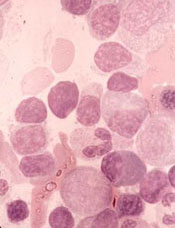
The US Food and Drug Administration (FDA) has granted fast track designation to zanubrutinib for the treatment of Waldenström’s macroglobulinemia (WM).
Zanubrutinib (BGB-3111) is a BTK inhibitor being developed by BeiGene to treat various B-cell malignancies.
BeiGene is preparing to submit to the FDA, in the first half of 2019, a new drug application seeking accelerated approval of zanubrutinib for patients with WM.
The application will be supported by results from a phase 1 study. Results from this trial were presented at the 14th International Conference on Malignant Lymphoma (14-ICML) last year.
Researchers are also evaluating zanubrutinib in phase 2 (NCT03332173) and phase 3 (NCT03053440) trials of WM patients. In the phase 3 trial, researchers are comparing zanubrutinib to the BTK inhibitor ibrutinib.
Phase 1 results
As of March 31, 2017, 48 WM patients were enrolled in the phase 1 study. Thirty-eight patients had relapsed/refractory disease, and 10 patients were treatment-naïve.
There was a dose-escalation phase and a dose-expansion phase. The dose-expansion phase included doses of 160 mg twice a day or 320 mg once a day.
The most common (>10%) adverse events, (AEs) of any attribution were petechiae/purpura/contusion (35%), upper respiratory tract infection (31%), constipation (25%), diarrhea (19%), epistaxis (19%), nausea (17%), cough (15%), anemia (15%), headache (15%), neutropenia (13%), and rash (13%).
Most of these events were grade 1 or 2 in severity. The exceptions were grade 3/4 anemia and neutropenia (8% each) as well as grade 3/4 diarrhea and headache (2% each).
Five serious AEs were considered possibly related to zanubrutinib—1 case each of hemothorax, atrial fibrillation, colitis, febrile neutropenia, and headache. Three AEs led to treatment discontinuation—1 case each of bronchiectasis, prostate adenocarcinoma, and adenocarcinoma of pylorus.
At the time of the data cutoff, 42 patients were evaluable for response. At a median follow-up of 12.3 months (range, 4.4 to 30.5 months), the overall response rate was 90% (38/42).
The major response rate was 76% (32/42), with very good partial responses in 43% (18/42) of patients and partial responses in 33% (14/42) of patients. There were no complete responses and 2 cases of disease progression.
About fast track designation
The FDA’s fast track development program is designed to expedite clinical development and submission of applications for products with the potential to treat serious or life-threatening conditions and address unmet medical needs.
Fast track designation facilitates frequent interactions with the FDA review team, including meetings to discuss the product’s development plan and written communications about issues such as trial design and use of biomarkers.
Products that receive fast track designation may be eligible for accelerated approval and priority review if relevant criteria are met. Such products may also be eligible for rolling review, which allows a developer to submit individual sections of a product’s application for review as they are ready, rather than waiting until all sections are complete.