User login
Predicting outcomes in AML patients

Photo by Darren Baker
An international competition has produced models that can help predict outcomes in patients with acute myeloid leukemia (AML), according to researchers.
For the competition, known as the DREAM 9 challenge, 31 teams of computational researchers attempted to predict outcomes using data from hundreds of patients with AML.
DREAM, which stands for Dialogue for Reverse Engineering Assessment and Methods, is a platform for crowd-sourced studies that focus on developing computational tools to solve biomedical problems.
Essentially, it’s a competition that serves as a large, long-standing, international scientific collaboration.
“We used DREAM as a way to get general insight into making more accurate predictive models of clinical outcomes,” said Amina Qutub, PhD, of Rice University in Houston, Texas.
She and her colleagues described this effort in PLOS Computational Biology.
For the DREAM 9 challenge, each team was presented with training data from 191 AML patients, which included demographic information, such as age and gender, and more complex proteomic and phosphoprotein data that describes signaling protein pathways believed to play a role in AML.
The teams were also presented with a test set of 100 AML patients and were asked to predict response to therapy, remission duration, or overall survival for these patients.
The top-performing models—by Team EvoMed of Arizona State University and Team Chipmunks of the Ontario Institute for Cancer Research—were able to predict patient response to therapy with an accuracy of close to 80%.
Both of these models were impacted by the perturbation of PIK3CA and NPM1, which singles out these proteins as candidates for further study, according to researchers.
Another discovery resulting from this competition was that, overall, the 31 models were not as effective for predicting outcomes in patients classified as “resistant to therapy” than for responsive patients.
The median model prediction accuracy was 42% for resistant patients and 73% for responsive patients. ![]()
FDA lifts hold on phase 2 JCAR015 trial

Image from NIAID
The US Food and Drug Administration (FDA) has removed the clinical hold on the phase 2 ROCKET trial, a study of the chimeric antigen receptor (CAR) T-cell therapy JCAR015 in adults with relapsed or refractory B-cell acute lymphoblastic leukemia.
The trial will continue with a revised protocol, under which patients will receive only cyclophosphamide as conditioning.
The ROCKET trial was placed on hold after 3 patients died of cerebral edema.
Juno Therapeutics, the company developing JCAR015, believes the deaths were likely a result of adding fludarabine to the conditioning regimen.
Patients initially received conditioning with cyclophosphamide alone, but investigators decided to add fludarabine in hopes of increasing efficacy. Adding fludarabine to conditioning had been shown to increase the efficacy of 2 other CAR T-cell therapies, JCAR014 and JCAR017, in phase 1/2 trials.
However, in the ROCKET trial, the addition of fludarabine was associated with an increase in the incidence of severe neurotoxicity and the 3 deaths from cerebral edema.
Although other factors may have contributed to the deaths, Juno said fludarabine was the most likely culprit. So the company asked the FDA if it could continue the ROCKET trial using conditioning with cyclophosphamide alone.
In response, the FDA requested that Juno submit a revised patient informed consent form, revised investigator brochure, revised trial protocol, and a copy of a presentation the company made to the FDA.
The FDA said it would review these documents within 30 days of receiving them, but the review only took a few days. The agency agreed to lift the clinical hold and allow the trial to proceed with the revised protocol.
ROCKET is not the first trial of JCAR015 to be placed on clinical hold. The phase 1 trial of the therapy was placed on hold in 2014, after 2 patients died of cytokine release syndrome.
That hold was lifted following changes to enrollment criteria and dosing. Results from this trial were presented at ASCO 2015 and ASCO 2016. ![]()
Cell of origin impacts aggressiveness of AML

New research suggests the cell of origin controls the aggressiveness and outcome of acute myeloid leukemia (AML) driven by MLL-AF9.
Using a mouse model, investigators found that MLL-AF9 expression in long-term hematopoietic stem cells (HSCs) causes invasive, chemoresistant AML that expresses genes related to epithelial-mesenchymal transition (EMT).
The researchers also found these genes were associated with poor survival in humans with AML.
“The prognosis thus depends on the particular hematopoietic stem or precursor cells in which the genetic alteration occurs and what genes are expressed,” said Antoine Peters, PhD, of Friedrich Miescher Institute for Biomedical Research in Basel, Switzerland.
Dr Peters and his colleagues related these findings in Cancer Cell.
The investigators compared the results of MLL-AF9 expression in long-term HSCs and granulocyte-macrophage progenitors (GMPs) in vitro. They found that colonies derived from long-term HSCs exhibited a greater capacity to migrate and expressed genes known to be involved in cell migration and tissue invasion.
With in vivo experiments, the researchers found that long-term HSCs were more potent in inducing AML than GMPs. Twenty percent of long-term HSC-derived AMLs proved especially aggressive, exhibiting extensive tissue infiltration, resistance to chemotherapy, and the expression of EMT genes such as EVI1, ERG, and ZEB1.
The investigators then tested the function of EMT-related transcription factors in leukemic cell migration and invasion. And they found that knocking down ZEB1 significantly reduced leukemic blast invasion.
Next, the researchers classified mouse and human leukemias according to EVI1/Evi1 and ERG/Erg expression and compared transcriptional profiles between species.
This revealed 111 genes that were more highly expressed in EVI1highERGhigh AML and Evi1highErghigh iMLL-AF9 long-term-HSC-early AML. These genes were dubbed ‘‘cluster I.’’
The investigators also identified 40 genes that were more highly expressed in EVI1lowERGlow AML samples and in iMLL-AF9 GMP-derived AML. These were called ‘‘cluster II.’’
Finally, the team evaluated how the 2 gene clusters related to overall survival in patients with 11q23+ AML.
Patients with high expression of cluster I genes tended to have poor survival rates, while patients with high levels of cluster II genes had variable survival rates and a propensity for poor outcomes if they had co-expression of a “substantial fraction” of cluster I genes.
The researchers noted that many cluster I genes are implicated in cell migration, invasion, EMT, and inflammation.
“The expression of genes such as EVI1, ERG, or ZEB1 now allows us to classify patients into different groups according to prognosis and, if necessary, to adapt treatment,” said Jürg Schwaller, MD, of University Children’s Hospital and University of Basel in Switzerland.
“Our findings should also enable us to develop new, more personalized therapies for these patients.” ![]()
Study reveals increase in susceptibility to malaria
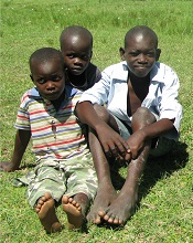
Photo by Gabrielle Tenenbaum
Progress in reducing the malaria burden in Africa may have had the paradoxical effect of increasing malaria transmission among older children in recent years, according to research published in PLOS Medicine.
Researchers analyzed data on children admitted to a hospital in Kenya over a 25-year period and found that malaria transmission decreased from 1998 to 2009 but began to rise after that, and older children accounted for most of this increase.
The researchers believe this may be a result of a decrease in acquired immunity among the older children.
Philip Bejon, PhD, of the University of Oxford in the UK, and his colleagues conducted this study. The team analyzed malaria screening data from pediatric emergency admissions to Kilifi County Hospital between 1990 and 2014 and linked it to data on residence and insecticide-treated bednet (ITN) use.
During the period studied, 69,104 children, ages 3 months to 13 years, had malaria screening data available.
The proportion of hospital admissions found positive for malaria decreased from a high of 56% in 1998 to a low of 7% in 2009, but then rose again to 24% in 2014.
The researchers said they were unable to determine the cause of the decline in malaria infections between 1998 and 2009, which pre-dated the dramatic scale-up in ITN distribution and use in the area.
However, they noted that the subsequent increases in malaria infections coincided with a shift in burden from younger to older children, and children who lived in areas with high usage of ITNs were less likely to present with malaria than children who lived in areas with low usage of ITNs.
The researchers said the upswing in malaria transmission among older children following a period of low malaria transmission may be due to decreased acquired immunity because these children were less likely to have been exposed to malaria early in life.
The team also noted that varying levels of access to care and ITN use may have introduced bias in this study, and these data were taken from a single geographic setting. However, they still believe the findings suggest malaria elimination will require continued—and increasing—vigilance. ![]()
Immunotherapy may benefit relapsed HSCT recipients

Photo from Business Wire
Results of a phase 1 study suggest that repeated doses of the immunotherapy drug ipilimumab is a feasible treatment option for patients with hematologic diseases who relapse after allogeneic hematopoietic stem cell transplant (HSCT).
Seven of the 28 patients studied responded to the treatment, but immune-mediated toxic effects and graft-vs-host disease (GVHD) occurred as well.
These results were published in NEJM.
Ipilimumab, which is already approved to treat unresectable or metastatic melanoma, works by blocking the immune checkpoint CTLA-4. Blockade of CTLA-4 has been shown to augment T-cell activation and proliferation.
“We believe [,in the case of relapse after HSCT,] the donor immune cells are present but can’t recognize the tumor cells because of inhibitory signals that disguise them,” said study author Matthew Davids, MD, of the Dana-Farber Cancer Institute in Boston, Massachusetts.
“By blocking the checkpoint, you allow the donor cells to see the cancer cells.”
Dr Davids and his colleagues tested this theory in 28 patients who had relapsed after allogeneic HSCT. The patients had acute myeloid leukemia (AML, n=12), Hodgkin lymphoma (n=7), non-Hodgkin lymphoma (n=4), myelodysplastic syndromes (MDS, n=2), multiple myeloma (n=1), myeloproliferative neoplasm (n=1), or acute lymphoblastic leukemia (n=1).
Patients had received a median of 3 prior treatment regimens, excluding HSCT (range, 1 to 14), and 20 patients (71%) had received treatment for relapse after transplant. Eight patients (29%) previously had grade 1/2 acute GVHD, and 16 (57%) previously had chronic GVHD.
The median time from transplant to initial treatment with ipilimumab was 675 days (range, 198 to 1830), and the median time from relapse to initial treatment with ipilimumab was 97 days (range, 0 to
1415).
Patients received induction therapy with ipilimumab at a dose of 3 mg/kg or 10 mg/kg every 3 weeks for a total of 4 doses. Those who had a clinical benefit received additional doses every 12 weeks for up to 60 weeks.
Safety
Five patients discontinued ipilimumab due to dose-limiting toxic effects. Four of these patients had GVHD, and 1 had severe immune-related adverse events.
Dose-limiting GVHD presented as chronic GVHD of the liver in 3 patients and acute GVHD of the gut in 1 patient.
Immune-related adverse events included death (n=1), pneumonitis (2 grade 2 events, 1 grade 4 event), colitis (1 grade 3 event), immune thrombocytopenia (1 grade 2 event), and diarrhea (1 grade 2 event).
Efficacy
There were no responses in patients who received ipilimumab at 3 mg/kg. Among the 22 patients who received ipilimumab at 10 mg/kg, 5 had a complete response, and 2 had a partial response.
Six other patients did not qualify as having responses but had a decrease in their tumor burden. Altogether, ipilimumab reduced tumor burden in 59% of patients.
The complete responses occurred in 4 patients with extramedullary AML and 1 patient with MDS developing into AML. Two of the AML patients remained in complete response at 12 and 15 months, and the patient with MDS remained in complete response at 16 months.
At a median follow-up of 15 months (range, 8 to 27), the median duration of response had not been reached. Responses were associated with in situ infiltration of cytotoxic CD8+ T cells, decreased activation of regulatory T cells, and expansion of subpopulations of effector T cells.
The 1-year overall survival rate was 49%.
The investigators said these encouraging results have set the stage for larger trials of checkpoint blockade in this patient population. Further research is planned to determine whether immunotherapy drugs could be given to high-risk patients to prevent relapse. ![]()
Study links adipocytes and myeloma progression

A study published in Cancer Letters suggests that adipocytes contribute to the growth and progression of multiple myeloma (MM).
Researchers co-cultured MM cells with adipocytes derived from patients defined as normal weight, overweight, obese, or super obese.
This revealed a positive correlation between adipocytes and indicators of MM progression, which was most pronounced in cells from obese and super obese individuals.
“Once a person with cancer is out of the normal weight category, their BMI [body mass index] is contributing to multiple myeloma growth and progression,” said study author Katie DeCicco-Skinner, PhD, of American University in Washington, DC.
For this study, Dr DeCicco-Skinner and her colleagues obtained human adipose-derived stem cells from 29 patients who underwent liposuction and were defined as normal weight (BMI = 20–25 kg/m2), overweight (25–30 kg/m2), obese (30–35 kg/m2), or super obese (35–40 kg/m2).
Once the cells matured into adipocytes, they were co-cultured with the MM cell lines RPMI-8226 and NCI-H929.
The researchers found that adipocytes from overweight, obese, and super obese patients had increased levels of proteins linked to cancer, including PPAR-gamma, cytochrome C, interleukin-6, and leptin.
In addition, adipocyte-conditioned media from obese and super obese patients significantly increased MM cell adhesion.
And conditioned media from overweight, obese, and super obese patients increased angiogenic potential and expression of matrix metalloproteinase-2.
“We know multiple myeloma cells will anchor into bone marrow, and fat cells in the bone marrow will support the growth and spread of the cancer,” Dr DeCicco-Skinner said.
“In our study, as BMI increased, we started seeing an increase in the ability of multiple myeloma cells to adhere, which causes the cancer to better anchor. With angiogenesis, cancer cells cannot exist without their own blood supply. We also found the amount of blood vessels that developed was directly proportional to a patient’s BMI.”
Dr DeCicco-Skinner and her colleagues had assumed that cancer proliferation would benefit from a higher-than-normal BMI because of the epidemiological link between obesity and cancer. But the relationship between MM and the BMI of obese and super obese patients was drastic.
“We found that fat cells from obese or morbidly obese patients secreted a high amount of inflammatory proteins, which contributed to tumor progression,” Dr DeCicco-Skinner said.
These findings suggest a new approach for MM treatment, according to the researchers. They say physicians may want to consider tailoring drugs based on a patient’s BMI because a drug may not be as effective in obese or super obese patients.
“A patient may need to receive drugs to block inflammatory or other obesity-specific proteins, in addition to standard anticancer drugs they receive,” Dr DeCicco-Skinner said.
“Obesity increasingly plays a role in cancer cases, as the numbers of those who are obese rise. Improving our understanding of how fat cells and cancer cells communicate with each other, and how the communication changes during obesity, is critical.” ![]()
INR monitoring systems to be pulled from market

The Alere INRatio® and INRatio®2 PT/INR Monitoring Systems are being withdrawn from the US market because they may provide inaccurate international normalized ratio (INR) results.
These handheld blood coagulation systems, which consist of a small monitor and disposable test strips, are used to monitor patients taking warfarin.
The manufacturer of these systems, Alere Inc., said it is working with the US Food and Drug Administration (FDA) to determine the most appropriate timing for product discontinuation.
The company also said it will provide guidance on transitioning patients using the INRatio systems to an alternate solution to allow them to continue anticoagulation monitoring in the least disruptive manner possible.
Background on the withdrawal
In December 2014, Alere initiated a voluntary correction to inform users of the INRatio® and INRatio®2 PT/INR Monitoring Systems that patients with certain medical conditions should not be tested with the systems.
The company found that, in certain cases, the systems provided an INR result that was clinically significantly lower than a result obtained using a reference INR system (laboratory method).
In fact, there has been speculation that this issue may have affected the results of the ROCKET AF trial, a comparison of rivaroxaban and warfarin in which INRs were measured with the INRatio systems.
The FDA is currently investigating this possibility, although the European Medicines Agency has said the study’s overall results were not affected by the issue.
After Alere reported the issue with the INRatio systems to the FDA, the company began conducting its own investigation.
Over the past 2 years, Alere invested in the research and development of software enhancements to address the potential of the INRatio systems to deliver results that differ from laboratory INR measurement.
Alere said it is confident the software enhancements it developed and submitted to the FDA at the end of 2015 effectively address this issue.
However, the FDA said the company’s studies do not adequately demonstrate the effectiveness of the software modification. The agency therefore advised Alere to submit a proposed plan to voluntarily remove the INRatio systems from the market.
The company decided to follow this recommendation and plans to provide a timeline to discontinue the product line. Alere said it will provide further information on patient transition to patients and healthcare providers.
For more information regarding this product discontinuation, visit http://www.inr-care.com. ![]()
The Alere INRatio® and INRatio®2 PT/INR Monitoring Systems are being withdrawn from the US market because they may provide inaccurate international normalized ratio (INR) results.
These handheld blood coagulation systems, which consist of a small monitor and disposable test strips, are used to monitor patients taking warfarin.
The manufacturer of these systems, Alere Inc., said it is working with the US Food and Drug Administration (FDA) to determine the most appropriate timing for product discontinuation.
The company also said it will provide guidance on transitioning patients using the INRatio systems to an alternate solution to allow them to continue anticoagulation monitoring in the least disruptive manner possible.
Background on the withdrawal
In December 2014, Alere initiated a voluntary correction to inform users of the INRatio® and INRatio®2 PT/INR Monitoring Systems that patients with certain medical conditions should not be tested with the systems.
The company found that, in certain cases, the systems provided an INR result that was clinically significantly lower than a result obtained using a reference INR system (laboratory method).
In fact, there has been speculation that this issue may have affected the results of the ROCKET AF trial, a comparison of rivaroxaban and warfarin in which INRs were measured with the INRatio systems.
The FDA is currently investigating this possibility, although the European Medicines Agency has said the study’s overall results were not affected by the issue.
After Alere reported the issue with the INRatio systems to the FDA, the company began conducting its own investigation.
Over the past 2 years, Alere invested in the research and development of software enhancements to address the potential of the INRatio systems to deliver results that differ from laboratory INR measurement.
Alere said it is confident the software enhancements it developed and submitted to the FDA at the end of 2015 effectively address this issue.
However, the FDA said the company’s studies do not adequately demonstrate the effectiveness of the software modification. The agency therefore advised Alere to submit a proposed plan to voluntarily remove the INRatio systems from the market.
The company decided to follow this recommendation and plans to provide a timeline to discontinue the product line. Alere said it will provide further information on patient transition to patients and healthcare providers.
For more information regarding this product discontinuation, visit http://www.inr-care.com.
COMP recommends orphan status for drug to treat PNH

The European Medicines Agency’s Committee for Orphan Medicinal Products (COMP) has issued a positive opinion recommending orphan designation for Coversin for the treatment of paroxysmal nocturnal hemoglobinuria (PNH).
Coversin is a second-generation complement inhibitor that acts on complement component-C5, preventing release of C5a and formation of C5b-9 (also known as the membrane attack complex).
Coversin is a recombinant small protein (16,740 Da) derived from a native protein found in the saliva of the Ornithodoros moubata tick.
The drug is being developed by Akari Therapeutics.
In vitro experiments have shown that Coversin inhibits red blood cell lysis in PNH, and the drug can achieve full complement inhibition in the blood of PNH patients who are resistant to the drug eculizumab.
In a phase 1a trial of healthy volunteers, Coversin completely inhibited complement C5 activity within 12 hours of administration.
Akari Therapeutics is currently conducting a phase 1b study of Coversin in healthy volunteers and is administering the drug to a patient with eculizumab-resistant PNH. Thus far, Coversin has prevented hemolytic episodes and improved disease symptoms in this patient. And the only drug-related adverse event has been occasional local and transient irritation at the injection site.
Coversin is also being studied in atypical hemolytic uremic syndrome and Guillain Barré syndrome.
About orphan designation
The COMP adopts an opinion on the granting of orphan drug designation, and that opinion is submitted to the European Commission for a final decision.
Orphan designation provides regulatory and financial incentives for companies to develop and market therapies that treat a life-threatening or chronically debilitating condition affecting no more than 5 in 10,000 people in the European Union, and where no satisfactory treatment is available.
Orphan designation provides a 10-year period of marketing exclusivity in the European Union if the drug receives regulatory approval.
The designation also provides incentives for companies seeking protocol assistance from the European Medicines Agency during the product development phase and direct access to the centralized authorization procedure.
Drug could be disease-modifying for SCD, team says
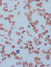
Image by Graham Beards
Researchers say the small molecule GBT440 could be a disease-modifying agent for patients with sickle cell disease (SCD).
Preclinical data showed that GBT440 can reduce sickling, extend the circulating half-life of red blood cells (RBCs), and decrease excessive erythropoiesis in SCD.
GBT440 binds specifically to hemoglobin and is designed to inhibit sickle hemoglobin (HbS) polymer formation.
“One promising strategy for preventing red blood cell sickling and subsequently modifying sickle cell disease over the long term involves inhibiting polymerization of HbS in red blood cells,” said David R. Archer, PhD, of Emory University School of Medicine in Atlanta, Georgia.
“This can be achieved by increasing the proportion of oxygenated HbS in those cells. We believe our preclinical results provide strong evidence that GBT440 inhibits HbS polymerization and red blood cell sickling, which is important because it addresses the underlying pathophysiology of sickle cell disease and has the potential to change its devastating clinical course.”
Dr Archer and his colleagues reported these results in the British Journal of Haematology. The research was supported by Global Blood Therapeutics, Inc., the company developing GBT440.
The researchers reported that, in vitro, GBT440 dose-dependently increased the affinity of HbS for oxygen, delayed polymerization of HbS, and reduced the number of sickled RBCs in whole blood from SCD patients.
In an animal model of SCD, GBT440 inhibited RBC sickling, prolonged the half-life of RBCs, and reduced reticulocyte counts.
The researchers said the drug also exhibited favorable pharmacokinetic properties in various animal species, suggesting the potential for once-daily oral dosing in SCD patients.
“Our preclinical work has developed a foundation of evidence that GBT440 is a potent inhibitor of the polymerization of HbS,” said Ted W. Love, MD, chief executive officer of Global Blood Therapeutics, Inc.
“We continue to build on these data with our ongoing phase 1/2 study, which has shown that GBT440 was well-tolerated over 90 days of dosing and that all SCD patients who received multiple doses of GBT440 exhibited improvements in one or more clinical markers of hemolysis and anemia. Our next step is to initiate a pivotal trial in adults with SCD later this year.”
Method reveals cells of origin in AML

in the bone marrow
Whole-genome profiling of open chromatin is a reliable way to identify the cells of origin in acute myeloid leukemia (AML), according to research published in Nature Communications.
“Knowing the cell of origin of cancer cells can provide insight into tumor subtypes and possibly diagnostic and therapeutic benefit,” said study author Jennifer Trowbridge, PhD, of the Jackson Laboratory for Mammalian Genetics in Bar Harbor, Maine.
“But existing methods to identify cell of origin from bulk tumor cell samples have been unsuccessful.”
Dr Trowbridge and her colleagues hypothesized that analyzing open chromatin in bulk tumor cells might provide a better method for identifying cancer cells of origin because of the cell-type specificity of chromatin structure.
The researchers worked with a mouse model of AML driven by expression of MLL-AF9, a fusion oncogene formed by a chromosome translocation between chromosomes 9 and 11.
The team began with 5 distinct, normal cell types found in the bone marrow in both mice and humans: long-term hematopoietic stem cells (HSCs), short-term HSCs, multipotent progenitors, common myeloid progenitors, and granulocyte macrophage progenitors.
The AML that developed from these different cells of origin had different penetrance and aggressiveness when engrafted in mice. The stem cell-derived lines proved the most aggressive and the committed progenitor lines the least aggressive.
These patterns were also reflected in the frequency of leukemia-initiating cells in each cell line, with HSCs having the highest frequency and committed progenitors having the lowest.
The researchers then set out to profile the open chromatin in these distinct AML samples and compare them to open chromatin patterns in normal cells using computational models.
The team identified open chromatin signatures and gene expression patterns in AML samples that may allow stem-cell-derived AML to be distinguished from progenitor-cell-of-origin AML.
These results support findings in human data suggesting the stage of a progenitor cell when it transforms to leukemia impacts clinical progression, with earlier-stage cell-of-origin cancers being more aggressive.
The researchers noted that, with further study of open chromatin in normal human stem and progenitor cell types as well as AML patient cohorts, this profiling approach could reveal precise regions with prognostic significance based on cell of origin; in other words, a valuable cancer biomarker.