User login
A 53-year-old African American woman with progressive shortness of breath for 1 month and a productive cough with intermittent blood-tinged sputum sought treatment at our emergency room. Over the past 2 months, she had lost 20 pounds—despite a normal appetite—and had noticed multiple “bumps” on her body.
The patient had no changes in her bowel habits, nor had she experienced nausea, vomiting, hematochezia, hemoptysis, odynophagia, dysphagia, chills, fever, or night sweats. Her past medical and surgical histories were unremarkable.
She had a 40-year history of smoking 1 to 3 packs of cigarettes per day, and a 30-year history of drinking a 1-liter bottle of hard liquor a day. She also occasionally used marijuana.
While in the ER, the patient was comfortable breathing on room air and afebrile with stable vital signs. We noticed decreased breath sounds bilaterally, as well as rhonchi. The patient had diffuse lymphadenopathy, as well as multiple, fixed, painless subcutaneous nodules on the neck, arms, flanks, back, and proximal right thigh. There was a 4 × 3.5 cm exophytic beefy-red tumor on the left inner arm (FIGURE 1). Some smaller nodules had surface hyperpigmentation, while others were subcutaneous, with no epidermal surface changes. All of the lesions were asymptomatic.
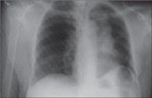
We ordered a chest radiograph (FIGURE 2) and blood work. Abnormal hematologic test results were: alkaline phosphatase (236 IU/L), gamma-glutamyl transferase (435 IU/L), and serum Ca++ (8.2 mg/dL).
FIGURE 1
Tumor on left inner arm
FIGURE 2
Chest x-ray brings Dx into focus
What is your diagnosis?
Dx: SCC of the lungs with skin metastases
Our patient’s chest radiograph—which revealed a left pulmonary mass—combined with her blood work and lesional biopsies, led us to diagnose primary squamous cell carcinoma (SCC) of the lung with skin metastases.
Approximately 30% of all lung cancers are classified as SCC.1 Pulmonary SCC metastatic to the skin tends to remain localized within the skin in the areas of the abdomen, anterior chest, back, scalp, and neck.2-6 Though cutaneous metastases of internal malignancies occur in 0.7% to 9.0% of all cancer patients, the incidence of skin metastases from lung cancer ranges from 1% to 12%.6-8
Lesions are painless and moveable
Most cutaneous metastases consist of single or multiple lesions that are painless, movable, and nonspecific. These lesions may also be firm to palpation, ulcerated, or exudative and able to penetrate the dermis or subcutaneous layer of skin with intact overlying epidermis.7-9
Skin eruptions may be the first sign of trouble
Prompt identification and diagnosis of these lesions are tantamount in tumor staging and therapy.
Schwartz reported that “in a series of patients with skin metastases seen in dermatologic consultation, the underlying cancer had been undiagnosed in 60% with lung cancer, in 53% with renal cancer, and in 40% with ovarian cancer.”7 Similar findings have been reported elsewhere.4,8 Thus, metastatic carcinoma to the skin should always be included in the differential diagnosis for newly eruptive skin lesions, regardless of whether the patient has ever been diagnosed with any form of cancer.10
A grim prognosis
Current therapies for skin metastases from primary lung carcinomas include surgical excision, symptomatic therapy, and chemotherapy—with and without radiation. After a patient’s course of treatment, skin metastases may be an important diagnostic marker for tumor recurrence.
Kamiyoshihara et al8 found that when skin metastases are detected, only 23% of the underlying primary cancers are operable, noting that most “portent a fatal outcome.”
Our patient declined further treatment
A CT of the patient’s chest showed a left hilar mass. A radionucleotide bone scan revealed metastases to the right elbow. We estimated that our patient had 1 year to live. In light of her very dismal prognosis, our patient declined any further work-up or treatment.
Our patient died 5 months later.
Correspondence
David A. Kasper DO, MBA, 2064 West Market St., Potts-ville, PA 17901; [email protected]
1. Parkin DM, Pisani P, Ferlay J. Global cancer statistics. CA Cancer J Clin. 1999;49:33.-
2. Ambrogi V, Nofroni I, Tonini G, Mineo TC. Skin metastases in lung cancer: analysis of a 10-year experience. Oncol Rep. 2001;8:57-61.
3. Altundag K, Yalcin S, Ozkaya O, Guler N. Synchronous squamous cell carcinoma of the stomach, the lung and the skin. Onkologie. 2004;27:291-293.
4. Brownstein MH, Helwig EB. Patterns of cutaneous metastasis. Arch Dermatol. 1972;105:862-868.
5. Reingold IM. Cutaneous metastases from internal carcinoma. Cancer. 1966;19:162-168.
6. Terashima T, Kanazawa M. Lung cancer with skin metastasis. Chest. 1994;106:1448-1450.
7. Schwartz RA. Cutaneous metastatic disease. J Am Acad Dermatol. 1995;33:161-182.
8. Kamiyoshihara M, Sakata K, Otani Y, Kawashima O, Takahashi T, Morishita Y. Solitary skin metastasis after lung cancer resection. Jpn J Thorac Cardiovasc Surg. 2002;50:343-346.
9. Kaplan RP. Specific cutaneous manifestations of internal malignancy. Adv Dermatol. 1986;1:3-42.
10. Nakamura H, Shimizu T, Kodama K, Shimizu H. Metastasis of lung cancer to the finger: a report of two cases. Int J Dermatol. 2005;44:47-49.
A 53-year-old African American woman with progressive shortness of breath for 1 month and a productive cough with intermittent blood-tinged sputum sought treatment at our emergency room. Over the past 2 months, she had lost 20 pounds—despite a normal appetite—and had noticed multiple “bumps” on her body.
The patient had no changes in her bowel habits, nor had she experienced nausea, vomiting, hematochezia, hemoptysis, odynophagia, dysphagia, chills, fever, or night sweats. Her past medical and surgical histories were unremarkable.
She had a 40-year history of smoking 1 to 3 packs of cigarettes per day, and a 30-year history of drinking a 1-liter bottle of hard liquor a day. She also occasionally used marijuana.
While in the ER, the patient was comfortable breathing on room air and afebrile with stable vital signs. We noticed decreased breath sounds bilaterally, as well as rhonchi. The patient had diffuse lymphadenopathy, as well as multiple, fixed, painless subcutaneous nodules on the neck, arms, flanks, back, and proximal right thigh. There was a 4 × 3.5 cm exophytic beefy-red tumor on the left inner arm (FIGURE 1). Some smaller nodules had surface hyperpigmentation, while others were subcutaneous, with no epidermal surface changes. All of the lesions were asymptomatic.
We ordered a chest radiograph (FIGURE 2) and blood work. Abnormal hematologic test results were: alkaline phosphatase (236 IU/L), gamma-glutamyl transferase (435 IU/L), and serum Ca++ (8.2 mg/dL).
FIGURE 1
Tumor on left inner arm
FIGURE 2
Chest x-ray brings Dx into focus
What is your diagnosis?
Dx: SCC of the lungs with skin metastases
Our patient’s chest radiograph—which revealed a left pulmonary mass—combined with her blood work and lesional biopsies, led us to diagnose primary squamous cell carcinoma (SCC) of the lung with skin metastases.
Approximately 30% of all lung cancers are classified as SCC.1 Pulmonary SCC metastatic to the skin tends to remain localized within the skin in the areas of the abdomen, anterior chest, back, scalp, and neck.2-6 Though cutaneous metastases of internal malignancies occur in 0.7% to 9.0% of all cancer patients, the incidence of skin metastases from lung cancer ranges from 1% to 12%.6-8
Lesions are painless and moveable
Most cutaneous metastases consist of single or multiple lesions that are painless, movable, and nonspecific. These lesions may also be firm to palpation, ulcerated, or exudative and able to penetrate the dermis or subcutaneous layer of skin with intact overlying epidermis.7-9
Skin eruptions may be the first sign of trouble
Prompt identification and diagnosis of these lesions are tantamount in tumor staging and therapy.
Schwartz reported that “in a series of patients with skin metastases seen in dermatologic consultation, the underlying cancer had been undiagnosed in 60% with lung cancer, in 53% with renal cancer, and in 40% with ovarian cancer.”7 Similar findings have been reported elsewhere.4,8 Thus, metastatic carcinoma to the skin should always be included in the differential diagnosis for newly eruptive skin lesions, regardless of whether the patient has ever been diagnosed with any form of cancer.10
A grim prognosis
Current therapies for skin metastases from primary lung carcinomas include surgical excision, symptomatic therapy, and chemotherapy—with and without radiation. After a patient’s course of treatment, skin metastases may be an important diagnostic marker for tumor recurrence.
Kamiyoshihara et al8 found that when skin metastases are detected, only 23% of the underlying primary cancers are operable, noting that most “portent a fatal outcome.”
Our patient declined further treatment
A CT of the patient’s chest showed a left hilar mass. A radionucleotide bone scan revealed metastases to the right elbow. We estimated that our patient had 1 year to live. In light of her very dismal prognosis, our patient declined any further work-up or treatment.
Our patient died 5 months later.
Correspondence
David A. Kasper DO, MBA, 2064 West Market St., Potts-ville, PA 17901; [email protected]
1. Parkin DM, Pisani P, Ferlay J. Global cancer statistics. CA Cancer J Clin. 1999;49:33.-
2. Ambrogi V, Nofroni I, Tonini G, Mineo TC. Skin metastases in lung cancer: analysis of a 10-year experience. Oncol Rep. 2001;8:57-61.
3. Altundag K, Yalcin S, Ozkaya O, Guler N. Synchronous squamous cell carcinoma of the stomach, the lung and the skin. Onkologie. 2004;27:291-293.
4. Brownstein MH, Helwig EB. Patterns of cutaneous metastasis. Arch Dermatol. 1972;105:862-868.
5. Reingold IM. Cutaneous metastases from internal carcinoma. Cancer. 1966;19:162-168.
6. Terashima T, Kanazawa M. Lung cancer with skin metastasis. Chest. 1994;106:1448-1450.
7. Schwartz RA. Cutaneous metastatic disease. J Am Acad Dermatol. 1995;33:161-182.
8. Kamiyoshihara M, Sakata K, Otani Y, Kawashima O, Takahashi T, Morishita Y. Solitary skin metastasis after lung cancer resection. Jpn J Thorac Cardiovasc Surg. 2002;50:343-346.
9. Kaplan RP. Specific cutaneous manifestations of internal malignancy. Adv Dermatol. 1986;1:3-42.
10. Nakamura H, Shimizu T, Kodama K, Shimizu H. Metastasis of lung cancer to the finger: a report of two cases. Int J Dermatol. 2005;44:47-49.

