User login
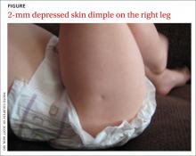
A MOTHER BROUGHT HER 6-MONTH-OLD BOY in for a routine well-child check and asked that we look at a skin dimple in his right leg (FIGURE). The mother, who was a healthy 43-year-old, indicated that she hadn’t noticed the dimple until he was several weeks old, and since then it hadn’t changed significantly. She said that the dimple didn’t seem to bother him, and he was able to move all of his extremities without difficulty.
The baby was born by normal spontaneous vaginal delivery and was discharged from the hospital at 2 days of age. He’d had no significant illnesses, all of his immunizations were up to date, and he’d been meeting all of his development milestones. There were no family members with similar findings.
The baby was developmentally appropriate for his age and his physical exam was unremarkable, including his neurological exam. The only significant finding was a 2-mm circular dimple on the lateral aspect of his proximal right leg.
WHAT IS YOUR DIAGNOSIS?
HOW WOULD YOU TREAT THIS PATIENT?
Diagnosis: Amniocentesis scar
Our patient had a classic skin dimple resulting from an inadvertent puncture during routine amniocentesis.
Amniocentesis is generally performed under real-time ultrasonography during the second trimester, when the fetus occupies approximately half of the amniotic cavity and the ratio of viable to nonviable cells in the amniotic fluid is greatest. It is typically performed on women of advanced maternal age (as was the case with our patient), who are at greater risk of delivering babies with genetic disorders. While amniocentesis is generally considered safe, complications—including puncture of placental and fetal vessels, peripheral nerve damage, fistula formation, pneumothorax, ocular trauma, and fetal death—have been reported.1
Scar formation from puncture of the fetus is estimated to occur in 1% to 3% of all amniocentesis procedures performed,2 although this may be an underestimation because the scars are often inconspicuous.3 The nonpigmented, depressed, dimplelike lesions generally measure 1 to 2 mm in diameter1 (although linear scars have also been reported4), and are not associated with any limitations in movement or underlying signs of injury. The chest, abdomen, back, and extremities are the most commonly affected sites.1
The main risk factors for needle puncture during amniocentesis are a history of repetitive needle insertion attempts and limited operator experience.2 Most lesions are present at birth, although others may become more evident as the infant gains weight and the scar retracts to form a dimple.5 The lesions are benign and require no further work-up.
Amniocentesis wasn’t performed? Time to look further
Most lesions caused by amniocentesis are present at birth, although others become more evident as the baby gains weight and the scar retracts to form a dimple. When assessing a newborn, the gluteal folds should be separated to look for any dimpling. Small “sacral dimples” that overlie the coccyx and have well-visualized, intact bases are considered benign normal variants and do not require further work-up.6,7 However, if there are multiple dimples, midline dimpling more than 2.5 cm from the anus, or dimples associated with excess hair, pigmentation, skin tags, or vascular anomalies, further evaluation (ultrasound or magnetic resonance imaging) and neurosurgical referral may be necessary to evaluate for a closed neural tube defect.8
Abnormalities that should be considered part of the differential for midline skin dimpling include:
- congenital dermal sinus tracts (remnants of incomplete neural tube closure during fetal development)
- diastematomyelia (a longitudinal split in the spinal cord, usually the result of an osseous or fibrous band that forms 2 hemicords)
- tethered spinal cord (an abnormal attachment of the cord to surrounding structures).8
Skin dimpling has also been reported in association with congenital rubella; in such cases, the dimples have been located on the patella and other bony prominences.9
Nothing to worry about
Our patient’s skin dimple was not along the spine, and the mother acknowledged having had amniocentesis during her pregnancy, so no further work-up was indicated. We simply reassured her that the dimple on her son’s leg was completely benign.
CORRESPONDENCE
Scott Akin, MD, FAAFP,
1364 Clifton Road, NE, Box M-7, Atlanta, GA 30322
[email protected]
1. Raimer SS, Raimer BG. Needle puncture scars from midtrimester amniocentesis. Arch Dermatol. 1984;120:1360-1362.
2. Epley SL, Hanson JW, Cruiksank DP. Fetal injury with midtrimester diagnostic amniocentesis. Obstet Gynecol. 1979;53:77-80.
3. Bruce S, Duffy JO, Wolf JE Jr. Skin dimpling associated with midtrimester amniocentesis. Pediatr Dermatol. 1984;2:140-142.
4. Broome DL, Wilson MG, Weiss B, et al. Needle puncture of fetus: a complication of second-trimester amniocentesis. Am J Obstet Gynecol. 1976;126:247-252.
5. Ahluwalia J, Lowenstein E. Skin dimpling as a delayed manifestation of traumatic amniocentesis. Skinmed. 2005;4:323-324.
6. Ben-Sira L, Ponger P, Miller E, et al. Low-risk lumbar skin stigmata in infants: the role of ultrasound screening. J Pediatr. 2009;155:864-869.
7. Medina LS, Crone K, Kuntz KM. Newborns with suspected occult spinal dysraphism: a cost-effectiveness analysis of diagnostic strategies. Pediatrics. 2001;108:e101.
8. Zywicke HA, Rozzelle CJ. Sacral dimples. Pediatr Rev. 2011; 32:109-113.
9. Hammond K. Skin dimples and rubella. Pediatrics. 1967;39:291-292.
A MOTHER BROUGHT HER 6-MONTH-OLD BOY in for a routine well-child check and asked that we look at a skin dimple in his right leg (FIGURE). The mother, who was a healthy 43-year-old, indicated that she hadn’t noticed the dimple until he was several weeks old, and since then it hadn’t changed significantly. She said that the dimple didn’t seem to bother him, and he was able to move all of his extremities without difficulty.
The baby was born by normal spontaneous vaginal delivery and was discharged from the hospital at 2 days of age. He’d had no significant illnesses, all of his immunizations were up to date, and he’d been meeting all of his development milestones. There were no family members with similar findings.
The baby was developmentally appropriate for his age and his physical exam was unremarkable, including his neurological exam. The only significant finding was a 2-mm circular dimple on the lateral aspect of his proximal right leg.
WHAT IS YOUR DIAGNOSIS?
HOW WOULD YOU TREAT THIS PATIENT?
Diagnosis: Amniocentesis scar
Our patient had a classic skin dimple resulting from an inadvertent puncture during routine amniocentesis.
Amniocentesis is generally performed under real-time ultrasonography during the second trimester, when the fetus occupies approximately half of the amniotic cavity and the ratio of viable to nonviable cells in the amniotic fluid is greatest. It is typically performed on women of advanced maternal age (as was the case with our patient), who are at greater risk of delivering babies with genetic disorders. While amniocentesis is generally considered safe, complications—including puncture of placental and fetal vessels, peripheral nerve damage, fistula formation, pneumothorax, ocular trauma, and fetal death—have been reported.1
Scar formation from puncture of the fetus is estimated to occur in 1% to 3% of all amniocentesis procedures performed,2 although this may be an underestimation because the scars are often inconspicuous.3 The nonpigmented, depressed, dimplelike lesions generally measure 1 to 2 mm in diameter1 (although linear scars have also been reported4), and are not associated with any limitations in movement or underlying signs of injury. The chest, abdomen, back, and extremities are the most commonly affected sites.1
The main risk factors for needle puncture during amniocentesis are a history of repetitive needle insertion attempts and limited operator experience.2 Most lesions are present at birth, although others may become more evident as the infant gains weight and the scar retracts to form a dimple.5 The lesions are benign and require no further work-up.
Amniocentesis wasn’t performed? Time to look further
Most lesions caused by amniocentesis are present at birth, although others become more evident as the baby gains weight and the scar retracts to form a dimple. When assessing a newborn, the gluteal folds should be separated to look for any dimpling. Small “sacral dimples” that overlie the coccyx and have well-visualized, intact bases are considered benign normal variants and do not require further work-up.6,7 However, if there are multiple dimples, midline dimpling more than 2.5 cm from the anus, or dimples associated with excess hair, pigmentation, skin tags, or vascular anomalies, further evaluation (ultrasound or magnetic resonance imaging) and neurosurgical referral may be necessary to evaluate for a closed neural tube defect.8
Abnormalities that should be considered part of the differential for midline skin dimpling include:
- congenital dermal sinus tracts (remnants of incomplete neural tube closure during fetal development)
- diastematomyelia (a longitudinal split in the spinal cord, usually the result of an osseous or fibrous band that forms 2 hemicords)
- tethered spinal cord (an abnormal attachment of the cord to surrounding structures).8
Skin dimpling has also been reported in association with congenital rubella; in such cases, the dimples have been located on the patella and other bony prominences.9
Nothing to worry about
Our patient’s skin dimple was not along the spine, and the mother acknowledged having had amniocentesis during her pregnancy, so no further work-up was indicated. We simply reassured her that the dimple on her son’s leg was completely benign.
CORRESPONDENCE
Scott Akin, MD, FAAFP,
1364 Clifton Road, NE, Box M-7, Atlanta, GA 30322
[email protected]
1. Raimer SS, Raimer BG. Needle puncture scars from midtrimester amniocentesis. Arch Dermatol. 1984;120:1360-1362.
2. Epley SL, Hanson JW, Cruiksank DP. Fetal injury with midtrimester diagnostic amniocentesis. Obstet Gynecol. 1979;53:77-80.
3. Bruce S, Duffy JO, Wolf JE Jr. Skin dimpling associated with midtrimester amniocentesis. Pediatr Dermatol. 1984;2:140-142.
4. Broome DL, Wilson MG, Weiss B, et al. Needle puncture of fetus: a complication of second-trimester amniocentesis. Am J Obstet Gynecol. 1976;126:247-252.
5. Ahluwalia J, Lowenstein E. Skin dimpling as a delayed manifestation of traumatic amniocentesis. Skinmed. 2005;4:323-324.
6. Ben-Sira L, Ponger P, Miller E, et al. Low-risk lumbar skin stigmata in infants: the role of ultrasound screening. J Pediatr. 2009;155:864-869.
7. Medina LS, Crone K, Kuntz KM. Newborns with suspected occult spinal dysraphism: a cost-effectiveness analysis of diagnostic strategies. Pediatrics. 2001;108:e101.
8. Zywicke HA, Rozzelle CJ. Sacral dimples. Pediatr Rev. 2011; 32:109-113.
9. Hammond K. Skin dimples and rubella. Pediatrics. 1967;39:291-292.
