User login
A 54-year-old woman was admitted to the emergency department with a swollen right leg, fever, and altered mental status. Her family brought her in after finding her confused and lethargic. She was incontinent of stool and urine and complained of a rash with blisters on her right thigh. The patient had noted a pimple in her groin more than 5 days earlier; over the past few days she has been complaining of increasing leg pain. She related to her sisters that she had an appointment with her gynecologist in the next few days to have the lesion drained.
The patient had no fever, chest pain, shortness of breath, nausea, or vomiting. Her medical history included type 2 diabetes mellitus, hypertension, and cortical atrophy with mild mental retardation. She had been living independently in her own apartment, and was last seen by her sisters 6 days before with no apparent complaints. She had been wheelchair-bound for 6 months due to a fractured ankle from which she has not been able to completely rehabilitate.
The medications she was taking included glyburide, raloxifene (Evista), and furosemide (Lasix). Surgical history was significant only for a cholecystectomy. She did not smoke or drink alcohol. Upon presentation to the ED she appeared ill, with a blood pressure of 124/50 mm Hg, pulse 110, respiratory rate 18, and temperature of 102°F. Her fingerstick blood sugar was 573. She was able to answer simple questions but was not oriented to time or place. Her skin was hot and dry. Chest exam revealed clear lungs with tachypnea and a 2/6 systolic murmur. Her abdomen was slightly obese, soft, and nontender with normal bowel sounds and a well-healed right upper quadrant incision. Her genitourinary exam revealed a purulent drainage in the groin near her vulva.
Her right leg was markedly swollen, erythematous, and had a brownish-red discoloration that extended from her groin circumferentially to her knee. The skin had a “woody” feel when palpated and large bullae were present (FIGURE 1).
The decision to obtain x-rays of her pelvis and femur was made to assess the extent of her infection (FIGURES 2 AND 3).
FIGURE 1
Cellulitis in the leg
FIGURE 2
Radiograph of thigh and hip area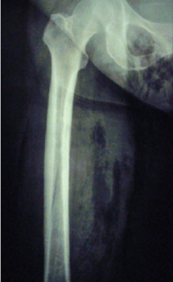
FIGURE 3
Radiograph of knee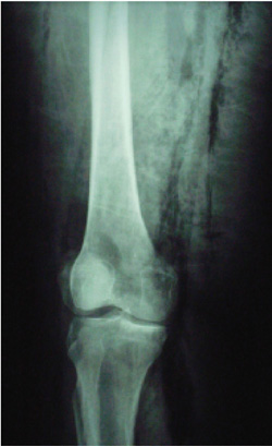
What is the differential diagnosis for this patient?
What tests might help delineate the extent of her infection?
Diagnosis: Acute necrotizing fasciitis
The patient was diagnosed with acute necrotizing fasciitis, a rare, often fatal, soft-tissue bacterial infection. According to the Centers for Disease Control and Prevention, only 500 to 1500 cases of necrotizing fasciitis are diagnosed each year in the US.1
Epidemiology
Peripheral vascular disease, diabetes, and a compromised immune system are significant risk factors for necrotizing fasciitis.2 Diabetes is present in 18% to 60% of cases;1,3 in addition, 19% to 77% of patients use intravenous drugs.1,3,4 Other significant predisposing factors include alcohol abuse (9%–31%),1,4 obesity,1,4 and malnutrition.3 Although risk factors are numerous, half of all cases of streptococcal necrotizing fasciitis occur in previously healthy individuals. Pathogenic agents can be introduced as a result of minor trauma, insect bites, or surgical incisions.
In this case the patient noted a “pimple” in the groin area and complained of pain for 5 days. By the time she reached the hospital she had mental status changes, fever, appeared toxic, and had signs of early septic shock. We can identify in this case the probable port of entry as the lesion in the groin that was visualized on physical exam to be draining pus.
Pathophysiology
Necrotizing fasciitis involves the superficial layer of skin, subcutaneous tissues, and fascia. The infection spreads rapidly along these layers, causing edema and compression of vasculature, which rapidly progresses to tissue necrosis and sepsis. Even with new broad-spectrum antibiotics, mortality can be as high as 75% in patients who become septic and develop renal failure.
Necrotizing fasciitis occurs when a mixed variety of organisms, both aerobic and anaerobic, invade the subcutaneous tissue and fascia.5 Most necrotizing soft-tissue infections are polymicrobial, with only a small percentage involving a single organism. In immune-compromised patients, Pseudomonas spp and gram-negative enteric organisms can be found. The organisms isolated most often in polymicrobial necrotizing soft-tissue infections are combinations of staphylococci (especially Staphylococcus epidermis with beta-hemolytic streptococcus), enterococci, Enterobacteriaceae spp (commonly Escherichia coli, Proteus mirabilis, Klebsiella pneumoniae, and Pseudomonas aeruginosa), streptococci, Bacterioides prevotella spp, anaerobic gram-positive cocci, and Clostridium spp.6
Patient presentation
The clinical history and a meticulous physical examination are essential in establishing an early diagnosis of necrotizing infections.5 Necrotizing fasciitis can be easily misdiagnosed as only cellulitis. Most often, a patient with necrotizing fasciitis appears ill, with constitutional symptoms of fever, chills, hypotension, dehydration, and rapid heart rate. You can also see erythema with bullae formation, serosanguineous fluids drainage, induration, and violaceous discoloration. Pain and crepitation may be noted.3,5,7 Rapid progression of edema and pain out of proportion to examination is seen in the early stages. The parts of the skin affected by the disease can become numb with progression of the infection; this is thought to be due to infarction of the cutaneous nerves located in necrotic subcutaneous fascia and soft tissue.5
Causative factors in this patient included diabetes and obesity. Diabetic neuropathy may have also delayed presentation and dulled her perception of pain. Diabetic microvascular disease may also have contributed to a faster progression of tissue hypoxia.
Diagnostic methods: Lab tests, biopsy, x-rays
Laboratory testing for necrotizing fasciitis is thought by most experts to be non-specific. Another investigative team found that 76% of patients with necrotizing soft-tissue infections had low platelet count or PT and PTT with higher than normal values; prolonged PT is associated with increase mortality.6 Hypocalcemia, hypoproteinemia, anemia, and acidosis have also been noted.
Diagnosis must be considered early when necrotizing fasciitis is suspected. Although the gold standard for diagnosis is biopsy or wound exploration and surgical debridement,6 diagnosis can be made early when necrotizing fasciitis is suspected.
The role of soft-tissue radiographs in the diagnosis of necrotizing fasciitis is unclear. Plain films can provide information such as soft-tissue thickening and internal gas formation. Unfortunately, plain radiographs typically show no specific abnormality until the necrotizing process is well advanced.
Treatment of necrotizing fasciitis
Resuscitation
Adequate fluid resuscitation and stabilization of any patient suspected of having necrotizing fasciitis is the first line of therapy. Large-bore IV lines or a central line may be necessary. Adequate monitoring should include a Foley catheter and pulse oximetry. Correction of any metabolic abnormalities needs to be addressed.
Antibiotics
Antibiotic treatment should be started as soon as possible, although no study has shown antibiotics to significantly alter mortality. A Gram stain of the infected material would be helpful to guide further antibiotic choices. However, initial therapy should be directed at both aerobic and anaerobic organisms.
Triple therapy is recommended: penicillin or ampicillin for Clostridia, Streptococci, and Peptostreptococcus; clindamycin or metronidazole for anaerobes, Bacteroides fragilis, Fusobacterium, and Peptostreptococcus; and gentamicin or another aminoglycoside for Enterobacteriaceae. Imipenem or meropenem can be used as the initial agent for high beta-lactamase resistance, wide-spectrum efficacy, and inhibition of endotoxin release from aerobic bacilli. Tetanus prophylaxis with absorbed tetanus toxoid and passive immune coverage with tetanus hyperimmune globulin is indicated for a patient whose history of immunization is unclear or unavailable.6
Surgery
Urgent surgical consultation is necessary. Early recognition and prompt aggressive debridement of all necrotic tissue is critical for survival—in fact, it is the only therapy demonstrated to improve the rate of survival.7 Necrotic tissue serves as a culture medium and creates an anaerobic environment, which hinders an adequate immune response. Sufficient debridement consists of exposure to all margins of viable tissue. Antibiotics are important but are secondary to urgent removal of the toxic tissue.
Hyperbaric oxygen therapy
All necrotizing infections are associated with ischemia, reduced tissue oxygen tension, and a decrease in host cellular immunity. The physciological rationale for increasing oxygen is that tension ischemia may be reversed and host defense mechanisms improved. Hyperbaric oxygen is generally considered an important adjunct in the treatment of clostridial myonecrosis or gas gangrene.
Studies have failed to show statistically significant outcome differences with respect to mortality and length of hospitalization.3 Some studies show improvement of survival rates or limb salvage; others show no difference in outcomes with hyperbaric oxygen. Note that these studies show no consistency in patient population or number of visits to the operating room. More evidence is needed, preferably by way of randomized controlled trials, before routine or wide-spread use of hyperbaric oxygen can be recommended.
The patient’s treatment and outcome
The emergency department physicians initiated intravenous antibiotics and obtained an urgent surgical consultation. In addition, they sent for blood cultures and other laboratory tests.
In the operating room, surgeons debrided her skin and removed all necrotic muscles and skin in her perineum and entire medial thigh during the first surgery. Eighteen hours later she returned to the operating room—the infection had spread to once-viable tissue from the symphysis pubis to the knee. The family was consulted concerning a more radical surgical approach, a hip disarticulation or hemipelvectomy. They declined. The patient was made comfortable; she died 12 hours later. Her wound culture later grew E coli, Proteus vulgaris, Coryne-bacterium, Enterococcus, Staphylococcus spp, and Peptostreptococcus.
CORRESPONDENCE
Susan Dufel, MD, Department Trauma and Emergency Medicine, University of Connecticut, 80 Seymour Street, Hartford CT 06102. E-mail: [email protected]
1. Faucher LD, Morris SE, Edelman LS, et al. Burn center management of necrotizing soft-tissue surgical infections in unburned patients. Am J Surg 2001;182:563-569.
2. Childers BJ, Potyondy LD, Nachreiner R, et al. Necrotizing fasciitis: a fourteen-year retrospective study of 163 patients. Am Surg 2002;68:109-116.
3. Kuncir EJ, Tillou A, St Hill CR. Necrotizing soft-tissue infections. Emerg Med Clin North Am 2003;21:1075-1087.
4. Bosshardt TL, Henderson VJ, Organ CH, Jr. Necrotizing soft-tissue infections. Arch Surg 1996;131:846-854.
5. Majeski J, John JF, Jr. Necrotizing soft tissue infections: a guide to early diagnosis and initial therapy. South Med J 2003;96:900-906.
6. Headley AJ. Necrotizing fasciitis: a primary care review. Am Fam Phys 2003;68:323-328.
7. Chin-Ho Wong, Haw-Chong Chang, Pasupathy S. Necrotizing fasciitis: clinical presentation, microbiology, determinants of mortality. J Bone Joint Surg 2003;85A(8):1454-1460.
A 54-year-old woman was admitted to the emergency department with a swollen right leg, fever, and altered mental status. Her family brought her in after finding her confused and lethargic. She was incontinent of stool and urine and complained of a rash with blisters on her right thigh. The patient had noted a pimple in her groin more than 5 days earlier; over the past few days she has been complaining of increasing leg pain. She related to her sisters that she had an appointment with her gynecologist in the next few days to have the lesion drained.
The patient had no fever, chest pain, shortness of breath, nausea, or vomiting. Her medical history included type 2 diabetes mellitus, hypertension, and cortical atrophy with mild mental retardation. She had been living independently in her own apartment, and was last seen by her sisters 6 days before with no apparent complaints. She had been wheelchair-bound for 6 months due to a fractured ankle from which she has not been able to completely rehabilitate.
The medications she was taking included glyburide, raloxifene (Evista), and furosemide (Lasix). Surgical history was significant only for a cholecystectomy. She did not smoke or drink alcohol. Upon presentation to the ED she appeared ill, with a blood pressure of 124/50 mm Hg, pulse 110, respiratory rate 18, and temperature of 102°F. Her fingerstick blood sugar was 573. She was able to answer simple questions but was not oriented to time or place. Her skin was hot and dry. Chest exam revealed clear lungs with tachypnea and a 2/6 systolic murmur. Her abdomen was slightly obese, soft, and nontender with normal bowel sounds and a well-healed right upper quadrant incision. Her genitourinary exam revealed a purulent drainage in the groin near her vulva.
Her right leg was markedly swollen, erythematous, and had a brownish-red discoloration that extended from her groin circumferentially to her knee. The skin had a “woody” feel when palpated and large bullae were present (FIGURE 1).
The decision to obtain x-rays of her pelvis and femur was made to assess the extent of her infection (FIGURES 2 AND 3).
FIGURE 1
Cellulitis in the leg
FIGURE 2
Radiograph of thigh and hip area
FIGURE 3
Radiograph of knee
What is the differential diagnosis for this patient?
What tests might help delineate the extent of her infection?
Diagnosis: Acute necrotizing fasciitis
The patient was diagnosed with acute necrotizing fasciitis, a rare, often fatal, soft-tissue bacterial infection. According to the Centers for Disease Control and Prevention, only 500 to 1500 cases of necrotizing fasciitis are diagnosed each year in the US.1
Epidemiology
Peripheral vascular disease, diabetes, and a compromised immune system are significant risk factors for necrotizing fasciitis.2 Diabetes is present in 18% to 60% of cases;1,3 in addition, 19% to 77% of patients use intravenous drugs.1,3,4 Other significant predisposing factors include alcohol abuse (9%–31%),1,4 obesity,1,4 and malnutrition.3 Although risk factors are numerous, half of all cases of streptococcal necrotizing fasciitis occur in previously healthy individuals. Pathogenic agents can be introduced as a result of minor trauma, insect bites, or surgical incisions.
In this case the patient noted a “pimple” in the groin area and complained of pain for 5 days. By the time she reached the hospital she had mental status changes, fever, appeared toxic, and had signs of early septic shock. We can identify in this case the probable port of entry as the lesion in the groin that was visualized on physical exam to be draining pus.
Pathophysiology
Necrotizing fasciitis involves the superficial layer of skin, subcutaneous tissues, and fascia. The infection spreads rapidly along these layers, causing edema and compression of vasculature, which rapidly progresses to tissue necrosis and sepsis. Even with new broad-spectrum antibiotics, mortality can be as high as 75% in patients who become septic and develop renal failure.
Necrotizing fasciitis occurs when a mixed variety of organisms, both aerobic and anaerobic, invade the subcutaneous tissue and fascia.5 Most necrotizing soft-tissue infections are polymicrobial, with only a small percentage involving a single organism. In immune-compromised patients, Pseudomonas spp and gram-negative enteric organisms can be found. The organisms isolated most often in polymicrobial necrotizing soft-tissue infections are combinations of staphylococci (especially Staphylococcus epidermis with beta-hemolytic streptococcus), enterococci, Enterobacteriaceae spp (commonly Escherichia coli, Proteus mirabilis, Klebsiella pneumoniae, and Pseudomonas aeruginosa), streptococci, Bacterioides prevotella spp, anaerobic gram-positive cocci, and Clostridium spp.6
Patient presentation
The clinical history and a meticulous physical examination are essential in establishing an early diagnosis of necrotizing infections.5 Necrotizing fasciitis can be easily misdiagnosed as only cellulitis. Most often, a patient with necrotizing fasciitis appears ill, with constitutional symptoms of fever, chills, hypotension, dehydration, and rapid heart rate. You can also see erythema with bullae formation, serosanguineous fluids drainage, induration, and violaceous discoloration. Pain and crepitation may be noted.3,5,7 Rapid progression of edema and pain out of proportion to examination is seen in the early stages. The parts of the skin affected by the disease can become numb with progression of the infection; this is thought to be due to infarction of the cutaneous nerves located in necrotic subcutaneous fascia and soft tissue.5
Causative factors in this patient included diabetes and obesity. Diabetic neuropathy may have also delayed presentation and dulled her perception of pain. Diabetic microvascular disease may also have contributed to a faster progression of tissue hypoxia.
Diagnostic methods: Lab tests, biopsy, x-rays
Laboratory testing for necrotizing fasciitis is thought by most experts to be non-specific. Another investigative team found that 76% of patients with necrotizing soft-tissue infections had low platelet count or PT and PTT with higher than normal values; prolonged PT is associated with increase mortality.6 Hypocalcemia, hypoproteinemia, anemia, and acidosis have also been noted.
Diagnosis must be considered early when necrotizing fasciitis is suspected. Although the gold standard for diagnosis is biopsy or wound exploration and surgical debridement,6 diagnosis can be made early when necrotizing fasciitis is suspected.
The role of soft-tissue radiographs in the diagnosis of necrotizing fasciitis is unclear. Plain films can provide information such as soft-tissue thickening and internal gas formation. Unfortunately, plain radiographs typically show no specific abnormality until the necrotizing process is well advanced.
Treatment of necrotizing fasciitis
Resuscitation
Adequate fluid resuscitation and stabilization of any patient suspected of having necrotizing fasciitis is the first line of therapy. Large-bore IV lines or a central line may be necessary. Adequate monitoring should include a Foley catheter and pulse oximetry. Correction of any metabolic abnormalities needs to be addressed.
Antibiotics
Antibiotic treatment should be started as soon as possible, although no study has shown antibiotics to significantly alter mortality. A Gram stain of the infected material would be helpful to guide further antibiotic choices. However, initial therapy should be directed at both aerobic and anaerobic organisms.
Triple therapy is recommended: penicillin or ampicillin for Clostridia, Streptococci, and Peptostreptococcus; clindamycin or metronidazole for anaerobes, Bacteroides fragilis, Fusobacterium, and Peptostreptococcus; and gentamicin or another aminoglycoside for Enterobacteriaceae. Imipenem or meropenem can be used as the initial agent for high beta-lactamase resistance, wide-spectrum efficacy, and inhibition of endotoxin release from aerobic bacilli. Tetanus prophylaxis with absorbed tetanus toxoid and passive immune coverage with tetanus hyperimmune globulin is indicated for a patient whose history of immunization is unclear or unavailable.6
Surgery
Urgent surgical consultation is necessary. Early recognition and prompt aggressive debridement of all necrotic tissue is critical for survival—in fact, it is the only therapy demonstrated to improve the rate of survival.7 Necrotic tissue serves as a culture medium and creates an anaerobic environment, which hinders an adequate immune response. Sufficient debridement consists of exposure to all margins of viable tissue. Antibiotics are important but are secondary to urgent removal of the toxic tissue.
Hyperbaric oxygen therapy
All necrotizing infections are associated with ischemia, reduced tissue oxygen tension, and a decrease in host cellular immunity. The physciological rationale for increasing oxygen is that tension ischemia may be reversed and host defense mechanisms improved. Hyperbaric oxygen is generally considered an important adjunct in the treatment of clostridial myonecrosis or gas gangrene.
Studies have failed to show statistically significant outcome differences with respect to mortality and length of hospitalization.3 Some studies show improvement of survival rates or limb salvage; others show no difference in outcomes with hyperbaric oxygen. Note that these studies show no consistency in patient population or number of visits to the operating room. More evidence is needed, preferably by way of randomized controlled trials, before routine or wide-spread use of hyperbaric oxygen can be recommended.
The patient’s treatment and outcome
The emergency department physicians initiated intravenous antibiotics and obtained an urgent surgical consultation. In addition, they sent for blood cultures and other laboratory tests.
In the operating room, surgeons debrided her skin and removed all necrotic muscles and skin in her perineum and entire medial thigh during the first surgery. Eighteen hours later she returned to the operating room—the infection had spread to once-viable tissue from the symphysis pubis to the knee. The family was consulted concerning a more radical surgical approach, a hip disarticulation or hemipelvectomy. They declined. The patient was made comfortable; she died 12 hours later. Her wound culture later grew E coli, Proteus vulgaris, Coryne-bacterium, Enterococcus, Staphylococcus spp, and Peptostreptococcus.
CORRESPONDENCE
Susan Dufel, MD, Department Trauma and Emergency Medicine, University of Connecticut, 80 Seymour Street, Hartford CT 06102. E-mail: [email protected]
1. Faucher LD, Morris SE, Edelman LS, et al. Burn center management of necrotizing soft-tissue surgical infections in unburned patients. Am J Surg 2001;182:563-569.
2. Childers BJ, Potyondy LD, Nachreiner R, et al. Necrotizing fasciitis: a fourteen-year retrospective study of 163 patients. Am Surg 2002;68:109-116.
3. Kuncir EJ, Tillou A, St Hill CR. Necrotizing soft-tissue infections. Emerg Med Clin North Am 2003;21:1075-1087.
4. Bosshardt TL, Henderson VJ, Organ CH, Jr. Necrotizing soft-tissue infections. Arch Surg 1996;131:846-854.
5. Majeski J, John JF, Jr. Necrotizing soft tissue infections: a guide to early diagnosis and initial therapy. South Med J 2003;96:900-906.
6. Headley AJ. Necrotizing fasciitis: a primary care review. Am Fam Phys 2003;68:323-328.
7. Chin-Ho Wong, Haw-Chong Chang, Pasupathy S. Necrotizing fasciitis: clinical presentation, microbiology, determinants of mortality. J Bone Joint Surg 2003;85A(8):1454-1460.