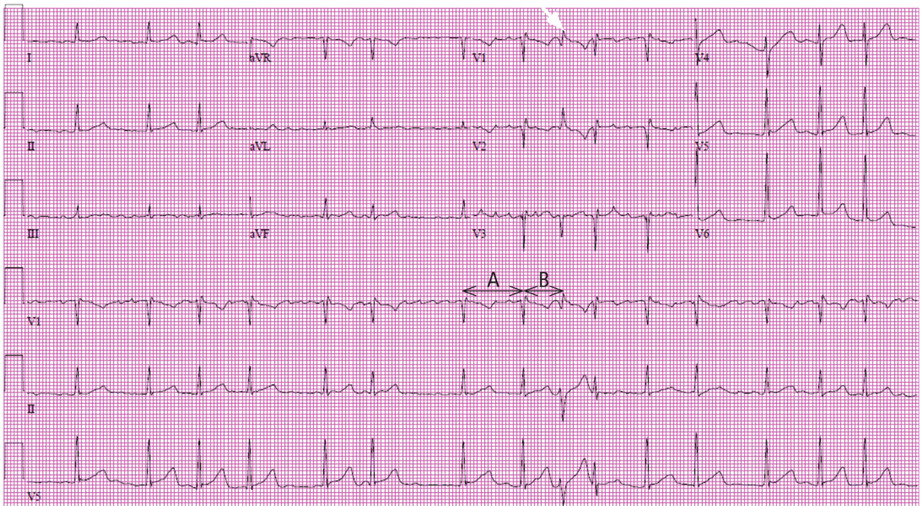
In the article “Presumed premature ventricular contractions” by Drs. Moises Auron and Donald Underwood (Cleve Clin J Med 2011; 78:812–813), Figure 1 was incorrectly labelled. The corrected figure and legend appear below. The authors wish to thank Philippe Akhrass, MD, from the State University of New York, Brooklyn, and Shahrokh Rafii, MD, from Brookdale University Hospital and Medical Center, Brooklyn, NY, for pointing out this error.
Figure 1. The electrocardiogram shows atrial fibrillation. Following the seventh beat, the cycle length “A” is longer than the subsequent cycle “B,” giving a long-short sequence that ends in an aberrantly conducted beat that has terminal broadening and a right-bundle-branch-type pattern (white arrow). This is a typical Ashman sequence. The next beat in sequence is slightly aberrant but is returning to the baseline QRS configuration.