User login
Sharon Worcester is an award-winning medical journalist for MDedge News. She has been with the company since 1996, first as the Southeast Bureau Chief (1996-2009) when the company was known as International Medical News Group, then as a freelance writer (2010-2015) before returning as a reporter in 2015. She previously worked as a daily newspaper reporter covering health and local government. Sharon currently reports primarily on oncology and hematology. She has a BA from Eckerd College and an MA in Mass Communication/Print Journalism from the University of Florida. Connect with her via LinkedIn and follow her on twitter @SW_MedReporter.
ASH preview: Key themes include tackling CAR T obstacles, sickle cell advances, VTE
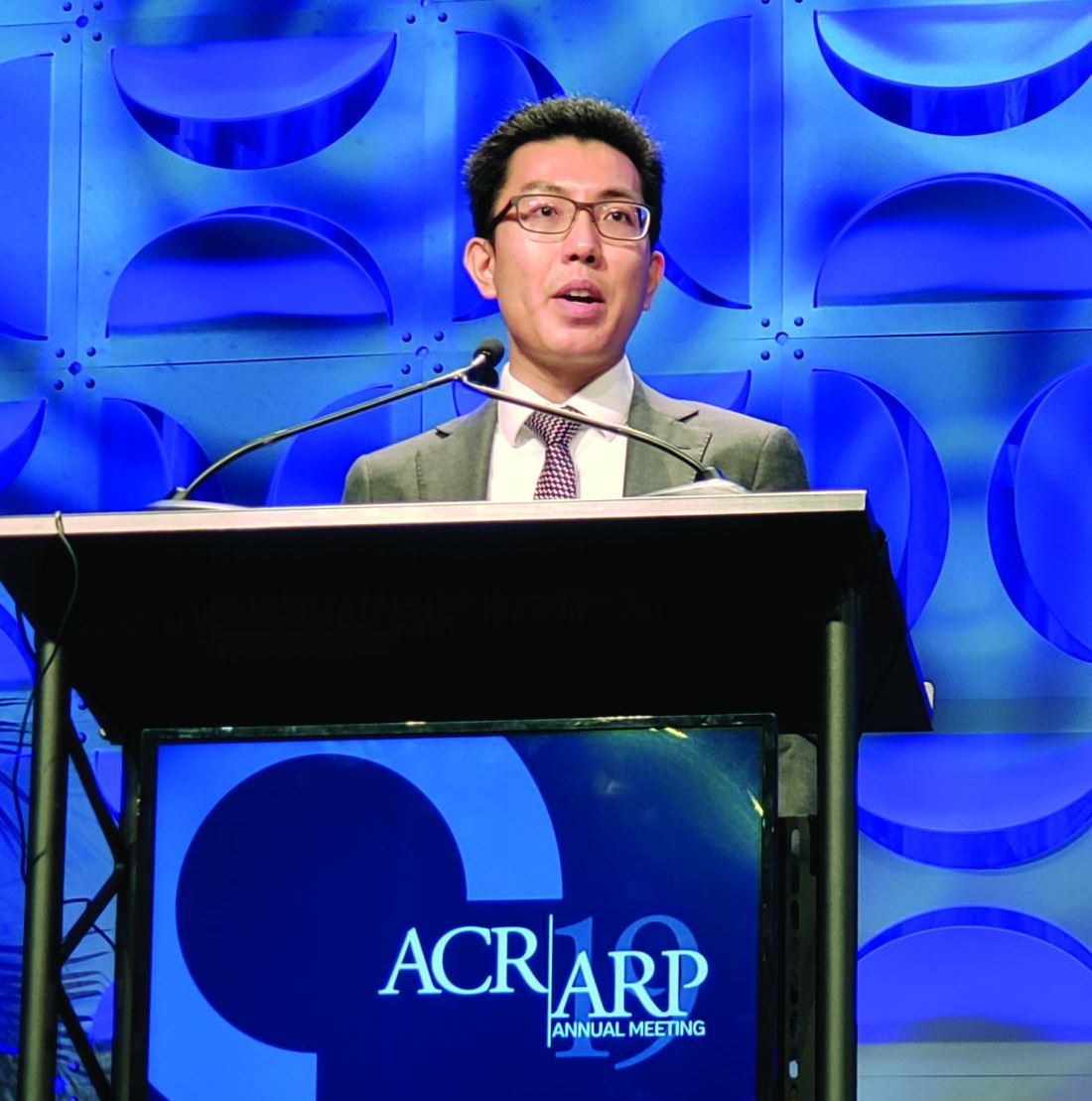
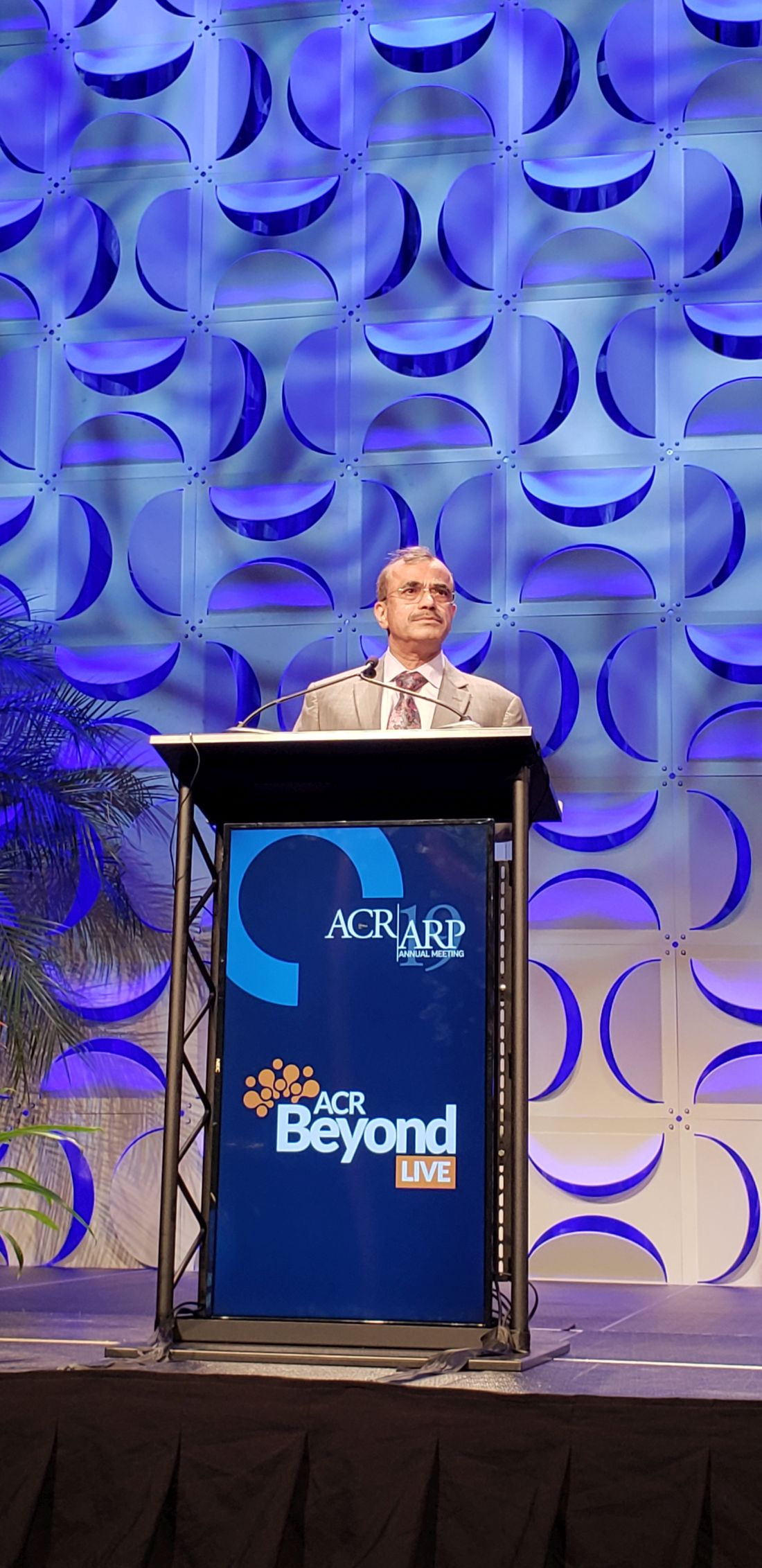
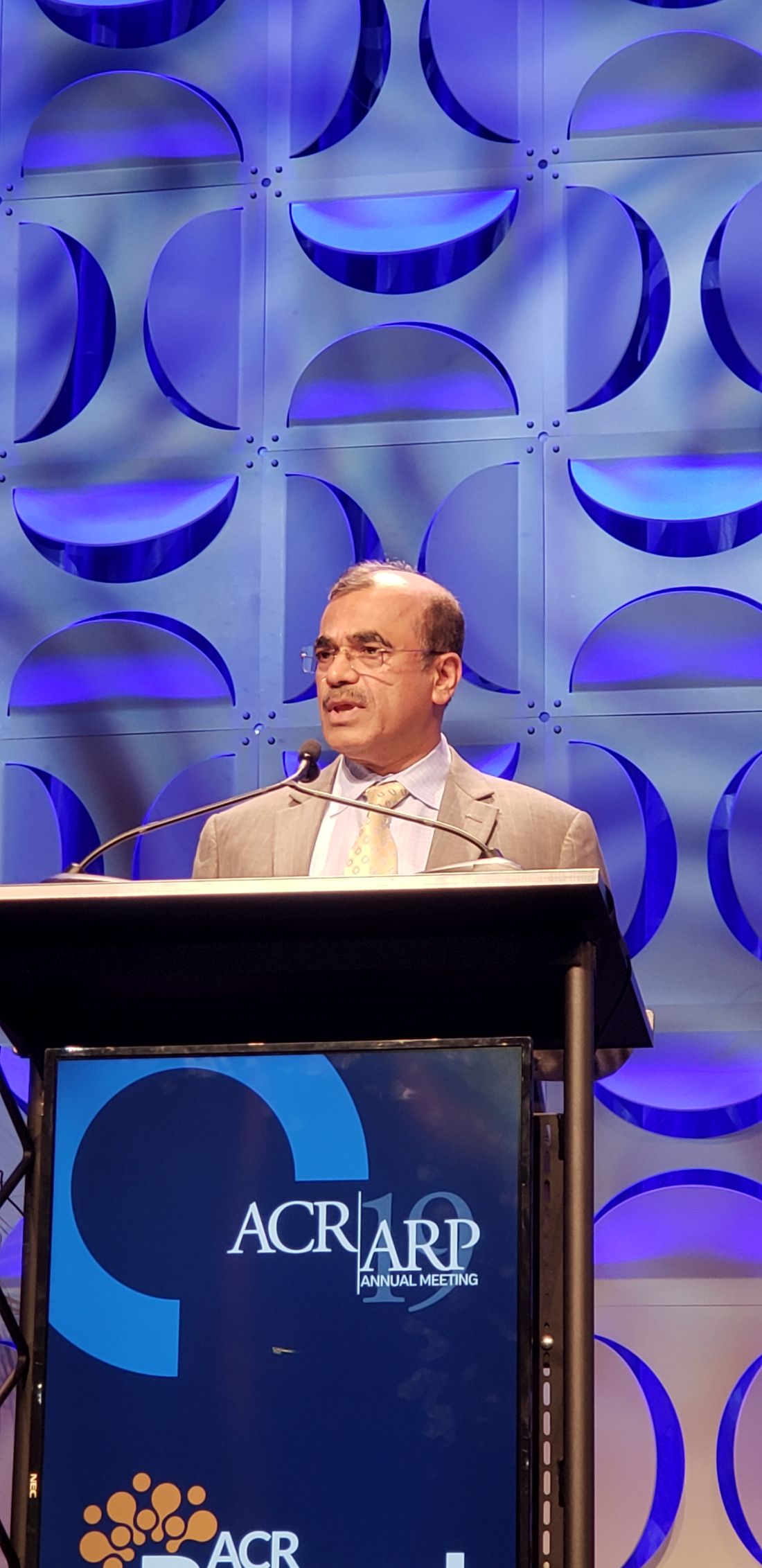
Chimeric antigen receptor (CAR) T-cell therapies have garnered a great deal of attention given their “incredible efficacy” in treating B-cell malignancies, and new findings are taking aim at the drawbacks of therapy, such as the time, expense, and toxicity involved, according to Robert A. Brodsky, MD.
One example, from a study slated for presentation during a plenary session at the upcoming annual meeting of the American Society of Hematology involves the investigational T-cell bispecific antibody mosunetuzumab, which targets both CD20 on the surface of malignant B cells, and CD3 on cytotoxic T cells, engaging the T cells and directing their cytotoxicity against B cells.
In a study (Abstract 6) of 218 non-Hodgkin lymphoma patients, including 23 who had already received CAR T-cell therapy and had relapsed or were refractory to the treatment, 64% responded, 42% had a complete response, and the median duration of response is now out to 9 months, Dr. Brodsky, ASH secretary and director of the division of hematology at Johns Hopkins University, Baltimore, said during a premeeting press conference.
“It’s basically an antibody using the patient’s own T cell to do what a CAR-T cell would do – [a] very exciting study and large study,” he said. “It is an off-the-shelf product, it completely gets around the problem of the time to generate the CAR T-cell product, and because it’s going to be much simpler and faster to produce, it’s likely going to be much cheaper than CAR T cells.”
The preliminary results also suggest it is less toxic than CAR T-cell therapy, he added.
Two other CAR T-cell therapy–related studies highlighted during the press conference address its use for multiple myeloma. One, the phase 1b/2 CARTITUDE study (Abstract 577) uses CAR T cells against the B-cell maturation antigen (BCMA) in the relapsed/refractory setting.
Of 25 patients treated with chemotherapy followed by CAR T-cell infusion and followed for a median of 3 months, 91% responded, two achieved a complete remission, and “many other responses were very deep responses,” Dr. Brodsky said, noting that the second featured multiple myeloma trial (Abstract 930) looked at bispecific CAR T-cell therapy targeting BCMA and CD38 in an effort to reduce resistance to the therapy.
“Again, very interesting preliminary results,” he said, noting that of 16 patients followed for a median of 36 weeks, 87.5% responded, the treatment was well tolerated, and progression-free survival at 9 months was 75%.
In addition to the “key theme” of overcoming CAR T-cell therapy obstacles, three other themes have emerged from among the thousands of abstracts submitted for presentation at ASH. These, as presented during the press conference, include new venous thromboembolism (VTE) therapies and approaches to research; inclusive medicine, with abstracts focused on age- and race-related issues in clinical trials; and new advances in the treatment of sickle cell disease. All of these have potentially practice-changing implications, as do the six late-breaking abstracts selected from 93 abstracts submitted for consideration for oral presentation at ASH, Dr. Brodsky said.
One of the “truly practice-changing” late-breakers is a randomized phase 3 trial (Abstract LBA-1) comparing the bispecific antibody blinatumomab to chemotherapy for post-re-induction therapy in high- and intermediate-risk acute lymphoblastic leukemia (ALL) at first relapse in children, adolescents and young adults.
The study demonstrated the superiority of blinatumomab for efficacy and tolerability, which is particularly encouraging given the challenges in getting relapsed ALL patients back into remission so they can undergo bone marrow transplant, Dr. Brodsky said.
Of 208 patients randomized, 73% vs. 45% in the blinatumomab vs. chemotherapy arms were able to get to transplant – and therefore to potential cure, he said.
“Of note, the blinatumomab arm was less toxic and there was marked improvement in disease-free and overall survival, so this is clearly going to become a new standard of care for relapsed and refractory ALL,” he added.
Large population-based study underscores link between gout, CVD event risk
ATLANTA – Gout is associated with an increased risk of both fatal and nonfatal cardiovascular disease events, according to a large population-based health data linkage study in New Zealand.
“Overall, the survival was quite good within both cohorts, but ... there is a clear and statistically significant difference in the survival between the people with gout and those without gout,” Ken Cai, MBBS, reported at the annual meeting of the American College of Rheumatology, noting that a similarly “significant and clear” difference was seen in nonfatal CVD events between the groups.
Of 968,387 individuals included in the analysis, 34,056 had gout, said Dr. Cai, a rheumatology clinical fellow at the University of Auckland (New Zealand). After adjusting for population-level estimated 5-year CVD risk for cardiovascular death, nonfatal myocardial infarction, stroke, or other vascular event, the adjusted hazard ratios were 1.20 for fatal and 1.32 for nonfatal first CVD events in patients with gout. The CVD risk score used in the analysis accounted for age, gender, ethnicity, level of social deprivation, diabetes status, previous hospitalization for atrial fibrillation, and baseline dispensing of blood pressure–lowering, lipid-lowering, and antiplatelet/anticoagulant medications.
“To allow for any other differences between the gout and nongout cohorts with respect to gender, age, ethnicity, and social deprivation, we further adjusted for these factors again, even though they had been accounted for within our CVD risk score,” he said, noting that “gout continued to demonstrate an increased adjusted hazard ratio” for fatal and nonfatal events after that adjustment (HRs, 1.40 and 1.35, respectively)
Additional analysis in the gout patients showed that CVD risk was similarly increased both in those who had been dispensed allopurinol at least once in the prior 5 years and those who had not (adjusted HRs for fatal events, 1.41 and 1.33; and for nonfatal, first CVD events, 1.34 and 1.38, respectively), and “there was no significant difference between these two groups, compared to people without gout,” he said.
Adjustment for serum urate levels in gout patients also showed similarly increased risk for fatal and nonfatal events for those with levels less than 6 mg/dL and those with levels of 6 mg/dL or greater (adjusted HRs of 1.32 and 1.42 for fatal events, and 1.27 and 1.43 for nonfatal first CVD events, respectively).
Again, no significant difference was seen in the risk of events between these two groups and those without gout, Dr. Cai said, noting that patients with no serum urate monitoring also had an increased risk of events (adjusted HR of 1.41 for fatal events and 1.29 for nonfatal, first CVD events).
Gout and hyperuricemia have previously been reported to be independent risk factors for CVD and CVD events, and urate-lowering therapy such as allopurinol have been thought to potentially be associated with reduced risk of CVD, he said, noting that the relationships are of particular concern in New Zealand, where gout affects more than 4% of the adult population.
“Maori, who are the indigenous people of New Zealand, and Pasifika people are disproportionately affected by gout; 8.5% of Maori, and 13.9% of Pasifika adults have gout,” he said, adding that an estimated one-third of Maori and Pasifika adults over age 65 years have gout.
To further assess the relationships between gout and CVD risk, he and his colleagues used validated population-level risk-prediction equations and linked National Health Identifier (NHI) data, he said.
National registries of medicines dispensing data, hospitalization, and death were linked to the Auckland/Northland regional repository of laboratory results from Jan. 1, 2012 to Dec. 31, 2016.
“We included all New Zealand residents aged 20 years or older who were in contact with publicly funded services in 2011 and were alive at the end of December, 2011,” he said, adding that those with a previous hospitalization for CVD or heart failure prior to the end of December 2011 were excluded, as were those with primary residence outside of the region for the prior 3 years and those missing predictor variable data.
Although the findings are limited by an inability to adjust for smoking status, body mass index, and blood pressure – as such data are not collected at the national level, and by the population-based nature of the study, which does not allow determination about causation, they nevertheless reinforce the association between gout and an increased estimated risk of CVD events, Dr. Cai said.
“Even after adjustment for estimated 5-year CVD risk and the additional weighting of risk factors within it, gout independently increased the hazard ratio for fatal and nonfatal events,” he said. “In our study, this effect was not ameliorated by allopurinol use or serum urate lowering to treatment target.”
Similar studies are needed in other populations, he said.
Dr. Cai reported grant support from Arthritis Australia.
SOURCE: Cai K et al. Arthritis Rheumatol. 2019;71(suppl 10), Abstract 2732.
REPORTING FROM ACR 2019
SPIRIT-H2H results confirm superiority of ixekizumab over adalimumab for PsA
ATLANTA – Ixekizumab (Taltz) provided significantly greater improvement in joint and skin symptoms, compared with adalimumab (Humira), in biologic-naive patients with active psoriatic arthritis (PsA), according to final 52-week safety and efficacy results from the randomized SPIRIT-H2H study.
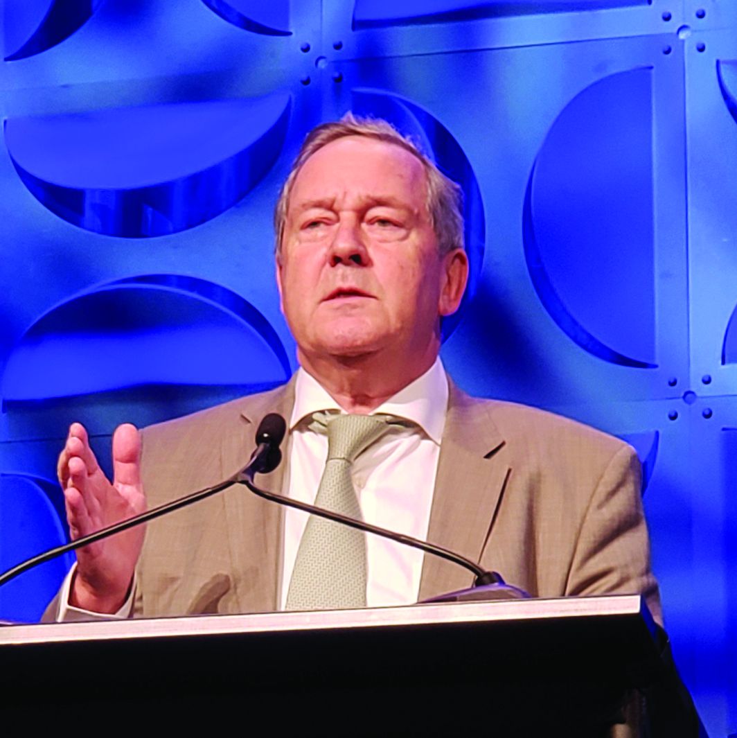
The high-affinity monoclonal antibody against interleukin-17A also performed at least as well as the tumor necrosis factor (TNF)–inhibitor adalimumab across multiple PsA domains and regardless of methotrexate use, Josef Smolen, MD, reported during a late-breaking abstract session at the annual meeting of the American College of Rheumatology.
Multiple biologic disease-modifying antirheumatic drugs (bDMARDs) are available for the treatment of PsA, but few studies have directly compared their efficacy and safety, said Dr. Smolen of the Medical University of Vienna. He noted that the SPIRIT-H2H study aimed to compare ixekizumab and adalimumab and also to address “one of the most clinically relevant questions for clinicians,” which relates to the efficacy of bDMARDs with and without concomitant methotrexate.
Ixekizumab is approved for adults with active PsA and moderate to severe plaque psoriasis, but TNF inhibitors like adalimumab have long been considered the gold standard for PsA treatment, he explained.
Of 283 patients with PsA randomized to receive ixekizumab and 283 randomized to receive adalimumab, 87% and 84%, respectively, completed week 52 of the head-to-head, open-label study comparing the bDMARDs. Treatment with ixekizumab achieved the primary endpoint of simultaneous improvement of 50% on ACR response criteria (ACR50) and 100% on the Psoriasis Area and Severity Index (PASI100) in 39% of patients, which was significantly higher than the rate of 26% with adalimumab, Dr. Smolen said.
Ixekizumab also performed at least as well as adalimumab for the secondary outcome measures of ACR50 response (50% in both groups) and PASI100 response (64% vs. 41%), as well as for all other outcomes measures, including multiple musculoskeletal PsA domains, he said.
“Remarkably ... at 1 year, more than one-third of the patients achieved an ACR70 in both groups, and half of the patients achieved an ACR50,” he added, noting that the ACR100 responses were in line with previous investigations.
Stratification by methotrexate use showed that the simultaneous ACR50 and PASI100 response rates were improved with ixekizumab versus adalimumab both in users and nonusers of methotrexate (39% vs. 30% and 40% vs. 20%, respectively). This finding highlights the ongoing debate about whether TNF inhibitors should or should not be used with methotrexate for PsA.
“This study was not adequately powered to say that, but there is some indication, and I think that this is food for thought for future further analysis because the data in the literature are discrepant in this respect,” Dr. Smolen said.
In non-methotrexate users in SPIRIT-H2H, the ACR20 responses were 53% with ixekinumab vs. 40% with adalimumab, ACR50 responses were 72% vs. 60%, and ACR70 responses were 41% vs. 27%, respectively, he said noting that the difference for ACR70 was statistically significant, and that the ACR70 response with ixekinumab was about the same as the ACR50 for adalimumab.
As for ACR20, ACR50, and ACR70 responses in methotrexate users, “the lines criss-crossed” early on, he said, but all were “slightly superior” with adalimumab than with ixekizumab at 52 weeks (75% vs. 68%, 56% vs. 48%, and 39% vs. 32%, respectively).
Study participants had a mean age of 48 years and had active PsA with at least 3/66 tender joints, at least 3/68 swollen joints, at least 3% psoriasis body surface area involvement, no prior treatment with bDMARDs, and prior inadequate response to one or more conventional synthetic DMARDs. Treatment was dosed according to drug labeling through 52 weeks.
The safety profiles of both agents were consistent with previous reports; treatment-emergent adverse events occurred in 73.9% of ixekizumab and 68.6% of adalimumab patients, and serious adverse events occurred in 4.2% and 12.4%, respectively.
“On the other hand, ixekizumab had more injection site reactions: 11% vs. close to 4%,” he said, noting that 4.2% of the ixekizumab patients and 7.4% of the adalimumab patients discontinued treatment because of adverse events. No deaths occurred in either group.
As reported previously in Annals of the Rheumatic Diseases, ixekizumab was superior to adalimumab for simultaneous achievement of ACR50 and PASI100 at 24 weeks, and these final 52-week results confirm those results, he said.
The study was funded by Eli Lilly, which markets ixekizumab. Dr. Smolen reported research grants and/or honoraria from Eli Lilly and AbbVie, which markets adalimumab, as well as many other pharmaceutical companies.
SOURCE: Smolen J et al. Arthritis Rheumatol. 2019;71(suppl 10), Abstract L20.
REPORTING FROM ACR 2019
COAST-X top-line results: Ixekizumab improves nonradiographic axSpA vs. placebo
ATLANTA – Adding ixekizumab (Taltz) to conventional background medications significantly improved the signs and symptoms of nonradiographic axial spondyloarthritis (axSpA) in the randomized, double-blind, placebo-controlled phase 3 COAST-X trial.
The high-affinity interleukin-17A monoclonal antibody ixekizumab “has shown efficacy in ankylosing spondylitis – also called radiographic axial spondyloarthritis – [and] it recently was approved by the [Food and Drug Administration] for the treatment of active ankylosing spondylitis,” said Atul Deodhar, MD, explaining that COAST-X sought to assess its efficacy in patients with active nonradiographic axSpA and objective evidence of inflammation. He presented the results of the trial at the annual meeting of the American College of Rheumatology.
Of 303 adults with an established diagnosis of axSpA who met Assessment of Spondyloarthritis International Society (ASAS) classification criteria and who were enrolled in the 52-week trial, 105 were randomized to receive background medications plus placebo, and 102 and 96 received background medications plus ixekizumab every 2 or 4 weeks, respectively. The primary endpoint of a 40% improvement in ASAS response criteria (ASAS 40) was reached at week 16 by 19% of the placebo-group patients and by 35% and 40% of the 2- and 4-week ixekizumab-group patients, and at week 52 by 13%, 30%, and 31% of the patients in the groups, respectively, Dr. Deodhar reported.
Additionally, “all major secondary endpoints were met for each ixekizumab regimen, both at week 16 and week 52,” said Dr. Deodhar, professor of medicine in the division of arthritis and rheumatic diseases at Oregon Health & Science University, Portland.
For example, Ankylosing Spondylitis Disease Activity Score (ASDAS) at week 16 declined by 0.6 with placebo, 1.3 with 2-week ixekizumab dosing, and 1.1 points with 4-week ixekizumab dosing; Bath Ankylosing Spondylitis Disease Activity Index (BASDAI) and Functional Index changes were –1.5, –2.5, and –2.2, and –1.3, –2.3 and –2.0; Short Form–36 physical component score changes were 5.3, 8.0, and 8.1 points; and MRI sacroiliac joint Spondyloarthritis Research Consortium of Canada score changes were –0.3, –4.5 and –3.4, in the groups, respectively.
“ASDAS less than 2.1 – low disease activity – was achieved by 32% and 27% [in the 2- and 4-week ixekizumab groups] versus 12% in the placebo [group],” he said, noting that similarly significant results were seen at week 52.
Notably, the differences in ASAS 40 response rates between the treatment and placebo groups were observed beginning at week 1, and “a notable proportion” of patients who escaped to the open-label 2-week ixekizumab group, as allowed per study protocol starting at week 16, had an ASAS 40 response at the time of escape; the ASAS 40 response rates at that time were 6.5%, 16.7%, 25% in the groups, respectively, and the rates increased further on open-label ixekizumab, he said.
Study participants were adults diagnosed with axSpA by a physician and treated for at least 3 months. Inclusion criteria also included BASDAI score of at least 4, back pain score of at least 4, inflammation as evidenced by sacroiliitis on MRI or elevated C-reactive protein levels of greater than 5 mg/L, and inadequate response or intolerance to at least two NSAIDs.
Ixekizumab in both treatment groups was given at a dose of 80 mg, and changes to conventional background medications, including NSAIDs, conventional synthetic disease-modifying antirheumatic drugs, analgesics, and low-dose corticosteroids, were allowed, as was escape to open-label ixekizumab given every 2 weeks at investigators’ discretion after week 16.
Ixekizumab treatment was well tolerated; the frequency of serious adverse events and AEs leading to treatment discontinuation was low and similar across all arms, Dr. Deodhar said.
For example, treatment-emergent AEs occurred in 55.7%, 77.5%, and 65.6% of patients, serious AEs occurred in 1.0%, 1.0%, and 2.1%, and AE-related discontinuations occurred in 1.9%, 1.0%, and 1.0% or patients in the groups, respectively.
No deaths occurred and no new safety signals were identified.
“The results demonstrate, for the first time, that blocking IL-17A is a potential treatment option for patients with nonradiographic axSpA,” he concluded.
COAST-X was sponsored by Eli Lilly. Dr. Deodhar and most coauthors reported receiving research grants and/or honoraria for consulting or speaking from Eli Lilly and other pharmaceutical companies. Four authors are current employees and shareholders of Eli Lilly.
SOURCE: Deodhar A et al. Arthritis Rheumatol. 2019;71(suppl 10), Abstract 2729.
REPORTING FROM ACR 2019
Biologic DMARDs appear as effective in elderly-onset RA as in young-onset RA
ATLANTA – Elderly-onset and young-onset rheumatoid arthritis patients initiating treatment with biologic disease-modifying antirheumatic drugs (bDMARDs) respond similarly with respect to clinical improvement at 48 weeks and adverse events, data from a large registry in Japan suggest.
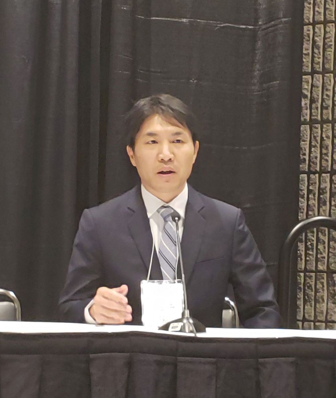
The findings have important implications – particularly for elderly-onset rheumatoid arthritis (RA) patients, who tend to present with higher disease activity levels and increased disability, but who nevertheless receive biologics less frequently, compared with young-onset RA patients, according to Sadao Jinno, MD, of the department of rheumatology and clinical immunology, Kobe University Graduate School of Medicine, Osaka, Japan, and colleagues.
The findings were presented in a poster at the annual meeting of the American College of Rheumatology.
Of 7,183 participants in the multicenter observational registry, 989 who initiated bDMARDs and who had a DAS28-erythrocyte sedimentation rate score of at least 3.2 at the time of initiation were included in the current analysis. The proportion of elderly-onset RA patients in the registry was 36.8%, and the proportion of elderly-onset RA patients using bDMARDs was significantly lower than that among young-onset RA patients (18.3% vs. 28.0%; P less than .001), Dr. Jinno and colleagues reported.
However, after adjustment for differences in baseline characteristics between the two age groups, no significant difference was seen in Clinical Disease Activity Index (CDAI) score at 48 weeks (odds ratio, 1.01), Dr. Jinno said during a press conference highlighting the findings.
A trend toward lower remission rates was observed in the early-onset patients (OR, 0.52; P = 1.10). The low-disease activity/remission rate was similar in the groups after adjustment for multiple confounders (OR, 0.86; P = 0.77), he said, adding that drug maintenance rates and adverse event–related discontinuation rates also were similar in the groups (hazard ratio, 0.95; P = 0.78 for drug maintenance; HR, 0.78; P = 0.22 for discontinuation).
Patients were enrolled in the multicenter observational registry between September 2009 and December 2017, and those with onset at age 60 years or older were considered to have elderly-onset RA.
“In my daily practice, I see a lot of patients with elderly-onset RA who are treated with biologics very effectively and safely,” he said. “So we wanted to see if there really is any difference [in outcomes] between the elderly-onset patients and the young-onset patients.”
The findings suggest they can be treated as effectively and safely as younger patients, he said.
In a press release, he further stated that
Conversely, it is important to keep in mind that dysfunction in elderly-onset RA patients may worsen without timely biologic treatment, he noted.
Future investigation will focus on whether elderly-onset RA patients respond differently to “various modes of biologics,” he said.
Dr. Jinno and colleagues reported having no relevant disclosures.
SOURCE: Jinno S et al. Arthritis Rheumatol. 2019;71(suppl 10), Abstract 1345.
REPORTING FROM ACR 2019
Weekly tocilizumab provides durable benefits for giant cell arteritis patients
ATLANTA –
Of 250 patients treated in the double-blind portion of the trial, 215 entered the extension period (part 2 of the trial), and 197 (92%) completed all 3 years of the trial. A total of 38 of 81 (47%) extension participants in clinical remission (CR) at week 52 after receiving tocilizumab (Actemra) weekly maintained it throughout the extension, and 13 of 36 (36%) treated every other week maintained it, John Stone, MD, reported at the annual meeting of the American College of Rheumatology.
Notably, more of those 51 patients treated with tocilizumab who maintained CR throughout the extension were treatment free at the end of the extension, compared with those originally assigned to the placebo groups (65% vs. 45%), and their median time to first flare after stopping treatment was longer, said Dr. Stone, director of clinical rheumatology at Massachusetts General Hospital and associate professor of medicine in the division of rheumatology at Harvard Medical School, both in Boston.
For example, times to first flare were 575 and 428 days for the weekly and biweekly tocilizumab dosing groups – which each had 26-week prednisone tapering – but they were only 162 days in a placebo group with 26-week prednisone tapering and 295 days for patients in another placebo group with 52-week prednisone tapering, Dr. Stone explained. Retreatment with tocilizumab restored CR in patients who experienced flares after discontinuing treatment, he added.
Importantly, cumulative glucocorticoid doses over the 3 years were also lower in the tocilizumab groups (2,647 and 3,782 mg/day with weekly and biweekly tocilizumab and 5,248, and 5,323 mg/day in the placebo groups, respectively), Dr. Stone said.
No new safety signals were observed during the extension. Rates of serious adverse events (SAEs) and serious infections per 100 patient-years during the entire 3 years of the study were comparable among those who never received tocilizumab and those who received at least 1 dose (23.2 and 25.4 for SAEs and 4.6 and 3.5 for serious infections).
Detailed results of the extension period
GiACTA enrolled patients with GCA and randomized them to receive weekly or biweekly treatment with the interleukin-6 receptor–alpha inhibitor tocilizumab at doses of 162 mg delivered subcutaneously with a 26-week prednisone taper, or to the placebo groups with a 26- or 52-week prednisone taper. The results, reported in 2017, demonstrated the superiority of tocilizumab over placebo for providing sustained glucocorticoid-free remission and ultimately led to approval from Food and Drug Administration of the biologic agent for the treatment of GCA.
The current analysis was carried out to determine long-term safety, to explore maintenance of efficacy after discontinuation, and to “gain insight into the long-term glucocorticoid-sparing effects of tocilizumab,” Dr. Stone said.
He noted that the extension period was not randomized because of the way that patients in the “original four randomized groups had sorted themselves out into very different groups by virtue of their response in part 1 [of the trial],” which made randomization impossible because of the varying categories of patients.
Patients in the extension were also treated at investigators’ discretion to prevent “catastrophic GCA flares, vision loss, and other problems” that could potentially occur with discontinuation of the blinded injections in part 1, he said.
Nevertheless, the analysis “provides some incredibly important information about the management of GCA now,” he said, adding that the dramatic effect of the original randomization was still profound at 3 years “despite the fact that investigators could treat the patients as they wished.”
“Even at 3 years there was still a profound difference in the cumulative glucocorticoid [use], compared to those patients who were randomized to one of the placebo versus prednisone groups,” he explained.
However, the most important question from this study relates to what happened when weekly tocilizumab was stopped: Of the 81 patients in part 2 who were in CR on tocilizumab after part 1 of the study, 59 (73%) were not started on any treatment by their investigator, and of those, 25 (42%) maintained treatment-free remission for 2 full years following discontinuation of tocilizumab, he said.
What happened when prednisone was stopped?
In the absence of a direct comparison group (since the extension wasn’t a randomized, controlled part of the trial), the best comparison to that is what happens when prednisone is stopped, he noted.
“And we did that experiment in part 1” of the trial, he said. Overall, 68% of patients in the prednisone/placebo groups flared in 1-year of follow-up, and most of those did so even before they stopped prednisone. In the group that received placebo plus a 52-week prednisone taper, 49% of patients flared and “every single one of those patients flared before they got off prednisone,” he said.
“So in that context, 2 years of treatment-free remission following weekly tocilizumab discontinuation seems very good,” he added.
Of the patients who flared in part 2 of the trial, 11 whose investigator elected to treat them with only tocilizumab reentered remission in a median of 15.8 days, 13 treated with tocilizumab and glucocorticoids reentered remission in a median of 8.5 days (1 didn’t reenter remission at all), and 15 treated with only glucocorticoids reentered remission in a median of 38 days.
“There are a number of biases in these data, but they do point out the fact that some patients can be treated with tocilizumab alone,” Dr. Stone said. “I would be very careful doing that.”
The trial was not powered for the comparison of weekly versus biweekly dosing, he said. However, “multiple lines of investigation and analysis support the idea that weekly tocilizumab is more effective at controlling disease,” he added. But “if you want to be assured that you are doing everything you can to control the disease, weekly tocilizumab would be better than every-other-week tocilizumab,” he said.
GiACTA was funded by Roche. Dr. Stone reported research grants from Roche/Genentech and consulting fees from Chugal, Roche/Genentech, and Xencor.
SOURCE: Stone J et al. Arthritis Rheumatol. 2019;71(suppl 10), Abstract 808.
REPORTING FROM ACR 2019
B-cell-poor RA responds better to tocilizumab than to rituximab
ATLANTA – Tocilizumab proved more effective than rituximab in B-cell-poor but not in B-cell-rich patients with RA who have had an inadequate response to disease-modifying antirheumatic drugs (DMARDs) and tumor necrosis factor inhibition in the randomized, open-label, 48-week, phase 4 R4-RA trial.
If validated, the findings of the trial – the first randomized, controlled, biopsy-driven trial in RA – could have “massive implications” for treatment selection and improved outcomes, Constantino Pitzalis, MD, reported during a press conference at the annual meeting of the American College of Rheumatology.
Of 164 RA patients who were failing or intolerant to conventional synthetic (cs) DMARD therapy and at least one tumor necrosis factor inhibitor (TNFi), 83 were randomized to receive rituximab and 81 received tocilizumab. Of those patients, 49.1% were considered B-cell poor (BCP) based on synovial tissue biopsies obtained at trial entry.
The BCP patients treated with tocilizumab were numerically more likely than those treated with rituximab to achieve the coprimary endpoint of Clinical Disease Activity Index (CDAI) improvement of at least 50% from baseline at 16 weeks (56.1% vs. 44.7%), and they were significantly more likely – twice as likely, in fact – to achieve the coprimary endpoint of CDAI improvement of at least 50% from baseline at 16 weeks as well as CDAI score less than 10, indicating a major treatment response (46.3% vs. 23.7%), said Dr. Pitzalis, head of the Centre for Experimental Medicine & Rheumatology at Queen Mary University of London.
BCP patients receiving tocilizumab also were significantly more likely to achieve a number of secondary endpoints, he noted.
In the B-cell-rich (BCR) population, no significant differences were seen in the majority of endpoints with tocilizumab versus rituximab, he said.
Study participants had a mean age of 55-56 years and were intolerant of or refractory to csDMARDs and at least one TNFi. They were recruited from 19 centers in Europe, were randomized and treated with standard doses of rituximab or tocilizumab, and were stratified based on histologic classification (BCP vs. BCR). Baseline characteristics were comparable among the treatment groups, Dr. Pitzalis said.
Adverse events occurred in 62 and 68 patients in the rituximab and tocilizumab groups, respectively, and serious adverse events occurred in 8 and 12 patients, respectively, thus 40% of all serious adverse events occurred in the rituximab group and 60% in the tocilizumab group. Infections and serious infections each occurred in three patients in each group, and two patients in each group discontinued treatment because of adverse events.
B cells are pivotal to RA pathogenesis, as demonstrated by the efficacy of the B-cell-depleting agent, rituximab. However, rituximab, which is licensed for use following failure of csDMARDs and TNFi therapy, is only effective for achieving a 50% improvement in ACR response criteria at 6 months in about 30% of such patients, Dr. Pitzalis noted. In a recent early RA cohort, he and his colleagues found synovial heterogeneity, with more than half of patients showing low or no synovial B-cell infiltration.
“So why would you give them rituximab?” he asked, explaining the rationale for the R4-RA trial: The hypothesis was that alternative biologic agents targeting alternative pathways may be more effective in BCP patients.
“The results showed quite clearly that rituximab was inferior to tocilizumab in this patient group,” he said. “We demonstrated that tocilizumab is more effective than rituximab in achieving low disease activity in patients who had, on synovial biopsy, low levels of B-cell infiltration.
“The study really highlights the importance of integrating molecular pathology into the clinical algorithms, because making the diagnosis is not sufficient. We really need to know what the pathology of the patient is so we can give the right drug to the right patients.”
Donald Thomas, MD, a rheumatologist in private practice in Silver Spring, Md., called the study “fascinating,” and noted during a question-and-answer period during the press conference that, during his lifetime, he “has probably wasted tens of thousands to hundreds of thousands of dollars,” using the trial-and-error approach to treatment.
“We can only treat by trial and error ... so being able to pinpoint therapy is just phenomenal,” he added, further noting in an interview after the press conference that finding the right treatment can be “such a struggle.”
“I literally have patients who have gone through 10 medicines and wasted thousands of dollars,” he said.
In response to a question from Dr. Thomas about the potential for rheumatologists to do their own synovial biopsies to help guide treatment in the event the findings are validated, Dr. Pitzalis said that is both feasible and an important goal.
“All the biopsies [in the study] were carried out by rheumatologists,” he noted. “We have trained over 150 rheumatologists worldwide, including at 15 centers in the United States. ... We want to empower the rheumatologists to do it.”
He added that “this is early data ... and will require validation in larger trials,” but said the point is that “we can’t continue to just give these drugs and see if the patient responds.”
R4-RA is funded by the National Institute of Health Research Efficacy and Mechanism Evaluation program. Dr. Pitzalis and some of the other study authors reported financial relationships with Roche/Genentech, which markets tocilizumab and rituximab, and other pharmaceutical companies.
SOURCE: Pitzalis C et al. Arthritis Rheumatol. 2019;71(suppl 10), Abstract 2911.
REPORTING FROM ACR 2019
Guselkumab improves psoriatic arthritis regardless of prior TNFi use
ATLANTA – Guselkumab improved outcomes in psoriatic arthritis patients regardless of past treatment with tumor necrosis factor inhibitors in the phase 3 DISCOVER-1 trial.
The anti-interleukin-23p19 monoclonal antibody is approved in the United States for the treatment of moderate to severe plaque psoriasis (PsO).
Benefits in psoriatic arthritis (PsA) were seen in both biologic-naive and tumor necrosis factor inhibitor (TNFi)–treated patients and occurred with both 4- and 8-week dosing regimens, Atul Deodhar, MD, reported during a plenary session at the annual meeting of the American College of Rheumatology.
For example, the primary endpoint of ACR 20 response at 24 weeks was achieved in 58.6% and 52.8% of patients randomized to receive 100 mg of guselkumab delivered subcutaneously either at baseline and every 4 weeks or at baseline, week 4, and then every 8 weeks, respectively, compared with 22.2% of those randomized to receive placebo, said Dr. Deodhar, professor of medicine at Oregon Health & Science University, Portland.
Greater proportions of patients in the guselkumab groups achieved ACR 20 response at week 16; ACR 50 response at weeks 16 and 24; ACR 70 response at week 24; Psoriasis Area and Severity Index 75, 90, and 100 responses at week 24; and minimal disease activity response at week 24, he said, adding that improvements were also seen with guselkumab versus placebo for the controlled major secondary endpoints of change from baseline in Health Assessment Questionnaire–Disability Index score, Short Form 36 Health Survey score, and investigator global assessment (IGA) of PsO response.
The response rates with guselkumab versus placebo were seen regardless of prior TNFi use, he said.
The study included 381 patients with active PsA, defined as three or more swollen joints, three or more tender joints, and C-reactive protein of 0.3 mg/dL or greater despite standard therapies. About 30% were exposed to up to two TNFi therapies and 10% were nonresponders or inadequate responders to those therapies.
Concomitant use of select nonbiologic disease-modifying antirheumatic drugs, oral corticosteroids, and NSAIDs was allowed, and patients with less than 5% improvement in tender plus swollen joints at week 16 could initiate or increase the dose of the permitted medications while continuing study treatment, Dr. Deodhar said.
The mean body surface area with PsO involvement was 13.4%; 42.5% of patients had an IGA of 3-4 for skin involvement. Mean swollen and tender joint counts were 9.8 and 19.3, respectively, indicating a population with moderate to severe disease, he added.
Serious adverse events, serious infections, and death occurred in 2.4%, 0.5%, and 0.3% of patients, respectively.
“Both guselkumab regimens were safe and well tolerated through week 24,” Dr. Deodhar said, noting that the safety profile was consistent with that established in the treatment of PsO and described in the label.
DISCOVER-1 was funded by Janssen Research & Development. Dr. Deodhar reported relationships (advisory board activity, consulting, and/or research grant funding) with several pharmaceutical companies including Janssen. Several coauthors are employees of Janssen.
SOURCE: Deodhar A et al. Arthritis Rheumatol. 2019;71(suppl 10), Abstract 807.
A progressive exercise intervention improved AGFR in breast cancer survivors
BARCELONA – A progressive aerobic and resistance exercise intervention improved the android:gynoid fat ratio (AGFR) in breast cancer survivors, which could provide important health benefits.
AGFR is associated with increased risk for cardiovascular disease and type 2 diabetes in breast cancer survivors, therefore exercise-induced AGFR improvement may reduce the risk for such comorbid conditions, Christina Dieli-Conwright, PhD, of the University of Southern California Norris Comprehensive Cancer Center, Los Angeles, and colleagues reported in a poster at the European Society for Medical Oncology Congress.
A significant decrease in AGFR from baseline was noted in 50 survivors of stage I-III breast cancer who participated in the exercise intervention, compared with 50 such survivors randomized to a usual care group (P less than .001), and strong correlations were found between AGFR and homeostatic model assessment of insulin resistance (HOMA-IR; r = 0.95; P less than .01), the investigators found.
Study participants had a mean age of 53 years, 54% were overweight (body mass index greater than 25 kg/m2), 63% were Hispanic, 90% had undergone a mastectomy, and 76% received chemotherapy and radiation therapy. Adherence to the intervention, which involved three weekly sessions of supervised, progressive, moderate-to-vigorous aerobic and resistance exercise for 16 weeks, was 95%.
AGFR was calculated using whole-body dual-energy x-ray absorptiometry and HOMA-IR was calculated using fasting insulin and glucose levels.
“Exercise reduces fat mass in breast cancer survivors, however, few studies have focused on AGFR,” the investigators wrote.
The findings of the current study suggest that a progressive aerobic and resistance exercise intervention is an effective strategy for decreasing AGFR in breast cancer survivors, they concluded.
The National Cancer Institute funded the study. The authors reported having no disclosures.
SOURCE: Dieli-Conwright C et al. ESMO 2019, Abstract 228P.
REPORTING FROM ESMO 2019
Exercise improved QoL, functioning in breast cancer survivors
BARCELONA – A supervised and adapted exercise program improved quality of life, physical functioning, and strength in breast cancer survivors participating in the MAMA MOVE Gaia study.
Of 19 women who initiated participation in the program, which included a 16-week control phase followed by a 16-week exercise training intervention phase, 15 completed the program, and, after the training intervention, they experienced a significant increase in handgrip strength and sit-to-stand repetitions, Ana Joaquim, MD, of Centro Hospitalar de Vila Nova de Gaia/Espinho, Portugal, and colleagues reported in a poster at the European Society for Medical Oncology Congress.
During the control phase of the prospective nonrandomized study, participants experienced no significant changes over time in any domain of quality of life as measured by the EORTC QLQ-C30 questionnaire, although a trend toward improved physical functioning was noted at an evaluation performed 8 weeks after the control phase, compared with one performed just prior to the intervention phase (77.3 to 85.3 points, P = .051), the investigators said.
After the intervention phase, however, handgrip strength improved significantly at both the limb where surgery was performed and at the nonoperated limb (from 22.2 to 25.6 kg.f and from 22.6 to 26.9 kg.f). Similar results were observed for a sit-to-stand test (improvement from 12 to 17 repetitions).
Participants in the single-arm clinical trial were assessed after 8 weeks of the control phase, immediately prior to the intervention period, 8 weeks after the control phase, and 16 weeks into the invention phase.
The intervention phase consisted of 3 60-minute sessions per week of combined moderate to vigorous aerobic and strength exercise, defined as exercise at 65%-85% of maximum heart rate or at 6-8 points on the OMNI scale. Mean compliance among the participants was 63.6%.
The participants had a median age of 59 and 15 of the 19 were diagnosed with invasive carcinoma. Following surgery, 13 underwent radiotherapy, 15 received chemotherapy, and 18 received hormone therapy.
“Treatments for early breast cancer have side effects that affect quality of life and cause deconditioning,” the investigators wrote, adding that “physical exercise might have a supportive and coadjuvant role in the rehabilitation of breast cancer survivors.”
The MAMA MOVE trial aimed to assess the potential benefits of a community-based supervised exercise training program, and the findings suggest such programs could help improve quality of life, particularly with respect to physical functioning, they concluded.
The MAMA MOVE Gaia study was funded by Liga Portuguesa Contra o Cancro. The investigators reported having no disclosures.
SOURCE: Joaquim A et al. ESMO 2019, Abstract 234P.
REPORTING FROM ESMO 2019


