User login
Nodules on nose and tattoos
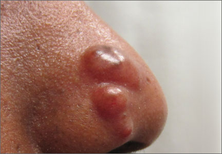
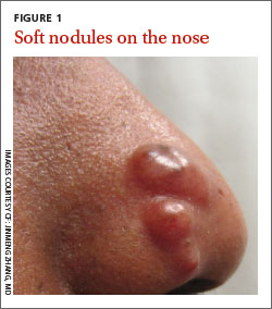
A 38-year-old African American man with no significant medical history presented to our dermatology clinic with a 5-month history of nodules on the right side of his nose (FIGURE 1). For several years, he’d also had nodules that gradually appeared on several red-inked tattoos shortly after he received each tattoo (FIGURE 2). He also had a 5-year history of nontender swelling of his fingers.
The patient denied any trauma to the areas with nodules or being in contact with anyone who was sick. He had no respiratory complaints, but chest x-rays from recent and past records showed stable bilateral intrathoracic lymphadenopathy without any lobar infiltration or pleural effusion.
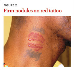
Physical examination revealed 3 reddish-brown soft nodules on the nasal ala and multiple, nontender, 3- to 5-mm firm nodules located in the red-inked areas of tattoos on his arms and neck. The tattoo nodules were asymptomatic and stable in size. He had clubbing of multiple digits and nail dystrophy. His distal fingers were edematous, but nontender. X-rays revealed lytic, lace-like lucencies of the middle and distal phalanges and erosions of the distal phalanges on both hands (FIGURE 3).
We biopsied the nodules on his nose and on one of his tattoos.
WHAT IS YOUR DIAGNOSIS?
HOW WOULD YOU TREAT THIS PATIENT?
Diagnosis: Sarcoidosis
Based on his clinical presentation and skin biopsy results, the patient was given a diagnosis of cutaneous sarcoidosis. A biopsy from the right side of his nose demonstrated sarcoidal granulomas. Acid-fast bacilli and periodic acid-Schiff stains were negative. A biopsy of one of the tattoo nodules showed sarcoidal granulomas, and close inspection revealed red tattoo pigment within the granulomatous inflammation.
X-rays showed bilateral hilar lymphadenopathy, which was consistent with pulmonary sarcoidosis, and the lace-like appearance of the middle and distal phalanges was consistent with skeletal sarcoidosis.
Systemic sarcoidosis is an idiopathic, granulomatous disease that affects multiple organ systems but primarily the lungs and lymphatic system.1 The estimated prevalence of systemic sarcoidosis ranges from less than 1 to 40 cases per 100,000 people, and the condition is more common among African Americans.1
Cutaneous sarcoidosis can occur as a manifestation of systemic sarcoidosis. It occurs in 20% to 35% of patients with systemic sarcoidosis2 and may present as asymptomatic red or skin-colored papules and firm nodules within tattoos, old scars, or permanent makeup. Cutaneous sarcoidosis in tattoos may be the first manifestation of sarcoidosis, and the time between acquiring the tattoo and developing sarcoidal nodules varies widely.
It is not clear why sarcoidal granulomas occur in tattoos. One possibility is that chronic low-grade exposure of the immune system to foreign materials such as tattoo ink leads to granulomatous hypersensitivity.2,3 Sarcoidosis usually occurs in red (cinnabar), black (ferric oxide), or blue-black areas of tattoos,4 in which the pigment acts as a nidus for granuloma formation.
A skin biopsy is helpful in making a diagnosis of cutaneous sarcoidosis. The histopathology shows noncaseating epithelioid granulomas.
Because skin manifestations may be the first and only sign of systemic sarcoidosis, patients with cutaneous sarcoidosis should be evaluated for systemic disease. Cutaneous sarcoidosis has been associated with bilateral hilar lymphadenopathy, pulmonary sarcoidosis, uveitis, arthritis, and dactylitis.3
Foreign-body reactions, syphilis are part of the differential Dx
Nonsarcoidal tattoo granulomas are a foreignbody reaction to the pigment used in tattooing and are characterized by lesions occurring only at the site of tattoos. This type of granulomatous reaction is most commonly seen in redpigmented tattoos, but can be seen in other tattoo colors as well. Macrophages containing pigment and “naked” granulomas are seen on histology.
Atypical mycobacterial skin infection can occur in tattoos that were created with contaminated ink or ink diluted with nonsterile water.5 Mycobacterial species such as Mycobacterium chelonae have been isolated from skin biopsies taken from the margins of new tattoos that developed a persistent erythematous eruption.5
Granuloma annulare is characterized by red or skin-colored plaques in annular and rope-like patterns with central clearing and nonscaly borders. A localized variant is frequently found on the extremities.6 There are associations between granuloma annulare, diabetes mellitus, and internal malignancy.7
Secondary syphilis classically presents with symmetric macules or papules distributed on the trunk and extremities. However, cutaneous manifestations vary widely. Lesions involving the palms and soles are important clues to a syphilis diagnosis, and patients often have malaise and fever.
Treatment includes topical, intralesional corticosteroids
The evidence for the treatment of cutaneous sarcoidosis is largely drawn from uncontrolled case series; there have been few double-blind, placebo-controlled studies.8 The first-line treatment for limited papules is a high-potency topical corticosteroid (eg, clobetasol 0.05% ointment applied twice weekly) and an intralesional corticosteroid (eg, triamcinolone, one 5-10 mg/mL injection every 4 weeks).8
Antimalarials such as hydroxychloroquine (200 mg twice a day for at least 6 months) or methotrexate (10-15 mg/week taken at once orally or as a subcutaneous or intramuscular injection) can also be helpful. Treatment with a midpotency topical corticosteroid such as triamcinolone 0.1% cream twice a day and doxycycline hyclate (100 mg twice a day for 4 months) has been reported to clear cutaneous lesions in tattoos.3
Oral corticosteroids are the gold standard for severe cutaneous sarcoidosis, but their multiple adverse effects, such as diabetes and adrenal suppression, may prevent prolonged use.8 For most cutaneous lesions, intralesional corticosteroids and/or hydroxychloroquine followed by methotrexate can be effective.8
The nodules on our patient’s nose were successfully treated with intralesional triamcinolone 5 mg/mL. No treatment was initiated for the tattoo nodules because they were asymptomatic and the patient was not concerned about their appearance. He continues to get new tattoos, but is minimizing the use of red ink.
The patient was also started on prednisone 10 mg/d, which improved his hand swelling. Rheumatologists were considering a steroidsparing immunosuppressive agent such as methotrexate; however, the patient was lost to follow-up.
CORRESPONDENCE
Jinmeng Zhang, MD, Division of Dermatology, Washington University School of Medicine, St. Louis, MO 63110; [email protected]
1. American Thoracic Society. Statement on sarcoidosis. Joint Statement of the American Thoracic Society, the European Respiratory Society (ERS) and the World Association of Sarcoidosis and Other Granulomatous Disorders (WASOG) adopted by the ATS Board of Directors and by the ERS Executive Committee, February 1999. Am J Respir Crit Care Med. 1999;160:736-755.
2. Guerra JR, Alderuccio JP, Sandhu J, et al. Granulomatous tattoo reaction in a young man. Lancet. 2013;382:284.
3. Antonovich DD, Callen JP. Development of sarcoidosis in cosmetic tattoos. Arch Dermatol. 2005;141:869-872.
4. Baumgartner M, Feldmann R, Breier F, et al. Sarcoidal granulomas in a cosmetic tattoo in association with pulmonary sarcoidosis. J Dtsch Dermatol Ges. 2010;8:900-902.
5. Kennedy BS, Bedard B, Younge M, et al. Outbreak of Mycobacterium
chelonae infection associated with tattoo ink. N Engl J Med. 2012;367:1020-1024.
6. Hsu S, Lehner AC, Chang JR. Granuloma annulare localized to the palms. J Am Acad Dermatol. 1999;41:287-288.
7. Thornsberry LA, English JC 3rd. Etiology, diagnosis, and therapeutic management of granuloma annulare: an update. Am J Clin Dermatol. 2013;14:279-290.
8. Lodha S, Sanchez M, Prystowsky S. Sarcoidosis of the skin: a review for the pulmonologist. Chest. 2009;136:583-596.
A 38-year-old African American man with no significant medical history presented to our dermatology clinic with a 5-month history of nodules on the right side of his nose (FIGURE 1). For several years, he’d also had nodules that gradually appeared on several red-inked tattoos shortly after he received each tattoo (FIGURE 2). He also had a 5-year history of nontender swelling of his fingers.
The patient denied any trauma to the areas with nodules or being in contact with anyone who was sick. He had no respiratory complaints, but chest x-rays from recent and past records showed stable bilateral intrathoracic lymphadenopathy without any lobar infiltration or pleural effusion.
Physical examination revealed 3 reddish-brown soft nodules on the nasal ala and multiple, nontender, 3- to 5-mm firm nodules located in the red-inked areas of tattoos on his arms and neck. The tattoo nodules were asymptomatic and stable in size. He had clubbing of multiple digits and nail dystrophy. His distal fingers were edematous, but nontender. X-rays revealed lytic, lace-like lucencies of the middle and distal phalanges and erosions of the distal phalanges on both hands (FIGURE 3).
We biopsied the nodules on his nose and on one of his tattoos.
WHAT IS YOUR DIAGNOSIS?
HOW WOULD YOU TREAT THIS PATIENT?
Diagnosis: Sarcoidosis
Based on his clinical presentation and skin biopsy results, the patient was given a diagnosis of cutaneous sarcoidosis. A biopsy from the right side of his nose demonstrated sarcoidal granulomas. Acid-fast bacilli and periodic acid-Schiff stains were negative. A biopsy of one of the tattoo nodules showed sarcoidal granulomas, and close inspection revealed red tattoo pigment within the granulomatous inflammation.
X-rays showed bilateral hilar lymphadenopathy, which was consistent with pulmonary sarcoidosis, and the lace-like appearance of the middle and distal phalanges was consistent with skeletal sarcoidosis.
Systemic sarcoidosis is an idiopathic, granulomatous disease that affects multiple organ systems but primarily the lungs and lymphatic system.1 The estimated prevalence of systemic sarcoidosis ranges from less than 1 to 40 cases per 100,000 people, and the condition is more common among African Americans.1
Cutaneous sarcoidosis can occur as a manifestation of systemic sarcoidosis. It occurs in 20% to 35% of patients with systemic sarcoidosis2 and may present as asymptomatic red or skin-colored papules and firm nodules within tattoos, old scars, or permanent makeup. Cutaneous sarcoidosis in tattoos may be the first manifestation of sarcoidosis, and the time between acquiring the tattoo and developing sarcoidal nodules varies widely.
It is not clear why sarcoidal granulomas occur in tattoos. One possibility is that chronic low-grade exposure of the immune system to foreign materials such as tattoo ink leads to granulomatous hypersensitivity.2,3 Sarcoidosis usually occurs in red (cinnabar), black (ferric oxide), or blue-black areas of tattoos,4 in which the pigment acts as a nidus for granuloma formation.
A skin biopsy is helpful in making a diagnosis of cutaneous sarcoidosis. The histopathology shows noncaseating epithelioid granulomas.
Because skin manifestations may be the first and only sign of systemic sarcoidosis, patients with cutaneous sarcoidosis should be evaluated for systemic disease. Cutaneous sarcoidosis has been associated with bilateral hilar lymphadenopathy, pulmonary sarcoidosis, uveitis, arthritis, and dactylitis.3
Foreign-body reactions, syphilis are part of the differential Dx
Nonsarcoidal tattoo granulomas are a foreignbody reaction to the pigment used in tattooing and are characterized by lesions occurring only at the site of tattoos. This type of granulomatous reaction is most commonly seen in redpigmented tattoos, but can be seen in other tattoo colors as well. Macrophages containing pigment and “naked” granulomas are seen on histology.
Atypical mycobacterial skin infection can occur in tattoos that were created with contaminated ink or ink diluted with nonsterile water.5 Mycobacterial species such as Mycobacterium chelonae have been isolated from skin biopsies taken from the margins of new tattoos that developed a persistent erythematous eruption.5
Granuloma annulare is characterized by red or skin-colored plaques in annular and rope-like patterns with central clearing and nonscaly borders. A localized variant is frequently found on the extremities.6 There are associations between granuloma annulare, diabetes mellitus, and internal malignancy.7
Secondary syphilis classically presents with symmetric macules or papules distributed on the trunk and extremities. However, cutaneous manifestations vary widely. Lesions involving the palms and soles are important clues to a syphilis diagnosis, and patients often have malaise and fever.
Treatment includes topical, intralesional corticosteroids
The evidence for the treatment of cutaneous sarcoidosis is largely drawn from uncontrolled case series; there have been few double-blind, placebo-controlled studies.8 The first-line treatment for limited papules is a high-potency topical corticosteroid (eg, clobetasol 0.05% ointment applied twice weekly) and an intralesional corticosteroid (eg, triamcinolone, one 5-10 mg/mL injection every 4 weeks).8
Antimalarials such as hydroxychloroquine (200 mg twice a day for at least 6 months) or methotrexate (10-15 mg/week taken at once orally or as a subcutaneous or intramuscular injection) can also be helpful. Treatment with a midpotency topical corticosteroid such as triamcinolone 0.1% cream twice a day and doxycycline hyclate (100 mg twice a day for 4 months) has been reported to clear cutaneous lesions in tattoos.3
Oral corticosteroids are the gold standard for severe cutaneous sarcoidosis, but their multiple adverse effects, such as diabetes and adrenal suppression, may prevent prolonged use.8 For most cutaneous lesions, intralesional corticosteroids and/or hydroxychloroquine followed by methotrexate can be effective.8
The nodules on our patient’s nose were successfully treated with intralesional triamcinolone 5 mg/mL. No treatment was initiated for the tattoo nodules because they were asymptomatic and the patient was not concerned about their appearance. He continues to get new tattoos, but is minimizing the use of red ink.
The patient was also started on prednisone 10 mg/d, which improved his hand swelling. Rheumatologists were considering a steroidsparing immunosuppressive agent such as methotrexate; however, the patient was lost to follow-up.
CORRESPONDENCE
Jinmeng Zhang, MD, Division of Dermatology, Washington University School of Medicine, St. Louis, MO 63110; [email protected]
1. American Thoracic Society. Statement on sarcoidosis. Joint Statement of the American Thoracic Society, the European Respiratory Society (ERS) and the World Association of Sarcoidosis and Other Granulomatous Disorders (WASOG) adopted by the ATS Board of Directors and by the ERS Executive Committee, February 1999. Am J Respir Crit Care Med. 1999;160:736-755.
2. Guerra JR, Alderuccio JP, Sandhu J, et al. Granulomatous tattoo reaction in a young man. Lancet. 2013;382:284.
3. Antonovich DD, Callen JP. Development of sarcoidosis in cosmetic tattoos. Arch Dermatol. 2005;141:869-872.
4. Baumgartner M, Feldmann R, Breier F, et al. Sarcoidal granulomas in a cosmetic tattoo in association with pulmonary sarcoidosis. J Dtsch Dermatol Ges. 2010;8:900-902.
5. Kennedy BS, Bedard B, Younge M, et al. Outbreak of Mycobacterium
chelonae infection associated with tattoo ink. N Engl J Med. 2012;367:1020-1024.
6. Hsu S, Lehner AC, Chang JR. Granuloma annulare localized to the palms. J Am Acad Dermatol. 1999;41:287-288.
7. Thornsberry LA, English JC 3rd. Etiology, diagnosis, and therapeutic management of granuloma annulare: an update. Am J Clin Dermatol. 2013;14:279-290.
8. Lodha S, Sanchez M, Prystowsky S. Sarcoidosis of the skin: a review for the pulmonologist. Chest. 2009;136:583-596.