User login
Pruritic blisters on legs and feet
A 23-year-old active duty US Navy sailor came to our medical center for treatment of 1–2 cm tense pruritic blisters on his dorsal feet, calves, and anterior lower extremities (FIGURE 1). He told us that several days earlier, he had noticed ill-defined “itchy red bumps” on both legs. He denied fever, night sweats, malaise, trauma, insect bites, contact with animals or plants, or recent illnesses. He did say, however, that he’d recently done outdoor fieldwork with the Marine Corps in southern California.
His medical history was unremarkable, and he was not taking any prescribed or over-the-counter medications or supplements. He had no family history of blistering or other autoimmune disorders.
On examination, we noticed clear fluid-filled vesicles and bullae on non-erythematous, non-urticarial bases that were haphazardly distributed on both legs and dorsal feet. Agminated or herpetiform configurations were not present. Ruptured bullae left erythematous to beefy red eroded bases, and there were numerous smaller red papules with vesicular surface changes. All of the lesions were below the knees; there was complete sparing of the trunk, upper extremities, intertriginous skin, head, and neck.
The remainder of the physical examination was unremarkable, and there was no lymphadenopathy. Complete blood count and chemistries were within normal limits, and the patient’s HIV status was negative.
We performed lesional and perilesional punch biopsies. The lesional biopsy demonstrated subepidermal blistering with a predominantly eosinophilic infiltrate in all layers of the dermis and within the blister. Direct immunofluorescence (DIF) was performed on the perilesional biopsy and was negative for IgA, IgG, IgM, C3, and fibrinogen. Gram stain, potassium hydroxide (KOH) prep, and wound culture were also negative.
FIGURE 1
Tense pruritic blisters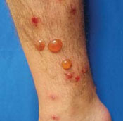
What is your diagnosis?
Diagnosis: Bullous arthropod bite reaction
Bullous arthropod bite reaction (BABR) occurs in sensitized individuals as a delayed hypersensitivity immune reaction to insect saliva.1 Patients typically present with grouped localized pruritic or asymptomatic blistering2 and otherwise appear well. Unless secondarily infected, the blisters are non-erythematous and non-purulent, and develop within hours to days of the bite.
Lesion location is key. The distribution and location of the lesions will tip you off to a BABR diagnosis. The lesions in BABR are usually grouped and localized to a specific area of the body, depending on the causative arthropod and the circumstances leading to the bites. For example:
- Lesions caused by Cheyletiella mites are typically found on the forearms, anterior thighs, and lower abdomen after an infested pet sits on an individual’s lap.2,3
- Blisters caused by flea bites are isolated to the lower extremities.4 (We suspect that flea bites were the culprit in our patient’s case.)
- Lesions caused by Cimex lectularius, more commonly known as bedbugs, may be found on the entire body and tend to occur in groups of 3.5
Insect bite? What insect bite?
Most patients will only complain of pruritus and will tell you that they don’t recall having had any insect bites.6 That said, the distribution of the lesions, lack of systemic illness, and otherwise unremarkable physical exam are sufficient for diagnosis.
Occasionally, a punch biopsy with DIF may be necessary to rule out more serious bullous disorders. In BABR, you may see both subepidermal and intraepidermal blistering, with perivascular and interstitial eosinophilic and lymphocytic infiltrates. Blisters separated by strands of keratinocytes create a characteristic multilocular appearance. Unlike autoimmune blistering disorders, DIF is negative in BABR.7 Gram stain, Tzanck smear, bacterial culture, and KOH prep may also provide additional information if infection is a concern.
For certain patients, the DX may be less clear-cut
Similar bullous lesions following insect bites have been reported in patients with HIV,8 chronic lymphocytic leukemia,9-12 EBV-associated Natural Killer leukemia/ lymphoma,13 and mantle cell lymphoma.14 There is ongoing debate as to whether the vesicular lesions in these patients truly represent an exaggerated response to an arthropod bite or mimic an insect-like bite reaction.10,12
Nevertheless, when you suspect a patient has BABR, be aware of its association with both hematologic malignancies and HIV. Appropriate evaluation, such as HIV screening and complete blood count, should be performed.
A condition that mimics contact dermatitis
Clinically and histologically, BABR can mimic the following:
- Contact dermatitis. With contact dermatitis, the blistering is more likely to appear in streaks or in a linear fashion.15 Lesions will be painful, as well as pruritic, and occur following direct contact with a plant or chemical allergen.
- Drug-induced pemphigoid. The patient’s history will increase your suspicion of drug-induced pemphigoid. Patients may be taking sulfur-containing drugs (furosemide), antibiotics (penicillins, fluoroquinolones), antihypertensives (ACE inhibitors, calcium channel blockers), neuroleptics, or sulfasalazine.7,16,17 The eruption is usually more generalized than BABR, and may involve the mucous membranes.
- Fungal infections. These infections will typically occur on the palms and soles. Infiltrate is typically neutrophilic, but can be eosinophilic.18
- Bullous scabies. Patients will have severe pruritus. Burrows and lesions can typically be found on moist areas (periumbilical, intertriginous skin).19,20
- Bullous pemphigoid. This is more commonly seen in elderly patients with comorbid conditions. Onset of blistering is gradual, and occurs predominantly on flexural skin.
Pemphigoid gestationis, erythema toxicum neonatorum, incontinentia pigmenti, and some pemphigus variants also have predominantly eosinophilic infiltration in the skin. These, however, are clinically distinct from BABR.
Focus on symptoms and prevention
BABR will resolve over time without aggressive intervention. Most patients are treated symptomatically with oral anti-histamines and topical steroids for pruritus.1 Prevention of further bites is important because of the risk of arthropod-transmitted diseases.21
Our patient couldn’t comfortably wear shoes
Our patient had extensive tense blistering on both legs that prevented him from comfortably wearing shoes (FIGURE 2). Using a #11 blade, we punctured all of the blisters at the most dependent portion of each lesion. We decompressed the lesions, but did not de-roof them so that the blistered skin could serve as a biological dressing. We applied topical mupirocin and wrapped both legs with a compressive dressing.
We gave the patient a 2-week tapering course of oral prednisone. At the 3-week follow-up, all of the blistered skin had completely healed with the exception of post-inflammatory hyperpigmentation. No new lesions developed. Our patient was well, with no recurrence of blistering, at his 6-month follow-up.
FIGURE 2
Wearing shoes was a problem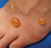
Correspondence
Kendall Lane, MD, Expeditionary Health Services Pacific, 3985 Cummings Rd, Suite 4, San Diego, CA 92136; [email protected]
1. North American arthropod envenomation and Parasitism. In: Auerbach PS, ed. Wilderness Medicine. 5th ed. Philadelphia, PA: Mosby; 2007;947–982.
2. Bjarke T, Hellgren L, Orstadius K. Cheyletiella parasitivorax dermatitis in man. Acta Derm Venereol 1973;53:217-224.
3. Shelley ED, Shelley WB. The diagnostic challenge of non-burrowing mite bites Cheyletiella yasguri. JAMA 1984;251:2690-2691.
4. Infestations and bites. In: Habif TP. Clinical Dermatology: A Color Guide to Diagnosis and Treatment. 4th ed. St Louis, Mo: Mosby; 2004;533–534.
5. Elston DM, Stockwell S. What’s eating you? Bed-bugs. Cutis 2000;65:262-264.
6. Steen C, Carbonaro P, Schwartz R. Arthropods in dermatology. J Am Acad Dermatol 2004;50:819-842.
7. The vesiculobullous reaction pattern. In: Weedon D. Skin Pathology. 2nd ed. Brisbane, Australia: Churchill Livingstone; 2002;129–191.
8. Ajithkumar K, Soshamma G, George B. Abnormal insect bite reactions: A manifestation of immunosuppression of HIV infection? Indian J Dermatol Venereol Leprol 2001;67:72-74.
9. Davis MD, Perniciaro C, Dahl PR, Randle HW, McEvoy MT, Leiferman KM. Exaggerated arthropod-bite lesions in patients with chronic lymphocytic leukemia: a clinical, histopathologic, and immunopathologic study of eight patients. J Am Acad Dermatol 1998;39:27-35.
10. Pendersen J, Carganello J, Van Der Weyden MB. Exaggerated reaction to insect bites in patients with chronic lymphocytic leukemia: clinical and histological findings. Pathology 1990;22:141-143.
11. Rosen LB, Frank BL, Rywlin AM. A characteristic vesticulobullous reaction in patients with chronic lymphocytic leukemia. J Am Acad Dermatol 1986;15:943-950.
12. Barzilai A, Shpiro D, Goldberg I, et al. Insect bite-like reaction in patients with hematologic malignant neoplasms. Arch Dermatol 1999;135:1503-1507.
13. Tokur Y, Ishihara S, Tagawa S, et al. Hypersensitivity to mosquito bites as the primary clinical manifestation of juvenile type of Epstein-Barr virus-associated natural killer cell leukemia/lymphoma. J Am Acad Dermatol 2001;45:569-578.
14. Khamaysi Z, Dodiuk-Gad RP, Weltfriend S, et al. Insect bite-like reaction associated with mantle cell lymphoma: clinicopathological, immunopathological, and molecular studies. Am J Dermatopathol 2005;27:290-295.
15. Allergic contact dermatitis. In: Habif TP. Clinical Dermatology: A Color Guide to Diagnosis and Treatment. 4th ed. St Louis, Mo: Mosby; 2004;84–97.
16. Kimyai-Asadi A, Usman A, Nousari HC. Ciprofloxacin-induced bullous pemphigoid. J Am Acad Dermatol 2000;42:847.-
17. Kashihara M, Danno K, Miyachi Y, Horiguchi Y, Imamura S. Bullous pemphigoid-like lesions induced by phenacetin. Report of a case and an immunopathologic study. Arch Dermatol 1984;120:1196-1199.
18. Mycoses and algal infections. In: Weedon D. Skin Pathology. 2nd ed. Brisbane, Australia: Churchill Livingstone; 2002: 663.
19. Nakamura E, Taniguchi H, Ohtaki N. A case of crusted scabies with a bullous pemphigoid-like eruption and nail involvement. J Dermatol 2006;33:196-201.
20. Ansarin H, Jalali MH, Mazloomi S, Soltani-Arabshahi R, Setarehshenas R. Scabies presenting with bullous pemphigoid-like lesions. Dermatol Online J 2006;12(1):19.-
21. Elston D. Prevention of arthropod-related disease. J Am Acad Dermatol 2004;947-954.
A 23-year-old active duty US Navy sailor came to our medical center for treatment of 1–2 cm tense pruritic blisters on his dorsal feet, calves, and anterior lower extremities (FIGURE 1). He told us that several days earlier, he had noticed ill-defined “itchy red bumps” on both legs. He denied fever, night sweats, malaise, trauma, insect bites, contact with animals or plants, or recent illnesses. He did say, however, that he’d recently done outdoor fieldwork with the Marine Corps in southern California.
His medical history was unremarkable, and he was not taking any prescribed or over-the-counter medications or supplements. He had no family history of blistering or other autoimmune disorders.
On examination, we noticed clear fluid-filled vesicles and bullae on non-erythematous, non-urticarial bases that were haphazardly distributed on both legs and dorsal feet. Agminated or herpetiform configurations were not present. Ruptured bullae left erythematous to beefy red eroded bases, and there were numerous smaller red papules with vesicular surface changes. All of the lesions were below the knees; there was complete sparing of the trunk, upper extremities, intertriginous skin, head, and neck.
The remainder of the physical examination was unremarkable, and there was no lymphadenopathy. Complete blood count and chemistries were within normal limits, and the patient’s HIV status was negative.
We performed lesional and perilesional punch biopsies. The lesional biopsy demonstrated subepidermal blistering with a predominantly eosinophilic infiltrate in all layers of the dermis and within the blister. Direct immunofluorescence (DIF) was performed on the perilesional biopsy and was negative for IgA, IgG, IgM, C3, and fibrinogen. Gram stain, potassium hydroxide (KOH) prep, and wound culture were also negative.
FIGURE 1
Tense pruritic blisters
What is your diagnosis?
Diagnosis: Bullous arthropod bite reaction
Bullous arthropod bite reaction (BABR) occurs in sensitized individuals as a delayed hypersensitivity immune reaction to insect saliva.1 Patients typically present with grouped localized pruritic or asymptomatic blistering2 and otherwise appear well. Unless secondarily infected, the blisters are non-erythematous and non-purulent, and develop within hours to days of the bite.
Lesion location is key. The distribution and location of the lesions will tip you off to a BABR diagnosis. The lesions in BABR are usually grouped and localized to a specific area of the body, depending on the causative arthropod and the circumstances leading to the bites. For example:
- Lesions caused by Cheyletiella mites are typically found on the forearms, anterior thighs, and lower abdomen after an infested pet sits on an individual’s lap.2,3
- Blisters caused by flea bites are isolated to the lower extremities.4 (We suspect that flea bites were the culprit in our patient’s case.)
- Lesions caused by Cimex lectularius, more commonly known as bedbugs, may be found on the entire body and tend to occur in groups of 3.5
Insect bite? What insect bite?
Most patients will only complain of pruritus and will tell you that they don’t recall having had any insect bites.6 That said, the distribution of the lesions, lack of systemic illness, and otherwise unremarkable physical exam are sufficient for diagnosis.
Occasionally, a punch biopsy with DIF may be necessary to rule out more serious bullous disorders. In BABR, you may see both subepidermal and intraepidermal blistering, with perivascular and interstitial eosinophilic and lymphocytic infiltrates. Blisters separated by strands of keratinocytes create a characteristic multilocular appearance. Unlike autoimmune blistering disorders, DIF is negative in BABR.7 Gram stain, Tzanck smear, bacterial culture, and KOH prep may also provide additional information if infection is a concern.
For certain patients, the DX may be less clear-cut
Similar bullous lesions following insect bites have been reported in patients with HIV,8 chronic lymphocytic leukemia,9-12 EBV-associated Natural Killer leukemia/ lymphoma,13 and mantle cell lymphoma.14 There is ongoing debate as to whether the vesicular lesions in these patients truly represent an exaggerated response to an arthropod bite or mimic an insect-like bite reaction.10,12
Nevertheless, when you suspect a patient has BABR, be aware of its association with both hematologic malignancies and HIV. Appropriate evaluation, such as HIV screening and complete blood count, should be performed.
A condition that mimics contact dermatitis
Clinically and histologically, BABR can mimic the following:
- Contact dermatitis. With contact dermatitis, the blistering is more likely to appear in streaks or in a linear fashion.15 Lesions will be painful, as well as pruritic, and occur following direct contact with a plant or chemical allergen.
- Drug-induced pemphigoid. The patient’s history will increase your suspicion of drug-induced pemphigoid. Patients may be taking sulfur-containing drugs (furosemide), antibiotics (penicillins, fluoroquinolones), antihypertensives (ACE inhibitors, calcium channel blockers), neuroleptics, or sulfasalazine.7,16,17 The eruption is usually more generalized than BABR, and may involve the mucous membranes.
- Fungal infections. These infections will typically occur on the palms and soles. Infiltrate is typically neutrophilic, but can be eosinophilic.18
- Bullous scabies. Patients will have severe pruritus. Burrows and lesions can typically be found on moist areas (periumbilical, intertriginous skin).19,20
- Bullous pemphigoid. This is more commonly seen in elderly patients with comorbid conditions. Onset of blistering is gradual, and occurs predominantly on flexural skin.
Pemphigoid gestationis, erythema toxicum neonatorum, incontinentia pigmenti, and some pemphigus variants also have predominantly eosinophilic infiltration in the skin. These, however, are clinically distinct from BABR.
Focus on symptoms and prevention
BABR will resolve over time without aggressive intervention. Most patients are treated symptomatically with oral anti-histamines and topical steroids for pruritus.1 Prevention of further bites is important because of the risk of arthropod-transmitted diseases.21
Our patient couldn’t comfortably wear shoes
Our patient had extensive tense blistering on both legs that prevented him from comfortably wearing shoes (FIGURE 2). Using a #11 blade, we punctured all of the blisters at the most dependent portion of each lesion. We decompressed the lesions, but did not de-roof them so that the blistered skin could serve as a biological dressing. We applied topical mupirocin and wrapped both legs with a compressive dressing.
We gave the patient a 2-week tapering course of oral prednisone. At the 3-week follow-up, all of the blistered skin had completely healed with the exception of post-inflammatory hyperpigmentation. No new lesions developed. Our patient was well, with no recurrence of blistering, at his 6-month follow-up.
FIGURE 2
Wearing shoes was a problem
Correspondence
Kendall Lane, MD, Expeditionary Health Services Pacific, 3985 Cummings Rd, Suite 4, San Diego, CA 92136; [email protected]
1. North American arthropod envenomation and Parasitism. In: Auerbach PS, ed. Wilderness Medicine. 5th ed. Philadelphia, PA: Mosby; 2007;947–982.
2. Bjarke T, Hellgren L, Orstadius K. Cheyletiella parasitivorax dermatitis in man. Acta Derm Venereol 1973;53:217-224.
3. Shelley ED, Shelley WB. The diagnostic challenge of non-burrowing mite bites Cheyletiella yasguri. JAMA 1984;251:2690-2691.
4. Infestations and bites. In: Habif TP. Clinical Dermatology: A Color Guide to Diagnosis and Treatment. 4th ed. St Louis, Mo: Mosby; 2004;533–534.
5. Elston DM, Stockwell S. What’s eating you? Bed-bugs. Cutis 2000;65:262-264.
6. Steen C, Carbonaro P, Schwartz R. Arthropods in dermatology. J Am Acad Dermatol 2004;50:819-842.
7. The vesiculobullous reaction pattern. In: Weedon D. Skin Pathology. 2nd ed. Brisbane, Australia: Churchill Livingstone; 2002;129–191.
8. Ajithkumar K, Soshamma G, George B. Abnormal insect bite reactions: A manifestation of immunosuppression of HIV infection? Indian J Dermatol Venereol Leprol 2001;67:72-74.
9. Davis MD, Perniciaro C, Dahl PR, Randle HW, McEvoy MT, Leiferman KM. Exaggerated arthropod-bite lesions in patients with chronic lymphocytic leukemia: a clinical, histopathologic, and immunopathologic study of eight patients. J Am Acad Dermatol 1998;39:27-35.
10. Pendersen J, Carganello J, Van Der Weyden MB. Exaggerated reaction to insect bites in patients with chronic lymphocytic leukemia: clinical and histological findings. Pathology 1990;22:141-143.
11. Rosen LB, Frank BL, Rywlin AM. A characteristic vesticulobullous reaction in patients with chronic lymphocytic leukemia. J Am Acad Dermatol 1986;15:943-950.
12. Barzilai A, Shpiro D, Goldberg I, et al. Insect bite-like reaction in patients with hematologic malignant neoplasms. Arch Dermatol 1999;135:1503-1507.
13. Tokur Y, Ishihara S, Tagawa S, et al. Hypersensitivity to mosquito bites as the primary clinical manifestation of juvenile type of Epstein-Barr virus-associated natural killer cell leukemia/lymphoma. J Am Acad Dermatol 2001;45:569-578.
14. Khamaysi Z, Dodiuk-Gad RP, Weltfriend S, et al. Insect bite-like reaction associated with mantle cell lymphoma: clinicopathological, immunopathological, and molecular studies. Am J Dermatopathol 2005;27:290-295.
15. Allergic contact dermatitis. In: Habif TP. Clinical Dermatology: A Color Guide to Diagnosis and Treatment. 4th ed. St Louis, Mo: Mosby; 2004;84–97.
16. Kimyai-Asadi A, Usman A, Nousari HC. Ciprofloxacin-induced bullous pemphigoid. J Am Acad Dermatol 2000;42:847.-
17. Kashihara M, Danno K, Miyachi Y, Horiguchi Y, Imamura S. Bullous pemphigoid-like lesions induced by phenacetin. Report of a case and an immunopathologic study. Arch Dermatol 1984;120:1196-1199.
18. Mycoses and algal infections. In: Weedon D. Skin Pathology. 2nd ed. Brisbane, Australia: Churchill Livingstone; 2002: 663.
19. Nakamura E, Taniguchi H, Ohtaki N. A case of crusted scabies with a bullous pemphigoid-like eruption and nail involvement. J Dermatol 2006;33:196-201.
20. Ansarin H, Jalali MH, Mazloomi S, Soltani-Arabshahi R, Setarehshenas R. Scabies presenting with bullous pemphigoid-like lesions. Dermatol Online J 2006;12(1):19.-
21. Elston D. Prevention of arthropod-related disease. J Am Acad Dermatol 2004;947-954.