User login
Hyperpigmented papules and plaques on chest
A 20-year-old black man came into our medical center with a mildly pruritic scaly rash affecting his neck and upper body for 2 weeks. Physical exam revealed well-demarcated, hyperpigmented hyperkeratotic papules coalescing to form large plaques on his central chest, back, and shoulders. He had a reticulated pattern on his shoulders and arms (FIGURE 1). His face, intertriginous skin, genitals, mucous membranes, and lower extremities were spared. The remainder of the physical exam was unremarkable.
Woods lamp and potassium hydroxide (KOH) preparation were negative. Labs, including fasting blood glucose and thyroid function test, were normal. Our patient denied any recent travel, fever, night sweats, or weight loss. He noted only that he used the weight benches at the gym. His medical and family histories were unremarkable, and he was not taking any medications or supplements.
FIGURE 1
Plaques on chest, reticulated pattern on arms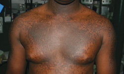
What is your diagnosis?
How would you manage this condition?
Dx: Confluent and reticulated papillomatosis
Confluent and reticulated papillomatosis of Gougerot and Carteaud (CRP) is a rare skin disorder characterized by benign blue-gray or brown hyperpigmented hyperkeratotic papules and plaques. The lesions initially occur on the trunk or central chest as 1- to 2-mm warty papules that become confluent to form plaques, spreading to the neck, abdomen, and upper extremities. Peripherally, the lesions are distributed in a reticular pattern. Although less common, CRP may be isolated to one part of the body, including the face and genitals; the mucous membranes are swpared.
With the exception of Japan, where a male predominance is seen, young women are more commonly affected.1 Patients are typically asymptomatic, or complain of mild pruritus and cosmetic concerns.
A disorder of keratinocyte maturation and differentiation?
The cause of CRP is unknown. Several theories have been entertained: an underlying endocrine disorder, a rare form of cutaneous amyloidosis, reaction to bacteria or fungus on the skin, and a keratinization abnormality.1 Most patients with CRP do not have an underlying endocrine disorder or any evidence of amyloidosis, making these theories less likely. KOH preparation for fungal elements is typically negative and patients do not respond to antifungal therapy.
A bacterial cause has been implicated because CRP responds to antibiotics, but many have argued that antibiotics are acting as an anti-inflammatory agent rather than an antibacterial medication.1 Others have suggested antibiotics may be acting to decrease epidermal proliferation by blocking protein and DNA synthesis and reducing keratinocyte production of cytokines.2
Overall, the most accepted theory is that it is a disorder of keratinocyte maturation and differentiation.1,3 Histological analysis, electron microscopy (EM), and immunohistochemical studies support this theory. Hyperkeratosis can be seen on histological examination. On EM, the stratum granulosum contains more lamellar granules and a larger transition cell layer, and immunochemical analysis reveals an increased expression of genetic markers associated with keratinization.1
The etiology of this keratinization abnormality is poorly understood.
CRP has been reported in several family members, prompting suggestions by some of a possible genetic component.4 Others have proposed staphylococcal enterotoxin B may promote certain immune factors causing abnormal keratinization.2
Is it CRP or tinea versicolor?
The differential for CRP includes:
- tinea or pityriasis versicolor
- tinea corporis
- seborrheic dermatitis
- keratosis follicularis (Darier disease)
- acanthosis nigricans
- macular amyloidosis.
Of all of these, however, CRP is most likely to be confused with tinea versicolor, as both are associated with hyper- or hypopigmented papules and plaques on the chest and back, as well as mild pruritus. In CRP, however, a Woods lamp and KOH will be negative, while with tinea versicolor, KOH will be positive and the Woods lamp may reveal yellow-green fluorescence.
If a patient’s KOH is negative and/ or the patient does not respond to treatment for tinea versicolor (which includes topical or oral antifungals), a trial of oral antibiotics for CRP may be reasonable. Response to an oral antibiotic, such as minocycline, will help to confirm the diagnosis. Although most patients with CRP do not have an endocrine disorder, it’s a good idea to keep this reported association in mind, and perform further testing, as needed.
Oral minocycline is the treatment of choice
The preferred treatment for CRP is oral minocycline (100 mg orally twice a day for 6 weeks).1,2 Oral azithromycin, erythromycin, clarithromycin, tetracycline, cefdinir,3 roxithromycin,5 doxycycline,2 and amoxicillin6 have also been used. Isotretinoin is an effective alternative to oral antibiotics, but clinicians often avoid it because of the adverse side effect profile.
With oral antibiotic therapy, the patient may completely clear and stay clear, or go on to have multiple recurrences or exacerbations. Topical retinoids have also been used with some success,4 but most reported cases have been successfully treated with oral antibiotics.1
Our patient required Tx for several months
We initially treated our patient with doxycycline (100 mg orally twice a day) for 1 month. The lesions cleared after 2 weeks and then recurred during week 4 of treatment. We discontinued the doxycycline, and started the patient on minocycline. The primary lesions resolved after 5 weeks of minocycline, though we noted residual post-inflammatory hyper-pigmentation (FIGURE 2). Our patient continued the medication for an additional 8 weeks. He was lost to follow-up.
FIGURE 2
Weeks later, hyperpigmentation remains
Correspondence
Kendall Lane, MD, LCDR, MC, United States Navy, Navy Medical Center San Diego, 34520 Bob Wilson Drive, Suite 300, San Diego, CA 92134
1. Schwartz R. Confluent and reticulated papillomatosis. Available at: http://www.emedicine.com/derm/topic82.htm. Accessed March 25, 2009.
2. Greenblatt D, Cintra M, Teixiera F, et al. Hyperpigmented plaques on a young man. J Am Acad Dermatol. 2007;56:896-898.
3. Scheinfeld N. Confluent and reticulated papillomatosis: a review of the literature. Am J Clin Dermatol. 2006;7:305-313.
4. Schwartzberg JB, Schwartzberg HA. Response of confluent and reticulate papillomatosis of Gougerot and Carteaud to topical tretinoin. Cutis. 2000;66:291-293.
5. Ito S, Hatamochi A, Yamazaki S. A case of confluent and reticulated papillomatosis that successfully responded to roxithromycin. J Dermatol. 2006;33:71-72.
6. Davis RF, Harman KE. Confluent and reticulated papillomatosis successfully treated with amoxicillin. Br J Dermatol. 2007;156:583-584.
A 20-year-old black man came into our medical center with a mildly pruritic scaly rash affecting his neck and upper body for 2 weeks. Physical exam revealed well-demarcated, hyperpigmented hyperkeratotic papules coalescing to form large plaques on his central chest, back, and shoulders. He had a reticulated pattern on his shoulders and arms (FIGURE 1). His face, intertriginous skin, genitals, mucous membranes, and lower extremities were spared. The remainder of the physical exam was unremarkable.
Woods lamp and potassium hydroxide (KOH) preparation were negative. Labs, including fasting blood glucose and thyroid function test, were normal. Our patient denied any recent travel, fever, night sweats, or weight loss. He noted only that he used the weight benches at the gym. His medical and family histories were unremarkable, and he was not taking any medications or supplements.
FIGURE 1
Plaques on chest, reticulated pattern on arms
What is your diagnosis?
How would you manage this condition?
Dx: Confluent and reticulated papillomatosis
Confluent and reticulated papillomatosis of Gougerot and Carteaud (CRP) is a rare skin disorder characterized by benign blue-gray or brown hyperpigmented hyperkeratotic papules and plaques. The lesions initially occur on the trunk or central chest as 1- to 2-mm warty papules that become confluent to form plaques, spreading to the neck, abdomen, and upper extremities. Peripherally, the lesions are distributed in a reticular pattern. Although less common, CRP may be isolated to one part of the body, including the face and genitals; the mucous membranes are swpared.
With the exception of Japan, where a male predominance is seen, young women are more commonly affected.1 Patients are typically asymptomatic, or complain of mild pruritus and cosmetic concerns.
A disorder of keratinocyte maturation and differentiation?
The cause of CRP is unknown. Several theories have been entertained: an underlying endocrine disorder, a rare form of cutaneous amyloidosis, reaction to bacteria or fungus on the skin, and a keratinization abnormality.1 Most patients with CRP do not have an underlying endocrine disorder or any evidence of amyloidosis, making these theories less likely. KOH preparation for fungal elements is typically negative and patients do not respond to antifungal therapy.
A bacterial cause has been implicated because CRP responds to antibiotics, but many have argued that antibiotics are acting as an anti-inflammatory agent rather than an antibacterial medication.1 Others have suggested antibiotics may be acting to decrease epidermal proliferation by blocking protein and DNA synthesis and reducing keratinocyte production of cytokines.2
Overall, the most accepted theory is that it is a disorder of keratinocyte maturation and differentiation.1,3 Histological analysis, electron microscopy (EM), and immunohistochemical studies support this theory. Hyperkeratosis can be seen on histological examination. On EM, the stratum granulosum contains more lamellar granules and a larger transition cell layer, and immunochemical analysis reveals an increased expression of genetic markers associated with keratinization.1
The etiology of this keratinization abnormality is poorly understood.
CRP has been reported in several family members, prompting suggestions by some of a possible genetic component.4 Others have proposed staphylococcal enterotoxin B may promote certain immune factors causing abnormal keratinization.2
Is it CRP or tinea versicolor?
The differential for CRP includes:
- tinea or pityriasis versicolor
- tinea corporis
- seborrheic dermatitis
- keratosis follicularis (Darier disease)
- acanthosis nigricans
- macular amyloidosis.
Of all of these, however, CRP is most likely to be confused with tinea versicolor, as both are associated with hyper- or hypopigmented papules and plaques on the chest and back, as well as mild pruritus. In CRP, however, a Woods lamp and KOH will be negative, while with tinea versicolor, KOH will be positive and the Woods lamp may reveal yellow-green fluorescence.
If a patient’s KOH is negative and/ or the patient does not respond to treatment for tinea versicolor (which includes topical or oral antifungals), a trial of oral antibiotics for CRP may be reasonable. Response to an oral antibiotic, such as minocycline, will help to confirm the diagnosis. Although most patients with CRP do not have an endocrine disorder, it’s a good idea to keep this reported association in mind, and perform further testing, as needed.
Oral minocycline is the treatment of choice
The preferred treatment for CRP is oral minocycline (100 mg orally twice a day for 6 weeks).1,2 Oral azithromycin, erythromycin, clarithromycin, tetracycline, cefdinir,3 roxithromycin,5 doxycycline,2 and amoxicillin6 have also been used. Isotretinoin is an effective alternative to oral antibiotics, but clinicians often avoid it because of the adverse side effect profile.
With oral antibiotic therapy, the patient may completely clear and stay clear, or go on to have multiple recurrences or exacerbations. Topical retinoids have also been used with some success,4 but most reported cases have been successfully treated with oral antibiotics.1
Our patient required Tx for several months
We initially treated our patient with doxycycline (100 mg orally twice a day) for 1 month. The lesions cleared after 2 weeks and then recurred during week 4 of treatment. We discontinued the doxycycline, and started the patient on minocycline. The primary lesions resolved after 5 weeks of minocycline, though we noted residual post-inflammatory hyper-pigmentation (FIGURE 2). Our patient continued the medication for an additional 8 weeks. He was lost to follow-up.
FIGURE 2
Weeks later, hyperpigmentation remains
Correspondence
Kendall Lane, MD, LCDR, MC, United States Navy, Navy Medical Center San Diego, 34520 Bob Wilson Drive, Suite 300, San Diego, CA 92134
1. Schwartz R. Confluent and reticulated papillomatosis. Available at: http://www.emedicine.com/derm/topic82.htm. Accessed March 25, 2009.
2. Greenblatt D, Cintra M, Teixiera F, et al. Hyperpigmented plaques on a young man. J Am Acad Dermatol. 2007;56:896-898.
3. Scheinfeld N. Confluent and reticulated papillomatosis: a review of the literature. Am J Clin Dermatol. 2006;7:305-313.
4. Schwartzberg JB, Schwartzberg HA. Response of confluent and reticulate papillomatosis of Gougerot and Carteaud to topical tretinoin. Cutis. 2000;66:291-293.
5. Ito S, Hatamochi A, Yamazaki S. A case of confluent and reticulated papillomatosis that successfully responded to roxithromycin. J Dermatol. 2006;33:71-72.
6. Davis RF, Harman KE. Confluent and reticulated papillomatosis successfully treated with amoxicillin. Br J Dermatol. 2007;156:583-584.