User login
He Tried So Hard to Avoid It …
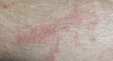
A 38-year-old man presents to dermatology with what he assumes is poison ivy: an itchy, blistery rash that usually appears in the summer, more years than not. Each year, it’s a bit worse in terms of extent and symptomatology, despite his efforts to avoid the problem altogether.
The rash pretty consistently manifests on his leg, although there are other areas of involvement. Equally predictable, at this point, is his wife’s reaction: She banishes him to the couch for fear of “catching” whatever he has.
After so many years’ experience with the condition, the patient is understandably alert to coming in contact with the offending plant. He has even taken photos of it, to demonstrate how abundantly it grows in his yard.
EXAMINATION
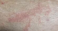
The patient’s lesions are classic collections of vesicles crisscrossing his legs in linear configurations. There is faint underlying erythema. Smaller but similar lesions are scattered over his arms and trunk; the patient is sure he spread the rash with his scratching.
The plant in the patient’s photos has five dart-shaped leaves with uniformly serrated margins extending from a single stem. It grows as a vine on fences and masonry. He has scrupulously avoided contact with it and therefore cannot understand how he keeps developing the rash.
What is the diagnosis?
DISCUSSION
It has been said that humor is tragedy plus time; certain skin diseases, such as shingles or poison ivy, certainly provide fodder. In the midst of an attack, however, these conditions can produce not only miserable symptoms of itching, pain, and sleeplessness, but also considerable mental anguish regarding contagion.
Repeated polls of the general public reveal an almost universal belief that poison ivy is contagious and that scratching spreads it around the body. Research, and a thorough knowledge of the pathophysiology involved, have long since disproven both concepts. So while the inclination to distance oneself from an affected person is understandable, there is actually no need to do so. For the patient, facing six weeks or more of isolation is no laughing matter (at least, until long after the fact).
This particular case highlights one other pertinent issue with poison ivy: As the photos established, the patient had carefully avoided the wrong plant. The five dart-shaped serrated leaves suggest Virginia creeper—certainly not poison ivy. This may sound like a minor issue, but it is quite possible that while the patient was steering clear of a harmless plant, he was in fact coming in contact with the one he should have been concerned about.
True poison ivy, Toxicodendron radicans, can develop as a bush, a vine that stays low to the ground, a small tree, or even a climbing vine that can reach 30 feet or higher into mature trees, especially along waterways or in low, boggy land. The vines can attain a thickness of more than 3 in and have a surface covered by tiny “rootlets,” giving them a shaggy look.
Regardless of the plant’s form, its leaves are always found in threes, protruding from the same stem. The leaves are diamond shaped, can reach a length of 10 in, and often have a single notch on the margin that is said to resemble the outline of a thumb. The plant produces white berries in late summer or early fall.
It has been estimated that the number of poison ivy plants has doubled since 1960, for at least two reasons. First, an increase in population has led to more land being cleared for housing; many properties now border woodlands, which are an ideal environment for this plant.
Second, and more surprising, poison ivy thrives in a CO2-rich environment. Carbon dioxide in the atmosphere has increased significantly in the past 50 years and will continue to do so. Experts predict that the density of poison ivy will double again in the next 20 years as a result. The potency of the urushiol, the offending substance in the stems and leaves, is expected to increase as well.
The patient (height, 6’3” and weight, > 300 lb) was treated with a 60-mg IM injection of triamcinolone, a two-week, 40-mg taper of prednisone, and twice-daily application of betamethasone cream. This, of course, followed a discussion of the risks versus benefits of such a course of action.
TAKE-HOME LEARNING POINTS
• Climbing vines with five serrated leaves coming off the same stem are probably Virginia creeper and not poison ivy.
• Poison ivy is not contagious, cannot be spread by scratching, and is not poisonous in any way.
• The rash produced by poison ivy exposure can be severe and can last six weeks or more without treatment.
• The number of poison ivy plants has doubled in the past 50 years and is expected to double again within 20 years. The potency of the plant’s allergen is also expected to increase.
Foot Rash + Gnarly Toenails = Man in Need of a Diagnosis
ANSWER
The correct answer is to perform a KOH examination (choice “b”), which takes just five minutes and offers the chance to establish the fungal origin of the rash. Although the patient’s skin is quite dry, the use of a moisturizer (choice “a”) is unlikely to address the overall problem. A punch biopsy (choice “c”) would be a logical choice if the KOH failed to solve the mystery. The use of combination creams (choice “d”) that contain a steroid (triamcinolone) and an antifungal (nystatin) is essentially an admission of the lack of a definitive diagnosis. For reasons discussed below, this strategy has almost no chance of helping.
DISCUSSION
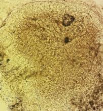
In this case, the KOH prep showed numerous hyphal elements, confirming suspicions of a fungal origin. One potential source of these organisms was the patient’s feet, where fungal infection had been present for years (“more than 30,” questioning revealed).
A common scenario is one in which the patient applies a steroid cream to a bit of dry skin just above the feet, which allows the fungi to gain a “foothold” from which to spread upward onto the leg; this progress is assisted through scratching and additional steroid application. If no firm diagnosis is ever established, definitive treatment cannot be undertaken and the problem never resolves.
In my opinion, there is never a reason to prescribe a product containing nystatin. In 1950, when it was discovered by researchers working in New York State laboratories (after which it was named), its efficacy against Candida species represented a notable advance, given the limited drug choices available for that purpose. But it has little, if any, activity against the dermatophytes causing our patient’s problems. And the steroid (triamcinolone) in this combination product, far from adding any therapeutic benefit, effectively diminishes any natural immune response.
The other reason to refrain from prescribing nystatin is that, since its discovery, at least three generations of products that treat both fungi and yeast (the azoles, such as clotrimazole, econazole, and fluconazole) have become available and have been found to be very effective.
The more important issue in this case, however, is finally having an accurate diagnosis: tinea corporis, probably caused by the most common dermatophyte, Trichophyton rubrum. The patient’s body is obviously a very happy home for this ubiquitous organism, to the extent that our chances of eliminating it are quite small. But we can at least make the patient more comfortable.
Treatment entailed ketoconazole foam (applied bid to his legs) and a two-month course of oral terbinafine (250 mg/d), which cleared up most of the skin problem. For his overgrown toenails, the patient was advised to establish care with a podiatrist for regular trimming.
In terms of a differential, this patient might have had psoriasis or eczema—and may still have one or both, since there’s no law against having more than one condition in the same location. In time, we may have to reconsider our solitary diagnosis.
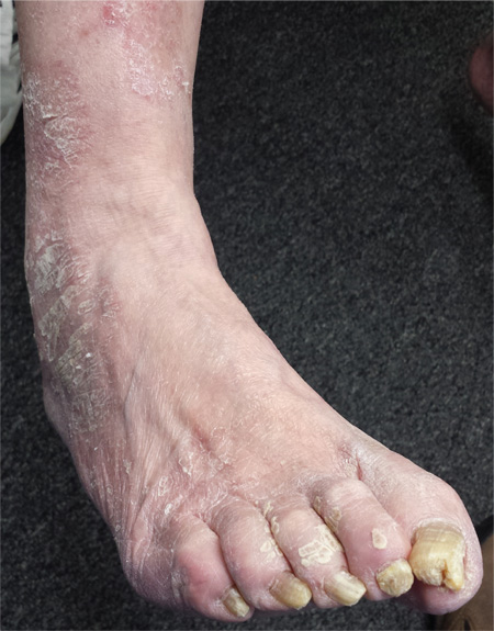
For several years, a 66-year-old man has had an itchy rash on his right leg; recently, it has become more bothersome. In general, he has noticed that when cold weather arrives, the rash improves slightly, but it inevitably worsens again as winter progresses. Over the years, the providers he has consulted have prescribed a number of topical products—among them, antifungal and steroid creams. Each of these products seems to help for a short period, then stops; at that point, the patient switches to a different product, with similar mixed results. The patient says he doesn’t have any other skin problems. Examination reveals patches of dry skin scattered from the knee to the top of the patient’s foot. Most have a faintly erythematous surface and arciform borders. These patches blend into a similar rash that covers the sides of both feet. All 10 toenails are grossly dystrophic, yellowed, and overgrown. The skin on the patient’s other leg is somewhat dry but otherwise unaffected.
Wife Says Husband Needs to Use Moisturizer
A 72-year-old man self-refers to dermatology at the insistence of his wife, who wants confirmation of her assertion that he needs to use a moisturizer on his arms. “They’re just so dry!” she says. “They’d look so much better if he’d use moisturizer every day, like I do.”
Her husband says he tried it and it didn’t help, so he stopped. “Besides, I can’t stand the greasy feeling it leaves behind!”
The patient claims to be in decent health aside from mild arthritis. He is retired from his job with the local power company, which kept him outdoors for many years. His employer required workers to wear long-sleeved khaki shirts, but, like all his fellow employees, the patient rolled up the sleeves in hot weather, exposing his forearms.
EXAMINATION
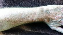
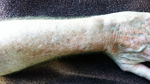
The patient has type II skin, blue eyes, and light brown hair. The skin on the dorsa of both arms is flecked with innumerable tan to orange macules from the mid forearms to (and including) the hands. Many old scars are seen in these same areas.
Closer examination reveals that the skin in these areas is quite thin and dry. Fine telangiectasias and focal scaling are also noted. The effect is much less pronounced from about the elbow up and is totally absent on both volar forearms.
What is the diagnosis?
DISCUSSION
The patient’s admittedly dry skin is not his primary problem, and all the moisturizers in the world will not make much difference. The term we use for his problem is dermatoheliosis , literally “sun condition,” which includes predictable indicators of past overexposure to UV sources. Specific lesions include solar lentigines, the tan to orange freckles that are commonly called “age spots” or “liver spots.” (Given enough sun exposure, a 25-year-old can develop solar lentigines, so age is not the cause. Likewise, the liver is not involved at all.)
Chronic overexposure to the sun thins the skin (solar atrophy), making it more susceptible to minor trauma and resultant scarring and also making the tortuous red blood vessels (telangiectasias) visible. Other indications of sun damage include actinic keratoses (flaky white papules), solar elastosis (creamy yellow to white “chicken skin”), and solar purpura. The combination of these superimposed colors (white, red, tan, orange, pink) in the context of dermatoheliosis is sometimes termed poikilodermatous change.
Although dermatoheliosis is a problem in terms of the potential for skin cancer and for cosmetic reasons, it will not be helped much by moisturizers. Sunscreen, too, will have minimal impact so long after the damage has been done. Laser resurfacing could erase most of these skin changes, but it would also knock out all the normal skin pigment, making the area contrast sharply with the rest of the patient’s skin.
A combination of fade cream (4% hydroquinone, applied bid) and sunscreen could reduce the brown discoloration to a degree. Twice-daily application of 5-fluorouracil cream for two weeks would erase much of the redness, but only for a few months. Various other methods have been tried with modest success.
The main outcome of this patient’s visit was to establish the need for periodic examination. His wife’s opinion notwithstanding, the utility of moisturizers is dubious.
TAKE-HOME LEARNING POINTS
• Dermatoheliosis is the collective term for skin changes associated with chronic overexposure to the sun.
• The white, red, pink, and yellow discoloration seen on this kind of sun-damaged skin is known as poikilodermatous change.
• In addition to poikiloderma, patients with advanced dermatoheliosis also have solar atrophy, multiple scars, telangiectasias, actinic keratoses, dry skin, and often solar purpura.
• Moisturizers play a minimal role in treating dermatoheliosis (providing skin-smoothing effects, if anything).
• Such patients need to be examined periodically for skin cancer.
• Younger patients with dermatoheliosis are well advised to use better sun protection.
Hole in Jaw Has Drained Fluid for 20 Years
ANSWER
The correct answer is all of the above (choice “d”). The patient’s actual diagnosis, sinus tract of odontogenic origin (choice “a”), will be discussed further.
Branchial cleft cyst (choice “b”) is always in the differential for neck masses, and squamous cell carcinoma (choice “c”) should always be considered in cases of nonhealing lesions—although 20 years is an unlikely timeframe for that diagnosis! Additional differential possibilities include thyroglossal duct cyst and pyogenic granuloma.
DISCUSSION
Sinus tracts of odontogenic origin, also called dentocutaneous sinus tracts, are primarily caused by periapical abscesses. As the purulent material accumulates in the confined space around the apical area, pressure increases; this sets in motion a tunneling process that terminates in an outlet, often inside the mouth but also (often enough) on the skin.
In the latter instance, known as extraoral sinus, the opening forms along the chin or submental area. In 80% of cases, the source is the mandibular teeth.
Dermocutaneous sinuses of maxillary origin, though not unknown, are decidedly unusual. They can drain anywhere on the maxilla, including around the nose. In edentulous patients, retained tooth fragments or segments of apical abscesses can act as the nidus for this process.
When a draining sinus manifests more acutely or occurs in a patient from a high-risk area (eg, Mexico or Central America), other diagnoses must be considered. These include scrofula, in which regional nymph nodes, infected by Mycobacterium tuberculosis or atypical mycobacterial organism, break down and drain. The indolent nature and chronicity of this patient’s problem effectively ruled out this diagnosis.
Culture of the fluid draining from the abscess would reveal a number of organisms (mostly of the strep family) but would not show the actual causative bacteria, since they are typically anaerobic. Biopsy of the surrounding tissue is occasionally necessary, when squamous cell carcinoma or other neoplastic process is suspected.
TREATMENT
The patient was advised to see a dentist, who will likely obtain a panoramic radiograph of her teeth, with particular attention to the affected area.
If an abscess is identified, as expected, treatment would entail root canal or extraction. The sinus tract would then heal rather quickly.
Antibiotics would be of limited use without elimination of the pocket. However, when patients complain of discomfort or outright pain, antibiotics (eg, penicillin V potassium or amoxicillin/clavulanate) can help to reduce the inflammation and offer some relief.
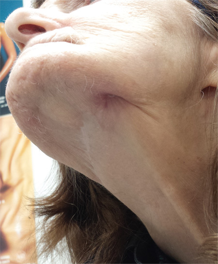
A 74-year-old woman is referred to dermatology by the primary care provider at her nursing home. She has a small hole on her left jaw that has drained foul-smelling material for more than 20 years. Although the site has never been painful, it occasionally swells and becomes slightly sensitive before slowly returning to its usual small size over a period of weeks. The patient is in generally poor health, with early dementia, chronic congestive heart failure, and diabetes. All her teeth were removed almost 30 years ago. She is afebrile and in no acute distress. On the submental aspect of her left jaw, there is a round, 6-cm area of skin that is retracted and fixed around a centrally placed sinus opening (measuring about 2 to 3 mm). A scant amount of purulent-looking fluid can be expressed from the spot. The area is faintly pink, but there is no evidence of increased warmth or tenderness on palpation.
Child With “Distressing” Problem
ANSWER
The correct answer is nevus sebaceous (choice “a”). This benign hamartomatous lesion is derived from local tissue and grows at the same rate.
It differs considerably from the other items in the differential, including aplasia cutis congenita (choice “b”). In this condition, a focal area of epidermis simply fails to develop, leaving a permanent hairless scar that contrasts sharply with the raised, mammillated plaque of nevus sebaceous.
Epidermal nevus (choice “c”) is usually a collection of tan to brown superficial nevoid papules that can be linear, agminated, or plaque-like. These lesions lack the color and mammillated surface of those seen in nevus sebaceous.
Neonatal lupus (choice “d”) can present at birth with hairless, cicatricial inflamed lesions. However, these tend to resolve quickly, often leaving focal scarring alopecia but no plaque formation.
DISCUSSION
Nevus sebaceous (NS), first described by Jadassohn in 1895, has long been recognized as an unusual but by no means rare congenital lesion. Occurring equally in both sexes and comprising sebaceous glands in a nevoid morphologic context, NS is considered a variant of sebaceous nevi and verrucous epidermal nevi in some circles. All three are derived from overgrowth of local, normal tissues that typically grow at the same rate as surrounding structures.
The vast majority of NS lesions are found in the scalp, although they can also develop on the ear or neck and, rarely, elsewhere on the body. This patient’s plaque—with its uniform surface; tiny, smooth, shiny papules; and (perhaps most important) total lack of hair—is typical. Other classic features are congenital onset and permanent nature, which distinguish them from the rest of the differential.
Focal malignant transformation of NS lesions has been reported—in fact, this author has seen two such cases in 30 years. Both were small basal cell carcinomas, although cases of melanoma and other malignancies have been reported.
Such changes are rare enough that most experts consider prophylactic removal to be unwarranted. Watching the lesions for change over the years is certainly reasonable, as is protecting them from sun exposure.
Surgical removal—usually performed by a plastic surgeon—is occasionally necessary for cosmetic reasons. This is particularly so when NS covers a portion of the face, or when the cosmetic implications of having a hairless plaque in the scalp are sufficiently distressing.
This patient and her parents were educated about the nature of the diagnosis and apprised of their options.
Editor's note: For a similar presentation with a very different diagnosis, see the March 2015 DermaDiagnosis case (http://bit.ly/1ye69Ym).
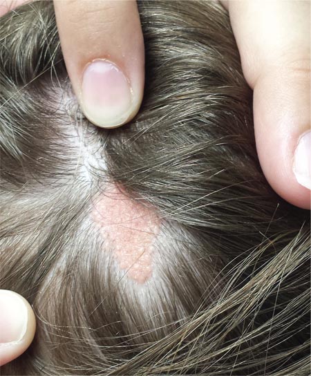
A “bald spot” is the chief complaint of a 12-year-old girl brought for evaluation by her mother. The lesion in her left parietal scalp has been there since birth, slowly growing but producing no symptoms. Although the child’s primary care provider has reassured the family that the “birthmark” is benign, they remain concerned. Furthermore, the patient has become increasingly distressed by the hairlessness. The child is otherwise healthy. There is no history of excessive sun exposure. The lesion is a roughly oval, uniformly pink, hairless 3.6-cm plaque with a faintly mammillated surface and well-defined margins. It is only visible when the surrounding hair is parted sufficiently to reveal it. Examination of the rest of the patient’s skin is unremarkable.
One Palm, Two Soles, Three Guesses on Diagnosis
A 27-year-old man, who works as an electrician, self-refers for evaluation of changes on his left palm. He first noticed them three years ago, when a scaly patch appeared on his volar wrist. At the time, his mother sent him an unidentified cream from Mexico, which he used on the rash. Although it resolved, within a few months, his entire palm was similarly affected.
The problem has persisted despite a number of treatment attempts—with, among other things, OTC creams and lotions, topical steroids, antifungals, and two courses of oral antibiotics. He even tried a vegetarian diet. Nothing has worked.
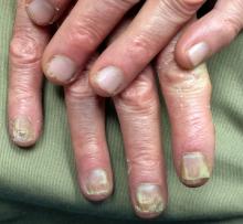
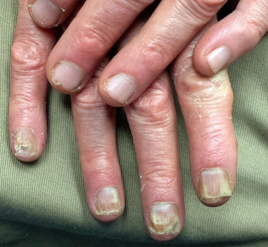
The hand is mildly symptomatic and faintly irritated. He reports similar symptoms on the soles of both feet, which he says date to several years before his hand problem. The fingernails on the affected hand have also changed.
The skin and nails of his right hand have remained completely normal throughout this experience. His health is otherwise excellent.
EXAMINATION
The skin on his left palm and on both soles is identical: uniformly scaly and pink, and in sharp contrast to the normal, smooth skin of his right palm. All five fingernails of his left hand are yellowed, thickened, and dystrophic. Surprisingly, all toenails are normal in appearance. His right hand is unaffected.
What is the diagnosis?
DISCUSSION
This curious phenomenon, known as “two-feet, one-hand disease,” represents a variant of dermatophytosis that is usually remarkably mild in terms of symptoms but often lasts years (if not decades) once the patient develops it. The causative organism is the same dermatophyte that causes jock itch and athlete’s foot: Trichophyton rubrum, the most commonly isolated fungal organism causing disease in humans.
There has never been a satisfactory explanation for why this fairly common condition spares one hand completely. It has been posited that the disease more often affects the dominant hand, but studies haven’t supported that theory. Neither has one hand (left or right) been shown to predominate.
What we do know is that the organisms feed on nonliving skin, which is why they cause minimal symptoms. The palms, soles, and nails represent more tissue for them to feed on and a way to escape the reach of the immune system.
Susceptibility is a key factor in why some people, but not others, develop two-foot-one-hand disease. It is thought that the inability to fend off this organism is related to an inherited, qualitative deficiency of cell-mediated immunity—coupled in some cases with environmental factors such as heat, humidity, and choice of shoes.
We also know that as common and well known as this diagnosis is in dermatology circles, it is far more obscure to nondermatology providers. As a result, the uninitiated provider tends to treat it as he or she would eczema or psoriasis (both of which belong in the differential): with steroids, which are the very thing certain to worsen the condition.
In some cases, before seeking professional care, patients attempt to treat their problem with steroids prescribed for another condition—or even for another patient. The condition will appear to improve but then worsen over time, as the steroids suppress what little immune response exists. The overall effect is thus to worsen the situation considerably, making the condition more difficult to treat.
In the absence of a definite diagnosis, even when antifungals are tried, they are often relatively weak products (eg, tolnaftate or undecylenic acid) that afford little if any relief. The patient (and sometimes provider) then interprets this as a sign that the problem is not of fungal origin and proceeds to other, truly ineffective treatments.
The two key factors that solve the mystery of two-foot-one-hand disease are: knowledge of its existence and the performance of a KOH examination of scrapings from the palm or sole. In this case, KOH revealed numerous fungal hyphae, confirming the diagnosis. Then—and only then—could effective treatment be provided.
This patient was treated with miconazole cream (bid application) and a month-long course oral terbinafine (250 mg/d). The good news is that this will help control the disease. The bad news? The problem will continue to lurk in the background, because we can do little or nothing to make the patient less susceptible. At least he now knows what he has, and he has the weapons to ward it off in the future.
TAKE-HOME LEARNING POINTS
• “Two feet, one hand” disease is a form of dermatophytosis that affects both feet and one hand while completely sparing the other hand. To date, no one has explained this pattern of distribution.
• As with many dermatologic conditions, the diagnosis of two-feet-one-hand disease is simple if you know it exists and completely obscure if you don’t.
• Individual susceptibility appears to explain why some people develop it and many more (including family/household members of affected individuals) don’t.
• Besides knowledge of its existence, the key to diagnosis is the confirmatory KOH prep.
• This is a perfect example of the principle of “correct diagnosis dictates correct treatment.”
A Ripple Effect of Groin Skin Issues
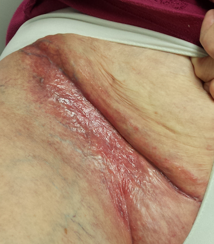
For several months, a 60-year-old woman has had a rash on the right side of her groin, which her primary care provider diagnosed as a “probable yeast infection.” The patient was given a prescription for nystatin cream; when this did not help, she was prescribed a combination clotrimazole/betamethasone cream. Initial signs of improvement prompted her to continue application daily.
However, although the original rash cleared, other skin changes occurred in the area. These alarmed the patient’s primary care provider, who observed them during a follow-up exam. In response, the patient requested a referral to dermatology.
EXAMINATION
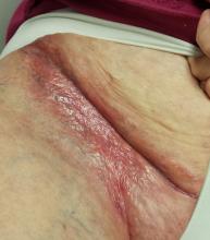
The entire right crural area is bright red and shiny. Multiple blood vessels are seen coursing over the surface of the area. There is no edema, increased warmth, or tenderness on palpation.
Punch biopsy shows marked epidermal atrophy and fails to show any evidence of cellular atypia.
What is the diagnosis?
DISCUSSION
When the patient was advised to stop using the combination cream, the “new” problem resolved quickly and her skin returned to normal. Although the source of the original rash remains unknown, what is clear is that the prescribed steroid induced marked atrophy. This then became “the problem.”
The groin is particularly susceptible to these types of changes. Why? Three reasons are occlusion, thin skin, and hydration. Read on for further explanation!
Occlusion, intentional or not, is known to potentiate the effects of topical steroids. The nature of intertriginous skin (skin folds) is that it effectively occludes the medicated skin, magnifying positive and negative effects. This means that the steroid works better and faster, but it also makes atrophy more likely (and equally quick to develop). When steroids are used for sufficient duration in susceptible areas—groin, axillae, under the breasts—focal areas of epidermis can wither totally, leaving an actual hole in the skin through which subcutaneous fat can be seen.
Furthermore, intertriginous skin is inherently thin (as are eyelids, neck skin, and the face). As a result, it takes less time for these changes to develop. The groin is especially prone to this problem because well-hydrated skin facilitates better penetration of the steroid.
This situation would likely have been avoided if the patient had used the prescribed product for only a week or two. A better option might have been a prescription for a weaker steroid (eg, hydocortisone 2.5%), although even that can be problematic if the medication is used for an extended period.
Though not quite as effective, topical NSAID preparations (calcineurin inhibitors such as pimecrolimus and tacrolimus) cause no such problems. They can help to reduce steroid use—for example, the patient can apply the steroid every third day and the calcineurin inhibitor on the other days.
The vehicle (the base in which the steroid is mixed) matters as well. Steroid ointments are inherently “self-occlusive,” especially in intertriginous areas—a fact that calls for increased caution in their use.
The atrophic look seen in this patient’s groin could have had other causes, such as T-cell or B-cell lymphoma. These were effectively ruled out by the biopsy.
TAKE-HOME LEARNING POINTS
• Epidermal atrophy is especially common on intertriginous skin (skin folds).
• The stronger the steroid (classes 1 through 7, with 1 being the strongest), the faster the atrophy occurs.
• Occlusion—caused by opposing skin folds or by other material (eg, socks, plastic wrap)—potentiates the positive and negative effects of topical steroids.
• Thin, moist skin allows better penetration by topical steroids, making them work better but also making atrophy more likely.
• Calcineurin inhibitors (eg, tacrolimus or pimecrolimus) have no such adverse effects and can help to reduce overuse of topical steroids in susceptible areas.
As Problem Spreads, Man Seeks Help
ANSWER
The correct answer is Schamberg disease (choice “b”), a benign form of capillaritis; see Discussion for more information.
Scurvy patients can present with ecchymosis (among other findings that were missing in this case). But scurvy (choice “a”) is rare, and by the time the disease is evident, the patient is typically quite ill.
Cutaneous T-cell lymphoma (choice “c”) can manifest as purpuric annular lesions. However, these would be unlikely to take the distributive pattern seen in this case, and they usually have an atrophic surface.
Thrombocytopenia (choice “d”) and other coagulopathies, although rightly considered, would probably manifest in other ways as well (ie, not just cutaneously).
Continue for the discussion >>
DISCUSSION
Schamberg disease is typical of a whole class of conditions in which red blood cells (RBCs) are extravasated from slightly damaged capillaries. This results in nonblanchable purpura and subsequent hemosiderin staining caused by phagocytosis of the RBCs by macrophages. Clinically, this family of diseases present as cayenne pepper–colored macules, most of which are annular in configuration.
Schamberg is, by far, the most common of these conditions. This presentation was typical: manifestation on the knees and ankles followed by upward spread (hence the condition’s other name, progressive pigmentary purpura). Usually resolving on their own within months, these lesions are almost always asymptomatic—but nonetheless alarming to the patient.
Other equally benign, self-limited forms of capillaritis include those in which lesions are pruritic (eg, purpura of Doucas and Kapetanakis) or lichenoid (purpura of Gougerot-Blum). Another example is lichen aureus, in which only one or two lesions, more gold than brown, appear on the legs of younger patients.
There are many theories as to these conditions’ cause, the most common of which is increased intravascular pressure secondary to dependence. However, if this were so, we’d likely see a great deal more cases, since many patients have problems related to venous insufficiency.
In some cases, skin biopsy (usually 4-mm punch) is necessary to rule out more serious diseases, such as an early form of cutaneous T-cell lymphoma. When a coagulopathy is suspected, blood work is necessary to confirm or rule out the diagnosis. In this case, there was no reason to suspect coagulopathy (or scurvy), since no other signs were seen.
This patient was educated about his diagnosis and provided Web-based resources he could consult. Various treatments—topical steroids, increased vitamin C intake, and increased exposure to UV light—have been tried but with disappointing results.
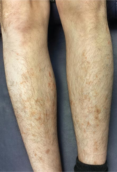
For several months, a 30-year-old man has had an asymptomatic rash on his legs. The lesions first appeared on his lower legs and ankles; over the subsequent months, they have spread upward. Now, the rash reaches to just below his knees. During this time, he has had two bouts of strep throat, both adequately treated. He denies any other skin problems and has no relevant family history. The patient denies alcohol or drug abuse and is not taking any prescription medications. Prior to referral to dermatology, he was seen in two urgent care clinics; at one, he received a diagnosis of fungal infection and at the other, of “vitamin deficiency.” He was given a month-long course of terbinafine (250 mg/d) that produced no change in his rash. He achieved the same (non)result from an increased intake of vitamins. Examination reveals annular reddish brown macules, measuring 1 to 3 cm, sparsely distributed from the knees to just above the ankles on both legs. The lesions are a bit more densely arrayed on the anterior legs. There is no palpable component to any of them and no discernable surface scale. Digital pressure fails to blanch the lesions. The hairs and follicles on the patient’s legs appear normal. There are no notable skin changes elsewhere, and the patient is alert, oriented, and in no distress.
Attempt at “Wart” Removal Backfires
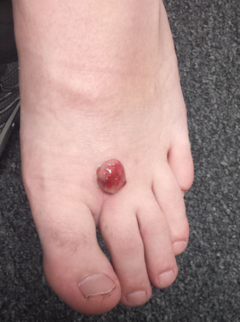
Two months ago, a tender lesion manifested on the dorsum of a 14-year-old boy’s right foot. Before it appeared, there was a tiny papule in the same spot; the boy’s mother says they thought it was a wart and attempted to remove it. As a result, the papule bled and became inflamed before quickly growing to its present form.
Besides being tender to touch, the lesion bleeds with minimal trauma. Attempts to remove it—using silver nitrate, liquid nitrogen, and triple-antibiotic cream—have all failed to have any positive impact.
The patient is reportedly otherwise healthy. He takes no medications of any kind.
EXAMINATION
The lesion is a 2-cm, domelike, red shiny nodule located on the mid-dorsum of the right foot. It is attached to the underlying skin by a thick sessile base. There is no surrounding erythema or other skin changes.
The site is anesthetized with local infiltrate of 1% lidocaine with epinephrine before deep shave biopsy is used to remove the lesion. The base is curetted, then cauterized for hemostasis.
PATHOLOGY
Microscopic examination shows a tangle of capillaries arranged in lobules separated by septae of connective tissue. A brisk inflammatory reaction and faint erosions are seen on the lesion’s surface.
What is the diagnosis?
DISCUSSION
First described by Ponce and Dor in 1897, pyogenic granuloma (PG) was thought to represent a kind of infection but turned out to be neither infectious nor granulomatous. It is now understood to represent the body’s attempt to heal a wound or other trauma (albeit this process goes awry for unknown reasons).
PG has a range of morphologic presentations; this case, with a berry-like friable papule, is fairly typical. PG is common on fingertips, lips, and faces (in children) and can even appear on gingival mucosae (particularly in first-trimester pregnancy). Other common areas of manifestation include ingrown toenails (lesion forms where the distal edge of the nail cuts into the lateral aspect of the nail bed) and the umbilical stump of newborns. An extremely common scenario is the child who cannot leave a wart or skin tag alone, picking and squeezing the lesion repeatedly until a PG forms.
PG is reactive in nature and not neoplastic. It is essentially vascular (other names for it include lobular capillary hemangioma and eruptive hemangioma), and the amount of blood that can flow from these lesions tends to frighten patients and parents.
While PG is completely benign, it is part of a differential of “look-alikes” that include nodular melanoma and metastatic cancer. For this reason, once removed, PGs are always sent for pathologic examination.
Removal should be done by deep shave, followed by electrodessication and curettage to prevent recurrence. Cryosurgery or silver nitrate application, though frequently done, is seldom effective. Significantly, neither yields a definitive diagnosis.
Certain drugs are associated with PG formation. These include isotretinoin, some chemotherapy drugs, and retroviral medications.
TAKE-HOME LEARNING POINTS
• Pyogenic granuloma (PG) is neither pyogenic nor granulomatous and is instead a reactive process.
• PG represents the body’s incomplete attempt to heal a wound.
• The trauma is often repetitive, particularly in PG associated with ingrown toenails or digitally manipulated by the patient.
• A berry-like appearance, sudden growth, and friability are all characteristic of PG.
• PG, though extremely common, needs to be removed to provide relief and to establish the lesion’s benign nature by pathologic examination.
Common Presentation for Complex Condition
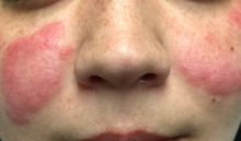
A month ago, a 33-year-old woman noticed skin changes on her arms and face. The affected areas have recently begun to itch and burn—particularly, the patient notes, since she spent an extended period in the sun over the weekend. She has used an antifungal cream (nystatin) on the rash, to no avail.
The patient denies joint pain, fever, and malaise. She has a sister with multiple sclerosis, but her family history is otherwise unremarkable. The patient’s only medication is oral contraceptives, which she has taken since the birth of her first and only child last year.
EXAMINATION
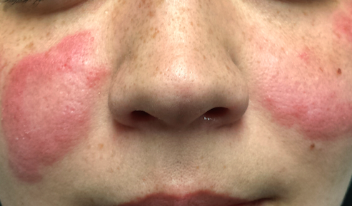
The patient is afebrile and in no particular distress. A florid red rash covers both cheeks, sparing the nose entirely. The margins are somewhat indurated and redder than the clearing centers. The follicular orifices are somewhat patulous, and a fine scale covers the affected areas of the face. The lateral brachial and triceps (sun-exposed) areas of both arms are similarly affected.
Punch biopsy reveals classic signs of lupus: vacuolar alteration of the basal cell layer, perivascular infiltrate around appendicial structures, and modest epidermal atrophy. Bloodwork yields no evidence of systemic lupus erythematosus (SLE).
What is the diagnosis?
DISCUSSION
Lupus, as a general topic, can be utterly confusing. Here are some facts that might help you make sense of the subject:
Purely cutaneous forms of lupus are commonly seen in dermatology; they manifest in sun-exposed skin as scaly annular lesions with clearing centers. Somewhat confusingly, however, cases of systemic lupus erythematosus (SLE) can present with similar cutaneous signs—and furthermore, patients with purely cutaneous lupus (subacute cutaneous lupus) may exhibit some systemic symptoms (just not enough to meet the strict criteria for SLE). Lupus in general is far more common in women than in men.
The “butterfly rash” seen in this case is uncommon but can occur in either cutaneous or systemic lupus. In most cases, though, this particular rash is a manifestation of seborrhea, psoriasis, rosacea or eczema—not lupus at all.
There are, of course, many other types of lupus. Another common form is discoid lupus (DLE), which manifests with round, scaly lesions on the head, neck, or ears; these are often misidentified as actinic keratosis, eczema, or psoriasis. DLE can be localized or generalized, purely cutaneous or a manifestation of SLE.
The key to diagnosis lies in first considering lupus in the differential and then biopsying the lesion (or sending the patient to someone who will). Once the clinical diagnosis is histologically confirmed (typical results include vacuolar interface dermatitis with sparse lymphocytic perivascular infiltrate), an immunologic workup is warranted. Also, depending on the predominant organ systems involved, the patient should be thoroughly evaluated by a dermatology or rheumatology specialist (or both).
Lupus is an autoimmune process, but in some ways it’s more useful to think of it as a form of vasculitis—which is why it can affect almost any organ system. The very first lupus patient I ever saw was in the psych ward having a psychotic break, which turned out to be secondary to a lupus-induced cerebritis. Since then, I’ve seen it affect the pericardium, kidneys, lungs, and joints. Lupus is even a major item in the differential of alopecia! SLE in particular is associated with an increase in thrombotic events and accounts for most early deaths from lupus (ie, within the first five years of diagnosis, when the cause of death is usually renal or pulmonary).
The patient in this case proved to have only cutaneous disease. She’ll respond nicely to a combination of sun protection and oral antimalarials (hydroxychloroquine) but will probably have recurrences every spring. Although unlikely to ever develop SLE, she is statistically more likely to develop other autoimmune diseases.
TAKE-HOME LEARNING POINTS
• Cutaneous lupus is more common than you might imagine. Lesions and eruptions in sun-exposed skin should prompt consideration of that item in the differential.
• Many forms of lupus have been identified, including neonatal lupus and overlapping syndromes involving lupus and lichen planus or even pemphigus.
• Though UV exposure is not always the cause, almost every type of lupus is worsened by UV light exposure.
• The differential for lupus is vast but includes psoriasis, sarcoidosis, dermatomyositis, and drug eruptions.
