User login
An Agreeable Girl With a Stubborn Rash
ANSWER
The correct answer is impetigo (choice “a”). Impetigo is almost always secondary to another condition, such as contact or irritant dermatitis, eczema, or dry skin.
DISCUSSION
Impetigo is a superficial bacterial infection usually caused by a combination of strep and staph organisms. It requires a break in the skin to provide a point of entry for the organisms. In young children, scratching and picking at eczema, along with lip licking, exacerbate the barrier-breaching process.
The organisms that cause impetigo are typically benign, but this was not always the case. Prior to WWI, certain strains of strep were capable of triggering an immune response that resulted in kidney damage. These “nephritogenic” strains of the Streptococcus family caused acute post-streptococcal glomerulonephritis (Bright disease), which, at that time, killed thousands each year. Fortunately, these strains are rare now.
In the pre-antibiotic days, when the average person bathed once a week, impetigo was highly contagious and serious enough that whole households were quarantined because of it.
Today, impetigo, once diagnosed, is relatively simple to manage. Mild cases can be treated with application of mupirocin ointment or cream three times a day. In this particular case, a 10-day course of an oral antibiotic (trimethoprim sulfa) was added, and the rash rapidly cleared.

Distraught parents of a 5-year-old girl are at their wit’s end dealing with their daughter’s perioral rash, which first appeared several months ago. Although they’ve consulted three different primary care providers, who rendered several diagnoses and numerous treatments, the rash continues to worsen. The parents worry about scarring, but they are more concerned that the rash may never clear at all.
Her treatments have included oral erythromycin, oral amoxicillin, topical anti-yeast cream, and various petroleum-based and hydrocortisone-containing OTC lip balms. In a moment of desperation, the parents even applied their son’s psoriasis cream (betamethasone) and diaper cream. These, too, had no effect.
Contactants had been considered as a possible source, causing the family to switch toothpaste brands and toothbrushes and eliminate mouthwash use—again, with no change.
Family history includes an atopic brother (eczema, asthma, seasonal allergies). The parents confirm that the patient has very sensitive skin and can’t tolerate many soaps and moisturizers. Before the rash manifested, they noticed she had a tendency to compulsively lick her lips.
The patient is quite fair-skinned, with red hair and blue eyes. The rash, which covers her entire perioral area, is impressively florid, red, and scaly. Focally, several areas of honey-colored crusts can be seen. The vermillion surfaces of the lips are unaffected except for slight focal fissuring. No nodes can be felt in the head or neck. The patient is in good spirits despite all this, and certainly not in any distress.
How to Nail the Diagnosis
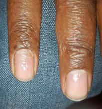
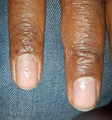
A 33-year-old African-American man is referred to dermatology—somewhat reluctantly—by his primary care provider for evaluation. A “fungal infection” has affected his fingernail on and off for several years, persisting despite four months of terbinafine treatment (250 mg/d).
The patient denies any antecedent trauma to the finger. He does have a history of hand eczema, mostly affecting the palmar surface of his hand, and atopy, marked by seasonal allergies and sensitive skin.
EXAMINATION
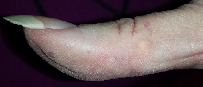
The patient’s fourth fingernail on his right hand is significantly dystrophic, with transverse ridges and modest onychorrhexis. The cuticle is detached from the nail plate, but the distal nail plate appears normal.
There are signs of his dyshidrotic hand eczema, with several 2 to 3 mm intradermal vesicles in the distal palm. Some spill onto the sides of his fingers.
What is the diagnosis?
DISCUSSION
To the unwary provider, every fingernail problem is fungal in nature; they simply have no other items in their differential, which is why terbinafine is overprescribed for nonfungal conditions. Even when we suspect fungal infection, the appropriate course would be to sample the nail plate and send it for culture or pathologic examination—not just throw another prescription at it.
However, in cases such as this one, there are other explanations. This patient’s nail issues are secondary to his eczema and are likely related to the disconnect between the cuticle and the nail plate. This gap allows debris (bits of food, dirt, etc) access to the nail matrix, thereby causing a misshapen nail. The key to diagnosis is the transverse ridging and detached cuticles. A history of hand eczema bolsters this impression—infection is not involved.
Application of a mid-strength steroid ointment to the cuticle area encourages it to reattach to the nail. This gives the nail a chance to grow in normally.
TAKE-HOME LEARNING POINTS
• Fungal infections are significantly (18 times) more common on toenails than on fingernails.
• Eczema on the hand or body can also lead to transverse ridges in fingernails, especially when the cuticle detaches from the nail plate.
• Other items in the differential include psoriasis, lichen planus, and nail changes associated with alopecia areata.
The Birthmark That Wasn't
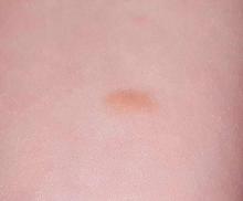
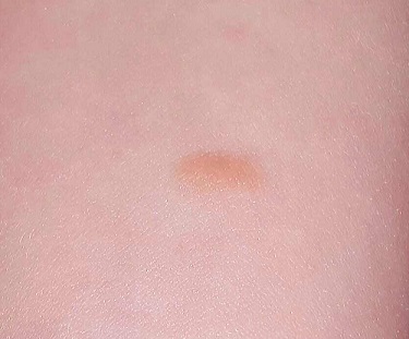
A 3-year-old girl is brought to dermatology by her mother, who requests evaluation of a lesion that manifested shortly after the child’s birth. The family’s primary care provider has always dismissed her lesion as a birthmark, but her parents are concerned by its tendency to abruptly change for no apparent reason. It swells up, becomes itchy and red, and then returns to normal within minutes.
According to her parents, the patient is otherwise quite healthy. She has never experienced any breathing problems and is not atopic.
EXAMINATION
The lesion—an orange, 2-cm, round nodule with a smooth surface—is located on the volar aspect of her forearm. When forcefully stroked, it immediately begins to swell and redden, resembling a wheal. The patient verifies that the lesion itches when touched but is not tender. Before the examination ends, the lesion returns to normal.
Her type I skin is otherwise unremarkable.
What is the diagnosis?
DISCUSSION
Mastocytoma is the term for this localized accumulation of mast cells. When traumatized, histamine (among other active components) is released, causing sudden swelling and itching.
Mast cells are normal white cells involved in the natural function of the immune system, but they can be involved in rare but serious pathologic processes. For example, some children develop dozens of lesions (a condition called urticarial pigmentosa); under extreme circumstances, they can release enough histamine to induce shock and even respiratory distress.
Rarely, mast cells can undergo malignant transformation in the bone marrow, leading to mast cell leukemia. Another rare complication is mastocytosis, in which organs and tissue are invaded by mast cells, destroying the function of the organs and causing problems related to histamine release.
While simple, benign mastocytomas are common and easy to identify visually, skin biopsy is the key to diagnosing this family of diseases. With a small, stable lesion such as this patient’s, spontaneous resolution alleviates the need for treatment. Surgical removal does serve as a permanent cure, however, and as an accurate way to distinguish mastocytoma from mast cell tumors and juvenile xanthogranuloma—the other items in the differential.
TAKE-HOME LEARNING POINTS
• Mastocytomas are benign lesions composed of mast cells.
• Present at or soon after birth, mastocytomas are usually orangish brown and round to oval, and measure, on average, 1 to 3 cm.
• When traumatized by friction, they swell due to the release of histamine from the mast cells. They return to normal within minutes to hours.
• Mastocytomas only rarely require removal, since they are benign and resolve on their own.
1-800-Zap-My-Zits
ANSWERThe correct diagnosis is discoid lupus erythematosus (DLE; choice “c”). For those unfamiliar with DLE, it is often mistaken for the other items listed. Biopsy can distinguish among them.
Fungal infection (dermatophytosis; choice “a”) of the face is unusual and would have responded in some way to the antifungal cream. Likewise, the use of steroid creams would have markedly worsened a fungal infection.
Although this could have been psoriasis (choice “b”), it’s rare for that condition to be confined to the face. It almost always appears elsewhere—the scalp, elbows, knees, and/or nails.
Dermatomyositis (choice “d”), an autoimmune condition, can certainly present with a bimalar rash. However, it is usually accompanied by additional symptoms, such as progressive weakness and muscle pain.
DISCUSSION
DLE can represent a stand-alone diagnosis, or it can be a manifestation of systemic lupus erythematosus (SLE). When present in this bimalar form, the lesions are often mistaken for the “butterfly rash” commonly seen in SLE.
This patient was thoroughly tested for SLE, and no evidence of it was found. Biopsy did, however, show changes consistent with DLE (interface dermatitis with increased mucin formation, among others).
The treatment for DLE is rather simple: It consists of sun protection and oral hydroxychloroquine. This helps reduce inflammation, although the patient will still have residual scarring.
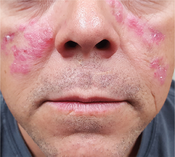
A 52-year-old man is referred to dermatology by his primary care provider for evaluation of facial lesions that first appeared almost a year ago. The patient, who works as a welder, has noticed that sun exposure tends to exacerbate the problem. He denies joint pain, fever, and malaise. He self-diagnosed the condition as acne and ordered a product from a TV ad, but this cream only made things worse. The asymptomatic lesions persist, despite application of a number of prescription products (2.5% hydrocortisone cream, adapalene gel, and antifungal creams, including tolnaftate and clotrimazole). The eruption—comprised of discrete, round, scaly lesions—covers a good portion of the bimalar areas of his face. The lesions are purplish red, and on closer inspection, you observe patulous follicular orifices. Some of the older lesions have focal atrophy. The rest of the examination is unremarkable.
This'll Really Get Under Your Skin
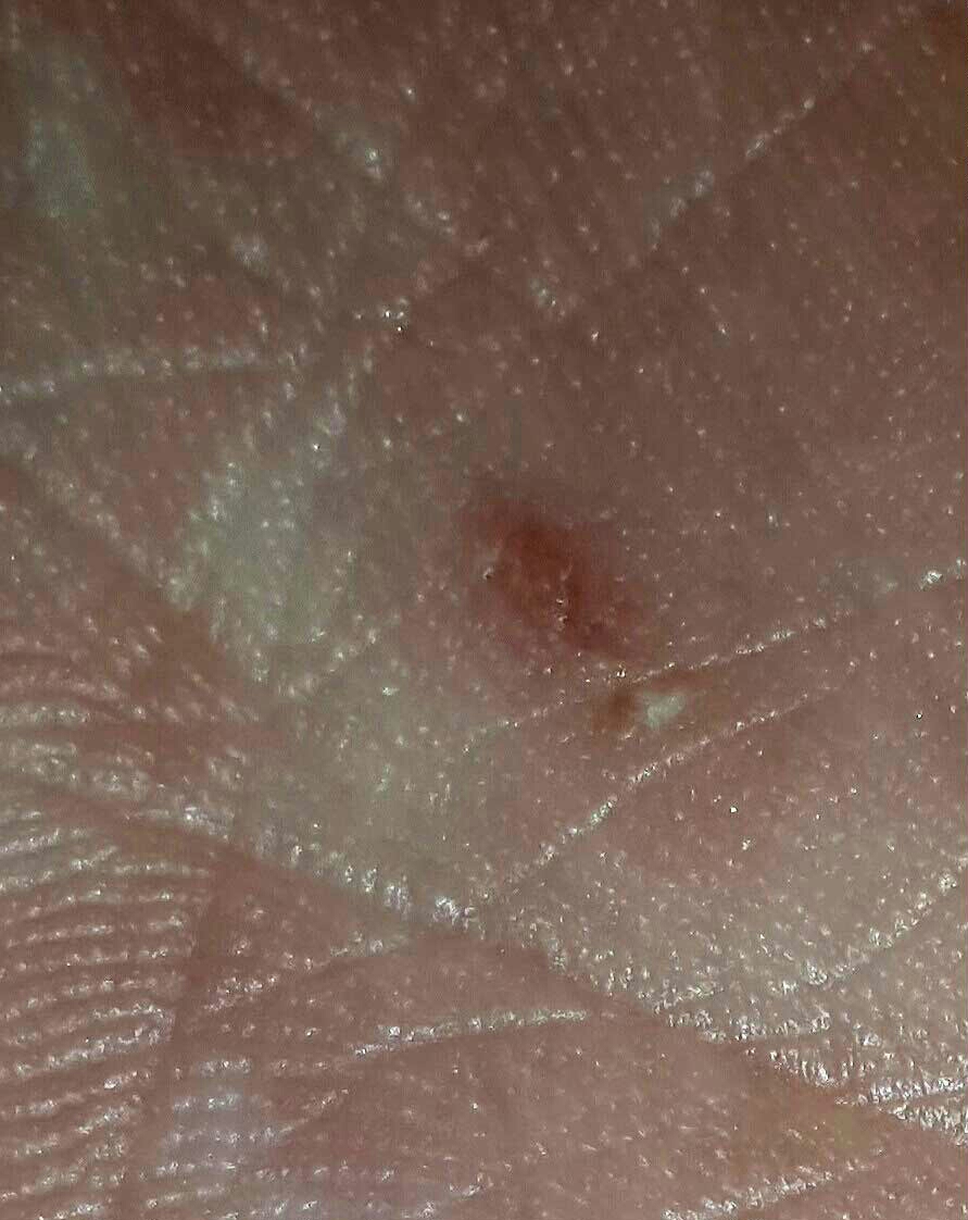
A 23-year-old woman presents to dermatology with an itchy rash she has had for several months. Although it manifested on her wrists and finger, the rash moves around and causes itching on her legs, trunk, and arms at various times. It has not affected her breasts or axillae.
The patient has been seen in primary care several times and received the usual topical steroids, antihistamines, and at least three courses of prednisone—none affording much relief.
She denies that anyone else in her household is itching. During her last visit to primary care, they treated her with topical permethrin cream, which was to be left on overnight then washed off. No relief was forthcoming.
EXAMINATION
Scattered areas of faint eczematoid rashes can be seen across her thighs and arms. There are two or three tiny excoriated papules on both volar wrists, but no intact vesicles are observed.
A closer inspection of her palms reveals one tiny linear vesicle on the mid right palm. Vigorous scraping with a #10 blade produces material, which is placed on a slide, covered, filled with potassium hydroxide 10%, and examined under a microscope.
What is the diagnosis?
DISCUSSION
Microscopic evaluation revealed a scabies adult, still moving among the dead cells. The paucity of organisms was probably a result of the prior treatment with permethrin, which confused the issue. When scabies is suspected, there’s only one way to confirm it: Perform a KOH prep. Otherwise, it’s just guesswork.
This particular patient was under the (faulty) impression that because her permethrin treatment failed, she didn’t have scabies. But a single treatment with topical antiscabetic cream is rarely curative. It must be done twice, seven to 10 days apart, to kill organisms newly hatched from eggs lain in the skin. In many cases, oral ivermectin is needed as well.
The itching experienced with scabies is due to an allergic response to scabetic material (droppings, tissue juice). The resultant eczematous rash can be very challenging to deal with.
Three other people live in the patient’s house, and it’s likely that these family members have been infested over several months’ time. They need treatment as well and will require two applications of cream.
Thought should also be given to how the patient acquired the infestation, lest she get it again. This may result in the need to avoid contact with certain people or confront them about their possible role as the source.
TAKE-HOME LEARNING POINTS
• The itching and rash of scabies result from an allergic reaction to the scabetic elements deposited in the skin. This reaction can closely resemble eczema.
• A single treatment of permethrin cream is unlikely to clear scabies. In order to have an effect on the eggs lain in the skin (which hatch in seven to 10 days), two treatments are necessary.
• Many cases of scabies will survive permethrin treatment, so consider adding ivermectin (oral antiscabetic medication) to the regimen.
• All family members living in the same house must be treated at the same time, lest they re-infest one another.
• KOH prep is the gold standard for diagnosing scabies.
A Lifetime Of Sunshine
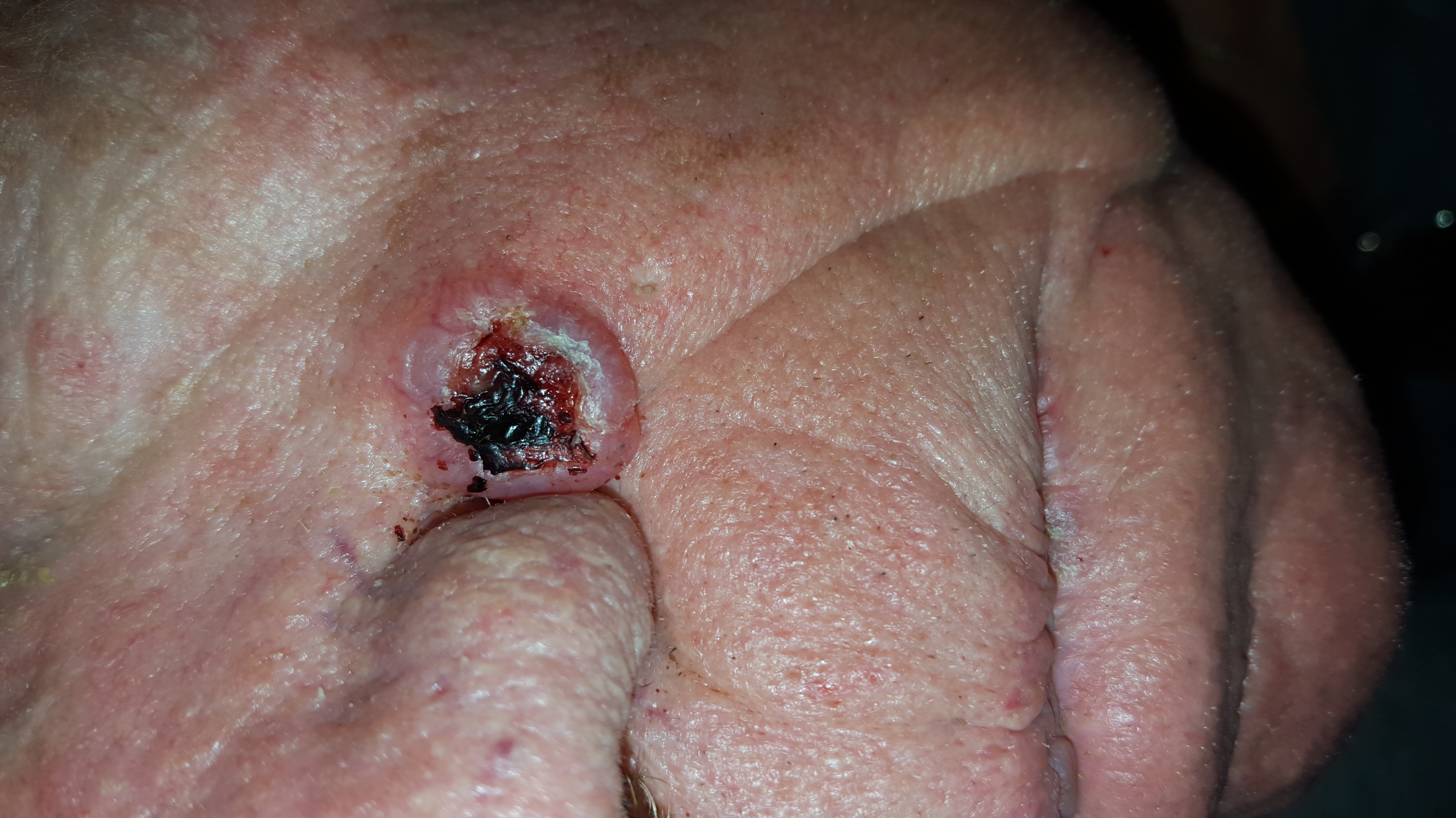
A 68-year-old man is seen for evaluation of a facial sore, which has been present for “at least a month.” It concerns him because, while the lesion is not painful, it won’t heal. The patient denies any preceding trauma to the area but does admit to having several skin cancers removed in his lifetime.
His lesion has been previously diagnosed as “pyoderma” and treated unsuccessfully with topical mupirocin and oral cephalexin.
He has held a number of jobs throughout his life, all of which involved being out in the sun all day—often seven days a week. And it wasn’t until he turned 45 that he finally started wearing a hat.
EXAMINATION
The lesion is an impressive, round, 1.2-cm nodule with extensive central ulceration. It has a rolled, translucent border and is tucked into the upper left nasolabial fold, directly adjacent to the alar bulb. It appears in the context of his fair, sun-damaged skin; he has several scars consistent with his history of skin cancer removal.
His skin elsewhere, while quite sun-damaged, is free of worrisome lesions.
A simple shave biopsy of the lesion is performed.
What is the diagnosis?
DISCUSSION
The biopsy showed changes consistent with basal cell carcinoma (BCC). It would be difficult to find a more typical example of BCC, the most common of all the sun-caused skin cancers (which include squamous cell, melanoma, and Merkel cell carcinoma). And yet, as this case illustrates, classic signs of BCC are often missed or overlooked, leading to “infection” or “pyoderma” diagnoses by providers who are unfamiliar with BCC.
I often tell medical providers that “it will be a rare day when you don’t see a BCC” because they are that common. But what I probably should say is “it will be a rare day when you don’t have a chance to see a BCC.” By that, I mean that you do have to look for them—not all are as obvious as this man’s—and you do need to have an understanding of who is susceptible and what the lesion will look like.
Older, sun-damaged individuals (mostly men) are most at risk for BBCs. Fair skin, freckles, red hair, and blue eyes are additional high-risk characteristics. This varies from melanoma patients, who tend to be much younger and whose lesions look nothing like a basal cell.
For BCC identification, focus on the areas directly and chronically exposed to the sun, such as the tops of the ears, the face, nose, chest, back, and arms.
BCCs can take on many appearances; they look as though they would hurt, but rarely do—a fact that should be noted as significant. Typically, they break down, scab, and bleed as they grow larger. They are usually very slow-growing and often take years to become noticeable. In other words, you have to think of it first, and look for it next. The fact that almost 1.5 million new BCCs are diagnosed each year in the US should heighten your suspicions considerably.
In this case, the patient will need extensive surgery, which may be deforming. Given his history of sun exposure, he will likely develop more skin cancers in the future.
TAKE-HOME LEARNING POINTS
• Basal cell carcinoma (BCC) is by far the most common type of sun-caused skin cancer; it will be seen frequently if properly looked for.
• Identification of BCCs will be more successful if the search is concentrated on the patients most likely to develop one and the areas where they are most likely to appear.
• This patient’s noduloulcerative BCC is the most common type and developed in a classic location. There is no type of infection that presents in this fashion.
• A simple shave biopsy confirms the diagnosis of BCC.
If at First You Don’t Succeed … Don’t Just Treat Again
ANSWER
Punch biopsy (choice “d”) is the correct answer for one simple reason: Correct diagnosis dictates correct treatment. What we’re missing is a diagnosis we can rely on.
DISCUSSION
This case demonstrates a major difference in outlook between the generalist and the specialist. The former is more interested in treating the problem, while the latter first wants to know what the problem is, then tailors the treatment to that problem and/or reassures the patient of the problem’s benign nature.
Had these lesions been of fungal origin, terbinafine would have had a positive effect. Furthermore, fungal infections are caused by organisms that only affect the outer layer of skin and create scaling, which was notably missing in this case.
Round to oval lesions suggest a number of diagnostic possibilities, only one of which is fungal (dermatophytosis). Others include T-cell lymphoma, psoriasis (though its lesions are almost always scaly), sarcoidosis, Hansen disease, lupus, and lichen planus. In cases like this one, these options need to be sorted through—and the only sure way to do that is with biopsy.
This patient’s biopsy showed a palisaded granulomatous process consistent with granuloma annulare (GA), a very commonly diagnosed benign condition. Since there are no ideal treatments for GA, he opted to do nothing, although he agreed to present for a biannual check-up. He was happy just to rule out all the things he didn’t have and thereby reduce his worries.
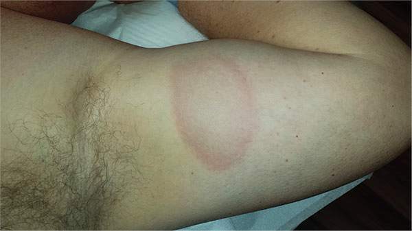
A 39-year-old man presents with asymptomatic lesions on both arms. When they manifested about six months ago, the patient diagnosed himself with “ringworm” and began treating them with an OTC clotrimazole cream his pharmacist recommended. Twice-daily application for two weeks did not result in a change, so the patient consulted his primary care provider (PCP), who also thought the problem was fungal. The PCP prescribed oral terbinafine (250 mg/d), which the patient took for a month without improvement. He then requested a referral to dermatology. The patient denies fever, malaise, shortness of breath, or unexplained weight loss. He is not taking any prescription medications. Both medial triceps have almost identical lesions: brownish red and oval, with well-defined margins. The margins are slightly raised relative to the central portions. The lesions, which measure 8 x 10 cm, exhibit no epidermal changes (eg, scale or papularity); they are totally intradermal. The rest of the examination is unremarkable.
Acne: Maybe She's Born With It?
A recent treatment regimen worsened this 13-year-old Native American girl’s “acne.” Within hours of beginning the treatment (application of benzoyl peroxide–containing gel and twice-daily use of a benzoyl peroxide–based cleanser), she reported intense burning and redness in the treated areas. She stopped this regimen after a week, and her primary care provider referred her for evaluation.
The lesions have been on her face since age 2 and are not usually problematic.
The patient has a notable history of atopy, with seasonal allergies and occasional bouts of asthma. She claims to have no other health problems but does admit to being overweight for most of her life; this is an issue for most of her family members, as well. Several of her relatives developed diabetes as adults.
EXAMINATION
The patient is 5 ft tall and weighs 250 lb. She has type IV skin, consistent with her Native American heritage. The posterior aspects of both cheeks are quite red and covered with hyperkeratotic, rough, pinpoint papules. There are no comedones or pustules there or elsewhere on her body.
No other blemishes or lesions are seen on her face, but there are hundreds of hyperkeratotic papules on her bilateral triceps. The skin in all her intertriginous (skin-on-skin) areas is quite dark and has a faintly papular, velvety appearance.
What is the diagnosis?
DISCUSSION
This girl’s facial and arm condition is keratosis pilaris (KP), an extremely common and harmless problem that affects up to 70% of newborns, though it may not fully express until age 1 or 2. Caused by an overproduction of perifollicular keratin, KP is inherited in an autosomal dominant pattern and is often seen in conjunction with atopic dermatitis and related conditions (eg, eczema, xerosis, asthma, icthyosis). KP can affect skin anywhere on the body except glabrous skin (palms and soles).
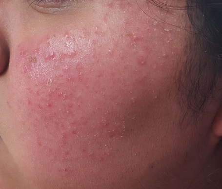
Variants of KP are also common; the one affecting this patient is keratosis pilaris rubra facei. This condition is often confused with acne, but treating it as such worsens irritation—especially in the wintertime, when humidity levels are low.
The presence of the patient’s condition from such an early age, the lack of comedones or pustules, and the discovery of more typical KP lesions on her arms all served to rule out acne and rosacea (the other item in the differential). The intertriginous hyperpigmentation represents acanthosis nigricans, reflecting her obese and (probable) prediabetic state, and is unrelated to her KP.
She will be treated with a one-week course of 2.5% hydrocortisone cream (bid application) and cessation of benzoyl peroxide–containing products. The condition should improve rapidly and become less prominent over time. Given the nature of KP, I have also found it helpful to encourage parents of affected children to research the condition online, to reinforce the information I’ve provided in the clinic.
TAKE-HOME LEARNING POINTS
• Keratosis pilaris (KP) is extremely common, affecting up to 70% of all newborns.
• Keratosis pilaris rubra facei, a common variant, affects the posterior two-thirds of the cheeks. Virtually all patients with this form will also exhibit KP on more typical areas (eg, the triceps).
• While there is no cure, moisturization helps to ease symptoms, as do other products (eg, salicylic acid–containing creams).
• KP is often mistaken for acne; unfortunately, treatment as such is counterproductive.
A Case of Cold Feet Hands
A 51-year-old woman is referred to dermatology for multiple problems she has had for several months—and, in some cases, years. The most immediate is a new nodule on the side of one thumb. Although it is asymptomatic, the fact that it simply “appeared” worries her, because her favorite uncle recently died of melanoma.
Several new lesions also materialized on her finger pads and chest. These do not cause symptoms either, but their newness makes them worrisome.
When asked about her health in general, the patient initially states it is “fine”—until her husband corrects her. She then recalls that she is being seen by various specialists, including a gastroenterologist for dysphagia-like symptoms and a rheumatologist for vague joint pains and what sounds like Raynaud disease.
EXAMINATION
On the lateral right thumb is a shallow, intradermal, white lesion. It is firm and round and measures 5 mm. No overlying skin changes (eg, redness or disruption of the skin surface) are observed. Under local anesthesia, a small incision is made in the lesion’s surface, allowing for exploration with small forceps; granular, gritty, white material is extracted.
On the affected thumb and two other fingers, telangiectasias can be seen. There are additional patches on the patient’s chest, but none around her mouth.
No sign of Raynaud disease is seen. However, the patient is adamant that this only appears when her hands are quite cold.
What is the diagnosis?
DISCUSSION
Connecting these dots leads to the likelihood of CREST syndrome, a variant of systemic sclerosis (SS); the acronym stands for calcinosis, Raynaud disease, esophageal dysmotility, sclerodactyly, and telangiectasia. The classification of CREST in the spectrum of SS is still open to discussion, but it is widely recognized as a real and distinct condition.
SS shares with CREST the involvement of anticentromere antibodies, but there are significant differences in the clinical course of each condition. Raynaud disease is often the first sign of CREST and can precede the other findings by years. Unlike SS, CREST typically spares the kidneys, and if it affects the lungs at all, it is through pulmonary artery hypertension and not fibrosis (the latter of which is seen in SS). Both conditions, however, can present with swelling of the hands and dysphagia.
Interpretation of these disparate findings requires the skill of a rheumatologist, who often shares management with other specialists.
As for differential diagnoses: Any one of these components can be a stand-alone diagnosis or can manifest with other diseases or syndromes. But when found together, they are highly suggestive of CREST. In this patient’s case, more testing needs to be done before a definitive diagnosis can be made.
TAKE-HOME LEARNING POINTS
• CREST syndrome is considered a variant of systemic sclerosis but has significantly different features and clinical course.
• The components of CREST (calcinosis, Raynaud disease, esophageal dysmotility, sclerodactyly, and telangiectasia) are often viewed as isolated phenomena by those unfamiliar with the condition.
• Raynaud disease is often the initial sign of CREST, preceding the rest by years.
• The telangiectasias seen with CREST often manifest on the hands and chest but can also be found on mucosal surfaces.
Moles: Their Role in Skin Cancer Diagnosis
ANSWER
The correct answer is none of the above (choice “d”). These lesions are all intradermal nevi, which have little, if any, risk for malignant transformation. Deeper nevi are considered quite safe, unless significant change has occurred. Despite the unlikelihood, however, it is risky to declare a 0% chance of skin cancer.
DISCUSSION
Slow growth and increased prominence are not the kinds of changes to look for in skin lesions. Rather, look for marked asymmetry (eg, the growth of a new, darker, macular component) or other change in color or consistency.
Hairs on these lesions are quite normal and are actually reassuring in confirming their benign nature. Skin cancers seldom support hair growth.
Most melanomas don’t come from moles. Instead, they are “de novo” lesions, literally coming from nothing, out of clear skin. It is true that the more moles someone has, the greater his or her risk for skin cancer, though not necessarily in one of the moles. When melanomas do develop from nevi (a collection of a certain type of melanocyte), this usually occurs in superficial types, such as compound or junctional nevi. From an objective standpoint, in this patient’s case, family history means nothing.
What does matter is to pay as much attention to the owner as to the lesion. The more fair-skinned and sun-damaged (freckles, blue eyes, red hair) the patient is, the more worrisome a lesion can be.
This patient had none of those traits, and she will likely have one of her lesions surgically excised to ensure she’s satisfied with the resulting scar. Of course, the tissue sample will be sent for pathologic examination, as any specimen should be.
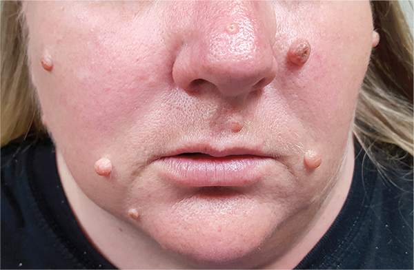
A 39-year-old woman self-refers for evaluation of moles she’s had on her face “all her life.” They have become more prominent with age, and many now have hairs growing in them. They are often traumatized by contact with fingernails or clothing. The patient worries that they might “turn into cancer” the way her grandfather’s moles did. The patient looks her stated age, is moderately overweight, and has more than her share of moles (some of which exceed 6 mm in diameter.) For the most part, they are skin-colored, and several are hair-bearing. Further questioning reveals that her moles manifested during puberty and have not been present “all her life.” Her type II skin is otherwise unremarkable and free of sun damage.

