User login
Serum urate level governs management of difficult-to-treat gout
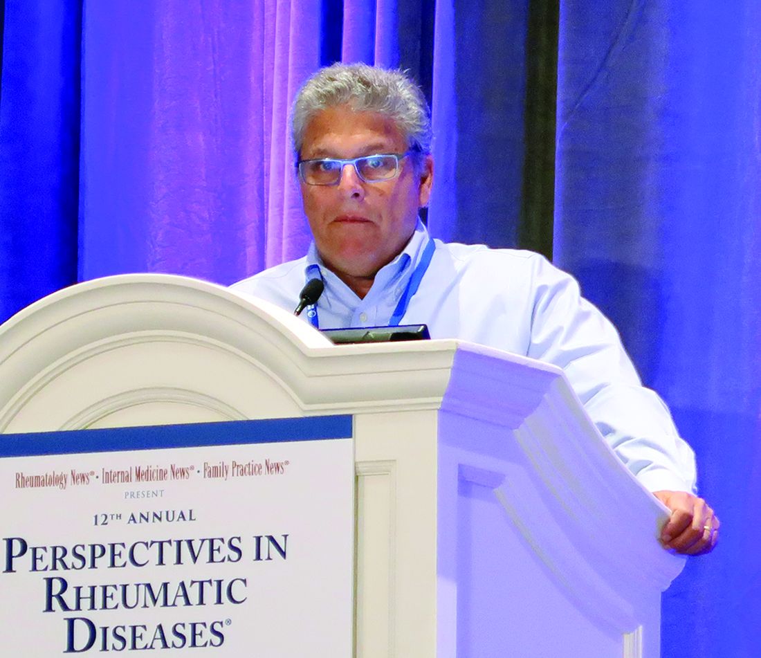
LAS VEGAS – Management of difficult-to-treat gout calls for a familiar therapeutic goal: lowering the serum urate level to less than 6 mg/dL. Underused treatment approaches, such as escalating the dose of allopurinol or adding probenecid, can help almost all patients reach this target, said Brian F. Mandell, MD, PhD, professor of rheumatic and immunologic disease at the Cleveland Clinic.
“The major reason for treatment resistance has nothing to do with the drugs not working,” Dr. Mandell said at the annual Perspectives in Rheumatic Diseases held by Global Academy for Medical Education. “And it does not even have to do ... with patient compliance. It is actually due to us and lack of appropriate monitoring and dosing of the medicines. We do not push the dose up.”
The urate saturation point in physiologic fluids with protein is about 6.8 mg/dL. Physicians and investigators have used 6 mg/dL as a target serum urate level in patients with gout for decades. “The bottom line is lowering the serum urate for 12 months reduces gout flares. There is absolutely no reason to question the physicochemical effect of lowering serum urate and dissolving the deposits and ultimately reducing attacks,” Dr. Mandell said. Urate lowering therapy takes time to reduce flare frequency and tophi, however. “It does not happen in 6 months in everyone,” he said.
Addressing intolerance and undertreatment
Clinicians may encounter various challenges when managing patients with gout. In cases of resistant gout, the target serum urate level may not be reached easily. At first, gout attacks and tophi may persist after levels decrease to less than 6 mg/dL. Complicated gout may occur when comorbidities limit treatment options or when tophi cause dramatic mechanical dysfunction.
“There is one way to manage all of these [scenarios], and that is to lower the serum urate,” Dr. Mandell said. “That is the management approach for chronic gout.”
Because this approach does not produce quick results, patients with limited life expectancy may not be appropriate candidates, although they still may benefit from prophylaxis against gout attacks, treatment of attacks, and surgery, he said.
Intolerance to a xanthine oxidase inhibitor is one potential treatment obstacle. If allopurinol causes gastrointestinal adverse effects or hypersensitivity reactions, switching to febuxostat (Uloric) may overcome this problem. Desensitizing patients with a mild allergy to allopurinol is another possible tactic. In addition, treating patients with a uricosuric such as probenecid as monotherapy or in combination with a xanthine oxidase inhibitor may help, Dr. Mandell said.
Increasing the dose of the xanthine oxidase inhibitor beyond the maximal dose listed by the Food and Drug Administration – 800 mg for allopurinol or 80 mg for febuxostat – is an option, Dr. Mandell said. In Europe, the maximal dose for allopurinol is 900 mg, and physicians have clinical experience pushing the dose of allopurinol to greater than 1,000 mg in rare instances, he noted. “There is not a dose-limiting toxicity to allopurinol,” he said. There is a bioavailability issue, however, and splitting the dose at doses greater than 300 mg probably is warranted, he added.
If these approaches fail to lower the serum urate level to below 6 mg/dL, rigid dietary changes may be a next step. Adjusting other medications also may be an option. For example, physicians might weigh using losartan as a blood pressure medicine instead of a thiazide.
Finally, physicians can debulk urate deposits with pegloticase. “Dramatically lower the body load of serum urate, and then come back and use your traditional drugs,” he said. After treatment with enzyme replacement therapy, patients almost invariably require lower doses of allopurinol or febuxostat, he said.
Also, in severe cases when the time necessary for traditional urate-lowering therapy to work may not make it the most appropriate route, aggressive therapy with pegloticase may be warranted, Dr. Mandell said.
The FAST trial
The ongoing Febuxostat versus Allopurinol Streamlined Trial (FAST) has provided data about undertreatment with allopurinol and the effects of increasing the dose. The prospective, randomized, open-label study is comparing the cardiovascular safety of allopurinol and febuxostat in patients with symptomatic hyperuricemia. It enrolled patients who were on allopurinol in normal clinical practice. To enter, patients had to have a serum urate level below 6 mg/dL. If patients’ levels were not below 6 mg/dL, investigators increased the dose of allopurinol to try to reach that target (Semin Arthritis Rheum. 2014 Aug;44[1]:25-30).
“Basically, this part of the study is a dose-escalation trial for efficacy,” Dr. Mandell said. “Of 400 patients taking allopurinol, 36% still had a urate above 6 [mg/dL]. ... If you uptitrated the dose, 97% of people were able to get to 6. Uptitration works. You just actually need to do it.” The results indicate that a 100-mg increase in allopurinol dose decreases serum urate by about 1 mg/dL.
Allopurinol hypersensitivity and chronic kidney disease
Patients with chronic kidney disease may have increased risk of allopurinol hypersensitivity. For a while, researchers postulated that oxypurinol, the active component of allopurinol, built up and caused toxicity in some patients with chronic kidney disease. As a result, researchers suggested adjusting the dose for patients with chronic kidney disease.
One problem with this approach is that only about 20% of patients with chronic kidney disease would reach the treatment target with the suggested doses, Dr. Mandell said. “You are exposing them to some potential risk with a very low chance of actually getting any efficacy at all,” he said.
Furthermore, allopurinol hypersensitivity behaves like an allergic reaction, not a toxicity reaction. Small studies suggest that starting allopurinol at a low dose and slowly increasing the dose may be safe in patients with chronic kidney disease. Allopurinol is not nephrotoxic, and some data indicate that it may be nephroprotective, he said.
Dr. Mandell reported that in recent years he was a clinical investigator and consultant for Horizon and a consultant for Takeda and Ardea/AstraZeneca/Ironwood.
Global Academy for Medical Education and this news organization are owned by the same parent company.
EXPERT ANALYSIS FROM PRD 2019
FDA approves diroximel fumarate for relapsing MS
The Food and Drug Administration has approved diroximel fumarate (Vumerity) for the treatment of relapsing forms of multiple sclerosis (MS) in adults, including clinically isolated syndrome, relapsing-remitting disease, and active secondary progressive disease, according to an Oct. 30 announcement from its developers, Biogen and Alkermes.
The approval is based on pharmacokinetic studies that established the bioequivalence of diroximel fumarate and dimethyl fumarate (Tecfidera), and it relied in part on the safety and efficacy data for dimethyl fumarate, which was approved in 2013. Diroximel fumarate rapidly converts to monomethyl fumarate, the same active metabolite as dimethyl fumarate.
Diroximel fumarate may be better tolerated than dimethyl fumarate. A trial found that the newer drug has significantly better gastrointestinal tolerability, the developers of the drug announced in July. In addition, the drug application for diroximel fumarate included interim data from EVOLVE-MS-1, an ongoing, open-label, 2-year safety study evaluating diroximel fumarate in patients with relapsing-remitting MS. Researchers found a 6.3% rate of treatment discontinuation attributable to adverse events. Less than 1% of patients discontinued treatment because of gastrointestinal adverse events.
Serious side effects of diroximel fumarate may include allergic reaction, progressive multifocal leukoencephalopathy, decreases in white blood cell count, and liver problems. Flushing and stomach problems are the most common side effects, which may decrease over time.
Biogen plans to make diroximel fumarate available in the United States in the near future, the company said. Prescribing information is available online.
AChR autoantibody subtype testing may improve accuracy of myasthenia gravis evaluations
AUSTIN, TEX. – When testing for acetylcholine receptor (AChR) autoantibodies in patients with suspected myasthenia gravis, testing for binding antibodies and for modulating antibodies is more accurate than testing for either subtype alone, researchers reported at the annual meeting of the American Association of Neuromuscular and Electrodiagnostic Medicine. Testing for both subtypes may be the most accurate approach, regardless of whether patients have coexisting neuromuscular disorders, the researchers said.
“The advent of improved methods of detecting AChR autoantibodies has greatly facilitated the diagnosis of myasthenia gravis,” said Pritikanta Paul, MBBS, a neuromuscular fellow at Mayo Clinic in Rochester, Minn., and his colleagues. AChR antibody assays frequently are part of evaluations for myasthenia gravis, but clinicians lack consensus as to which antibody subtypes – binding, blocking, or modulating – should be tested. Clinicians test for binding antibodies most commonly, while studies have found blocking antibodies to be “least useful as an initial diagnostic test,” Dr. Paul and his colleagues said.
To assess how combinatorial antibody testing and the presence of coexisting neuromuscular disorders affect testing’s sensitivity and specificity, the researchers reviewed clinical and electrophysiologic testing data from 360 patients with suspected myasthenia gravis who underwent serologic autoantibody testing between 2012 and 2015.
Titers of AChR binding antibodies greater than 0.02 nmol/L were considered positive, as were AChR modulating antibodies more than 20%. The researchers used a greater than 10% decrement of the compound muscle action potential to repetitive nerve stimulation at 2 Hz or positive response on single-fiber EMG as electrophysiologic confirmation of myasthenia gravis.
In all, 123 of the 360 patients had a final clinical and electrophysiologic diagnosis of myasthenia gravis, including 23 with ocular myasthenia gravis and 100 with generalized myasthenia gravis.
The sensitivity of testing for AChR binding autoantibodies was 92%, and the sensitivity of testing for modulating autoantibodies was 90%. In comparison, the sensitivity of testing for either antibody subtype or both was 94%.
Among 45 patients with myasthenia gravis and coexisting neuromuscular disorders, including peripheral neuropathy, mononeuropathies, radiculopathy, and motor neuron disease, the sensitivities of testing for binding antibodies, modulating antibodies, and either or both were 96%, 91%, and 96%, respectively.
Of the 237 patients who did not have myasthenia gravis, 89 had electrophysiologic confirmation of alternative diagnoses. Among these 89 patients, AChR autoantibody testing yielded 11 false positives. Three patients tested positive for both binding and modulating antibodies, six for binding antibodies only, and two for modulating antibodies only. Those with false-positive results had diagnoses that were “diverse and clinically distinguishable from myasthenia gravis,” including myalgia, neuropathy, blurred vision, epilepsy, encephalopathy, and hemifacial spasm, the researchers said.
The researchers had no relevant disclosures.
SOURCE: Paul P et al. AANEM 2019, Abstract 236.
REPORTING FROM AANEM 2019
Edasalonexent may slow progression of Duchenne muscular dystrophy
CHARLOTTE, N.C. – presented at the annual meeting of the Child Neurology Society.
The NF-kB pathway is “fundamental to the pathogenesis and biology of DMD,” said Richard Finkel, MD, chief of neurology at Nemours Children’s Health System in Orlando and principal investigator for the phase 2 study, known as MoveDMD.
A lack of dystrophin, combined with the mechanical stress of muscle contraction, activates the NF-kB pathway and inhibits muscle regeneration. “It is known that there is inflammation and fibrosis and release of cytokines early in life” in patients with DMD, Dr. Finkel said.
Independent of mutation
Edasalonexent is an NF-kB inhibitor that is being developed by Catabasis as a therapy for patients with DMD regardless of the genetic mutation that is causing the disease. It may be used as monotherapy or with other dystrophin-targeted treatments, Dr. Finkel said.
In a mouse model of DMD, an analog of the drug reduced muscle inflammation and increased the force of diaphragm muscle. To assess edasalonexent’s safety, pharmacokinetics, and effects on functional measures and MRI in patients with DMD, Dr. Finkel and colleagues conducted the MoveDMD trial. Investigators enrolled boys aged 4 years to younger than 8 years who were not receiving treatment with corticosteroids.
Researchers first examined drug safety and pharmacokinetics in 17 boys who received the treatment for 1 week. The investigators then followed 16 of these patients off treatment for as long as 6 months. This off-treatment period was followed by a phase 2, placebo-controlled period, during which the 16 patients and another 15 patients received edasalonexent 67 mg/kg/day, edasalonexent 100 mg/kg/day, or placebo for 12 weeks. Patients subsequently entered an open-label extension study.
Dr. Finkel presented a comparison of outcomes during the off-treatment period with outcomes during the open-label extension. “We used these boys as their own internal control, if you wish,” he said.
Creatine kinase levels decreased soon after treatment, as did other markers of muscle disease. The drug “seems to have an early and sustained biomarker response,” Dr. Finkel said.
Annualized rate of change on lower leg muscle MRI-T2 decreased. “There is a relative reduction and stabilization from week 12 all the way out through the open-label extension to 72 weeks,” he said. “It suggests that there is an early and sustained response in stabilization of the MRI as a biomarker.”
Timed function tests
A comparison of the annualized rates of change on timed function tests – including the 10-meter walk/run, time-to-stand, and four-stair-climb, and the North Star Ambulatory Assessment – during the off-treatment and on-treatment periods indicated slowing of disease progression with treatment. “Shortly after starting on drug ... there was a relative stabilization in each of these measures,” Dr. Finkel said.
In addition, the researchers observed an early signal of possible cardiac benefit. Mean heart rate at baseline was 99 bpm. On treatment, it decreased to 92 bpm. “Boys with DMD die typically of cardiomyopathy, so it is important to try to address the cardiac status,” he said.
The drug was safe and well tolerated. Most participants experienced mild gastrointestinal issues, which typically were transient. One serious adverse event during the trial occurred in a patient receiving placebo. Patients tended to have a stable body mass index during treatment, Dr. Finkel said.
During the open-label extension, patients had “clinically meaningful slowing of disease progression on edasalonexent,” relative to the off-treatment period, Dr. Finkel said. Investigators plan to further study edasalonexent for the treatment of DMD in a phase 3 trial. The phase 3 study, PolarisDMD, recently completed enrollment at 40 sites. Results could be available in about a year, Dr. Finkel said.
The study was sponsored by Catabasis. Dr. Finkel disclosed consulting work and grants or research support from Catabasis and other companies.
SOURCE: Finkel R et al. CNS 2019. Abstract PL1-3.
REPORTING FROM CNS 2019
Baricitinib may benefit patients with Aicardi-Goutières syndrome
CHARLOTTE, N.C. – Scores on a novel AGS scale improved, and skin and liver complications resolved in children with AGS who received treatment with baricitinib, according to results presented at the annual meeting of the Child Neurology Society.
AGS is caused by various heritable disorders of the innate immunity that result in excessive interferon production. AGS characteristically manifests as an early-onset encephalopathy that causes intellectual and physical disability, but patients may have a wide range of clinical phenotypes. The disease may involve the skin, liver, lungs, heart, and other organs, as well as the brain.
A multisystem disorder
“The neurologic features, while they are the most compelling for us, are really only the tip of the iceberg,” said Adeline Vanderver, MD, program director of the leukodystrophy center, and the Jacob A. Kamens Endowed Chair in Neurologic Disorders and Translational Neurotherapeutics at Children’s Hospital of Philadelphia. “Nearly every single organ system in the body is affected, from either direct interferon injury or from a secondary vasculopathy related to the interferonopathy.”
Dr. Vanderver presented results from the compassionate use study, which assessed whether the JAK inhibitor baricitinib (Olumiant) may decrease interferon signaling in AGS and limit the morbidity of the disease.
The phase 1, open-label trial “included compassionate use of baricitinib in AGS under the argument that these children did not have time to wait for approval of the drug,” said Dr. Vanderver. In 2018, the Food and Drug Administration approved baricitinib for moderate to severe rheumatoid arthritis in adults with an inadequate response to methotrexate.
The phase 1 trial in AGS included 35 patients with mutation-defined AGS and evidence of inflammatory disease that could be targeted by JAK inhibition. The trial population was 36% female. The average age of disease onset was 0.8 years, and patients’ average age at treatment was 6.1 years. The investigators assessed safety and laboratory data every 3 months and conducted clinical assessments every 6 months.
The heterogeneity of AGS phenotypes within families and across genotypes makes treatment trials in this disorder a challenge, Dr. Vanderver said. Outcome measures may have ceiling or floor effects that fail to capture the range of severity of AGS symptoms. Dr. Vanderver and colleagues developed a novel AGS scale to capture the scope of neurologic function in patients with AGS
.
When the researchers applied the AGS scale to a historical cohort of patients, most had stable scores about 6 months after disease onset. “After the first 6 months of the disease, the disease tends to be much more static, as the children have sustained significant neurologic injury,” Dr. Vanderver said.
They applied the novel AGS scale post hoc as an exploratory endpoint in the phase 1 trial. In addition, parents recorded information in a diary about skin involvement, irritability, seizures, and fever. “Over time, we see a reduction, although not always a statistically significant reduction, in symptom burden,” Dr. Vanderver said. The AGS clinical diary scores reflect “what the parents were telling us – that they felt like their children were feeling better during treatment,” she said.
Several patients had skin conditions that improved with treatment. One patient with dermatitis or eczema had the skin abnormality resolve within 3 days. A patient with full-body panniculitis began healing for the first time after about a month of treatment. Seasonal variations and dose adjustments led to fluctuations in some of the skin conditions. Nevertheless, the results suggested significant improvement in skin manifestations in patients with AGS, Dr. Vanderver said.
Patients generally had stable AGS scale scores in the year before treatment, although a couple of patients who were closer to disease onset had precipitous decline in neurologic function, she said. “We had a statistically significant increase in that scale of neurologic function in our patients during the period of the study, even in patients who had sometimes had years of disease duration,” said Dr. Vanderver.
Dr. Vanderver cautioned that she does not want to overstate the changes in function. Patients with AGS may have less potential for recuperation, compared with patients with other conditions. “A child with significant disruptive CNS disease may not recuperate normal functioning,” Dr. Vanderver said, “but it can be clinically meaningful to families if children start having better head control, smile, communicate, even if they might not regain all their motor milestones.”
In addition, a small subset of patients who had potentially life threatening liver complications from the disease experienced rapid normalization and improvement of liver function. “This blockade can be important not just for neurologic function but also to maintain normal physiologic homeostasis of other organs that are affected by the interferonopathy,” Dr. Vanderver said.
Interferon signaling scores decreased in the days after starting treatment and subsequently leveled out.
Serious adverse events that occurred during the trial, such as hospitalizations, were attributable to AGS. One child died from unrecognized pulmonary hypertension, which is now known to be a complication of AGS but was not at the time.
Harnessing a side effect
The most significant and recurrent laboratory abnormality was thrombocytosis. “That is a known complication of this family of drugs that in many cases allowed us to improve previous treatment-resistant thrombocytopenia, so we kind of like that side effect in most cases, but in two cases it did ... result in dose adjustments, although we never had to stop the medication for that.”
The study offers proof of principle that AGS is treatable, Dr. Vanderver said. A phase 2 trial is enrolling patients closer to disease onset. Early treatment of AGS may remain a challenge until there is newborn screening for the disease, she said.
Dr. Vanderver receives grant and in-kind support for translational research without personal compensation from Eli Lilly, Takeda, Illumina, Biogen, Homology, and Ionis. In addition, Dr. Vanderver serves on the scientific advisory boards of the European Leukodystrophy Association and the United Leukodystrophy Foundation, as well as in an unpaid capacity for Takeda, Ionis, Biogen, and Illumina.
Eli Lilly provided support for the phase 1 study. In addition, the study received support from the AGS Association Americas Family Foundation, National Human Genome Research Institute, National Institute of Neurological Disorders and Stroke, and the Children’s Hospital of Philadelphia Research Institute.
SOURCE: Vanderver A et al. CNS 2019. Abstract PL1-6.
REPORTING FROM CNS 2019
Celiac disease may underlie seizures
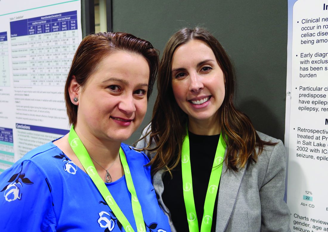
CHARLOTTE, N.C. – , according to a retrospective chart review presented at the annual meeting of the Child Neurology Society. Associations between celiac disease and seizures may have implications for screening and treatment, said study author Shanna Swartwood, MD, a fellow in the department of pediatric neurology at University of Utah in Salt Lake City.
“Screening for [celiac disease] early in patients with epilepsy, specifically with temporal EEG findings and intractable epilepsy, is warranted given the improvement of seizure burden that may result from exclusion of gluten from the diet,” said Dr. Swartwood and colleagues.
About 10% of patients with celiac disease have clinical neurologic manifestations, such as seizures. To characterize features of epilepsy in a pediatric population with celiac disease and to examine the effect of a gluten-free diet on seizure burden, Dr. Swartwood and colleagues reviewed patients treated at Primary Children’s Hospital in Salt Lake City since 2002. They identified patients with ICD-10 codes for seizures or epilepsy and celiac disease and reviewed 187 charts in all.
In all, 40 patients with seizures had biopsy-proven celiac disease, and 22 had a diagnosis of celiac disease based on the presence of antibodies. Among those with biopsy-proven celiac disease, 43% had intractable seizures. Among those with antibody-positive celiac disease, 31% had intractable seizures.
Among patients with intractable epilepsy, seizure onset preceded the diagnosis of celiac disease by an average of 5 years. For patients with nonintractable epilepsy, the first seizure occurred 1 year before the celiac disease diagnosis on average, but some patients received a celiac disease diagnosis first.
Focal seizures with secondary generalization and generalized tonic clonic seizures were the most common seizure types in this cohort. Epileptiform activity most often was seen in the temporal lobe. While other studies in patients with celiac disease have found occipital epileptiform activity to be the most common, only one patient in this cohort had activity in that location, Dr. Swartwood noted.
Patients with intractable seizures who adhered to a gluten-free diet “had a fairly robust response in terms of seizure improvement,” she said. Seizure improvement, including a decrease in seizure frequency or a decrease in antiepileptic medication dosage, occurred in seven of nine patients in the biopsy-proven group and in two of three patients in the antibody-positive group who adhered to a gluten-free diet and had intractable seizures. One patient was able to stop antiepileptic medication, and one patient had a complete resolution of seizure activity.
The researchers plan to further study the relationship between celiac disease and epilepsy, including whether various HLA subtypes of celiac disease correlate with seizures, said coinvestigator Cristina Trandafir, MD, PhD, assistant professor of pediatric neurology at University of Utah.
The chart review included relatively few patients with limited data. Nevertheless, the results suggest that there may be “substantial lag time” from first seizure to celiac disease diagnosis and that “earlier diagnosis and earlier placement on a gluten-free diet may be beneficial,” Dr. Swartwood said. Celiac disease may be asymptomatic, and screening for celiac disease with a blood test may make sense for patients with intractable seizures, she said.
The researchers had no relevant disclosures.
REPORTING FROM CNS 2019
Pediatric epilepsy surgery may improve cognition and behavior
CHARLOTTE, NC – according to a study presented at the annual meeting of the Child Neurology Society. The presence of comorbidities such as mood disorders and autism may influence the likelihood of perceived improvement, whereas the type of surgery may not.
“The parents and the families of the patients perceive that, even if the patients are not completely seizure free, the behavior and cognitive outcomes are better if there is some sort of seizure improvement,” said Trishna Kantamneni, MD, director of pediatric epilepsy at UC Davis in Sacramento.
To assess behavioral and cognitive outcomes following pediatric epilepsy surgery and to identify factors that predict improvement, Dr. Kantamneni and colleagues at the Cleveland Clinic Epilepsy Center retrospectively reviewed 126 patients younger than 18 years who underwent epilepsy surgery for medically refractory epilepsy during 2009-2016.
The primary outcome measure was the Impact of Childhood Neurologic Disability Scale (ICNDS), a parent-reported scale that assesses the behavior, cognition, and physical or neurologic disability of children with epilepsy. Parents completed the ICNDS preoperatively and at 6, 12, and 24 months after surgery. The researchers constructed separate linear mixed effects models to identify predictors of postoperative changes in ICNDS score.
Of the 126 patients, 62.7% were male, the median duration of epilepsy was 4.7 years, and 69.8% were seizure-free at the 2-year follow-up. Postoperative ICNDS scores were available for 103 patients at 6 months and for 54 patients at 24 months.
Before surgery, the average total ICNDS score was 55.7. At 6 months after surgery, the average score was 34.6, and at 24 months, it was 32.1, representing significant improvement from baseline.
In addition, behavior, cognition, and epilepsy subscores also improved post operatively, and the improvement persisted through 24 months. ICNDS scores significantly improved “even in patients who were not seizure-free after surgery,” by an average of about 22 points, the researchers said.
The absence of comorbid autism, cognitive impairment, and global developmental impairment and the absence of anxiety, depression, and ADHD were predictors of improved total ICNDS scores. Tumor pathology and being seizure free at 2 years also predicted improved scores. Duration and type of epilepsy, the number of antiepileptic drugs that patients were taking before surgery, and lobe of surgery were not predictive of improved ICNDS scores.
Dr. Kantamneni had no relevant disclosures.
SOURCE: Kantamneni T et al. CNS 2019, Abstract 51.
REPORTING FROM CNS 2019
Patients with Charcot-Marie-Tooth disease describe wide range of care
AUSTIN, TEX. – Patients with Charcot-Marie-Tooth disease (CMT) receive a range of supportive care that includes physical therapy, surgery, medications, orthoses, and walking aids, according to patient-reported data presented at the annual meeting of the American Association of Neuromuscular and Electrodiagnostic Medicine. Patients describe approaches to CMT management that are broadly consistent with guidelines, researchers said.
“The range of different CMT treatments was wide,” reported Tjalf Ziemssen, MD, PhD, a researcher at Technische Universität Dresden in Germany, and colleagues. “ These results indicate that pain may have a substantial impact on people with CMT.”
The data also suggest that “lower-limb problems and mobility issues have a considerable impact on people with CMT,” they said.
CMT is a rare, progressive neuropathy that leads to distal muscle weakness, muscle atrophy, and sensory loss. There is no cure, and patients rely on supportive care. Until recently, few studies have assessed the impact of CMT on patients’ lives.
An ongoing, international, 2-year observational study is collecting data from adults with CMT. Patients report data via an app called CMT & Me.
To examine patient-reported treatment patterns and care standards for CMT in the United States and the United Kingdom, Dr. Ziemssen and colleagues analyzed data through Aug. 5, 2019, about 9.5 months into the study. Their interim analysis included data from 439 patients, including 222 patients in the United Kingdom and 217 in the United States.
More than 70% of participants visit a family doctor each year, and a similar proportion visit a neurologist. About 40% visit physical therapists, orthotists, or podiatrists. Other health care professionals seen by patients include occupational therapists (20%), orthopedic surgeons (nearly 20%), and pain specialists (about 15%).
About 70% of participants had received rehabilitation therapy such as physical therapy or occupational therapy, and about 70% had used medications, most frequently nonopioid analgesics (about 50%) and antidepressants (about 30%).
More than 80% used orthoses or walking aids, most commonly ankle or leg braces, insoles, or walking sticks.
In addition, about half of respondents had undergone a surgery for CMT. The most common procedures were osteotomy, hammertoe correction, and plantar fascia release.
Together, patients saw about a dozen types of health care professionals. “Small proportions of participants had visited each professional, which suggests that the care requirements of CMT patients are varied,” the researchers said.
The study was sponsored by Pharnext. Dr. Ziemssen and coauthors received compensation for participating in the study. Other coauthors are employees of Pharnext or Vitaccess, the company that developed the app used in the study.
SOURCE: Ziemssen T et al. AANEM 2019. Abstract 83. Treatment of Charcot-Marie-Tooth Disease in the United Kingdom and United States.
REPORTING FROM AANEM 2019
Take drug, patient-level factors into account for when to end antiresorptive therapy
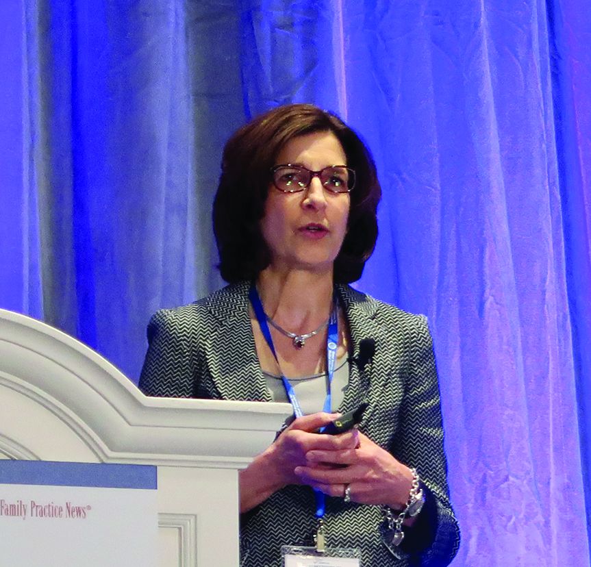
LAS VEGAS – according to an overview presented by Marcy B. Bolster, MD.
Recently published studies may help guide decisions about initiating and discontinuing treatment with bisphosphonates or denosumab (Prolia), the antiresorptive therapies. Understanding the ideal duration of bisphosphonate drug holidays “is a work in progress,” Dr. Bolster, from Harvard Medical School in Boston, said at the annual Perspectives in Rheumatic Diseases held by Global Academy for Medical Education.
No holiday with denosumab
Data indicate that twice yearly denosumab remains safe at 10 years, but studies have found a rapid loss of bone mineral density and an increased risk for vertebral fractures after treatment is discontinued (J Bone Miner Res. 2018 Feb;33[2]:190-8).
“Therefore, it is not appropriate for denosumab to be utilized with a drug holiday. If a patient is placed on denosumab, then consideration needs to be given for what to follow the course of denosumab,” Dr. Bolster said. “It is important to review with our patients the essential scheduled dosing of every 6 months, that the patient should not miss doses, and that we are not going to be able to initiate a drug holiday without starting another medicine.”
Patients likely to require hospitalization may not be good candidates for denosumab therapy because they may not be able to adhere to the dosing regimen, she said.
Denosumab vs. bisphosphonates: Real-world data
Trials have found greater increases in bone mineral density with denosumab, compared with the bisphosphonate drug alendronate, but that finding does not necessarily equate with reduced fracture risk, Dr. Bolster said. A recent population-based study examined fracture risk in approximately 92,000 people over age 50 years. Most were women, and their mean age was 71 years (JAMA Netw Open. 2019 Apr 5;2[4]:e192416).
The researchers compared the incidence of hospitalization for hip fracture among new denosumab users and new alendronate users during the 3 years after starting treatment. At 3 years, hip fractures occurred in 3.7% of the denosumab group and in 3.1% of the alendronate group. The rate of any fracture was 9% for each group. Although the study design had limitations, the analysis found “no difference between denosumab and alendronate in terms of fracture-risk reduction,” Dr. Bolster said. “Both agents are good agents.”
A recent meta-analysis compared fracture risk with denosumab and any bisphosphonate treatment using data from 10 trials that included more than 5,000 patients (J Clin Endocrinol Metab. 2019 May 1;104[5]:1753-65).
At 12 and 24 months, denosumab produced greater increases in bone mineral density at the spine, hip, and femoral neck. “In fact, there was a greater increase in bone density seen in those on denosumab who had had prior bisphosphonate use,” Dr. Bolster said. In 9 out of 10 trials, however, fracture rate did not differ between patients who received denosumab or any bisphosphonate at 12 or 24 months.
Bisphosphonate drug holidays
An increased risk of atypical femoral fracture with long-term bisphosphonate therapy has driven research on the effects of bisphosphonate drug holidays. “When we start a drug holiday, it requires continued close monitoring of the patient’s risk factors,” as well as monitoring whether a new fracture occurs during the holiday, Dr. Bolster said.
“We have very little data to guide the duration of a drug holiday,” she said. One study examined changes in bone density and bone turnover markers during a drug holiday after treatment with oral alendronate or intravenous zoledronic acid (J Bone Miner Res. 2019 May;34[5]:810-6).
The investigators conducted a post hoc analysis of data from the FLEX and HORIZON trials. Although alendronate was used for a longer duration, compared with zoledronic acid (5 years vs. 3 years), alendronate had a more rapid offset of drug effect after 3 years. The difference may relate to compliance rates with oral therapy during the treatment period, Dr. Bolster said.
The study did not examine fracture rates, which is the outcome that ultimately matters at the end of the day, she said.
Data suggest that bisphosphonate holidays are associated with increased risk of hip fracture. An analysis of Medicare data by Curtis et al. found that “hip fracture rates were lowest among those who remained on bisphosphonates,” Dr. Bolster said. Hip fracture rates increased with the length of the drug holiday, and a drug holiday of between 2 and 3 years was associated with 39% increased risk. The analysis included data from more than 156,000 women, about 40% of whom stopped bisphosphonates for more than 6 months. A total of 3,745 hip fractures occurred during follow-up.
Individualize treatment
“Duration of therapy should be individualized to the patient,” Dr. Bolster said. Physicians should assess the patient’s risk factors and take into account fragility fractures before and during treatment, bone density, and comorbidities.
“In terms of duration for drug holiday, does the patient now have osteopenia after treatment?” she said. “It is uncommon for bone density to change significantly during treatment, but occasionally we have a patient who goes from osteoporosis to osteopenia.”
The resumption of treatment should be based on established guidelines and individual patient factors, she said. For some postmenopausal woman, estrogen or raloxifene may not be ideal treatments when resuming therapy because these medications may increase cardiovascular or thrombotic risks. Denosumab may not be a good option for some patients because of the limitations surrounding its ability to be discontinued. The anabolic agents teriparatide and abaloparatide “may be good options to consider after a drug holiday, or even to give to patients during the drug holiday,” Dr. Bolster said. “The drug holiday does not have to be a treatment holiday. It really just needs to be an antiresorptive holiday.”
Dr. Bolster owns stock in Johnson & Johnson and is on an advisory board for Gilead.
Global Academy for Medical Education and this news organization are owned by the same parent company.
EXPERT ANALYSIS FROM PRD 2019
Study examines utility of repeat outpatient electrodiagnostic testing
AUSTIN, TEX. – During a 3-year period, 5.7% of patients referred to an electromyography laboratory returned for at least one additional electrodiagnostic study, according to research presented at the annual meeting of the American Association of Neuromuscular and Electrodiagnostic Medicine. A preliminary analysis suggests that repeat testing for the same indication does not change symptom or disease management in about one-third of cases.
Physicians may request repeat electrodiagnostic studies to monitor previous, new, or progressing symptoms in the same or different body segments. “While the utility of [electrodiagnostic] studies for clinical care has been established, testing is associated with some patient risk, time, and cost,” the researchers wrote. “To date, there have been no known studies investigating the utility of repeat [electrodiagnostic] testing in the outpatient setting.”
To study referral patterns and outcomes following repeat electrodiagnostic testing, Aimee K. Boegle, MD, PhD, an instructor in neurology at Beth Israel Deaconess Medical Center in Boston, and colleagues examined all outpatient electromyography and nerve conduction studies performed between 2015 and 2017 in the neurology department at their institution. The investigators excluded patients who underwent inpatient electrodiagnostic studies from their analysis.
Approximately 4,800 patients underwent electrodiagnostic testing, 276 of whom underwent testing more than once.
Among patients who underwent two studies, 55% were referred by a different physician for the second study. Median neuropathy was the most common referring and final diagnosis among patients who underwent repeat electrodiagnostic testing, Dr. Boegle said. This finding was not surprising because carpal tunnel syndrome is among the most common reasons for referral overall.
Median neuropathy was the referring diagnosis in 31% and the final diagnosis in 30%, cervical radiculopathy in 15% and 14%, ulnar neuropathy in 14% and 17%, lumbosacral radiculopathy in 12% and 10%, and polyneuropathy in 8% and 10%.
The neurology and orthopedics departments made the most referrals for repeat electrodiagnostic studies (49.4% and 29.3%, respectively), followed by primary care physicians/internal medicine (13%).
About 24% of the returning patients underwent testing for the same indication as their initial referral.
A preliminary analysis of 26 patients who underwent a repeat study for the same indication found no change in treatment in 34%. When a study prompted intervention, a conservative course of management such as a splint or physical therapy was used in 42%. About 8% received a pharmacologic intervention, such as a medication change or steroid injections. Another 8% received a surgical intervention and about 8% received further work-up.
The researchers had no relevant disclosures.
SOURCE: Boegle AK et al. AANEM 2019, Abstract 85.
REPORTING FROM AANEM 2019
