User login
Challenges in Sports Medicine and Orthopedics
Dr Patterson, editor of “Challenges in Sports Medicine and Orthopedics,” is a sports medicine physician at Florida Sports Injury in Clermont, Florida. Dr Patterson is board certified in family medicine and spinal cord injury medicine, and is a member of the faculty of sports and exercise medicine of the Royal College of Surgeons in Ireland.
|
|
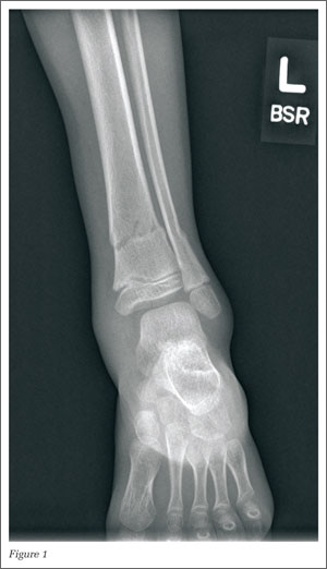
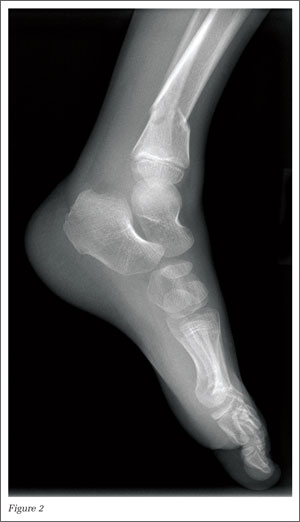
A 5-year-old boy presented to the ED after sustaining an injury to his left leg during play with a friend. He was unable to bear weight on the left foot and had a visible deformity to his lower extremity. The left foot was neurovascularly intact. Radiographs were completed (Figures 1 and 2).
What is your interpretation of the following radiographs?
Answer
|
|
The radiographs revealed a 20˚ anteriorly (apex posterior) angulated fracture through the metaphysis of the distal tibia and fibula. Angulated distal tibia fractures in adults are usually fixed with surgery; however, in children, displaced or angulated fractures to long bones such as the tibia stimulate a significant amount of growth. In treating pediatric patients, there is a greater amount of acceptable angulation the closer a fracture is to the end of bone.1
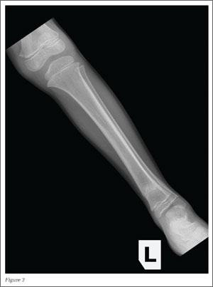
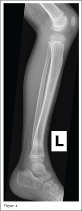
The patient in this case was placed in a long leg cast, and the fractures were reduced with three-point fixation technique. He remained in the cast for 5 weeks; thereafter, a below-the-knee orthopedic walking boot was placed for 3 weeks. The radiographs in Figures 3 and 4, taken 8 weeks after initiation of treatment, show a healed distal tibia and fibula fracture with an acceptable 7˚ of anterior angulation.
Dr Patterson, editor of “Challenges in Sports Medicine and Orthopedics,” is a sports medicine physician at Florida Sports Injury in Clermont, Florida. Dr Patterson is board certified in family medicine and spinal cord injury medicine, and is a member of the faculty of sports and exercise medicine of the Royal College of Surgeons in Ireland.
|
|
A 5-year-old boy presented to the ED after sustaining an injury to his left leg during play with a friend. He was unable to bear weight on the left foot and had a visible deformity to his lower extremity. The left foot was neurovascularly intact. Radiographs were completed (Figures 1 and 2).
What is your interpretation of the following radiographs?
Answer
|
|
The radiographs revealed a 20˚ anteriorly (apex posterior) angulated fracture through the metaphysis of the distal tibia and fibula. Angulated distal tibia fractures in adults are usually fixed with surgery; however, in children, displaced or angulated fractures to long bones such as the tibia stimulate a significant amount of growth. In treating pediatric patients, there is a greater amount of acceptable angulation the closer a fracture is to the end of bone.1
The patient in this case was placed in a long leg cast, and the fractures were reduced with three-point fixation technique. He remained in the cast for 5 weeks; thereafter, a below-the-knee orthopedic walking boot was placed for 3 weeks. The radiographs in Figures 3 and 4, taken 8 weeks after initiation of treatment, show a healed distal tibia and fibula fracture with an acceptable 7˚ of anterior angulation.
Challenges in Sports Medicine and Orthopedics
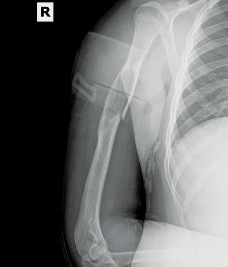
After playing football, an 18-year-old man presented to the ED with severe pain in his right arm. He stated that while throwing a pass, he felt a "snap" in his right arm and has been unable to move the arm since the injury. Radiographs were completed.

What is your interpretation of the radiographic image (Figure 1)?
Dr Patterson, editor of "Challenges in Sports Medicine and Orthopedics," is a sports medicine physician at Florida Sports Injury in Clermont, Florida. Dr Patterson is board certified in family medicine and spinal cord injury medicine, and is a member of the faculty of sports and exercise medicine of the Royal College of Surgeons in Ireland.
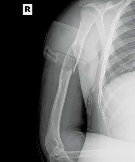
The radiograph (Figure 2) revealed a minimally displaced pathologic fracture (red arrow) through the midshaft of the humerus, which resulted from a space-occupying lesion (green arrow) in the humeral diaphysis. Since the radial nerve is commonly affected in this type of injury due to its close proximity to the humeral midshaft, careful neurologic assessment at the wrist and hand is essential. Injury to the nerve can occur during the fracture or reduction of the fracture, causing weakness in the extensors of the hand and numbness in the first dorsal web space. The incidence of radial nerve palsy in midshaft fractures of the humerus is 16%.1
In nondisplaced or minimally displaced fractures of the humeral midshaft, conservative management with a U-shaped (sugar-tong) splint from axilla to shoulder with elasticized wrap and sling is recommended. Surgical management is indicated in comminuted, significantly displaced, nonreducible, pathologic cases or in fractures resulting in neurovascular compromise. The patient in this case was referred to an orthopedic surgeon for open treatment.
- Smith WR, Agudelo JF, Parekh AA, Shank, JR. Musculoskeletal trauma surgery. In: Skinner HB, ed. Current Diagnosis & Treatment in Orthopedics. 4th ed. McGraw Hill Companies, Inc; 2006:121.
After playing football, an 18-year-old man presented to the ED with severe pain in his right arm. He stated that while throwing a pass, he felt a "snap" in his right arm and has been unable to move the arm since the injury. Radiographs were completed.
What is your interpretation of the radiographic image (Figure 1)?
Dr Patterson, editor of "Challenges in Sports Medicine and Orthopedics," is a sports medicine physician at Florida Sports Injury in Clermont, Florida. Dr Patterson is board certified in family medicine and spinal cord injury medicine, and is a member of the faculty of sports and exercise medicine of the Royal College of Surgeons in Ireland.
The radiograph (Figure 2) revealed a minimally displaced pathologic fracture (red arrow) through the midshaft of the humerus, which resulted from a space-occupying lesion (green arrow) in the humeral diaphysis. Since the radial nerve is commonly affected in this type of injury due to its close proximity to the humeral midshaft, careful neurologic assessment at the wrist and hand is essential. Injury to the nerve can occur during the fracture or reduction of the fracture, causing weakness in the extensors of the hand and numbness in the first dorsal web space. The incidence of radial nerve palsy in midshaft fractures of the humerus is 16%.1
In nondisplaced or minimally displaced fractures of the humeral midshaft, conservative management with a U-shaped (sugar-tong) splint from axilla to shoulder with elasticized wrap and sling is recommended. Surgical management is indicated in comminuted, significantly displaced, nonreducible, pathologic cases or in fractures resulting in neurovascular compromise. The patient in this case was referred to an orthopedic surgeon for open treatment.
- Smith WR, Agudelo JF, Parekh AA, Shank, JR. Musculoskeletal trauma surgery. In: Skinner HB, ed. Current Diagnosis & Treatment in Orthopedics. 4th ed. McGraw Hill Companies, Inc; 2006:121.
- Smith WR, Agudelo JF, Parekh AA, Shank, JR. Musculoskeletal trauma surgery. In: Skinner HB, ed. Current Diagnosis & Treatment in Orthopedics. 4th ed. McGraw Hill Companies, Inc; 2006:121.
Severe Left Shoulder Pain After a Fall
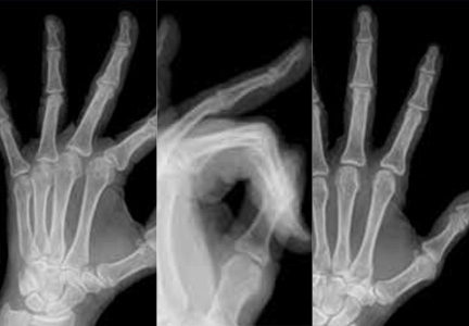
Flexion Deformity of the Fifth Digit
In the Hips, Increasing Pain and Decreasing Range of Motion
Athlete's Worsening Lower-Back Pain
Groin Pain in a Football Player
What is your interpretation of the radiographs, and how would treat the patient’s condition?
What is your interpretation of the radiographs, and how would treat the patient’s condition?
What is your interpretation of the radiographs, and how would treat the patient’s condition?