User login
A swollen knee
A 52-year-old African American homeless man visited a clinic at the local shelter, complaining of pain in his left knee for the past 2 days. He denied trauma or any history of knee injuries. He also denied any recent sexual activity. The patient was limping from significant pain. The patient’s clothes and hygiene were in poor condition. He worked in the fish market and had taken on the odor of his workplace. He admitted to a history of alcohol and substance abuse and was unwilling to discuss his psychiatric history. His chart indicated he had been diagnosed with schizophrenia.
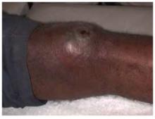
The patient was afebrile and had a swollen warm left knee (Figure 1). A crusty skin lesion with a small amount of purulence was seen over his patella. There was evidence of a joint effusion and the skin appeared red around the whole knee region. The patient could not fully flex his knee. The physical exam demonstrated normal ligamentous stability.
What procedure would be appropriate to perform?
What diagnostic tests would be helpful?
We offered to take the patient to the county hospital to obtain the appropriate diagnostic procedure and to initiate inpatient care. He adamantly refused treatment in a hospital, stating he was “too busy” to go the hospital and he only wanted “some pills.”
What else could you now do to care for this patient?
The differential diagnosis includes septic arthritis (such as staphylococcal and gonococcal infections of the knee joint), inflammatory types of arthritis, septic bursitis, cellulitis, and impetigo. Joint aspiration was performed in the clinic because the patient refused to go to the emergency room for diagnosis and treatment.
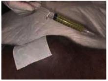
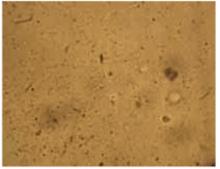
As seen in Figure 2, 10 mL of clear viscous yellow fluid was aspirated from the affected joint. Nine mL of aspirate was sent to a clinical laboratory for cell count, Gram stain, culture and sensitivity, crystal analysis, and microscopic examination. A drop of aspirate was placed on a wet mount and examined with plain light microscopy (Figure 3).
FIGURE 2
10 mL of clear viscous yellow fluid was aspirated from the joint.
Numerous refractile needle-shaped crystals were visualized.
Epidemiology of acute gout
Acute gout is predominantly a disease of the lower extremity, but any joint of any extremity may be involved. Ninety percent of patients experience acute attacks in the great toe at some time during the course of their disease. Next in order of frequency are the insteps, ankles, heels, knees, wrists, fingers, and elbows. The incidence of gout varies in populations from 0.20 to 0.35 per thousand per year, with an overall prevalence of 1.6 to 13.6 per thousand. The prevalence seems to increase substantially with age and increasing serum urate concentration.1 Prevalence is 3/100,000 for those 18 to 44 years of age; 21/100,000 for those 45 to 64, and 35/100,000 for those over 65. Men are 20 times more likely to have gout than women.2
Diagnosis
Most cases of gout are characterized by rapid onset of monoarticular arthritis resulting from deposition of urate crystals and a subsequent inflammatory response. The joint most commonly affected by gout is the first metatarsal phalangeal joint; the knee is the second most commonly involved joint. The diagnosis of acute gout was made based on the presence of the characteristic clinical history with urate crystals in the joint fluid.3
Management
Three treatments are available for patients suffering from acute gouty arthritis.
Colchicine inhibits microtubule formation and thus interferes with phagocytosis of the crystals, attenuating the inflammatory response. It also inhibits the release of chemotactic factors reducing the migration of neutrophils into the joint. In a randomized controlled trial, two thirds of patients treated with colchicine improved after 48 hours, but only one third of the patients receiving placebo demonstrated similar improvement. Improvement occurred earlier in the colchicine-treated patients. There were significant differences compared with placebo after 18–30 hours. All patients given colchicine (mean dose of 6.7 mg) developed diarrhea after a median time of 24 hours. Diarrhea occurred before relief of pain in most patients.4
The principal side effects of colchicine are gastrointestinal symptoms including abdominal pain and diarrhea. The dose associated with these symptoms is very close to the therapeutic dose.
Generally the initial dose is 1 mg, with 0.5 mg added every 2 hours until a total dose of up to 8 mg has been reached, or abdominal symptoms develop3 (Level of evidence, 1b; single RCT).4
Another option for treatment of acute gout is nonsteroidal anti-inflammatory agents. Indomethacin has been the standard for years but there is no proof that it is better than other NSAIDs5-8 (Level of evidence, 1b; a set of RCTs). The starting dose is 50 mg tid, tapered over approximately 1 week as symptoms subside. NSAIDs are also limited by their gastrointestinal and renal side effects.
Intra-articular injection of corticosteroids is an additional treatment for acute gout. The dose given depends on the size of the affected joint. The appropriate dose of methylprednisolone would be 5–10 mg for a small joint and 20–60 mg for a large joint such as the knee1 (Level of evidence, 5; expert opinion).
The patient’s treatment and outcome
The patient had a history of gastric ulcers and intolerance to NSAIDs. Therefore, he was started on oral colchicine for the probable gout. He was also given 500 mg of cephalexin po, qid, for the cellulitis and impetigo. He refused any blood tests, but accepted a follow-up appointment for the next day.
When we thought the patient had a septic joint, the optimal treatment would have involved hospitalization. The patient’s fear of hospitalization and losing his job made the choice of hospitalization a nonoption for this mentally ill homeless man. Instead of doing nothing, we began the diagnostic process in the outpatient setting.
If we had obtained purulent joint fluid it would have been incumbent upon us to press once again for hospitalization for septic arthritis. The appearance of yellow fluid and joint crystals gave us the option of attempting outpatient care.
Unfortunately, the patient never returned. Joint fluid Gram stain and culture results were negative, and the cell counts were consistent with inflammation. The hospital lab read the crystal analysis as negative even though our photograph documents the presence of crystals consistent with urate crystals.
Health care delivery must be flexible and creative in the setting of a shelter clinic. We can only hope that the oral medications he received allowed him full recovery from his acute illnesses.
1. Rudy D, Kurowski K. Family Medicine (House Officer Series). Philadelphia, Pa: Lippincott, Williams & Wilkins;1997.
2. Dambro M. Griffith’s 5 Minute Clinical Consult. 10th ed. Philadelphia, Pa: Lippincott, Williams & Wilkins;2002.
3. Emmerson BT. The management of gout. N Engl J Med 1996;334:445-51. Review.
4. Ahern MJ, Reid C, Gordon TP, et al. Does colchicine work? The results of the first controlled study in acute gout. Aust N Z J Med 1987;17:301-4.
5. Shrestha M, Morgan DL, Moreden JM, et al. Randomized double-blind comparison of the analgesic efficacy of intramuscular ketorolac and oral indomethacin in the treatment of acute gouty arthritis. Ann Emerg Med 1995;26:682-6.
6. Maccagno A, Di Giorgio E, Romanowicz A. Effectiveness of etodolac (‘Lodine’) compared with naproxen in patients with acute gout. Curr Med Res Opin 1991;12:423-9.
7. Altman RD, Honig S, Levin JM, Lightfoot RW. Ketoprofen versus indomethacin in patients with acute gouty arthritis: a multicenter, double blind comparative study. J Rheumatol 1988;15:1422-6.
8. Fraser RC, Davis RH, Walker FS. Comparative trial of azapropazone and indomethacin plus allopurinol in acute gout and hyperuricaemia. J R Coll Gen Pract 1987;37:409-11.
A 52-year-old African American homeless man visited a clinic at the local shelter, complaining of pain in his left knee for the past 2 days. He denied trauma or any history of knee injuries. He also denied any recent sexual activity. The patient was limping from significant pain. The patient’s clothes and hygiene were in poor condition. He worked in the fish market and had taken on the odor of his workplace. He admitted to a history of alcohol and substance abuse and was unwilling to discuss his psychiatric history. His chart indicated he had been diagnosed with schizophrenia.
The patient was afebrile and had a swollen warm left knee (Figure 1). A crusty skin lesion with a small amount of purulence was seen over his patella. There was evidence of a joint effusion and the skin appeared red around the whole knee region. The patient could not fully flex his knee. The physical exam demonstrated normal ligamentous stability.
What procedure would be appropriate to perform?
What diagnostic tests would be helpful?
We offered to take the patient to the county hospital to obtain the appropriate diagnostic procedure and to initiate inpatient care. He adamantly refused treatment in a hospital, stating he was “too busy” to go the hospital and he only wanted “some pills.”
What else could you now do to care for this patient?
The differential diagnosis includes septic arthritis (such as staphylococcal and gonococcal infections of the knee joint), inflammatory types of arthritis, septic bursitis, cellulitis, and impetigo. Joint aspiration was performed in the clinic because the patient refused to go to the emergency room for diagnosis and treatment.
As seen in Figure 2, 10 mL of clear viscous yellow fluid was aspirated from the affected joint. Nine mL of aspirate was sent to a clinical laboratory for cell count, Gram stain, culture and sensitivity, crystal analysis, and microscopic examination. A drop of aspirate was placed on a wet mount and examined with plain light microscopy (Figure 3).
FIGURE 2
10 mL of clear viscous yellow fluid was aspirated from the joint.
Numerous refractile needle-shaped crystals were visualized.
Epidemiology of acute gout
Acute gout is predominantly a disease of the lower extremity, but any joint of any extremity may be involved. Ninety percent of patients experience acute attacks in the great toe at some time during the course of their disease. Next in order of frequency are the insteps, ankles, heels, knees, wrists, fingers, and elbows. The incidence of gout varies in populations from 0.20 to 0.35 per thousand per year, with an overall prevalence of 1.6 to 13.6 per thousand. The prevalence seems to increase substantially with age and increasing serum urate concentration.1 Prevalence is 3/100,000 for those 18 to 44 years of age; 21/100,000 for those 45 to 64, and 35/100,000 for those over 65. Men are 20 times more likely to have gout than women.2
Diagnosis
Most cases of gout are characterized by rapid onset of monoarticular arthritis resulting from deposition of urate crystals and a subsequent inflammatory response. The joint most commonly affected by gout is the first metatarsal phalangeal joint; the knee is the second most commonly involved joint. The diagnosis of acute gout was made based on the presence of the characteristic clinical history with urate crystals in the joint fluid.3
Management
Three treatments are available for patients suffering from acute gouty arthritis.
Colchicine inhibits microtubule formation and thus interferes with phagocytosis of the crystals, attenuating the inflammatory response. It also inhibits the release of chemotactic factors reducing the migration of neutrophils into the joint. In a randomized controlled trial, two thirds of patients treated with colchicine improved after 48 hours, but only one third of the patients receiving placebo demonstrated similar improvement. Improvement occurred earlier in the colchicine-treated patients. There were significant differences compared with placebo after 18–30 hours. All patients given colchicine (mean dose of 6.7 mg) developed diarrhea after a median time of 24 hours. Diarrhea occurred before relief of pain in most patients.4
The principal side effects of colchicine are gastrointestinal symptoms including abdominal pain and diarrhea. The dose associated with these symptoms is very close to the therapeutic dose.
Generally the initial dose is 1 mg, with 0.5 mg added every 2 hours until a total dose of up to 8 mg has been reached, or abdominal symptoms develop3 (Level of evidence, 1b; single RCT).4
Another option for treatment of acute gout is nonsteroidal anti-inflammatory agents. Indomethacin has been the standard for years but there is no proof that it is better than other NSAIDs5-8 (Level of evidence, 1b; a set of RCTs). The starting dose is 50 mg tid, tapered over approximately 1 week as symptoms subside. NSAIDs are also limited by their gastrointestinal and renal side effects.
Intra-articular injection of corticosteroids is an additional treatment for acute gout. The dose given depends on the size of the affected joint. The appropriate dose of methylprednisolone would be 5–10 mg for a small joint and 20–60 mg for a large joint such as the knee1 (Level of evidence, 5; expert opinion).
The patient’s treatment and outcome
The patient had a history of gastric ulcers and intolerance to NSAIDs. Therefore, he was started on oral colchicine for the probable gout. He was also given 500 mg of cephalexin po, qid, for the cellulitis and impetigo. He refused any blood tests, but accepted a follow-up appointment for the next day.
When we thought the patient had a septic joint, the optimal treatment would have involved hospitalization. The patient’s fear of hospitalization and losing his job made the choice of hospitalization a nonoption for this mentally ill homeless man. Instead of doing nothing, we began the diagnostic process in the outpatient setting.
If we had obtained purulent joint fluid it would have been incumbent upon us to press once again for hospitalization for septic arthritis. The appearance of yellow fluid and joint crystals gave us the option of attempting outpatient care.
Unfortunately, the patient never returned. Joint fluid Gram stain and culture results were negative, and the cell counts were consistent with inflammation. The hospital lab read the crystal analysis as negative even though our photograph documents the presence of crystals consistent with urate crystals.
Health care delivery must be flexible and creative in the setting of a shelter clinic. We can only hope that the oral medications he received allowed him full recovery from his acute illnesses.
1. Rudy D, Kurowski K. Family Medicine (House Officer Series). Philadelphia, Pa: Lippincott, Williams & Wilkins;1997.
2. Dambro M. Griffith’s 5 Minute Clinical Consult. 10th ed. Philadelphia, Pa: Lippincott, Williams & Wilkins;2002.
3. Emmerson BT. The management of gout. N Engl J Med 1996;334:445-51. Review.
4. Ahern MJ, Reid C, Gordon TP, et al. Does colchicine work? The results of the first controlled study in acute gout. Aust N Z J Med 1987;17:301-4.
5. Shrestha M, Morgan DL, Moreden JM, et al. Randomized double-blind comparison of the analgesic efficacy of intramuscular ketorolac and oral indomethacin in the treatment of acute gouty arthritis. Ann Emerg Med 1995;26:682-6.
6. Maccagno A, Di Giorgio E, Romanowicz A. Effectiveness of etodolac (‘Lodine’) compared with naproxen in patients with acute gout. Curr Med Res Opin 1991;12:423-9.
7. Altman RD, Honig S, Levin JM, Lightfoot RW. Ketoprofen versus indomethacin in patients with acute gouty arthritis: a multicenter, double blind comparative study. J Rheumatol 1988;15:1422-6.
8. Fraser RC, Davis RH, Walker FS. Comparative trial of azapropazone and indomethacin plus allopurinol in acute gout and hyperuricaemia. J R Coll Gen Pract 1987;37:409-11.


