User login
VIDEO: Cell-free DNA mutations linked with recurrence of ovarian cancer
NATIONAL HARBOR, MD. – Next-generation sequencing of high-grade serous ovarian tumor specimens taken during interval debulking identified several mutations that matched those in cell-free DNA (cfDNA), Rebecca C. Arend, MD, said at the annual meeting of the Society of Gynecologic Oncology.
Furthermore, three of four patients whose ovarian cancer recurred had mutations in cfDNA that were previously detected in tumors, said Dr. Arend of the University of Alabama at Birmingham.
Researchers continue to search for ways to spare patients with ovarian cancer from serial biopsies, Dr. Arend noted during a video interview. As part of that work, she and her associates performed longitudinal next-generation sequencing of 50 genes in tumor and plasma specimens from 14 patients with high-grade serous ovarian cancer.
Mutations found only in tumors were relatively consistent before and after neoadjuvant chemotherapy, while mutations found only in cell-free DNA varied substantially.
The researchers also sequenced plasma samples from four patients at cancer recurrence. Three patients had at least one mutation that was previously detected in their tumor sample. Implicated genes included PIK3CA, TP53, KIT, and KDR.
Many studies have sought circulating tumor markers that reliably predict tumor recurrence. When it comes to cfDNA, “we are not there yet,” but the work is worthwhile, Dr. Arend stressed. Sequencing tumor and cfDNA specimens from patients at multiple points during their journey might one day help pinpoint mutations that reliably predict cancer recurrence, sparing patients from repeated biopsies and helping them efficiently enter clinical trials that target their specific mutation, she added.
Dr. Arend cited no funding sources and reported having no conflicts of interest.
The video associated with this article is no longer available on this site. Please view all of our videos on the MDedge YouTube channel
AT THE ANNUAL MEETING ON WOMEN’S CANCER
Key clinical point: Next-generation sequencing of cell-free DNA specimens (liquid biopsies) might one day help identify tumor recurrence in patients with high-grade serous ovarian cancer.
Major finding: Three of four patients whose cancer recurred had mutations in cell-free DNA that were previously detected in tumor specimens collected during interval debulking.
Data source: Next-generation sequencing of tumor and cell-free (plasma) DNA from 14 patients with high-grade serous ovarian cancer before and after platinum-based neoadjuvant chemotherapy.
Disclosures: Dr. Arend cited no funding sources and reported having no conflicts of interest.

VIDEO: No disease progression in endometrial cancer patients with isolated tumor cells
AT THE ANNUAL MEETING ON WOMEN’S CANCER
NATIONAL HARBOR, MD. – Patients with endometrial cancer and isolated tumor cells in sentinel lymph nodes had excellent short-term outcomes, even if they opted out of adjuvant chemotherapy or radiation therapy.
In a single-center prospective study, all 10 such patients remained alive and free of disease progression at 3 years, Marie Plante, MD, said in a video interview at the annual meeting of the Society of Gynecologic Oncology.
The finding suggests that isolated tumor cells in sentinel lymph nodes are not, by themselves, an indication for adjuvant therapy in women with endometrial cancer, said Dr. Plante of Laval University in Quebec City. The issue remains controversial, however, and merits larger cohort studies with longer-term follow-up, she acknowledged.
Ultrastaging of sentinel lymph node biopsies that are negative on hematoxylin and eosin staining has boosted the detection of low-volume metastases. In the case of endometrial cancer with isolated tumor cells, there is a dilemma about whether to recommend adjuvant therapy. Very few studies have examined this relatively rare patient subgroup.
This study included 519 patients undergoing hysterectomy, salpingo-oophorectomy, lymphadenectomy, and sentinel lymph node mapping for endometrial cancer. Pathologic ultrastaging identified 31 patients with isolated tumor cells (6%), of whom 11 received adjuvant chemotherapy, 14 received pelvic radiation therapy, and 10 received brachytherapy or observation only.
Only one patient with isolated tumor cells developed recurrent disease within 3 years. This patient had a 7-cm carcinosarcoma that recurred despite adjuvant chemotherapy and radiation therapy. There were no recurrences among patients with endometrioid histology, Dr. Plante noted.
Dr. Plante cited no funding sources and reported having no conflicts of interest.
The video associated with this article is no longer available on this site. Please view all of our videos on the MDedge YouTube channel
AT THE ANNUAL MEETING ON WOMEN’S CANCER
NATIONAL HARBOR, MD. – Patients with endometrial cancer and isolated tumor cells in sentinel lymph nodes had excellent short-term outcomes, even if they opted out of adjuvant chemotherapy or radiation therapy.
In a single-center prospective study, all 10 such patients remained alive and free of disease progression at 3 years, Marie Plante, MD, said in a video interview at the annual meeting of the Society of Gynecologic Oncology.
The finding suggests that isolated tumor cells in sentinel lymph nodes are not, by themselves, an indication for adjuvant therapy in women with endometrial cancer, said Dr. Plante of Laval University in Quebec City. The issue remains controversial, however, and merits larger cohort studies with longer-term follow-up, she acknowledged.
Ultrastaging of sentinel lymph node biopsies that are negative on hematoxylin and eosin staining has boosted the detection of low-volume metastases. In the case of endometrial cancer with isolated tumor cells, there is a dilemma about whether to recommend adjuvant therapy. Very few studies have examined this relatively rare patient subgroup.
This study included 519 patients undergoing hysterectomy, salpingo-oophorectomy, lymphadenectomy, and sentinel lymph node mapping for endometrial cancer. Pathologic ultrastaging identified 31 patients with isolated tumor cells (6%), of whom 11 received adjuvant chemotherapy, 14 received pelvic radiation therapy, and 10 received brachytherapy or observation only.
Only one patient with isolated tumor cells developed recurrent disease within 3 years. This patient had a 7-cm carcinosarcoma that recurred despite adjuvant chemotherapy and radiation therapy. There were no recurrences among patients with endometrioid histology, Dr. Plante noted.
Dr. Plante cited no funding sources and reported having no conflicts of interest.
The video associated with this article is no longer available on this site. Please view all of our videos on the MDedge YouTube channel
Key clinical point: Isolated tumor cells in sentinel lymph nodes did not signal disease progression or death among patients with low-risk endometrial cancer.
Major finding: After 3 years of follow-up, the rate of progression-free survival was 100% among patients who opted out of adjuvant chemotherapy or radiation therapy.
Data source: A single-center prospective study of 519 patients undergoing hysterectomy, salpingo-oophorectomy, lymphadenectomy, and sentinel lymph node mapping for endometrial cancer, including 31 patients with isolated tumor cells identified in sentinel lymph nodes.
Disclosures: Dr. Plante cited no funding sources and had no conflicts of interest.
Open-capsule PPIs linked to faster ulcer healing after Roux-en-Y
The use of proton pump inhibitors in opened instead of closed capsules was associated with a nearly fourfold shorter median healing time among patients who developed marginal ulcers after Roux-en-Y gastric bypass, in a single-center retrospective cohort study.
In contrast, the specific class of proton pump inhibitor (PPI) did not affect healing times, wrote Allison R. Schulman, MD, and her associates at Brigham and Women’s Hospital, Boston. The report is in the April issue of Clinical Gastroenterology and Hepatology (doi: 10.1016/j.cgh.2016.10.015). “Given these results and the high prevalence of marginal ulceration in this patient population, further study in a randomized controlled setting is warranted, and use of open-capsule PPIs should be considered as a low-risk, low-cost alternative,” they added.
Roux-en-Y gastric bypass is one of the most common types of gastric bypass surgeries in the world, and up to 16% of patients develop postsurgical ulcers at the gastrojejunal anastomosis, the investigators noted. Acidity is a prime suspect in these “marginal ulcerations” because bypassing the acid-buffering duodenum exposes the jejunum to acid from the stomach, they added. High-dose PPIs are the main treatment, but there is no consensus on the formulation or dose of therapy. Because Roux-en-Y creates a small gastric pouch and hastens small-bowel transit, closed capsules designed to break down in the stomach “even may make their way to the colon before breakdown occurs,” they wrote.
They reviewed medical charts from patients who developed marginal ulcerations after undergoing Roux-en-Y gastric bypass at their hospital from 2000 through 2015. A total of 115 patients received open-capsule PPIs and 49 received intact capsules. All were followed until their ulcers healed.
For the open-capsule group, median time to healing was 91 days, compared with 342 days for the closed-capsule group (P less than .001). Importantly, capsule type was the only independent predictor of healing time (hazard ratio, 6.0; 95% confidence interval, 3.7 to 9.8; P less than .001) in a Cox regression model that included other known correlates of ulcer healing, including age, smoking status, the use of nonsteroidal anti-inflammatory drugs, Helicobacter pylori infection, the length of the gastric pouch, and the presence of fistulae or foreign bodies such as sutures or staples.
The use of sucralfate also did not affect time to ulcer healing, reflecting “many previous studies showing a lack of definitive benefit to this medication,” the researchers said. The findings have “tremendous implications” for health care utilization, they added. Indeed, patients who received open-capsule PPIs needed significantly fewer endoscopic procedures (median, 1.2 versus 1.8; P = .02) and used fewer health care resources overall ($7,206 versus $11,009; P = .05) compared with those prescribed intact PPI capsules.
This study was limited to patients who developed ulcer symptoms and underwent repeated surveillance endoscopies after surgery, the researchers noted. Selection bias is always a concern with retrospective studies, but insurers always covered both types of therapy and the choice of capsule type was entirely up to providers, all of whom consistently prescribed either open- or closed-capsule PPI therapy, they added.
The investigators did not acknowledge external funding sources. Dr. Schulman and four coinvestigators reported having no competing interests. One coinvestigator disclosed ties to Olympus, Boston Scientific, and Covidien.
Proton pump inhibitors (PPIs) are frequently employed to treat marginal ulcers after Roux-en-Y gastric bypass (RYGB). In a retrospective study, Schulman et al. compared intact vs. “open” PPI capsules.

They state that “this may be overcome by use of a soluble form of PPI,” but don’t state what is meant by “soluble PPI” or how the open-capsule PPI was delivered. Among the PPIs they reported using to compare intact vs. open capsules was Protonix [pantoprazole] which is not produced as a capsule, and soluble Prevacid [lansoprazole], which is an orally disintegrating tablet that should provide characteristics similar to an “open capsule.”
PPI capsules provide PPI in enteric-coated granules, which are designed to protect the PPI from acid degradation in the stomach of individuals with intact gastrointestinal tracts and allow more of the PPI dose to reach the small intestine where it is absorbed. If capsules really fail to release their enteric-coated granules until very distally in RYGB patients, bypassing this step to allow earlier release of PPI makes intuitive sense; formulations such as suspensions and rapidly disintegrating tablets that deliver enteric-coated granules without capsules are currently available.
However, if this is an issue, administering a suspension of uncoated PPI with bicarbonate potentially might be the most attractive option, given more rapid absorption than PPI delivered as enteric-coated granules.
Loren Laine, MD, AGAF, professor of medicine, digestive diseases, Yale University, New Haven, Conn. He has no conflicts of interest.
Proton pump inhibitors (PPIs) are frequently employed to treat marginal ulcers after Roux-en-Y gastric bypass (RYGB). In a retrospective study, Schulman et al. compared intact vs. “open” PPI capsules.
They state that “this may be overcome by use of a soluble form of PPI,” but don’t state what is meant by “soluble PPI” or how the open-capsule PPI was delivered. Among the PPIs they reported using to compare intact vs. open capsules was Protonix [pantoprazole] which is not produced as a capsule, and soluble Prevacid [lansoprazole], which is an orally disintegrating tablet that should provide characteristics similar to an “open capsule.”
PPI capsules provide PPI in enteric-coated granules, which are designed to protect the PPI from acid degradation in the stomach of individuals with intact gastrointestinal tracts and allow more of the PPI dose to reach the small intestine where it is absorbed. If capsules really fail to release their enteric-coated granules until very distally in RYGB patients, bypassing this step to allow earlier release of PPI makes intuitive sense; formulations such as suspensions and rapidly disintegrating tablets that deliver enteric-coated granules without capsules are currently available.
However, if this is an issue, administering a suspension of uncoated PPI with bicarbonate potentially might be the most attractive option, given more rapid absorption than PPI delivered as enteric-coated granules.
Loren Laine, MD, AGAF, professor of medicine, digestive diseases, Yale University, New Haven, Conn. He has no conflicts of interest.
The use of proton pump inhibitors in opened instead of closed capsules was associated with a nearly fourfold shorter median healing time among patients who developed marginal ulcers after Roux-en-Y gastric bypass, in a single-center retrospective cohort study.
In contrast, the specific class of proton pump inhibitor (PPI) did not affect healing times, wrote Allison R. Schulman, MD, and her associates at Brigham and Women’s Hospital, Boston. The report is in the April issue of Clinical Gastroenterology and Hepatology (doi: 10.1016/j.cgh.2016.10.015). “Given these results and the high prevalence of marginal ulceration in this patient population, further study in a randomized controlled setting is warranted, and use of open-capsule PPIs should be considered as a low-risk, low-cost alternative,” they added.
Roux-en-Y gastric bypass is one of the most common types of gastric bypass surgeries in the world, and up to 16% of patients develop postsurgical ulcers at the gastrojejunal anastomosis, the investigators noted. Acidity is a prime suspect in these “marginal ulcerations” because bypassing the acid-buffering duodenum exposes the jejunum to acid from the stomach, they added. High-dose PPIs are the main treatment, but there is no consensus on the formulation or dose of therapy. Because Roux-en-Y creates a small gastric pouch and hastens small-bowel transit, closed capsules designed to break down in the stomach “even may make their way to the colon before breakdown occurs,” they wrote.
They reviewed medical charts from patients who developed marginal ulcerations after undergoing Roux-en-Y gastric bypass at their hospital from 2000 through 2015. A total of 115 patients received open-capsule PPIs and 49 received intact capsules. All were followed until their ulcers healed.
For the open-capsule group, median time to healing was 91 days, compared with 342 days for the closed-capsule group (P less than .001). Importantly, capsule type was the only independent predictor of healing time (hazard ratio, 6.0; 95% confidence interval, 3.7 to 9.8; P less than .001) in a Cox regression model that included other known correlates of ulcer healing, including age, smoking status, the use of nonsteroidal anti-inflammatory drugs, Helicobacter pylori infection, the length of the gastric pouch, and the presence of fistulae or foreign bodies such as sutures or staples.
The use of sucralfate also did not affect time to ulcer healing, reflecting “many previous studies showing a lack of definitive benefit to this medication,” the researchers said. The findings have “tremendous implications” for health care utilization, they added. Indeed, patients who received open-capsule PPIs needed significantly fewer endoscopic procedures (median, 1.2 versus 1.8; P = .02) and used fewer health care resources overall ($7,206 versus $11,009; P = .05) compared with those prescribed intact PPI capsules.
This study was limited to patients who developed ulcer symptoms and underwent repeated surveillance endoscopies after surgery, the researchers noted. Selection bias is always a concern with retrospective studies, but insurers always covered both types of therapy and the choice of capsule type was entirely up to providers, all of whom consistently prescribed either open- or closed-capsule PPI therapy, they added.
The investigators did not acknowledge external funding sources. Dr. Schulman and four coinvestigators reported having no competing interests. One coinvestigator disclosed ties to Olympus, Boston Scientific, and Covidien.
FROM CLINICAL GASTROENTEROLOGY AND HEPATOLOGY
Key clinical point: The use of proton pump inhibitors in opened instead of closed capsules was associated with a nearly fourfold shorter median healing time among patients who developed ulcers at the gastrojejunal anastomosis after Roux-en-Y gastric bypass.
Major finding: The median time to ulcer healing was 91.0 versus 342.0 days for the open- and closed-capsule groups, respectively (P less than .001).
Data source: A single-center retrospective study of 162 patients.
Disclosures: The investigators did not acknowledge external funding sources. Dr. Schulman and four coinvestigators reported having no competing interests. One coinvestigator disclosed ties to Olympus, Boston Scientific, and Covidien.
In beta thalassemia major, liver stiffness declines with deferasirox
Patients with beta thalassemia major who were chelated only with oral deferasirox experienced significant improvements in liver stiffness, both from baseline and compared with patients who interrupted deferasirox therapy because of pregnancy, according to a small prospective 5-year study.
Transient elastography showed that continuous therapy with deferasirox (median dose, 35 mg per kg) yielded an 0.85 kPa average improvement in liver stiffness compared with baseline (P = .02), reported Nikolaos Sousos of Aristotle University of Thessaloniki, Greece, and his associates (Br J Haematol. 2017 Jan 20. doi: 10.1111/bjh.14509).
In contrast, interrupting therapy for a median of 16 months because of successful pregnancy led to an average increase in liver stiffness of 1.84 kPa – a significant difference between groups (P = .005) even after the researchers controlled for gender, age, ferritin levels, and T2-weighted magnetic resonance imaging (MRI) measurements of iron deposition in the liver and heart.
Patients with beta thalassemia major often develop liver fibrosis because of excessive intestinal iron absorption, iron overload from transfusions, or hepatitis C virus infection, the investigators noted. The median age of the patients in this study was 32 years (range, 20-47 years), they were HCV negative, and they received regular transfusions to maintain hemoglobin levels above 95 g/L. The seven female participants who temporarily stopped deferasirox because of pregnancy all restarted therapy at least 8 months before their follow-up transient elastography liver stiffness measurement, the investigators reported.
T2-weighted MRI measurements of liver iron concentration also had improved at follow-up, reflecting “better control of iron overload,” although the difference from baseline was not statistically significant, the investigators noted. This result reinforces previous findings (Gastroenterology. 2011;141[4]:1202-11) that long-term deferasirox therapy can significantly improve liver fibrosis in patients with beta thalassemia, regardless of liver iron concentration, the researchers added. Together, those findings suggest that “improvement in liver fibrosis, rather than liver iron concentration should be the primary effect of chelation therapy,” they suggested.
The Research Committee of Aristotle University of Thessaloniki, Novartis Hellas (Novartis AG, Basel, Switzerland), and the Greek Thalassaemia Association funded the study. The authors declared having no competing interests.
FROM THE BRITISH JOURNAL OF HAEMATOLOGY
Key clinical point. For patients with beta thalassemia major, long-term chelation with deferasirox led to significant improvements in liver stiffness.
Major finding: Five years of continuous therapy yielded an 0.85 kPa average improvement in liver stiffness compared with baseline (P = .02).
Data source: A 5-year, single-center prospective study of 22 patients.
Disclosures: The Research Committee of Aristotle University of Thessaloniki, Novartis Hellas (Novartis AG, Basel, Switzerland), and the Greek Thalassaemia Association funded the study. The authors declared having no competing interests.
VIDEO: Point-of-care assay caught acetaminophen toxicity
A rapid point-of-care assay for acetaminophen-related liver toxicity had a sensitivity of 100% and a specificity of 86%, compared with etiologic diagnosis, based on the results of a multicenter study published in the April issue of Clinical Gastroenterology and Hepatology.
The test might help guide treatment decisions for these patients in the emergency department and intensive care unit, said Dean W. Roberts, PhD, of the University of Arkansas, Little Rock, and his associates.
About 45% of acute liver failure cases in the United States stem from acetaminophen toxicity, but the diagnosis can be hard to confirm because the drug has a short half-life and patients often cannot or will not report an overdose, which also may consist of multiple exposures, limiting the interpretability of the Rumack nonogram. High-pressure liquid chromatography with electrochemical detection (HPLC-EC) accurately detects acetaminophen-protein adducts (3-[cysteine-S-yl] acetaminophen) released by lysed hepatocytes into the peripheral circulation, but this test requires specialized equipment and skilled personnel, the researchers noted (Clin Gastroenterol Hepatol. 2016 Sep 15. doi: 10.1016/j.cgh.2016.09.007).
The point-of-care assay was positive in all 33 patients diagnosed with acetaminophen toxicity, for a test sensitivity of 100%, the researchers reported. The median band amplitude for cases was 584 (range, 222-1,027), significantly lower than that for patients with nonacetaminophen acute liver failure (3,678; range, 394-8,289; P less than .001) or for controls (8,971; range, 5,151-11,108; P less than .001). Band amplitude correlated inversely with adduct levels because AcetaSTAT is a competitive immunoassay – the presence of adducts decreases reactions at the test band, the investigators reported.
AcetaSTAT results were negative for 25 of 29 patients who were initially diagnosed with nonacetaminophen liver failure, for a test specificity of 86%, a positive predictive value of 89%, and a negative predictive value of 100%. Among the remaining four “false positives,” three tested near or above the toxicity threshold on HPLC-EC and were considered positive after further review, the investigators said. The fourth false-positive case was HPLC-EC–negative autoimmune hepatitis.
AcetaSTAT might not catch cases very early after acetaminophen overdose or that have only mild toxicity, the researchers noted. Nonetheless, it can help guide treatment decisions “at the point of clinical care,” they said. “Because the survival rate of acetaminophen acute liver failure is more favorable than that of other causes of acute live failure, assay results could impact future physician referral patterns and reduce medical costs associated with additional tests to determine the etiology of liver injury.”
The National Institute of Diabetes and Digestive and Kidney Diseases funded the study. Dr. Roberts and two coinvestigators are part owners of Acetaminophen Toxicity Diagnostics and have submitted a patent application for the AcetaSTAT serum assay used in this study. There were no other disclosures.
Source: American Gastroenterological Association
FROM CLINICAL GASTROENTEROLOGY AND HEPATOLOGY
Key clinical point:
Major finding: Compared with etiologic diagnosis, its sensitivity was 100%, specificity was 86%, positive predictive value was 89%, and negative predictive value was 100%.
Data source: Competitive immunoassays of serum samples from 19 healthy controls, 29 patients with nonacetaminophen acute liver failure, and 33 patients with acetaminophen-induced acute liver failure.
Disclosures: The National Institute of Diabetes and Digestive and Kidney Diseases funded the study. Dr. Roberts and two coinvestigators are part owners of Acetaminophen Toxicity Diagnostics and have submitted a patent application for the AcetaSTAT serum assay used in this study. There were no other disclosures.
Unique, multi-omic profile found in children with autism and functional GI disorders
The gut microbiomes of children with autism spectrum disorder (ASD) and functional gastrointestinal disorders (FGID) had significantly higher levels of several Clostridium species and lower concentrations of other bacteria compared with neurotypical children with and without FGIDs, which correlated with increases in inflammatory cytokines, decreased tryptophan, and increased serotonin, according to a small, single-center, cross-sectional study.
This “unique multi-omic profile [was] specific to ASD-FGID and ASD-FGID with abdominal pain,” wrote Ruth Ann Luna, PhD, of Texas Children’s Microbiome Center at Texas Children’s Hospital, Houston, and her associates. The report was published online in Cellular and Molecular Gastroenterology and Hepatology (doi: 10.1016/j.jcmgh.2016.11.008).
Children with ASD are at increased risk for FGIDs such as functional constipation, nonretentive fecal incontinence, functional abdominal pain, abdominal migraines, and irritable bowel syndrome, compared with their neurotypical peers. Changes in the gut microbiome can affect immunologic pathways and the balance between tryptophan and serotonin. This altered “microbial-gut-brain axis” has been reported in both ASD and FGID, suggesting “that altered gut-brain communications not only may play a role in the increased occurrence of FGIDs in ASD individuals, but could advance our understanding of potential risk factors for FGID in the ASD community,” the researchers wrote.
Previous studies of stool specimens have found higher levels of several species of Clostridium in pediatric ASD compared with neurotypical children. To confirm and expand on that work, the investigators examined microbial and neuroimmune markers in rectal biopsies and blood specimens from 14 children with ASD-FGID, 15 neurotypical children with FGID, and 6 asymptomatic neurotypical children. Participants were recruited from Nationwide Children’s Hospital in Columbus, Ohio. The researchers quantified microbial 16S ribosomal DNA community signatures, cytokines, chemokines, and serotonergic metabolites, and correlated results with parental responses to the Questionnaire on Pediatric Gastrointestinal Symptoms–Rome III version.
The ASD-FGID group had significantly higher numbers for ribosomal DNA sequences for Clostridium lituseburense (P = .002), Lachnoclostridium bolteae (P = .02), Lachnoclostridium hathewayi (P = .03), Clostridium aldenense (P = .04), and Oscillospira plautii (P = .04), compared with neurotypical children with and without FGID. Children with ASD-FGID also had significantly lower levels of Dorea formicigenerans (P = .006), Blautia luti (P = .02), and Sutterella species (P = .03). “Overall, our identification of clostridial species aligns with previous autism studies that have identified microbiome alterations,” the researchers noted.
They also looked specifically at abdominal pain. Children with ASD-FGID and abdominal pain had significantly higher gut mucosal levels of Turicibacter sanguinis (P = .03), Clostridium aldenense (P = .004), Clostridium lituseburense (P = .003), Oscillospira plautii (P = .01), Clostridium disporicum (P = .049), and Clostridium tertium (P = .045) than did any other subgroup, the investigators found. Patients with both ASD-FGID and abdominal pain also had significantly higher levels of C. aldenense (P = .03), O. plautii (P = .04), Tyzzerella species (P = .045), and Parasutterella excrementihominis (P = .04) than did ASD-FGID patients without abdominal pain.
Both C. disporicum and C. tertium correlated with increases in the proinflammatory cytokines IL6 and interferon-gamma. Levels of these cytokines were highest in patients with ASD-FGID, and IL6 was highest of all among children with ASD-FGID with abdominal pain. Another proinflammatory cytokine, IL17A, also correlated with Clostridia species that were enriched in children with ASD-FGID. Both IL6 and IL17A have been implicated in autism-like phenotypes in rodents, the researchers noted. Several other cytokines also were linked to ASD-FGID, and abdominal pain correlated significantly with increases in MCP-1 (P = .03) and eotaxin (P = .03).
Gut mucosal levels of tryptophan were significantly lower among children with ASD-FGID compared with neurotypical children, either with (P = .006) or without (P = .009) FGID. In contrast, gut mucosal levels of 5-HIAA, the primary metabolite of serotonin, were significantly higher among children with ASD-FGID compared with neurotypical children (P = .01). Increased 5-HIAA also correlated significantly with abdominal pain (P = .04). Six species of bacteria correlated significantly with tryptophan or serotonin, implicating the gut microbiome in the serotonin pathway.
“Although these initial findings are correlative, these data form the framework for future studies targeting tryptophan-serotonin metabolism and inflammatory pathways in FGID in ASD,” the researchers concluded.
The U.S. Department of Health and Human Services funded the work. The investigators had no relevant disclosures.
Autism-spectrum disorder is a serious and increasingly prevalent developmental behavior disorder often accompanied and aggravated by a range of gastrointestinal and cognitive dysfunctions. Its etiology probably involves maternal diet and inflammatory events that alter central nervous system neurodevelopment critical to the cognition of social interaction. Candidate causal products of these events include the cytokines IL-6 and IL-17A, and certain bioactive amines, notably serotonin. Functional gastrointestinal disorders share these same molecules as biomarkers and disease modifiers, probably elicited in part by the intestinal microbiome. Hence, the comorbidity in ASD suggests these two disease processes are etiologically related.
The study by Luna and colleagues tightens the case for a microbial hub and serotonin and cytokine spokes in the gastrointestinal dysfunction of ASD: elevated mucosal tissue levels of select microbial taxa, mainly members of the genus Clostridium, and mucosal production of cytokines and serotonin-pathway bioamines associated with these and other select microbial species. Important and challenging questions loom ahead. What are the direct mucosal cell types and functions targeted of this network for the microbiota, and via what microbial products? Might they elicit epithelial or mucosal hematopoietic cell cytokine production that in turn causes mucosal bioamine secretion? And, what associated microbiota and products are just secondarily altered and not causally involved? The exciting study of Luna and colleagues raises confidence for this path ahead, and its promise for clarifying ASD pathogenesis and uncovering targetable elements for intervention.
Jonathan Braun, MD, PhD, is professor and chair of pathology and laboratory medicine, UCLA David Geffen School of Medicine, UCLA Health System, Los Angeles. He has no conflicts of interest.
The gut microbiomes of children with autism spectrum disorder (ASD) and functional gastrointestinal disorders (FGID) had significantly higher levels of several Clostridium species and lower concentrations of other bacteria compared with neurotypical children with and without FGIDs, which correlated with increases in inflammatory cytokines, decreased tryptophan, and increased serotonin, according to a small, single-center, cross-sectional study.
This “unique multi-omic profile [was] specific to ASD-FGID and ASD-FGID with abdominal pain,” wrote Ruth Ann Luna, PhD, of Texas Children’s Microbiome Center at Texas Children’s Hospital, Houston, and her associates. The report was published online in Cellular and Molecular Gastroenterology and Hepatology (doi: 10.1016/j.jcmgh.2016.11.008).
Children with ASD are at increased risk for FGIDs such as functional constipation, nonretentive fecal incontinence, functional abdominal pain, abdominal migraines, and irritable bowel syndrome, compared with their neurotypical peers. Changes in the gut microbiome can affect immunologic pathways and the balance between tryptophan and serotonin. This altered “microbial-gut-brain axis” has been reported in both ASD and FGID, suggesting “that altered gut-brain communications not only may play a role in the increased occurrence of FGIDs in ASD individuals, but could advance our understanding of potential risk factors for FGID in the ASD community,” the researchers wrote.
Previous studies of stool specimens have found higher levels of several species of Clostridium in pediatric ASD compared with neurotypical children. To confirm and expand on that work, the investigators examined microbial and neuroimmune markers in rectal biopsies and blood specimens from 14 children with ASD-FGID, 15 neurotypical children with FGID, and 6 asymptomatic neurotypical children. Participants were recruited from Nationwide Children’s Hospital in Columbus, Ohio. The researchers quantified microbial 16S ribosomal DNA community signatures, cytokines, chemokines, and serotonergic metabolites, and correlated results with parental responses to the Questionnaire on Pediatric Gastrointestinal Symptoms–Rome III version.
The ASD-FGID group had significantly higher numbers for ribosomal DNA sequences for Clostridium lituseburense (P = .002), Lachnoclostridium bolteae (P = .02), Lachnoclostridium hathewayi (P = .03), Clostridium aldenense (P = .04), and Oscillospira plautii (P = .04), compared with neurotypical children with and without FGID. Children with ASD-FGID also had significantly lower levels of Dorea formicigenerans (P = .006), Blautia luti (P = .02), and Sutterella species (P = .03). “Overall, our identification of clostridial species aligns with previous autism studies that have identified microbiome alterations,” the researchers noted.
They also looked specifically at abdominal pain. Children with ASD-FGID and abdominal pain had significantly higher gut mucosal levels of Turicibacter sanguinis (P = .03), Clostridium aldenense (P = .004), Clostridium lituseburense (P = .003), Oscillospira plautii (P = .01), Clostridium disporicum (P = .049), and Clostridium tertium (P = .045) than did any other subgroup, the investigators found. Patients with both ASD-FGID and abdominal pain also had significantly higher levels of C. aldenense (P = .03), O. plautii (P = .04), Tyzzerella species (P = .045), and Parasutterella excrementihominis (P = .04) than did ASD-FGID patients without abdominal pain.
Both C. disporicum and C. tertium correlated with increases in the proinflammatory cytokines IL6 and interferon-gamma. Levels of these cytokines were highest in patients with ASD-FGID, and IL6 was highest of all among children with ASD-FGID with abdominal pain. Another proinflammatory cytokine, IL17A, also correlated with Clostridia species that were enriched in children with ASD-FGID. Both IL6 and IL17A have been implicated in autism-like phenotypes in rodents, the researchers noted. Several other cytokines also were linked to ASD-FGID, and abdominal pain correlated significantly with increases in MCP-1 (P = .03) and eotaxin (P = .03).
Gut mucosal levels of tryptophan were significantly lower among children with ASD-FGID compared with neurotypical children, either with (P = .006) or without (P = .009) FGID. In contrast, gut mucosal levels of 5-HIAA, the primary metabolite of serotonin, were significantly higher among children with ASD-FGID compared with neurotypical children (P = .01). Increased 5-HIAA also correlated significantly with abdominal pain (P = .04). Six species of bacteria correlated significantly with tryptophan or serotonin, implicating the gut microbiome in the serotonin pathway.
“Although these initial findings are correlative, these data form the framework for future studies targeting tryptophan-serotonin metabolism and inflammatory pathways in FGID in ASD,” the researchers concluded.
The U.S. Department of Health and Human Services funded the work. The investigators had no relevant disclosures.
FROM CELLULAR AND MOLECULAR GASTROENTEROLOGY AND HEPATOLOGY
Key clinical point: The mucosal microbiome of children with comorbid autism spectrum disorder and functional gastrointestinal disorders significantly differed from that of neurotypical children with and without FGIDs, and these differences correlated with altered levels of inflammatory cytokines, tryptophan, and serotonin.
Major finding: Children with ASD-FGID had significant increases in Clostridium lituseburense (P = .002), Lachnoclostridium bolteae (P = .02), Lachnoclostridium hathewayi (P = .03), Clostridium aldenense (P = .04), and Oscillospira plautii (P = .04), and significant decreases in Dorea formicigenerans (P = .006), Blautia luti (P = .020), and Sutterella species (P = .025). The ASD-FGID phenotype was characterized by significantly lower gut levels of tryptophan, with higher levels of the serotonin metabolite 5-HIAA, and with several proinflammatory cytokines. Several bacterial species correlated with tryptophan, serotonin, or proinflammatory cytokines.
Data source: A single-center cross-sectional study of 14 children with ASD-FGID and 21 neurotypical children, of whom 15 had FGIDs.
Disclosures: The U.S. Department of Health and Human Services funded the work. The investigators had no relevant disclosures.
Guidelines tackle long-term screening, management of myeloma
New guidelines recommend proactively screening for the late-term effects of both myeloma itself and the multiple therapies many patients receive.
“We are entering a watershed period in which patients are expecting to live in excess of 5 to 10 years after a diagnosis of myeloma, and issues of survivorship are becoming increasingly important,” wrote John A. Snowden, MD, of Sheffield (England) Teaching Hospitals NHS Foundation Trust and his associates on behalf of the UK Myeloma Forum and the British Society for Haematology.
Late effects of myeloma and therapies “constitute a unique syndrome,” the guideline authors emphasized. “Survivorship in myeloma therefore requires specialized screening, coordinated management and multidisciplinary care” (Br J Haematol. 2017 Jan 20. doi: 10.1111/bjh.14514).
Patients with myeloma should not receive live attenuated vaccines, the guidelines noted. Inactivated vaccinations should be timed to periods of minimal disease and after treatment recovery. The authors recommended influenza and varicella-zoster vaccines for both patients and household contacts. For patients, they also recommended Haemophilus influenzae type b vaccine and conjugate pneumococcal vaccine, followed by polysaccharide PPV23 at least 2 months later. They also suggested revaccination after HSCT.
About half of myeloma patients have renal impairment and should undergo routine tests of serum calcium, parathyroid hormone, and vitamin D, the authors stated. Moderate to severe renal impairment, renal-related hyperparathyroidism, and nephrotic syndrome merit specialty referrals, they added. They also advised carefully managing diabetes and hypertension to delay end-stage kidney disease, modifying doses of lenalidomide and bisphosphonate doses in renally impaired patients, avoiding nephrotoxic drugs when possible, and considering erythropoiesis-stimulating agents and iron supplementation for anemia.
Endocrine disorders are also common in myeloma, and the authors recommended annual screening for hypothyroidism, hypogonadism in males, and menopausal symptoms in younger females, especially after HSCT. They emphasized annual measurements of weight, height, body mass index, waist circumference, strength and frailty, blood pressure, HbA1c, and serum lipids, with referral to primary care when needed. For bone loss, they emphasized weight-bearing exercise, bisphosphonates, dietary changes, and calcium and vitamin D supplementation. They recommended specialist input on hormone therapy, if indicated.
Spinal cord or nerve-root compression often accompanies myeloma, and long-term survivors also may have peripheral neuropathy secondary to chemotherapy and other drug treatments, the guidelines noted. They recommended testing thyroid function and vitamin B12 levels, reducing or eliminating neurotoxic agents, offering gabapentin or pregabalin for symptom control, and referring patients to pain specialists and neurologists for peripheral neuropathy beyond grade I. They also advised annual ophthalmic screening because even intermittent high-dose corticosteroid therapy can lead to cataracts.
Cardiopulmonary abnormalities affect about half of myeloma patients and deserve heightened attention, the authors stressed. They recommended routinely screening cardiovascular risk factors, testing natriuretic peptide annually, and performing electrocardiograms, echocardiography, and pulmonary function tests in at-risk patients. They also advised diet, weight control, smoking cessation, physical activity, and specialist referral for patients with established cardiovascular or pulmonary disease.
Bisphosphonates can cause osteonecrosis of the jaw, and chemotherapy and other therapies can cause oral dryness. The guidelines emphasized – in addition to monitoring for these adverse outcomes – the importance of oral hygiene, artificial saliva rinses, annual dental exams, and specialty evaluations for nonhealing lesions.
Novel myeloma therapies often cause diarrhea, but chronic diarrhea should be evaluated by a gastroenterologist to rule out malignancies, underlying bowel disease, AL amyloidosis, and bile acid malabsorption, the authors stressed. They also recommended annual assessments of liver function tests, drug and alcohol history, and vitamin D, B12, folate, and ferritin levels. Nutritionists should provide input if patients are losing weight, they added.
Myeloma confers at least eight times the risk of myelodysplastic syndrome and acute myeloid leukemia, compared with the general population, the guidelines noted. Second primary malignancies can result from long-term exposure to lenalidomide and to such alkylating agents as melphalan. They advised considering myelodysplastic syndrome and acute myeloid leukemia in patients with persistent or worsening cytopenias, investigating symptoms that could indicate other malignancies, participating in cancer screening registries, and developing formal surveillance for second primary malignancies.
Additional recommendations included baseline geriatric assessment in elderly and frail patients; holistic assessments at the start of each line of treatment to pinpoint needs and concerns and to plan support services; and regular assessments of mood, anxiety, and cognitive status, with referrals for therapy, psychiatry, and support groups as needed. The authors also stressed the role of routine holistic needs-assessments to detect and track both pain and fatigue. Therapy should always include prehabilitation and rehabilitation, and clinicians should recommend ongoing regular physical activity and a healthy lifestyle, they emphasized.
To develop the guidelines, the experts searched Medline and the Cochrane databases for literature published between 2006 and March 31, 2016. They based key recommendations on evidence from randomized, controlled trials. When those data were not available, they resorted to other studies and to consensus expert opinion. The recommendations take cost-effectiveness into account, but are not based on formal health economic assessments, the experts noted.
Myeloma UK paid for an independent medical writer to help search the literature and draft the manuscript. Dr. Snowden also disclosed support from Sheffield Hospitals Charity.
FROM BRITISH JOURNAL OF HAEMATOLOGY
VIDEO: Bacterial DNA predicted infections associated with prednisolone in severe alcoholic hepatitis
High baseline levels of circulating bacterial DNA increased the odds of serious infections by nearly fivefold in patients receiving prednisolone for severe alcoholic hepatitis, even after controlling for MELD score and white blood cell count, investigators reported in the April issue of Gastroenterology (2016 Dec 31. doi: 10.1053/j.gastro.2016.08.029).
“Patients with severe alcoholic hepatitis given prednisolone are at greater risk for developing serious infections and infections after treatment than patients not given prednisolone, which may offset its therapeutic benefit,” Nikhil Vergis, MD, and his associates wrote in Gastroenterology. “Level of circulating bacterial DNA before treatment could identify patients at high risk of infection if given prednisolone, which could be used to select therapies for patients with severe alcoholic hepatitis.”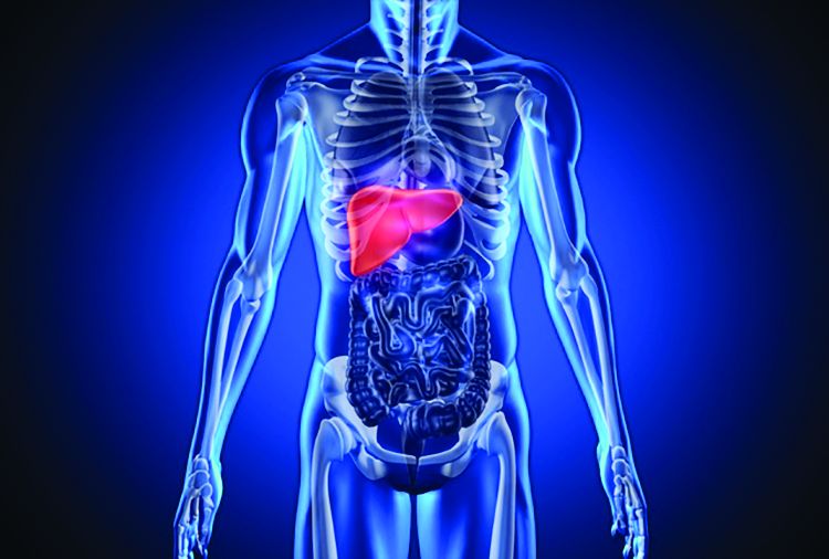
To further explore rates and predictors of infections in STOPAH, the researchers analyzed longitudinal data on incident infections for 1,092 trial participants who received either prednisolone (40 mg daily) or pentoxifylline (400 mg three times daily). For 731 patients, they also examined whether baseline circulating levels of 16s ribosomal bacterial DNA were associated with infections.
A total of 135 patients (12%) had an infection at baseline, 251 (23%) developed infections during treatment, and 89 (8%) developed infections after treatment, the investigators reported. Prednisolone therapy was not associated with infections during treatment, but was associated with a nearly 30% rise in the odds of serious posttreatment infections compared with pentoxifylline (odds ratio, 1.27; 95% confidence interval, 1.27-2.92; P = .002). Prednisolone recipients who developed infections were significantly more likely to die within 90 days than those who did not, even after controlling for end-stage liver disease or Lille score (OR, 2.5; 95% CI, 1.4-4.3; P = .002). Antibiotic therapy appeared to significantly reduce the risk of mortality among infected prednisolone recipients (13% vs. 52%; OR, 0.13; 95% CI 0.04-0.47; P = .002).
There was “a striking association between bacterial DNA and the development of infection within 7 days in patients treated with prednisolone,” the researchers reported. These patients had a median baseline circulating DNA level of 20.9 pg/mL, while prednisolone recipients who did not develop infections had a median baseline bacterial DNA level of 8.3 pg/mL (P = .004). Bacterial DNA predicted infections with an area under receiver operating characteristic curve of 0.70 (95% CI, 0.58-0.83; P = .003), which substantially exceeded the curve for white blood cell count (0.58).
A cut-off value of 18.5 pg/mL was 80% specific for predicting infection within 7 days of prednisolone therapy, the investigators also reported. Bacterial DNA level did not, however, predict infections within 7 days of pentoxifylline therapy, and pentoxifylline was not linked with infections that were serious, infections during treatment, or infections after treatment. (P =.08).
Using bacterial DNA levels to guide prednisolone prescription also appeared to reduce 90-day mortality in this patient population, although the effect achieved borderline statistical significance, the researchers said. “Larger prospective randomized studies are needed to definitely report whether bacterial DNA-guided therapy can [have an] impact on mortality in severe alcoholic hepatitis, and perhaps in other acute inflammatory conditions” in which immunosuppression is required, they added.
The National Institute for Health Research and Wellcome Trust and Medical Research Council provided funding. Dr. Vergis and 10 coinvestigators disclosed no conflicts of interest. Senior author Dr. Mark Thursz and one coinvestigator disclosed ties to Gilead, Bristol-Myers Squibb, AbbVie, Abbott, and Norgine.
Source: American Gastroenterological Association
FROM GASTROENTEROLOGY
Key clinical point: High baseline levels of circulating bacterial DNA predicted serious infections in patients receiving prednisolone for severe alcoholic hepatitis.
Major finding: The odds of serious posttreatment infections were significantly higher for prednisolone compared with pentoxifylline (OR, 1.27; P = .002). High baseline levels of circulating bacterial DNA predicted infection within 7 days of prednisolone therapy (adjusted OR, 4.68; P = .001).
Data source: An analysis of 1,092 patients with severe alcoholic hepatitis from the randomized, double-blind STOPAH trial.
Disclosures: The National Institute for Health Research and Wellcome Trust and Medical Research Council provided funding. Dr. Vergis and 10 coinvestigators reported having no competing interests. Senior author Mark Thursz and one coinvestigator disclosed ties to Gilead, Bristol-Myers Squibb, AbbVie, Abbott, and Norgine.
VIDEO: Infectious enteritis quadrupled short-term risk of IBS
More than 10% of patients developed irritable bowel syndrome (IBS) within a year after infectious enteritis, which gave them a more than fourfold greater risk than that of controls, according to a systematic review and meta-analysis of 45 studies.
“Protozoal and bacterial enteritis confer the greatest overall risk, although the magnitude of increased risk diminishes with time since exposure,” Fabiane B. Klem, MD, and Akhilesh Wadhwa, MD, of Mayo Clinic in Rochester, Minn., and their associates wrote in the April issue of Gastroenterology. Other significant risk factors for postinfectious IBS (PI-IBS) included female sex, clinically severe infections, antibiotic therapy, and comorbid psychological distress, they said.
Postinfectious IBS can last at least a decade after resolution of campylobacteriosis, shigellosis, salmonellosis, giardiasis, and norovirus infections, even when patients have no other risk factors for IBS, the researchers noted. To update and expand the most recent meta-analysis of this topic (Aliment Pharmacol Ther. 2007;26:535-44), the investigators searched Ovid Medline, EMBASE, Web of Science, and Cochrane Database of Systematic Reviews for studies published from 2006 through Aug. 31, 2015. This search yielded 45 studies, including 30 studies comparing infected patients with controls, who were usually matched by age, sex, and geographic location (Gastroenterology. 2017 Jan 6. doi: 10.1053/j.gastro.2016.12.039).
In all, 10.1% of patients with infectious enteritis developed IBS in the next 12 months (95% confidence interval, 7.2-14.1) – a 4.2-fold increase in risk, compared with that of controls (risk ratio, 4.2; 95% CI, 3.2-5.7). This risk subsequently dropped, but remained significantly elevated (RR, 2.3; 95% CI, 1.8-3.0), compared with controls. “Of patients with enteritis caused by protozoa or parasites, 41.9% developed IBS; of patients with enteritis caused by bacterial infection, 13.8% developed IBS,” the researchers emphasized. Patients with these infections remained at elevated risk of PI-IBS even after 1 year. Viral enteritis also significantly increased the risk of PI-IBS, but risk dropped to baseline levels after a year.
Among 10 pooled studies of IBS subtypes, 46% of patients had mixed IBS, 39% had diarrhea-predominant IBS, and 15% had constipation-predominant IBS. Female sex doubled the odds of PI-IBS (odds ratio, 2.2; 95% CI, 1.6-3.1) in 11 pooled studies. Significant clinical risk factors for PI-IBS included diarrhea lasting more than 7 days (eight studies; OR, 2.6; 95% CI, 1.5-4.6), bloody stool (four studies; OR, 1.9; 95% CI, 1.1-3.0), and antibiotic therapy during infectious enteritis (seven studies; OR, 1.7; 95% CI, 1.2-2.4).
Multiple reports linked PI-IBS to clinical psychological distress at the time of infectious enteritis. Specific risk factors included depression based on the Hospitalization Anxiety and Depression Scale (five studies; OR, 1.5; 95% CI, 1.2-1.9), anxiety based on the Hospital Anxiety and Depression Scale (four studies; OR, 2.0; 95% CI, 1.3-2.9), somatization (four studies; OR, 4.1; 95% CI, 2.7-6.0), and neuroticism (two studies; OR, 3.3; 95% CI, 1.6-6.6). Isolated studies also implicated hypochondriasis, extroversion, negative illness beliefs, stress, sleep disturbance, and adverse life events in the preceding year, the researchers said.
They found no evidence of publication bias, but noted a substantial amount of heterogeneity among studies. Also, some studies did not report multivariate analyses, so individual odds ratios might reflect “a conglomeration of factors,” they said.
The National Institutes of Health and the American Gastroenterological Association funded the work. The investigators reported having no conflicts of interest.
Source: American Gastroenterological Association
The phenomenon of IBS developing after a bout of gastroenteritis (postinfectious [PI]–IBS) was first reported in 1950 and subsequently elaborated by studies from Oxford (Q J Med. 1962;123:307-22), Sheffield (Gut. 1999;44:400-6), and Nottingham (BMJ 1997;314:779-82; Gut. 2000;47:804-11). It has proven to be a fertile area for research, which is the basis for this excellent meta-analysis.
The authors identified 45 studies, 29 in the last decade including a total of 21,421 participants with exposure to gastroenteritis. The pooled prevalence for PI-IBS was 11.5% (95% CI, 8.2%-15.8%) but with considerable heterogeneity, which the authors attempted to explain by a number of subgroup analyses. The authors report that protozoal infection seems to have a higher rate of PI-IBS than bacterial or viral infection, though some caution is warranted, since these figures rely on reports from just one outbreak of giardiasis in Bergen, Norway (Scand J Gastroenterol. 2012;47:956-61). However, if true, this might suggest that a different immune response could be responsible, a feature which others have suggested might predispose particular individuals to PI-IBS (Gut. 2016;65[8]1279-88).

The meta-analysis confirms the consistent increased risk in female patients (odds ratio, 1.69), anxiety (OR, 1.97), and somatization (greatest RR, 4.05), all common risks for the development of IBS but not specific to PI-IBS. Initial disease severity indicators, including bloody stool and more than 7 days of initial illness, which might indicate the severity of underlying damage to the gut, were shown to be significant risk factors. Animal studies of acute infection, particularly parasitic infestation, indicate that significant changes can be seen in both nerve and muscle, but routine histology in PI-IBS patients is normal. Infection produces a striking increase in gut permeability (Gut 2000;47:804-11), a feature of IBS whose molecular basis has been demonstrated by a series of elegant studies (Gut. 2017 Jan 12 [Epub ahead of print]; Gut. 2015;64:1379-88) demonstrating altered tight junctions and immune activation in IBS with diarrhea. The authors found treatment with antibiotics increased the risk of PI-IBS but whether this is attributable to confounding by indication is unclear.
This meta-analysis indicates that PI-IBS also potentially is the most common cause of IBS, given that both the Centers for Disease Control and Prevention in the United States and community surveys in the United Kingdom (BMJ. 1999;318:1046-50) indicate that gastroenteritis affects around 1 in 5 of the population each year. If the incidence of PI-IBS is around 10%, modeling suggests PI-IBS could account for the majority of new cases (J Neurogastroenterol Motil. 2012;18:200-4).
Dr. Robin Spiller is professor of gastroenterology, NIHR Nottingham Digestive Diseases Biomedical Research Unit, Nottingham Digestive Diseases Centre, University of Nottingham, England. He has no relevant conflicts of interest.
The phenomenon of IBS developing after a bout of gastroenteritis (postinfectious [PI]–IBS) was first reported in 1950 and subsequently elaborated by studies from Oxford (Q J Med. 1962;123:307-22), Sheffield (Gut. 1999;44:400-6), and Nottingham (BMJ 1997;314:779-82; Gut. 2000;47:804-11). It has proven to be a fertile area for research, which is the basis for this excellent meta-analysis.
The authors identified 45 studies, 29 in the last decade including a total of 21,421 participants with exposure to gastroenteritis. The pooled prevalence for PI-IBS was 11.5% (95% CI, 8.2%-15.8%) but with considerable heterogeneity, which the authors attempted to explain by a number of subgroup analyses. The authors report that protozoal infection seems to have a higher rate of PI-IBS than bacterial or viral infection, though some caution is warranted, since these figures rely on reports from just one outbreak of giardiasis in Bergen, Norway (Scand J Gastroenterol. 2012;47:956-61). However, if true, this might suggest that a different immune response could be responsible, a feature which others have suggested might predispose particular individuals to PI-IBS (Gut. 2016;65[8]1279-88).
The meta-analysis confirms the consistent increased risk in female patients (odds ratio, 1.69), anxiety (OR, 1.97), and somatization (greatest RR, 4.05), all common risks for the development of IBS but not specific to PI-IBS. Initial disease severity indicators, including bloody stool and more than 7 days of initial illness, which might indicate the severity of underlying damage to the gut, were shown to be significant risk factors. Animal studies of acute infection, particularly parasitic infestation, indicate that significant changes can be seen in both nerve and muscle, but routine histology in PI-IBS patients is normal. Infection produces a striking increase in gut permeability (Gut 2000;47:804-11), a feature of IBS whose molecular basis has been demonstrated by a series of elegant studies (Gut. 2017 Jan 12 [Epub ahead of print]; Gut. 2015;64:1379-88) demonstrating altered tight junctions and immune activation in IBS with diarrhea. The authors found treatment with antibiotics increased the risk of PI-IBS but whether this is attributable to confounding by indication is unclear.
This meta-analysis indicates that PI-IBS also potentially is the most common cause of IBS, given that both the Centers for Disease Control and Prevention in the United States and community surveys in the United Kingdom (BMJ. 1999;318:1046-50) indicate that gastroenteritis affects around 1 in 5 of the population each year. If the incidence of PI-IBS is around 10%, modeling suggests PI-IBS could account for the majority of new cases (J Neurogastroenterol Motil. 2012;18:200-4).
Dr. Robin Spiller is professor of gastroenterology, NIHR Nottingham Digestive Diseases Biomedical Research Unit, Nottingham Digestive Diseases Centre, University of Nottingham, England. He has no relevant conflicts of interest.
More than 10% of patients developed irritable bowel syndrome (IBS) within a year after infectious enteritis, which gave them a more than fourfold greater risk than that of controls, according to a systematic review and meta-analysis of 45 studies.
“Protozoal and bacterial enteritis confer the greatest overall risk, although the magnitude of increased risk diminishes with time since exposure,” Fabiane B. Klem, MD, and Akhilesh Wadhwa, MD, of Mayo Clinic in Rochester, Minn., and their associates wrote in the April issue of Gastroenterology. Other significant risk factors for postinfectious IBS (PI-IBS) included female sex, clinically severe infections, antibiotic therapy, and comorbid psychological distress, they said.
Postinfectious IBS can last at least a decade after resolution of campylobacteriosis, shigellosis, salmonellosis, giardiasis, and norovirus infections, even when patients have no other risk factors for IBS, the researchers noted. To update and expand the most recent meta-analysis of this topic (Aliment Pharmacol Ther. 2007;26:535-44), the investigators searched Ovid Medline, EMBASE, Web of Science, and Cochrane Database of Systematic Reviews for studies published from 2006 through Aug. 31, 2015. This search yielded 45 studies, including 30 studies comparing infected patients with controls, who were usually matched by age, sex, and geographic location (Gastroenterology. 2017 Jan 6. doi: 10.1053/j.gastro.2016.12.039).
In all, 10.1% of patients with infectious enteritis developed IBS in the next 12 months (95% confidence interval, 7.2-14.1) – a 4.2-fold increase in risk, compared with that of controls (risk ratio, 4.2; 95% CI, 3.2-5.7). This risk subsequently dropped, but remained significantly elevated (RR, 2.3; 95% CI, 1.8-3.0), compared with controls. “Of patients with enteritis caused by protozoa or parasites, 41.9% developed IBS; of patients with enteritis caused by bacterial infection, 13.8% developed IBS,” the researchers emphasized. Patients with these infections remained at elevated risk of PI-IBS even after 1 year. Viral enteritis also significantly increased the risk of PI-IBS, but risk dropped to baseline levels after a year.
Among 10 pooled studies of IBS subtypes, 46% of patients had mixed IBS, 39% had diarrhea-predominant IBS, and 15% had constipation-predominant IBS. Female sex doubled the odds of PI-IBS (odds ratio, 2.2; 95% CI, 1.6-3.1) in 11 pooled studies. Significant clinical risk factors for PI-IBS included diarrhea lasting more than 7 days (eight studies; OR, 2.6; 95% CI, 1.5-4.6), bloody stool (four studies; OR, 1.9; 95% CI, 1.1-3.0), and antibiotic therapy during infectious enteritis (seven studies; OR, 1.7; 95% CI, 1.2-2.4).
Multiple reports linked PI-IBS to clinical psychological distress at the time of infectious enteritis. Specific risk factors included depression based on the Hospitalization Anxiety and Depression Scale (five studies; OR, 1.5; 95% CI, 1.2-1.9), anxiety based on the Hospital Anxiety and Depression Scale (four studies; OR, 2.0; 95% CI, 1.3-2.9), somatization (four studies; OR, 4.1; 95% CI, 2.7-6.0), and neuroticism (two studies; OR, 3.3; 95% CI, 1.6-6.6). Isolated studies also implicated hypochondriasis, extroversion, negative illness beliefs, stress, sleep disturbance, and adverse life events in the preceding year, the researchers said.
They found no evidence of publication bias, but noted a substantial amount of heterogeneity among studies. Also, some studies did not report multivariate analyses, so individual odds ratios might reflect “a conglomeration of factors,” they said.
The National Institutes of Health and the American Gastroenterological Association funded the work. The investigators reported having no conflicts of interest.
Source: American Gastroenterological Association
FROM GASTROENTEROLOGY
Key clinical point.
Major finding: A total of 10.1% of patients with infectious enteritis developed IBS in the next 12 months, a 4.2-fold increase in risk, compared with that of controls.
Data source: A systematic review and meta-analysis of 45 studies.
Disclosures: The National Institutes of Health and the American Gastroenterological Association funded the work. The investigators reported having no conflicts of interest.
For mantle cell lymphoma, VR-CAP beat R-CHOP
For patients with newly diagnosed mantle cell lymphoma, duration and quality of response were superior with a regimen of bortezomib, rituximab, cyclophosphamide, doxorubicin, and prednisone (VR-CAP) when compared with rituximab, cyclophosphamide, doxorubicin, vincristine, and prednisone (R-CHOP), based on a post hoc analysis of the randomized, phase III LYM-3002 trial.
The difference was especially evident among patients who had a low- or medium-risk mantle cell lymphoma international prognostic index, Gregor Verhoef, MD, of University Hospital Leuven (Belgium) and his associates wrote in Haematologica.
In LYM-3002, 487 patients with newly diagnosed stage II-IV mantle cell lymphoma received six to eight 21-day cycles of intravenous VR-CAP or R-CHOP. Although overall response rates were similar for both groups, VR-CAP was associated with better duration of response and progression-free survival (PFS) and extended time to next treatment. To further explore these differences, the post hoc analysis stratified outcomes by response categories and analyzed depth of response based on computed tomography (CT) scans. Patients had a median age of about 65 years, and most were white males with stage-IV disease at diagnosis and an Eastern Cooperative Oncology Group performance status of 1 (Haematologica. 2017 Feb 9. doi: 10.3324/haematol.2016.152496).The superiority of VR-CAP held up across response categories. Complete responders to VR-CAP had more than twice the median PFS as did complete responders to R-CHOP (40.9 vs. 19.8 months; hazard ratio, 0.58; 95% confidence interval, 0.39-0.84; P = .004). Among partial responders, median PFS was 17.1 vs. 11.7 months, respectively (HR, 0.62; 95% CI, 0.43-0.89; P = .01). Respective median duration of overall response was 42.1 months for VR-CAP vs. 18.5 months among complete responders (HR, 0.42; P less than .001), and 20.2 vs. 9.6 months among partial responders (HR, 0.57; P = .006).
Median time to next treatment also favored VR-CAP over R-CHOP among both complete responders (not evaluable vs. 26.6 months; HR, 0.42; P less than .001) and partial responders (35.3 vs. 24.3 months; HR, 0.57; P = .006), the researchers said. Further, CT scans showed that proportionally more patients in each response category became lesion-negative on VR-CAP than on R-CHOP. Among complete responders, rates of lesion negativity were 72% and 59%, respectively. Among partial responders, rates were 48% and 28%.
The effects of VR-CAP were most evident among patients with a low or medium-risk mantle cell lymphoma international prognostic index. Perhaps high-risk status signifies more rapidly proliferative disease, which negates the deeper responses with VR-CAP, compared with R-CHOP, they added.
The LYM-3002 study was supported by Janssen Research & Development and Millennium Pharmaceuticals. Dr. Verhoef had no disclosures. Nine coinvestigators disclosed ties to Janssen, Roche, GlaxoSmithKline, and several other pharmaceutical companies.
FROM HAEMATOLOGICA
Key clinical point: A regimen of bortezomib, rituximab, cyclophosphamide, doxorubicin, and prednisone (VR-CAP) led to superior duration and quality of response when compared with rituximab, cyclophosphamide, doxorubicin, vincristine, and prednisone (R-CHOP) in patients with newly diagnosed mantle cell lymphoma.
Major finding: Among complete responders, median progression-free survival on VR-CAP was nearly twice that of R-CHOP (40.9 vs. 19.8 months; hazard ratio, 0.58; 95% confidence interval, 0.39-0.84; P = .004).
Data source: A post hoc analysis of a phase III trial comparing VR-CAP with R-CHOP in 487 patients with newly diagnosed, measurable stage II-IV mantle cell lymphoma (LYM-3002).
Disclosures: The LYM-3002 study was supported by Janssen Research & Development and Millennium Pharmaceuticals. Dr. Verhoef had no disclosures. Nine coinvestigators disclosed ties to Janssen, Roche, GlaxoSmithKline, and several other pharmaceutical companies.