User login
Anticoagulation Hub contains news and clinical review articles for physicians seeking the most up-to-date information on the rapidly evolving treatment options for preventing stroke, acute coronary events, deep vein thrombosis, and pulmonary embolism in at-risk patients. The Anticoagulation Hub is powered by Frontline Medical Communications.
Thrombolysis gap for stroke octogenarians disappears
NASHVILLE, TENN. – By 2010, U.S. octogenarians with acute ischemic stroke received intravenous thrombolytic treatment about as often as younger patients, showing that a sharp, age-related disparity in thrombolytic use a decade before had disappeared, based on comprehensive national data.
The 2010 data from the Nationwide Inpatient Sample further showed that the sex-based disparity in treatment with intravenous tissue plasminogen activator (TPA) seen in 2000 resolved as well from 2000 to 2010, but other disparities worsened with declines during the period in use of TPA at rural hospitals relative to urban hospitals and at nonteaching hospitals, compared with teaching hospitals, Dr. Michelle P. Lin said at the International Stroke Conference.
Perhaps most importantly, the statistics showed a “dramatic” increase in TPA use among all age groups during the decade ending in 2010, when thrombolytic therapy was administered to 3.5%-3.9% of adult patients regardless of their age, said Dr. Lin of the department of neurology at the University of Southern California in Los Angeles. In 2000, U.S. patients received TPA at less than a third of that rate.
Low TPA use in 2000 among patients aged 80 or older in part reflected the low number of octogenarians enrolled in the trials that documented the safety and efficacy of TPA for acute ischemicstroke patients, Dr. Lin said at the International Stroke Conference, sponsored by the American Heart Association.
Data from the National Inpatient Sample included information on the treatment received by 5,932,175 patients with acute ischemic stroke at more than 1,000 U.S. hospitals during 2000-2010. The age breakdown of the nearly 6 millionpatients showed 28% were aged 18-64 years, 37% were65-79, and 35% were 80 years or older.
In 2000, medical staffs administered intravenous treatment with TPA to 1.02% of these patients aged 18-64 years, 0.92% of patients aged 65-79 years, and 0.47% of patients aged 80 or older. By 2010, the annual rates of TPA use ran 3.61% in those 18-64 years, 3.87% among those 65-79 years, and 3.55% in patients 80 years or older. In an adjusted analysis, this translated into a greater than threefold increase in TPA use among the 18- to 64-year-olds, a nearly fourfold rise in patients 65-79 years, and a nearly sevenfold jump among those 80 or older, a 24% average annual increased rate among the oldest patients, who averaged 86 years old, Dr. Lin reported.
The data also showed that among the octogenarians during 2000-2005, TPA use in women lagged behind use in men by a relative 15%, but this completely disappeared during 2006-2010, when usage rates in men and women evened out. TPA use among African Americans, Hispanics, and Asians, compared with whites, remained significantly below the rate in whites throughout the decade, although the extent of the disparity narrowed for African Americans and Asians during the second half of the decade, compared with the first half.
Dr. Lin and her associates also analyzed TPA use relative to the demographic setting of the hospital and its teaching status. During 2000-2010, the relative usage of TPA at rural hospitals, compared with urban hospitals, fell from 65% of the comparator level to 36%. Among nonteaching hospitals, the rate of TPA use dropped from 52% of the teaching hospitals’ level to 49%.
Dr. Lin said she had no relevant financial disclosures.
[email protected]
On Twitter @mitchelzoler
AT THE INTERNATIONAL STROKE CONFERENCE
Key clinical point: By 2010, U.S. ischemic stroke patients aged 80 or older received thrombolytic treatment as often as younger patients.
Major finding: In 2010, thrombolysis was used to treat 3.55% of U.S. stroke patients 80 years or older, 3.87% of those 65-79, and 3.61% of those 18-64.
Data source: The U.S. National Inpatient Sample for 2000-2010, which included 5,932,175 adults with acute ischemic stroke.
Disclosures: Dr. Lin said she had no relevant financial disclosures.
General anesthesia linked to worsened stroke outcomes
NASHVILLE, TENN. – When acute ischemic stroke patients undergo an emergency endovascular procedure is it best done with general anesthesia or nongeneral anesthesia?
A post hoc analysis of data collected by a Dutch randomized, controlled trial of intra-arterial therapy suggested that nongeneral anesthesia was associated with substantially better patient outcomes, and the findings convinced the Dutch investigators who ran the study to stick with nongeneral anesthesia as their default approach.
In MR CLEAN (Multicenter Randomized Clinical Trial of Endovascular Treatment for Acute Ischemic Stroke in the Netherlands) (N. Engl. J. Med. 2015;372:11-20), 216 acute ischemic stroke patients underwent intra-arterial treatment following randomization. Among these patients, 79 were treated with general anesthesia and 137 with nongeneral anesthesia. The anesthesia choice was made on a case-by-case basis by each participating interventionalist.
The study’s primary endpoint – 90-day status rated as a good function based on a modified Rankin sale score of 0-2 – occurred in 38% of the intra-arterial patients treated with nongeneral anesthesia, 23% of the intra-arterial patients treated with general anesthesia, and 19% among the control patients who received standard treatment without intra-arterial intervention.
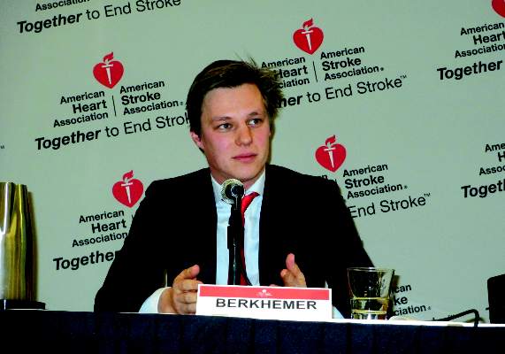
“The effect on outcome that we found with intra-arterial treatment in MR CLEAN was not observed in the subgroup of patients treated with general anesthesia,” said Dr. Olvert A. Berkhemer at the International Stroke Conference. The analysis also showed that patients in the general and nongeneral anesthesia subgroups had similar stroke severity as measured by their National Institutes of Health Stroke Scale score, said Dr. Berkhemer, a researcher at the Academic Medical Center in Amsterdam.
But U.S. stroke specialists who heard the report cautioned that unidentified confounders might explain the results, and they also expressed skepticism that the Dutch observations would deter U.S. interventionalists from continuing to use general anesthesia when they perform endovascular procedures.
“The big concern [about this analysis] is that there may have been some things about the general anesthesia patients that they did not account for. I suspect there is a huge bias, that general anesthesia patients were sicker,” said Dr. Bruce Ovbiagele, professor and chief of neurology at the Medical University of South Carolina in Charleston. “At my institution they have used nongeneral anesthesia, but I have been at other places where they usually use general anesthesia; it is variable,” Dr. Ovbiagele added.
Dr. Berkhemer’s analysis also showed that general anesthesia linked with a delayed start to treatment, but without resulting in a significant difference in time to reperfusion. He noted that “sometimes you cannot do the procedure without general anesthesia,” and in MR CLEAN 6 of the 137 intra-arterial patients who started with nongeneral anesthesia eventually received general anesthesia because of discomfort and pain, Dr. Berkhemer said at the conference, sponsored by the American Heart Association.
He speculated that patients who did not receive general anesthesia did better because they did not undergo acute episodes of reduced blood pressure caused by the hypotensive effect of general anesthesia.
The findings reinforced the approach already used in most of the Dutch centers that participated in MR CLEAN, where nongeneral anesthesia is preferred when possible. “In our center we always use nongeneral anesthesia, we are happy with that and we are not going to change,” said Dr. Diederik W.J. Dippel, lead investigator of MR CLEAN and professor of neurology at Erasmus University Medical Center in Rotterdam, The Netherlands. This prejudice against general anesthesia would make it hard to run a trial in The Netherlands that matched the two anesthesia approaches against each other, Dr. Dippel added.
On Twitter @mitchelzoler
Results from prior analyses had also shown better outcomes of acute ischemic stroke patients undergoing endovascular intervention when they avoided general anesthesia. A notable feature of Dr. Berkhemer’s analysis was that the stroke severity levels were well balanced between the patients who received general anesthesia and those who did not. But many other factors aside from stroke severity can affect whether or not a patient receives general anesthesia.
|
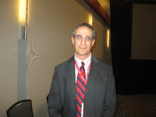
Dr. Larry B. Goldstein |
Although the outcome differences seen in this analysis were striking, many factors could have contributed. Patients received different drugs, patients may have had widely divergent clinical states despite their similar stroke severity, and the people performing the procedures were different. The many variables make it very hard to pinpoint the cause of the different outcomes.
At my institution, Duke, the interventionalists who work on acute ischemic stroke patients almost exclusively use general anesthesia. That’s because of their concern about how patients will act during a very delicate procedure. For example, stroke patients can have an aphasia that makes it hard for them to respond to requests to do something specific during the procedure. I am very skeptical that my colleagues will decide to switch to using no general anesthesia based on the results of this new analysis.
I agree with the Dutch investigators that a randomized, controlled trial of general anesthesia or no general anesthesia would be very hard to perform because individual interventionalists would need to believe there is equipoise between the two anesthesia approaches. Most interventionalists right now probably believe the approach they have always used remains best and so would be unwilling to participate in a randomized controlled trial.
Dr. Larry B. Goldstein is professor of neurology and chief of the stroke center at Duke University in Durham, N.C. He had no relevant disclosures. He made these comments in an interview.
Results from prior analyses had also shown better outcomes of acute ischemic stroke patients undergoing endovascular intervention when they avoided general anesthesia. A notable feature of Dr. Berkhemer’s analysis was that the stroke severity levels were well balanced between the patients who received general anesthesia and those who did not. But many other factors aside from stroke severity can affect whether or not a patient receives general anesthesia.

|
Dr. Larry B. Goldstein |
Although the outcome differences seen in this analysis were striking, many factors could have contributed. Patients received different drugs, patients may have had widely divergent clinical states despite their similar stroke severity, and the people performing the procedures were different. The many variables make it very hard to pinpoint the cause of the different outcomes.
At my institution, Duke, the interventionalists who work on acute ischemic stroke patients almost exclusively use general anesthesia. That’s because of their concern about how patients will act during a very delicate procedure. For example, stroke patients can have an aphasia that makes it hard for them to respond to requests to do something specific during the procedure. I am very skeptical that my colleagues will decide to switch to using no general anesthesia based on the results of this new analysis.
I agree with the Dutch investigators that a randomized, controlled trial of general anesthesia or no general anesthesia would be very hard to perform because individual interventionalists would need to believe there is equipoise between the two anesthesia approaches. Most interventionalists right now probably believe the approach they have always used remains best and so would be unwilling to participate in a randomized controlled trial.
Dr. Larry B. Goldstein is professor of neurology and chief of the stroke center at Duke University in Durham, N.C. He had no relevant disclosures. He made these comments in an interview.
Results from prior analyses had also shown better outcomes of acute ischemic stroke patients undergoing endovascular intervention when they avoided general anesthesia. A notable feature of Dr. Berkhemer’s analysis was that the stroke severity levels were well balanced between the patients who received general anesthesia and those who did not. But many other factors aside from stroke severity can affect whether or not a patient receives general anesthesia.

|
Dr. Larry B. Goldstein |
Although the outcome differences seen in this analysis were striking, many factors could have contributed. Patients received different drugs, patients may have had widely divergent clinical states despite their similar stroke severity, and the people performing the procedures were different. The many variables make it very hard to pinpoint the cause of the different outcomes.
At my institution, Duke, the interventionalists who work on acute ischemic stroke patients almost exclusively use general anesthesia. That’s because of their concern about how patients will act during a very delicate procedure. For example, stroke patients can have an aphasia that makes it hard for them to respond to requests to do something specific during the procedure. I am very skeptical that my colleagues will decide to switch to using no general anesthesia based on the results of this new analysis.
I agree with the Dutch investigators that a randomized, controlled trial of general anesthesia or no general anesthesia would be very hard to perform because individual interventionalists would need to believe there is equipoise between the two anesthesia approaches. Most interventionalists right now probably believe the approach they have always used remains best and so would be unwilling to participate in a randomized controlled trial.
Dr. Larry B. Goldstein is professor of neurology and chief of the stroke center at Duke University in Durham, N.C. He had no relevant disclosures. He made these comments in an interview.
NASHVILLE, TENN. – When acute ischemic stroke patients undergo an emergency endovascular procedure is it best done with general anesthesia or nongeneral anesthesia?
A post hoc analysis of data collected by a Dutch randomized, controlled trial of intra-arterial therapy suggested that nongeneral anesthesia was associated with substantially better patient outcomes, and the findings convinced the Dutch investigators who ran the study to stick with nongeneral anesthesia as their default approach.
In MR CLEAN (Multicenter Randomized Clinical Trial of Endovascular Treatment for Acute Ischemic Stroke in the Netherlands) (N. Engl. J. Med. 2015;372:11-20), 216 acute ischemic stroke patients underwent intra-arterial treatment following randomization. Among these patients, 79 were treated with general anesthesia and 137 with nongeneral anesthesia. The anesthesia choice was made on a case-by-case basis by each participating interventionalist.
The study’s primary endpoint – 90-day status rated as a good function based on a modified Rankin sale score of 0-2 – occurred in 38% of the intra-arterial patients treated with nongeneral anesthesia, 23% of the intra-arterial patients treated with general anesthesia, and 19% among the control patients who received standard treatment without intra-arterial intervention.
“The effect on outcome that we found with intra-arterial treatment in MR CLEAN was not observed in the subgroup of patients treated with general anesthesia,” said Dr. Olvert A. Berkhemer at the International Stroke Conference. The analysis also showed that patients in the general and nongeneral anesthesia subgroups had similar stroke severity as measured by their National Institutes of Health Stroke Scale score, said Dr. Berkhemer, a researcher at the Academic Medical Center in Amsterdam.
But U.S. stroke specialists who heard the report cautioned that unidentified confounders might explain the results, and they also expressed skepticism that the Dutch observations would deter U.S. interventionalists from continuing to use general anesthesia when they perform endovascular procedures.
“The big concern [about this analysis] is that there may have been some things about the general anesthesia patients that they did not account for. I suspect there is a huge bias, that general anesthesia patients were sicker,” said Dr. Bruce Ovbiagele, professor and chief of neurology at the Medical University of South Carolina in Charleston. “At my institution they have used nongeneral anesthesia, but I have been at other places where they usually use general anesthesia; it is variable,” Dr. Ovbiagele added.
Dr. Berkhemer’s analysis also showed that general anesthesia linked with a delayed start to treatment, but without resulting in a significant difference in time to reperfusion. He noted that “sometimes you cannot do the procedure without general anesthesia,” and in MR CLEAN 6 of the 137 intra-arterial patients who started with nongeneral anesthesia eventually received general anesthesia because of discomfort and pain, Dr. Berkhemer said at the conference, sponsored by the American Heart Association.
He speculated that patients who did not receive general anesthesia did better because they did not undergo acute episodes of reduced blood pressure caused by the hypotensive effect of general anesthesia.
The findings reinforced the approach already used in most of the Dutch centers that participated in MR CLEAN, where nongeneral anesthesia is preferred when possible. “In our center we always use nongeneral anesthesia, we are happy with that and we are not going to change,” said Dr. Diederik W.J. Dippel, lead investigator of MR CLEAN and professor of neurology at Erasmus University Medical Center in Rotterdam, The Netherlands. This prejudice against general anesthesia would make it hard to run a trial in The Netherlands that matched the two anesthesia approaches against each other, Dr. Dippel added.
On Twitter @mitchelzoler
AT THE INTERNATIONAL STROKE CONFERENCE
Key clinical point: Post hoc analysis of acute ischemic stroke patients treated intra-arterially suggests that avoiding general anesthesia produces better long-term outcomes.
Major finding: After 90 days, 38% of patients treated without general anesthesia and 23% of those who received general anesthesia had good outcomes.
Data source: Analysis of 216 patients in the Dutch MR CLEAN trial who underwent treatment for acute ischemic stroke.
Disclosures: Dr. Berkhemer, Dr. Dippel, and Dr. Ovbiagele had no disclosures.
Smart diet remains potent cardiovascular medicine
SNOWMASS, COLO. – Cutting dietary fat intake remains a highly effective strategy for reducing coronary heart disease risk – but only so long as the replacement nutrients aren’t even bigger offenders, Dr. Robert A. Vogel said at the Annual Cardiovascular Conference at Snowmass.
In the face of decades of public health admonitions to reduce saturated fat intake, most Americans have increased their consumption of trans fats and simple carbohydrates, especially sugar. And therein lies a problem. Trans fats are far more harmful than saturated fats in terms of cardiovascular risk. And excessive sugar consumption is a major contributor to abdominal obesity, metabolic syndrome, hypertension, and endothelial dysfunction.
“In the United States, sugar is a bigger source of hypertension than is salt,” asserted Dr. Vogel, a cardiologist at the University of Colorado, Denver.
The editors of Time magazine ignited a public controversy last year with a cover story arrestingly titled, “Eat Butter – Scientists labelled fat the enemy. Why they were wrong.” The editors were picking up on a British meta-analysis of 32 observational studies that concluded there is no clear evidence to support the notion that saturated fats are harmful to cardiovascular health and that swapping them out for consumption of polyunsaturated fatty acids (PUFAs) is beneficial (Ann. Intern. Med. 2014;160:398-406).
Dr. Vogel said those investigators are in fact correct: Many of the observational studies – going all the way back to the pioneering work by Dr. Ancel Keys in the 1950s – are flawed. They don’t convincingly prove the case for PUFAs as a healthier alternative. But there is persuasive evidence from well-conducted, randomized, controlled trials that this is indeed so, he added.
Several of these studies were done in an earlier era when it was possible to slip around the challenges and limitations of dietary studies in free-living populations. These trials wouldn’t be possible today for ethical reasons involving lack of informed consent.
For example, in the Finnish Mental Hospital Study conducted during 1959-1971, the food served at two mental institutions was altered. Patients at one hospital got 6 years of a diet high in PUFAs, then were crossed over to a typical Finnish diet. At the other mental hospital, patients were fed a normal Finnish diet for 6 years, then crossed over to the high-PUFA diet for 6 years. During the experimental-diet years, the coronary heart disease event rate was reduced by nearly 60% (Int. J. Epidemiol. 1979;8:99-118).
Similarly, in a prospective randomized trial conducted at a Los Angeles Veterans Affairs institution for older, cognitively impaired men, a no-choice shift to a diet high in PUFAs with reduced saturated fats resulted in roughly a 30% reduction in CHD events compared to the usual institutional diet (Lancet 1968;2:1060-2). A similar magnitude of CHD event reduction was seen with a high-PUFA dietary intervention in the Oslo Diet-Heart Study, a prospective secondary prevention trial (Circulation 1970;42:935-42).
In the contemporary era, the standout randomized dietary intervention trial is the Lyon Diet Heart Study, a 46-month prospective secondary prevention trial in which a Mediterranean diet low in saturated fat and high in alpha-linoleic acid, a PUFA, reduced the combined endpoint of cardiac death and nonfatal MI by 70%, compared with the usual post-MI prudent diet recommended at that time. Yet total cholesterol levels in the two study arms did not differ (Circulation 1999;99:779-85).
To put these results into context, Dr. Vogel noted that the Cholesterol Treatment Trialists Collaboration headquartered at the University of Oxford (England) has shown that for every 40 mg/dL of LDL-lowering achieved with statin therapy, the result is roughly a 20% reduction in CHD. In contrast, the classic nonpharmacologic diet studies resulted in 30%-70% relative risk reductions.
“Heart disease is a dietary disease,” the cardiologist emphasized. “When you compare diet intervention to LDL lowering with statins, you see that diet is very, very effective. But you have to know the details of the diet. You can’t take something out and put just anything in. It doesn’t work like that.”
For example, an analysis of data from the National Health and Nutrition Examination Survey concluded that individuals who consumed 25% of their calories from added sugar – that’s the equivalent of three 12-oz cans of a sugary cola per day – had a 175% increased risk of cardiovascular mortality during a median 14.6 years of follow-up, compared with those who got less than 10% of their calories from added sugar (JAMA Intern. Med. 2014;174:516-24).
And as for the impact of the trans fat that’s liberally present in many processed foods, the Nurses Health Study showed that for every 5% increase in energy intake from saturated fat – that’s equivalent to one 8-oz steak per day – the relative risk for CHD rose by a relatively modest 17%, while for a 5% increase in energy intake from trans fat – the equivalent of 4 oz of butter – CHD risk shot up by 382% (N. Engl. J. Med. 1997;337:1491-9).
Dr. Vogel reported serving as a paid consultant to the National Football League and the Pritikin Longevity Center and receiving a research grant from Sanofi.
EXPERT ANALYSIS FROM THE CARDIOVASCULAR CONFERENCE AT SNOWMASS
FDA: Limit testosterone use to men with specific medical conditions
Testosterone therapy is not appropriate for men with age-related low testosterone because it may be associated with an increased risk of cardiovascular events, the Food and Drug Administration has determined.
The agency will now require labeling changes to all prescription testosterone products, clarifying their appropriate use, and warning about a possible increased risk of heart attack and stroke in any patient taking the hormone. Testosterone is approved only for men with specific medical conditions, but is widely used off label for men with age-related low testosterone.
“Health care professionals should make patients aware of this possible risk when deciding whether to start or continue a patient on testosterone therapy,” an FDA statement said. “We are also requiring manufacturers of approved testosterone products to conduct a well-designed clinical trial to more clearly address the question of whether an increased risk of heart attack or stroke exists among users of these products. We are encouraging these manufacturers to work together on a clinical trial, but they are allowed to work separately if they so choose.”
The statement arose from a September 2014 recommendation by the FDA’s Bone, Reproductive, and Urologic Drugs Advisory Committee and Drug Safety and Risk Management Advisory Committee. The groups reviewed studies of aging men using testosterone – some of which reported an increased risk of heart attack, stroke, or death associated with testosterone treatment – and voted 20-1 that the current indication, as worded in the labeling for all testosterone products, should be tightened to make it clear that testosterone therapy is not indicated for men with age-related reductions in testosterone.
Any clinician who prescribes testosterone to a patient who later experiences a cardiovascular event should report that toFDA’s MedWatch Safety Information and Adverse Event Reporting Program.
On Twitter @alz_gal
Better stroke treatment moves tantalizingly within reach
Stroke is one of the most feared medical conditions, with the specter of suddenly finding oneself unable to talk, eat, walk, or live independently, according to study results.
In mid-February, results from three trials reported at the International Stroke Conference in Nashville, Tenn., changed the face of ischemic stroke treatment by proving that emergency endovascular catheterization to remove the embolus blocking cerebral blood flow produced better long-term outcomes than standard treatment with intravenous thrombolysis.
It wasn’t just that patients did better with endovascular embolectomy; it was how much they did better. In the two trials run in the United States and abroad, SWIFT PRIME and ESCAPE, the percentage of patients rated as not disabled (a modified Rankin Scale score of 0-1) when assessed after 90 days was 36% and 42% for patients treated with endovascular therapy in the two studies, compared with 17% and 19% in the two control arms. Embolectomy boosted the fraction of patients having the best stroke outcomes more than twofold, a breathtaking leap in efficacy.
Dr. Jeffrey L. Saver from UCLA, lead investigator for SWIFT PRIME, called it a “once-in-a-field” result, meaning that never again will stroke clinicians see this degree of incremental improvement by adding a new intervention.
The frustrating irony is how challenging delivery of this disease-altering treatment will be on a national scale. One problem is that it didn’t result from a single change in treatment, but from a careful mix of new diagnostic techniques with sophisticated CT imaging, new systems for expediting diagnosis, triage, transport, and treatment, in combination with new technology in the form of emboli-retrieving stents.
Stroke management specialists see a daunting series of issues to tackle as they attempt to roll out emergency endovascular interventions on a routine scale throughout much of the United States. Many more centers must open, modeled on the ones that succeeded in the trials. The centers need to be rationally positioned so they are close to patients but also give each center enough case volume to foster high interventional-skill levels. Staffing must be found for fast-moving stroke response teams that can make the diagnostics and interventions available around the clock and interpret the images to select appropriate patients. Ambulance systems have to be set up that take likely stroke patients to the centers that will best meet their treatment needs.
The stroke and public health communities will need to invest a lot of time, money, and leadership to make this happen, but it’s a clear mandate, given the promise endovascular treatment now holds to blunt the impact of one of medicine’s most feared maladies.
On Twitter @mitchelzoler
Stroke thrombolysis achieved inside an hour works best
NASHVILLE, TENN. – Fast thrombolytic treatment of acute ischemic stroke clearly helps patients, but being really, really fast is even better.
Patients treated with intravenous tissue plasminogen activator (tPA) within the first 60 minutes of their stroke onset, the putative “golden hour,” had significantly better outcomes at hospital discharge, compared with patients treated just an hour later, based on U.S. data from more than 65,000 acute ischemic stroke patients.
“In national U.S. clinical practice, treatment with intravenous tPA in the golden hour, compared with later, is associated with more frequent independent ambulation at discharge, discharge to home, and freedom from disability or dependence at discharge,” compared with patients treated at 61-180 minutes or later, Dr. Jeffrey L. Saver said at the International Stroke Conference.
“These findings support intensive efforts to accelerate patient presentation and treatment initiation, such as Target: Stroke Phase II and mobile CT ambulances, to maximize benefit of thrombolytic therapy for acute ischemic stroke,” said Dr. Saver, professor of neurology and director of the stroke center at the University of California, Los Angeles.
His study used data collected from 65,348 patients with acute ischemic stroke treated at any of 1,456 U.S. hospitals participating in the Get With the Guidelines–Stroke program during 2009-2013. Of those patients, 878 (1.3%) received tPA within the first hour following onset of their stroke, 10% within 61-90 minutes, 71% within 91-180 minutes, and 18% within 181-270 minutes.
Although the 878 patients treated within an hour of symptom onset constituted little more than 1% of all patients, the series was 10-fold larger than any prior report, which allowed a venture into “terra incognita” for insight into thrombolysis efficacy when used so early during a stroke, Dr. Saver noted. “Innovations in prehospital and emergency department systems increasingly enable intravenous tPA delivery in the first 60 minutes,” he said at the meeting, which was sponsored by the American Heart Association.
In a multivariate analysis that adjusted for many potential confounders, treatment in the first 60 minutes linked with statistically significant improvements, compared with patients treated at 1-4.5 hours, for several outcome measures at hospital discharge, including a 72% relative increase in being nondisabled – a modified Rankin score of 0 – and a 58% relative increase in being independent – a modified Rankin scale score of 0-2.
Dr. Saver highlighted how even a 30-minute drop in the time to treatment produced substantively better outcomes. The percentage of patients with a modified Rankin score of 0-1 at discharge was 38% in the 0- to 60-minute patients, 33% in those treated after 91-120 minutes had elapsed, and 28% in those treated 121-180 minutes after symptom onset.
Dr. Saver has been a consultant to BrainGate, Covidien, Grifols, and Stryker and has received research support from Covidien, Lundbeck, and Stryker.
On Twitter @mitchelzoler
AT THE INTERNATIONAL STROKE CONFERENCE
Key clinical point: Acute ischemic stroke patients who started intravenous tPA within an hour of onset had the best outcomes.
Major finding: Stroke thrombolysis within an hour produced 72% more nondisabled patients at hospital discharge, compared with treatment after 1-4.5 hours.
Data source: Review of 65,348 U.S. acute ischemic patients who arrived at hospitals participating in Get With the Guidelines during 2009-2013.
Disclosures: Dr. Saver has been a consultant to BrainGate, Covidien, Grifols, and Stryker and has received research support from Covidien, Lundbeck, and Stryker.
Poor response to statins predicts growth in plaque
For about one in five patients with known atherosclerotic coronary artery disease, standard-dose therapy with statins did not result in significant lowering of LDL cholesterol.
Furthermore, the results of this large pooled data sample showed that for statin hyporesponders, statin therapy did not prevent progression of intravascular plaque volume as measured by grayscale intravascular ultrasound.
Patients exhibit a wide range of response to standard statin dosing, and the effect of minimal LDL-C lowering on atherosclerotic disease progression had not previously been determined, according to Dr. Yu Kataoka of the University of Adelaide, Australia, and his colleagues (Arterioscler. Thromb. Vasc. Biol. 2015 [doi:10.1161/ATVBAHA.114.304477]).
Investigators pooled data from seven clinical trials that examined 647 total patients with angiographically confirmed CAD who were initiated on statins and followed by serial intravascular ultrasound. The present study analyzed baseline characteristics, serial lipid profile, and atheroma burden for the group.
In all, 130 patients of the 647 (20%) had minimal LDL-C lowering with statin therapy, showing nonsignificant lowering or even an increase in LDL-C levels during the study period. This group of hyporesponders differed in being slightly younger, more obese, less likely to have hypertension and dyslipidemia, and less likely to be receiving beta-blockers than were the statin responders. Other patient characteristics were similar between the two groups. A variety of agents were used, including atorvastatin, rosuvastatin, simvastatin, and pravastatin. Concurrent administration of other antiatherosclerotic agents was permitted and was similar between the groups. Atheroma burden at baseline was also similar between the two groups.
Measuring serial changes in atheroma burden showed a significant difference between statin responders and hyporesponders. The adjusted change in atheroma volume was –0.21% for the responders, compared with +0.83% for the hyporesponders (P = .006). Lumen volume decreased 11.64 mm3 for the responders, while the reduction was 16.54 mm3 for the hyporesponders (P = .006). Of those who responded to lipid therapy with LDL-C lowering, 29.8% had substantial atheroma regression, while 25.9% had substantial plaque progression; among hyporesponders, however, just 13.8% experienced significant plaque regression, while 37.7% had significant atheroma progression, both significant differences.
Dr. Kataoka and his colleagues emphasized that the factors contributing to poor statin response are not well understood. They noted that for this study, the pooled trials all showed adherence rates over 90%, eliminating patient compliance as a variable. Rigorous statistical techniques were used to control for comorbidities and coadministered medications. There are known genetic polymorphisms and phenotypic variations in statin metabolism, though these were not reported here. Although the results were not statistically significant, C-reactive protein levels were higher for the hyporesponse group, suggesting that another factor may be individual response to the anti-inflammatory effect that is among the known pleiotropic effects of this drug class.
In an interview, lead author Stephen Nicholls noted that many clinicians are still reluctant to treat to full effect. Citing the concept of “clinical inertia,” Dr. Nicholls pointed out that “Even when statins are prescribed, they are often at lower doses than ideal. That translated to more plaque growth, which leads directly to more heart attacks and more revascularization procedures.”
Study limitations included the potential residual confounding effects of pooling data from seven discrete clinical trials, though mixed modeling techniques attempted to correct for this effect. The present study also reported atheroma burden, but not actual clinical events. The study authors noted, however, that they had previously reported a direct relationship between atheroma progression and the occurrence of cardiovascular events.
Dr. Nicholls has received speaking honoraria and research support from many pharmaceutical companies, and from Infraredx. Dr. Steven E. Nissen of the Cleveland Clinic was a coinvestigator and has received research support from and is a consultant/adviser to numerous pharmaceutical companies; all honoraria or consulting fees go directly to charity so that he receives neither income nor a tax deduction. The other authors report no conflicts.
FROM ARTERIOSCLEROSIS, THROMBOSIS, AND VASCULAR BIOLOGY
Key clinical point: Patients on statins who had minimal LDL-C lowering also showed increased atheroma progression.
Major finding: Of 647 patients with CAD, 20% were hyporesponders to statin therapy and experienced greater progression of atheroma volume than statin responders (adjusted +0.83% vs. –0.21%, P = .006).
Data source: Pooled data from seven clinical trials, yielding 647 patients with angiographically confirmed CAD who were initiated on standard lipid dosing and followed by baseline and serial grayscale intravascular ultrasounds.
Disclosures: Dr. Nicholls has received speaking honoraria and research support from many pharmaceutical companies, and from Infraredx. Dr. Steven E. Nissen of the Cleveland Clinic was a coinvestigator and has received research support from and is a consultant/adviser to numerous pharmaceutical companies; all honoraria or consulting fees go directly to charity so that he receives neither income nor a tax deduction. The other authors report no conflicts.
Mediterranean diet linked with fewer strokes
NASHVILLE, TENN. – A diet that at least partially resembled the Mediterranean diet appeared able to forestall a significant number of strokes in results from a retrospective, epidemiologic study of more than 100,000 Californian women.
“Greater adherence to a Mediterranean diet pattern was associated with a 10%-18% reduced risk of total and ischemic stroke,” Dr. Ayesha Z. Sherzai said at the International Stroke Conference.
“Our finding emphasizes the importance of addressing diet as an important, modifiable risk factor for stroke,” said Dr. Sherzai, a neurologist at Columbia University in New York.
Although the findings Dr. Sherzai reported came entirely from women, “I believe that most of the reported data show that a Mediterranean diet protects against stroke in men as well as in women,” commented Dr. Ralph L. Sacco, professor and chairman of neurology at the University of Miami, in a video statement provided by the American Heart Association, which sponsored the conference.
Dr. Sherzai and her associates used data collected by the California Teachers Study, which enrolled and followed more than 130,000 women who worked as teachers or school administrators in the state during 1995. Their analysis focused on 104,268 of these women with complete data for the diet and stroke analysis and who remained California residents; data on incident ischemic and hemorrhagic strokes came from California’s hospital-discharge database for 1996-2011. During these years, the 104,268 women had 2,270 incident ischemic strokes and 895 incident hemorrhagic strokes.
The researchers obtained baseline diet data from a food frequency questionnaire each participant completed at enrollment in 1995. They rated each participant’s diet by its resemblance to the Mediterranean diet using a validated, 10-point scale first reported in 2003 (N. Engl. J. Med. 2003:348:2599-608). They then divided the women into five subgroups based on the extent to which their reported diet resembled a classic Mediterranean diet, with women who scored 0-2 least adherent (16% of the studied population) and women who scored 6-9 most adherent (24% of the population). Remaining women in the study included 18% with a score of 3, 21% who scored 4, and 20% who scored 5 (total equals 99% because of rounding). The Mediterranean diet is plant based, with high reliance on unrefined cereals, fruits, vegetables, and monounsaturated fats like olive oil with low consumption of meat, sugar/sweeteners, and saturated fat, Dr. Sherzai said.
In an analysis that adjusted for a variety of sociodemographic and clinical variables, women with a diet score of 6-9 has a statistically significant 17% reduced rate of all strokes, compared with women with a diet score of 0-2, Dr. Sherzai reported. Women with a diet score of 5 also had significantly fewer strokes, at a rate 12% below that of the women who scored 0-2.
For ischemic stroke only, women who scored 6-9 had 18% fewer strokes than women who scored 0-2, those who scored a 5 on their diet had 15% fewer strokes, and those whose diet scored a 4 had 16% fewer strokes than the comparator group, also statistically significant differences.
For the outcome of hemorrhagic stroke, none of the diet subgroups showed a statistically significant difference relative to the women who scored 0-2. But the trends went in the same direction, with women scoring 6-9 having 18% fewer strokes and women scoring a 5 having 12% fewer strokes.
On Twitter @mitchelzoler
AT THE INTERNATIONAL STROKE CONFERENCE
Key clinical point: Californian women who at least partially followed a Mediterranean diet had significantly fewer strokes, compared with women with other dietary patterns.
Major finding: Women with a Mediterranean-like diet had 17% fewer strokes than did women whose diet least resembled a Mediterranean diet.
Data source: Retrospective analysis of stroke incidence and diet in 104,268 women enrolled in the California Teachers Study starting in 1995 and followed through 2011.
Disclosures: Dr. Sherzai and Dr. Sacco had no disclosures.
Broadly implementing stroke embolectomy faces hurdles
NASHVILLE, TENN. – Results from three randomized controlled trials presented at the International Stroke Conference, plus the outcomes from a fourth trial first reported last fall, immediately established embolectomy as standard-of-care treatment for selected patients with acute ischemic stroke.
Stroke experts interviewed during the conference, however, said that making embolectomy routinely available to most U.S. stroke patients who would be candidates for the intervention will take months, if not years.
They envision challenges involving the availability of trained interventionalists, triage of patients to the right centers, and reimbursement issues as some of the obstacles to be dealt with before endovascular embolectomy aimed at removing intracerebral-artery occlusions in acute ischemic stroke patients becomes uniformly available.
Yet another challenge will arise when stroke-treatment groups that did not participate in the trials strive to replicate the success their colleagues reported by implementing the highly streamlined systems that were used in the trials for identifying appropriate stroke patients and for delivering treatment. Those systems were cited as an important reason why those studies succeeded in producing positive outcomes when similar embolectomy trials without the same efficiencies reported just a year or two ago failed to show benefit.
“The evidence makes it standard of care, but the challenge is that our systems are not set up. This is the big thing we will all go home to work on,” said Dr. Pooja Khatri, professor of neurology and director of acute stroke at the University of Cincinnati.
“You talk to everyone at this meeting, and what they want to go home and figure out is how can we deliver this care. It’s really challenging, at a myriad of levels,” said Dr. Colin P. Derdeyn, professor of neurology and director of the Center for Stroke and Cerebrovascular Disease at Washington University in St. Louis.
Growing endovascular availability
Arguably the most critical issue in rolling out endovascular stroke interventions more broadly is scaling up the number of centers that have the staff and systems in place to perform them. Clearly, the scope of providers able to deliver this treatment currently falls substantially short of what will be needed. “It’s kind of daunting to think about the [workforce] needs,” Dr. Khatri said in a talk at the conference, which was sponsored by the American Heart Association.
“In the United States, we’ve been building out a two-tier system, with comprehensive stroke centers capable of delivering this [endovascular embolectomy] treatment” and primary stroke centers capable of administering intravenous treatment with tissue plasminogen activator (TPA), the first treatment that patients eligible for embolectomy should receive, said Dr. Jeffrey L. Saver, professor of neurology and director of the stroke center at the University of California, Los Angeles, and lead investigator for one of the new embolectomy studies.
“Work groups have suggested about 60,000 U.S. stroke patients could potentially be treated with endovascular therapy, and we’d need about 300 comprehensive stroke centers to do this.” Dr. Saver estimated the current total of U.S. comprehensive stroke centers to be 75, a number that several others at the meeting pegged as more like 80, and they also noted that some centers are endovascular ready but have not received official comprehensive stroke center certification from the Joint Commission.
The extent to which availability of U.S. embolectomy remained limited through most of 2013 was apparent in data reported at the conference by Dr. Opeolu M. Adeoye, an emergency medicine physician and medical director of the telestroke program of the University of Cincinnati. During fiscal year 2013 (Oct. 2012 to Sept. 2013), 386,144 Medicare patients either older than 65 years or totally disabled had a primary hospital discharge diagnosis of stroke; of those, 5.1% had received thrombolytic therapy with intravenous TPA and 0.8% had undergone embolectomy. In a second analysis, he looked at stroke discharges and reperfusion treatments used in the 214 U.S. acute-care hospitals currently enrolled in StrokeNet, a program begun in 2013 by the National Institute of Neurological Disorders and Stroke to organize U.S. centers interested in participating in stroke trials.
During the same period, the 214 StrokeNet hospitals discharged 44,282 Medicare eligible patients who met the same age or disability criteria, with a TPA-treatment rate of 7.9% and an endovascular treatment rate of 2.2%. Although the StrokeNet hospitals treated roughly 11% of U.S. stroke patients in the specified demographic, they administered about 20% of the reperfusion treatment, Dr. Adeoye reported. He also highlighted that the 7.9% rate of TPA treatment among the StrokeNet hospitals correlated well with prior estimates that 6%-11% of stroke patients fulfill existing criteria for TPA treatment
A wide disparity existed in the rate of reperfusion use among StrokeNet hospitals. Sixty-seven hospitals, 31% of the StrokeNet group, treated at least 20 stroke patients with TPA during the study period, while 100 (47%) of the StrokeNet hospitals treated fewer than 10 acute stroke patients. The rate of those doing embolectomies was substantially lower, with 10 hospitals (5%) doing at least 20 endovascular procedures while 90% did fewer than 10.
Although Dr. Adeoye expressed confidence that the number of U.S. centers doing embolectomy cases will “change rapidly” following the new reports documenting the efficacy of the approach, he also acknowledged the challenges of growing the number of high-volume endovascular centers.
Centers that have been equivocal about embolectomy will now start doing it in a more concerted way, he predicted, but if cases get spread out and some sites do only a few patients a year, the quality of the procedures may suffer. “The more cases a site does, the better,” he noted, adding that regions that funnel all their stroke patients to a single endovascular site “do really well.”
“Right now, many hospitals want to do everything to get [fully] reimbursed and not send their patients down the line. There is a financial incentive to build up the stroke service at every hospital,” Dr. Derdeyn noted.
Another aspect to sorting out which centers start offering endovascular treatment will be the need to locate them in a rational way, as happened with trauma centers. Until now, placement of comprehensive stroke centers often depended on hospitals developing the capability as a marketing tool, noted Dr. Larry B. Goldstein, professor of neurology and director of the stroke center at Duke University in Durham, N.C. A hospital might achieve comprehensive stroke center certification, so a second center a few blocks away then follows suit seemingly to keep pace in a public-relations battle for cachet. The result has been an irrational clustering of centers with endovascular capability. He cited the situation in Cleveland, where comprehensive centers run by the Cleveland Clinic and Case Western stand a few dozen feet apart.
Challenges for triage
An analysis published last year by Dr. Adeoye and his colleagues showed that 56% of the U.S. population lived within a 60-minute drive of a hospital able to administer endovascular stroke treatment; by air, 85% had that access (Stroke 2014;45:3019-24). For TPA, 81% of people lived within a 60-minute drive of a center able to administer intravenous lytic treatment and 97% could reach these hospitals within an hour by air. While those numbers sound promising, though, fulfilling that potential depends on getting the right patients to the right hospitals.
Patient triage is perhaps the most vexing issue created by embolectomy’s success. For at least the short term, a limited number of centers will have the staffing and capacity to deliver endovascular embolectomy on a 24/7 basis to acute ischemic stroke patients who have a proximal blockage in a large cerebral artery. The relatively small number of sites able to offer embolectomy, and the much larger number of sites able to administer thrombolytic therapy with TPA, set the stage for some possible tension, or at least confusion, within communities over where an ambulance should bring an acute ischemic stroke patient.
“In some places they have trained the EMS [emergency medical services] to recognize severe strokes that are likely to benefit [from embolectomy], and they take those patients to places with endovascular capability. But there are some states with laws against doing this. There are major issues when EMS bypasses hospitals,” Dr. Derdeyn noted. “That’s the million-dollar question: How do you identify the stroke patients [with severe strokes who would benefit from embolectomy] and get them to where they need to go.” Like Dr. Adeoye, Dr. Derdeyn believes that endovascular treatment for stroke needs to be centralized at a relatively small number of high-volume centers.
“You can imagine that the fastest way to get stroke patients treated is to have them all go to one place, but that is much easier said than done,” Dr. Khatri said.
“Stent retrievers get cerebral arteries open, but that is not the biggest challenge. For the short term, the key issue is to get the correct patients to the correct hospitals where they can be treated by the correct team,” said Dr. Mayank Goyal, professor of diagnostic imaging at the University of Calgary (Alta.) and lead investigator for two of the three trials presented at the conference.
“You need a neurologist capable of deciding whether it really is a stroke, and pretty high-level imaging to identify the large-vessel occlusions that will benefit. Acquiring a CT angiography (CTA) image of the brain is a push-button process, but figuring out what the CTA says is not push button, especially the more sophisticated perfusion CT imaging to assess collateral circulation. I don’t see this capability happening in every catheterization laboratory,” Dr. Derdeyn said in an interview.
Another issue is volume. “Telemedicine may allow broader use of [more sophisticated] imaging, but if a place is only doing 20 endovascular procedures a year, they won’t have the best outcomes. Most small hospitals that today give patients TPA see 20 cases or fewer a year, and perhaps 5 patients will have a large-vessel occlusion,” Dr. Derdeyn said.
Before the new reports documenting the safety and efficacy of endovascular treatment, “we did not have the justification to bypass primary stroke centers and take patients directly to comprehensive stroke centers,” Dr. Khatri said. Now, “there is clear evidence that patients with severe strokes should not go to the nearby primary stroke center” but instead head directly for the centers capable of performing embolectomy. But Dr. Khatri also acknowledged that a complex calculation is needed to balance the trade off: Is it better to take a stroke patient more quickly to a nearby hospital that can only start TPA and perhaps later forward the patient to an embolectomy-ready hospital, or to transport the patient somewhat further to a facility able to deliver both TPA and embolectomy?
Dr. Khatri said that, in the Cincinnati area, “we have scheduled a retreat for March to start to plan how this will happen.” Her region includes just one comprehensive stroke center that already performs endovascular treatments for stroke, at the University of Cincinnati, which sits amid 16 other hospitals that perform acute stroke care and can administer TPA. “Ambulance-based triage will be a big issue,” she predicted.
Other aspects of improved triage will be training ambulance personal to better identify the more severe stroke patients who will most likely need endovascular treatment and improving communication between ambulance crews and receiving hospitals to further speed up a process that depends on quick treatment to get the best outcomes.
The ideal is “having paramedics call and tell us what is happening [in their ambulance] and give us as much information as possible so we can start planning for the patient’s arrival. Most hospitals don’t do this now; relatively few have their system well organized,” Dr. Goyal said in an interview. A finely orchestrated emergency transport system and hospital-based stroke team was part of the program developed at the University of Calgary by Dr. Goyal and his associates and which they credited with contributing to the successful embolectomy trial they led, called ESCAPE (Endovascular Treatment for Small Core and Anterior Circulation Proximal Occlusion with Emphasis on Minimizing CT to Recanalization Times)(N. Engl. J. Med. 2015 Feb. 11 [doi:10.1056/NEJMoa1414905]). Dr. Goyal said that he is now visiting hospitals around the world to assist them in setting up stroke-response systems that mimic what was successful in Calgary and the other centers that participated in ESCAPE.
Improving triage with better screening
A key to improved ambulance triage will be identifying a simple, evidence-based method for assessing stroke severity that ambulance personnel can use to determine what sort of care a patient needs and where the patient needs to go to. Although a couple of U.S. sites have begun pilot studies of field-based CT units that allow stroke patients to undergo imaging-based assessment in the field, clinical evaluation remains the main tool used in the ambulance.
One possible tool is the Los Angeles Motor Scale (LAMS), a stroke-assessment scoring system developed by Dr. Saver and his associates for ambulance use about a decade ago (Prehosp. Emerg. Care 2004;8:46-50). “A LAMS score of 4 or 5 [on a scale of 0-5] is a good starting point, and with time it might improve,” Dr. Goyal said.
The National Institutes of Health Stroke Scale (NIHSS) is a clinical assessment tool not designed for prehospital use, but a new analysis reported at the meeting showed value in using the NIHSS to identify stroke patients who are good candidates for endovascular treatment, further suggesting that a simple screening tool could potentially work in the ambulance to identify patients who probably need embolectomy.
The new analysis combined data from two randomized trials: The IMS (Interventional Management of Stroke) III trial, the results of which, published in early 2013, showed no incremental benefit of endovascular therapy plus TPA over TPA alone for patients with acute ischemic stroke (N. Engl. J. Med. 2013;368:893-903); and the MR CLEAN (Multicenter Randomized Clinical Trial of Endovascular Treatment for Acute Ischemic Stroke in the Netherlands) trial, the results of which, published in January, showed a significant incremental benefit from endovascular treatment – it was the first of the four studies recently reported to show this benefit (N. Engl. J. Med. 2015;372:11-20).
The combined data included all patients from both studies with a NIHSS score of at least 20, indicating a very severe stroke. This produced a total of 342 patients, of whom 191 received intravenous TPA plus endovascular treatment and 152 received only TPA. After 90 days, 24% of the patients treated with endovascular treatment and 14% of those treated only with TPA had a modified Rankin Scale score of 0-2, indicating no or limited disability, Dr. Joseph P. Broderick reported at the conference. After adjustments for age and other potential confounders, treatment with endovascular therapy produced a statistically significant 85% improvement in patients achieving an acceptable modified Rankin Scale score at 90 days, said Dr. Broderick, professor of neurology and director of the neuroscience institute at the University of Cincinnati.
“The NIHSS score is a surrogate for clot size. It is an imperfect measure, especially at lower levels, but when the score is 20 or higher it means the patient has a big clot” that will likely not fully respond to TPA but potentially will respond to embolectomy, Dr. Broderick said in an interview. “A patient with a NIHSS score of 20 or higher has about a 95% risk of having an ongoing major artery occlusion despite TPA treatment.”
“The challenge is that we don’t have a fully validated [prehospital] scoring system. Several groups are trying to create one; in the meantime we may come up with certain clinical thresholds” that can reliably guide ambulance crews on the best place to take each stroke patient, Dr. Khatri said.
Dr. Khatri has received research support from Penumbra and Genentech. Dr. Derdeyn has received honoraria from Penumbra and holds equity in Pulse Therapeutics. Dr. Saver has been a consultant to and received research support from Covidien. Dr. Adeoye has been a speaker for Genentech. Dr. Goldstein had no disclosures. Dr. Goyal has been a consultant to and received research support from Covidien and holds a patent on using CT angiography to diagnose stroke. Dr. Broderick has received research support from Genentech.
[email protected]
On Twitter @mitchelzoler
EXPERT ANALYSIS FROM THE INTERNATIONAL STROKE CONFERENCE
NSAIDs after MI raise bleeding risk
Even a short course of NSAIDs markedly raises the risk of major bleeding in patients receiving antithrombotic medication after having a myocardial infarction, according to a report published online Feb. 24 in JAMA.
In a nationwide Danish study, this risk was increased no matter which antithrombotic regimens the participants were taking and no matter which NSAIDs they were given. “There was no safe therapeutic window for concomitant NSAID use, because even short-term (0-3 days) treatment was associated with increased risk of bleeding,” said Dr. Anne-Marie Schjerning Olsen of Copenhagen University Hospital Gentofte, Hellerup (Denmark), and her associates.
More research is needed to confirm the findings of this observational study, but until then “physicians should exercise appropriate caution when prescribing NSAIDs for patients who have recently experienced MI,” they noted.
The only NSAID available over the counter in Denmark during the study period was ibuprofen, and it could only be purchased in low (20-mg) doses and in limited quantities (100 tablets). In countries like the United States, where ibuprofen and other NSAIDs are available without prescriptions and where there are few restrictions on the amounts that can be purchased, these study findings are even more worrying, Dr. Schjerning Olsen and her colleagues said.
Several current guidelines discourage the use of NSAIDs in people with a history of MI, including recommendations from the American Heart Association and the European Medicines Agency. But several sources have indicated that many such patients are being exposed to the drugs. To study the issue, the investigators analyzed data in four nationwide Danish health care registries. They identified roughly 62,000 adults (mean age 67.7 years) hospitalized for recent MI in 2002-2011 and put on antithrombotic medications, of whom nearly 21,000 (33.8%) also received at least one prescription for NSAID treatment.
During a median follow-up of 3.5 years, there were 5,288 major bleeding events in the study cohort, including 799 fatal bleeding events.
The incidence of major bleeding events was 4.2 per 100 person-years among patients given NSAIDs, compared with 2.2 per 100 person-years without NSAID therapy, for a hazard ratio of 2.0. Bleeding risk was markedly increased from the first day of exposure to NSAIDs (HR of 3.37 on days 0-3), and it persisted through 90 days. This pattern was consistent across all antithrombotic regimens and regardless of whether the prescribed NSAIDs were selective COX-2 inhibitors, such as rofecoxib or celecoxib, or nonselective COX-2 inhibitors, such as ibuprofen or diclofenac (JAMA 2015 Feb. 24 [doi:10.1001/jama.2015.0809]).
When major gastrointestinal bleeding events were considered individually, the incidence was 2.1 events per 100 person-years among NSAID users, compared with only 0.8 events per 100 person-years without NSAIDs, for an HR of 2.65. The incidence of combined cardiovascular events was 11.2 per 100 person-years among NSAID users, compared with 8.3 per 100 person-years without NSAIDs, for an HR of 1.4.
These results persisted through several sensitivity analyses. They remained consistent when patients with rheumatoid arthritis were excluded from the analysis; such patients are the primary users of NSAIDs in the age group of the study population. The findings also remained consistent when patients at high risk of bleeding due to comorbidities were excluded from the analysis, including those with malignancy, acute or chronic renal failure, or a history of bleeding events.
“Although it seems unlikely that physicians can completely avoid prescription of NSAIDs, even among high-risk patients, these results highlight the importance of considering the balance of benefits and risks before initiating any NSAID treatment,” Dr. Schjerning Olsen and her associates said.
The study was funded by the Danish Council for Independent Research, the William Harvey Research Institute at Barts, and the London School of Medicine and Dentistry. Dr. Schjerning Olsen reported having no financial conflicts of interest; one of her associates reported ties to Cardiome, Merck, Sanofi, Daiichi, and Bristol-Myers Squibb.
These findings are an important reminder that even though NSAIDs can be helpful and at times necessary medications, their use in patients with recent MI is related to clinically meaningful bleeding and ischemia risks.
The risks are even higher in countries like the U.S. than in Denmark, because NSAIDs are widely available here over the counter, and physicians may be unaware whether or not their patients are taking the drugs. Clinicians should advise all patients with CVD against using any NSAID except low-dose aspirin, especially those who’ve had a recent acute coronary syndrome.
Dr. Charles L. Campbell is in the division of cardiovascular medicine at the University of Tennessee, Chattanooga. Dr. David J. Moliterno is at Gill Heart Institute at the University of Kentucky, Lexington. They made these remarks in an editorial accompanying Dr. Schjerning Olsen’s report (JAMA 2015;313:801-2), and reported having no financial conflicts of interest.
Even a short course of NSAIDs markedly raises the risk of major bleeding in patients receiving antithrombotic medication after having a myocardial infarction, according to a report published online Feb. 24 in JAMA.
In a nationwide Danish study, this risk was increased no matter which antithrombotic regimens the participants were taking and no matter which NSAIDs they were given. “There was no safe therapeutic window for concomitant NSAID use, because even short-term (0-3 days) treatment was associated with increased risk of bleeding,” said Dr. Anne-Marie Schjerning Olsen of Copenhagen University Hospital Gentofte, Hellerup (Denmark), and her associates.
More research is needed to confirm the findings of this observational study, but until then “physicians should exercise appropriate caution when prescribing NSAIDs for patients who have recently experienced MI,” they noted.
The only NSAID available over the counter in Denmark during the study period was ibuprofen, and it could only be purchased in low (20-mg) doses and in limited quantities (100 tablets). In countries like the United States, where ibuprofen and other NSAIDs are available without prescriptions and where there are few restrictions on the amounts that can be purchased, these study findings are even more worrying, Dr. Schjerning Olsen and her colleagues said.
Several current guidelines discourage the use of NSAIDs in people with a history of MI, including recommendations from the American Heart Association and the European Medicines Agency. But several sources have indicated that many such patients are being exposed to the drugs. To study the issue, the investigators analyzed data in four nationwide Danish health care registries. They identified roughly 62,000 adults (mean age 67.7 years) hospitalized for recent MI in 2002-2011 and put on antithrombotic medications, of whom nearly 21,000 (33.8%) also received at least one prescription for NSAID treatment.
During a median follow-up of 3.5 years, there were 5,288 major bleeding events in the study cohort, including 799 fatal bleeding events.
The incidence of major bleeding events was 4.2 per 100 person-years among patients given NSAIDs, compared with 2.2 per 100 person-years without NSAID therapy, for a hazard ratio of 2.0. Bleeding risk was markedly increased from the first day of exposure to NSAIDs (HR of 3.37 on days 0-3), and it persisted through 90 days. This pattern was consistent across all antithrombotic regimens and regardless of whether the prescribed NSAIDs were selective COX-2 inhibitors, such as rofecoxib or celecoxib, or nonselective COX-2 inhibitors, such as ibuprofen or diclofenac (JAMA 2015 Feb. 24 [doi:10.1001/jama.2015.0809]).
When major gastrointestinal bleeding events were considered individually, the incidence was 2.1 events per 100 person-years among NSAID users, compared with only 0.8 events per 100 person-years without NSAIDs, for an HR of 2.65. The incidence of combined cardiovascular events was 11.2 per 100 person-years among NSAID users, compared with 8.3 per 100 person-years without NSAIDs, for an HR of 1.4.
These results persisted through several sensitivity analyses. They remained consistent when patients with rheumatoid arthritis were excluded from the analysis; such patients are the primary users of NSAIDs in the age group of the study population. The findings also remained consistent when patients at high risk of bleeding due to comorbidities were excluded from the analysis, including those with malignancy, acute or chronic renal failure, or a history of bleeding events.
“Although it seems unlikely that physicians can completely avoid prescription of NSAIDs, even among high-risk patients, these results highlight the importance of considering the balance of benefits and risks before initiating any NSAID treatment,” Dr. Schjerning Olsen and her associates said.
The study was funded by the Danish Council for Independent Research, the William Harvey Research Institute at Barts, and the London School of Medicine and Dentistry. Dr. Schjerning Olsen reported having no financial conflicts of interest; one of her associates reported ties to Cardiome, Merck, Sanofi, Daiichi, and Bristol-Myers Squibb.
Key clinical point: NSAIDs markedly raise major bleeding risk in patients taking antithrombotics after having an MI.
Major finding: The incidence of major bleeding events was 4.2 per 100 person-years among patients given NSAIDs, compared with 2.2 per 100 person-years without NSAID therapy, for a hazard ratio of 2.0.
Data source: An observational cohort study of bleeding risks in 61,971 MI patients across Denmark who were prescribed NSAIDs while receiving antithrombotic medications.
Disclosures: The study was funded by the Danish Council for Independent Research, the William Harvey Research Institute at Barts, and the London School of Medicine and Dentistry. Dr. Schjerning Olsen reported having no financial conflicts of interest; one of her associates reported ties to Cardiome, Merck, Sanofi, Daiichi, and Bristol-Myers Squibb.


