User login
Spatial Neglect Is Common Among Patients With TBI
PHILADELPHIA—Approximately 27% of patients with traumatic brain injury (TBI) have spatial neglect, according to a study presented at the 66th Annual Meeting of the American Academy of Neurology. The condition also may be a predictor of poor rehabilitation outcome.

Spatial neglect is a disorder of spatial cognition. It affects attentional control, spatial perception and representation, and motor action planning. Spatial neglect occurs in 30% to 50% of stroke survivors with unilateral brain damage. Besides stroke, TBI is another common cause of brain damage leading to spatial neglect. “We need to raise awareness of spatial neglect in the TBI population,” said Peii Chen, PhD, Research Scientist at the Kessler Foundation in West Orange, New Jersey.
Dr. Chen and colleagues conducted a retrospective analysis to estimate the prevalence of spatial neglect following TBI and to examine the condition’s effect on rehabilitation outcomes. They examined data for patients admitted between February 2012 and April 2013 to an acute inpatient rehabilitation program following TBI. Eligible patients were admitted to the program at fewer than 100 days after their injury and did not have severe disorder of consciousness. Participants were assessed for spatial neglect with the Catherine Bergego Scale (CBS) or the Star Cancellation test.
The researchers identified 359 admissions during the study period but excluded 187 admissions who failed to meet inclusion criteria. A total of 172 patients (78 women) were included in the analysis. Participants’ ages ranged between 17 and 95. Mean age was approximately 65.
Of 172 patients, 47 (27.3%) showed spatial neglect. The CBS was more sensitive at detecting spatial neglect than the Star Cancellation test. Thirty-four of 118 patients (28.8%) demonstrated symptoms of spatial neglect measured on the CBS, compared with 19 of 150 patients (12.7%) who took the Star Cancellation test.
The researchers used a regression model to examine whether spatial neglect affected clinical outcomes. After controlling for age and days after injury at admission, Dr. Chen and colleagues found that spatial neglect prolonged patients’ length of stay in the rehabilitation program. In addition, spatial neglect decreased functional independence measure (FIM) cognitive score at discharge and FIM total score at discharge.
Spatial neglect also reduced FIM motor score change per day and FIM total score change per day. Similar to previous findings of stroke patients, spatial neglect adversely affected rehabilitation outcome in the TBI population.
“Now that we have identified the prevalence and consequences of spatial neglect following TBI, it is important to explore effective treatment, using treatments that have been developed extensively with stroke survivors,” said Dr. Chen.
“Further prospective studies are required to understand the neural mechanisms underlying symptoms of spatial neglect following diffuse brain damage or TBI, which may differ from those following focal brain damage or stroke.”
—Erik Greb
Suggested Reading
Bonnì S, Mastropasqua C, Bozzali M, et al. Theta burst stimulation improves visuo-spatial attention in a patient with traumatic brain injury. Neurol Sci. 2013;34(11):2053-2056.
McKenna K, Cooke DM, Fleming J, et al. The incidence of visual perceptual impairment in patients with severe traumatic brain injury. Brain Inj. 2006;20(5):507-518.
PHILADELPHIA—Approximately 27% of patients with traumatic brain injury (TBI) have spatial neglect, according to a study presented at the 66th Annual Meeting of the American Academy of Neurology. The condition also may be a predictor of poor rehabilitation outcome.
Spatial neglect is a disorder of spatial cognition. It affects attentional control, spatial perception and representation, and motor action planning. Spatial neglect occurs in 30% to 50% of stroke survivors with unilateral brain damage. Besides stroke, TBI is another common cause of brain damage leading to spatial neglect. “We need to raise awareness of spatial neglect in the TBI population,” said Peii Chen, PhD, Research Scientist at the Kessler Foundation in West Orange, New Jersey.
Dr. Chen and colleagues conducted a retrospective analysis to estimate the prevalence of spatial neglect following TBI and to examine the condition’s effect on rehabilitation outcomes. They examined data for patients admitted between February 2012 and April 2013 to an acute inpatient rehabilitation program following TBI. Eligible patients were admitted to the program at fewer than 100 days after their injury and did not have severe disorder of consciousness. Participants were assessed for spatial neglect with the Catherine Bergego Scale (CBS) or the Star Cancellation test.
The researchers identified 359 admissions during the study period but excluded 187 admissions who failed to meet inclusion criteria. A total of 172 patients (78 women) were included in the analysis. Participants’ ages ranged between 17 and 95. Mean age was approximately 65.
Of 172 patients, 47 (27.3%) showed spatial neglect. The CBS was more sensitive at detecting spatial neglect than the Star Cancellation test. Thirty-four of 118 patients (28.8%) demonstrated symptoms of spatial neglect measured on the CBS, compared with 19 of 150 patients (12.7%) who took the Star Cancellation test.
The researchers used a regression model to examine whether spatial neglect affected clinical outcomes. After controlling for age and days after injury at admission, Dr. Chen and colleagues found that spatial neglect prolonged patients’ length of stay in the rehabilitation program. In addition, spatial neglect decreased functional independence measure (FIM) cognitive score at discharge and FIM total score at discharge.
Spatial neglect also reduced FIM motor score change per day and FIM total score change per day. Similar to previous findings of stroke patients, spatial neglect adversely affected rehabilitation outcome in the TBI population.
“Now that we have identified the prevalence and consequences of spatial neglect following TBI, it is important to explore effective treatment, using treatments that have been developed extensively with stroke survivors,” said Dr. Chen.
“Further prospective studies are required to understand the neural mechanisms underlying symptoms of spatial neglect following diffuse brain damage or TBI, which may differ from those following focal brain damage or stroke.”
—Erik Greb
Suggested Reading
Bonnì S, Mastropasqua C, Bozzali M, et al. Theta burst stimulation improves visuo-spatial attention in a patient with traumatic brain injury. Neurol Sci. 2013;34(11):2053-2056.
McKenna K, Cooke DM, Fleming J, et al. The incidence of visual perceptual impairment in patients with severe traumatic brain injury. Brain Inj. 2006;20(5):507-518.

Pseudobulbar Affect Is Common in Iraq and Afghanistan War Veterans With TBI
PHILADELPHIA—Between 60% and 70% of veterans of the wars in Iraq and Afghanistan who screened positive for traumatic brain injury (TBI) have symptoms of pseudobulbar affect (PBA), according to data presented at the 66th Annual Meeting of the American Academy of Neurology. Compared with those without PBA symptoms, veterans with PBA symptoms have higher rates of depression and post-traumatic stress disorder (PTSD) and use antidepressants, opioids, sedatives, and antiepileptic drugs more often.
Jennifer R. Fonda, a researcher at the Translational Research Center for Traumatic Brain Injury and Stress Disorders in the Veterans Affairs (VA) Boston Healthcare System, and colleagues conducted a cross-sectional study during which they searched VA clinical and demographic databases for veterans of Operation Enduring Freedom, Operation Iraqi Freedom, and Operation New Dawn who screened positive for TBI and who were receiving care from a VA facility in New England. The researchers excluded veterans with a diagnosis of bipolar disorder, schizophrenia, or other psychotic disorders except psychosis not otherwise specified due to trauma-related hallucinations.

Half of Veterans With Possible TBI Had Pain
Ms. Fonda’s group mailed the Center for Neurologic Study-Lability Scale (CNS-LS) questionnaire and a single question about involuntary episodes of laughing or crying to all eligible study participants. These questions were used to assess the presence of PBA symptoms. The researchers assessed health-related quality of life with the EuroQOL-5 Dimensions (EQ-5D).
Of the 4,283 veterans who were mailed the questionnaire, 758 (19.2%) responded. Respondents were older and more likely to be Caucasian, married, and college graduates than were nonrespondents. Respondents also were more likely to be diagnosed with depression and to have a prescription for antidepressants or antiepileptic drugs.
Among veterans who screened positive for TBI, 47% had PTSD, 50% had pain, 26% had major depression, 17% had headaches or migraine, and 17% had anxiety disorders. In addition, 37% of the study sample took antidepressants, 16% took sedatives or hypnotics, and 14.2% took opioids.
Prescription Costs Were Higher for Veterans With PBA Symptoms
In all, 69.6% of VA respondents who screened positive for TBI had a CNS-LS score of 13 or higher, which indicates the presence of PBA symptoms. Approximately 60% of respondents answered “yes” to the single PBA symptom screening question about involuntary episodes of laughing or crying. Among 591 respondents with complete data for CNS-LS score and the single screening question, 66% were positive for PBA symptoms on both measures and 19.5% were negative for PBA symptoms on both measures. More than 80% of participants who responded “yes” to the single screening question also reported some crying-related PBA symptoms. Mean scores for involuntary crying-related questions on CNS-LS were higher than for involuntary laughing-related questions.
Respondents with PBA symptoms reported lower health-related quality of life in all five functional domains of the EQ-5D than did individuals without PBA symptoms. More than 94% of veterans with a CNS-LS score of 13 or greater reported some problems with pain, discomfort, anxiety, or depression. More than 73% of veterans with a CNS-LS score of 13 or greater reported at least moderate problems with pain, discomfort, anxiety, or depression. About 49% of veterans with a CNS-LS score of 13 or greater reported at least moderate problems with usual activities.
Veterans with PBA had increased mean outpatient costs, compared with veterans without PBA. The increased costs resulted from increased specialty care, primary care, and rehabilitation. Inpatient costs were similar or modestly higher for veterans with PBA symptoms, compared with veterans without these symptoms. Prescription costs were more than doubled and outpatient costs were 20% higher for veterans with PBA symptoms, compared with veterans without these symptoms.
—Erik Greb
Suggested Reading
Ahmed A, Simmons Z. Pseudobulbar affect: prevalence and management. Ther Clin Risk Manag. 2013;9:483-489.
Brooks BR, Crumpacker D, Fellus J, et al. PRISM: a novel research tool to assess the prevalence of pseudobulbar affect symptoms across neurological conditions. PLoS One. 2013;8(8):e72232.
Colamonico J, Formella A, Bradley W. Pseudobulbar affect: burden of illness in the USA. Adv Ther. 2012;29(9):775-798.
PHILADELPHIA—Between 60% and 70% of veterans of the wars in Iraq and Afghanistan who screened positive for traumatic brain injury (TBI) have symptoms of pseudobulbar affect (PBA), according to data presented at the 66th Annual Meeting of the American Academy of Neurology. Compared with those without PBA symptoms, veterans with PBA symptoms have higher rates of depression and post-traumatic stress disorder (PTSD) and use antidepressants, opioids, sedatives, and antiepileptic drugs more often.
Jennifer R. Fonda, a researcher at the Translational Research Center for Traumatic Brain Injury and Stress Disorders in the Veterans Affairs (VA) Boston Healthcare System, and colleagues conducted a cross-sectional study during which they searched VA clinical and demographic databases for veterans of Operation Enduring Freedom, Operation Iraqi Freedom, and Operation New Dawn who screened positive for TBI and who were receiving care from a VA facility in New England. The researchers excluded veterans with a diagnosis of bipolar disorder, schizophrenia, or other psychotic disorders except psychosis not otherwise specified due to trauma-related hallucinations.
Half of Veterans With Possible TBI Had Pain
Ms. Fonda’s group mailed the Center for Neurologic Study-Lability Scale (CNS-LS) questionnaire and a single question about involuntary episodes of laughing or crying to all eligible study participants. These questions were used to assess the presence of PBA symptoms. The researchers assessed health-related quality of life with the EuroQOL-5 Dimensions (EQ-5D).
Of the 4,283 veterans who were mailed the questionnaire, 758 (19.2%) responded. Respondents were older and more likely to be Caucasian, married, and college graduates than were nonrespondents. Respondents also were more likely to be diagnosed with depression and to have a prescription for antidepressants or antiepileptic drugs.
Among veterans who screened positive for TBI, 47% had PTSD, 50% had pain, 26% had major depression, 17% had headaches or migraine, and 17% had anxiety disorders. In addition, 37% of the study sample took antidepressants, 16% took sedatives or hypnotics, and 14.2% took opioids.
Prescription Costs Were Higher for Veterans With PBA Symptoms
In all, 69.6% of VA respondents who screened positive for TBI had a CNS-LS score of 13 or higher, which indicates the presence of PBA symptoms. Approximately 60% of respondents answered “yes” to the single PBA symptom screening question about involuntary episodes of laughing or crying. Among 591 respondents with complete data for CNS-LS score and the single screening question, 66% were positive for PBA symptoms on both measures and 19.5% were negative for PBA symptoms on both measures. More than 80% of participants who responded “yes” to the single screening question also reported some crying-related PBA symptoms. Mean scores for involuntary crying-related questions on CNS-LS were higher than for involuntary laughing-related questions.
Respondents with PBA symptoms reported lower health-related quality of life in all five functional domains of the EQ-5D than did individuals without PBA symptoms. More than 94% of veterans with a CNS-LS score of 13 or greater reported some problems with pain, discomfort, anxiety, or depression. More than 73% of veterans with a CNS-LS score of 13 or greater reported at least moderate problems with pain, discomfort, anxiety, or depression. About 49% of veterans with a CNS-LS score of 13 or greater reported at least moderate problems with usual activities.
Veterans with PBA had increased mean outpatient costs, compared with veterans without PBA. The increased costs resulted from increased specialty care, primary care, and rehabilitation. Inpatient costs were similar or modestly higher for veterans with PBA symptoms, compared with veterans without these symptoms. Prescription costs were more than doubled and outpatient costs were 20% higher for veterans with PBA symptoms, compared with veterans without these symptoms.
—Erik Greb
Suggested Reading
Ahmed A, Simmons Z. Pseudobulbar affect: prevalence and management. Ther Clin Risk Manag. 2013;9:483-489.
Brooks BR, Crumpacker D, Fellus J, et al. PRISM: a novel research tool to assess the prevalence of pseudobulbar affect symptoms across neurological conditions. PLoS One. 2013;8(8):e72232.
Colamonico J, Formella A, Bradley W. Pseudobulbar affect: burden of illness in the USA. Adv Ther. 2012;29(9):775-798.

Bert Vargas, MD

Breathe2Relax

The Breathe2Relax app is centered around the basic concept that breathing into the belly—diaphragmatic breathing—provides deeper relaxation than does breathing into the chest. Designed for use by individuals with PTSD and TBI, the app helps with mood stabilization, anger control, and anxiety management. It is a portable stress management tool with breathing exercises documented to decrease the body’s “fight-or-flight” stress response.
Setup
Although you can jump right into the guided breathing exercises, you may find a richer experience by exploring the setup menu of the app before beginning. (TIP: Skip the Personalize button and go straight to Setup.) Setting up the app to your personal preferences will reduce the chances of growing agitated when you begin the breathing exercises.
Almost every element you encounter while using the app can be modified: scenery (eg, mountain meadows or the cosmos), background music (mostly instrumental and earth sounds, plus a choice for no music at all), inhale and exhale lengths set to tenths of a second, whether a breathing metronome and visual prompts will display during the exercise, and whether a voice will prompt breathing instructions. Additional modifications include how many cycles of inhalations/exhalations will be provided, whether you want to track your stress levels (these can be graphed later and tracked over time), and, importantly, whether you want this data to be saved and transmitted to T2 for survey data (there is a choice to remain anonymous) or to disenroll from the study and delete all stored information.
Breathe
Once all of your preferences are set, they will remain saved in the app until you change them. If you already know how to breathe diaphragmatically, click “Breathe” to begin a breathing session. If you are uncertain or need a refresher, click “Show Me How” to watch a 2-minute instructional video. As noted during this instruction, “Be patient. Although breathing sounds like it should be easy to do, diaphragmatic breathing takes practice.” It is recommended that this practice be done every day for maximum benefit, and it is okay to practice when you’re already feeling relaxed.
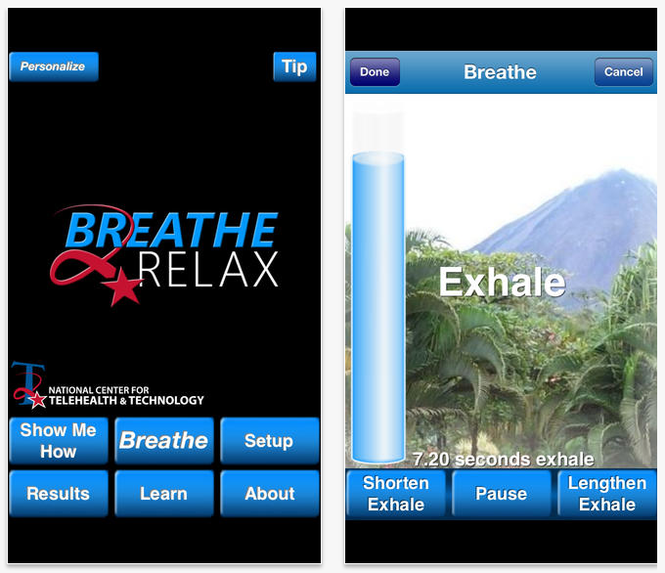
When you are ready to work through the breathing session, begin by rating your stress level. If you want to skip this step, go ahead and hit “Skip.” If you never want to enter this information when you enter the breathing session portion of the app, click the box that reads “Do Not Show Again” or return to the Setup menu and select “OFF” under Stress Tracking Information. However, by taking just a moment to note stress levels before (and after), you will see over time how you are performing and whether or not this is a beneficial exercise for you. Oftentimes, we don’t accurately perceive our own emotions and reactions, so graphing this output can help limit personal bias.
If you are following the breathing metronome, you will see it rise and fall with each breath until the session is complete. You will then be prompted to record your ending stress level and can click “Finish” to return to the home screen.
Results
The benefit of tracking stress levels during breathing sessions is revealed in a simple line graph. A thin line is produced using starting stress levels and a thick line is produced using ending stress levels. Sometimes the breathing session will result in a dramatic shift in stress level reduction, sometimes an insignificant reduction, and sometimes it may seemingly increase stress levels. If the latter happens, it is possible you were unable to focus on the exercise enough to properly engage in diaphragmatic breathing, or maybe one of the app settings has agitated you and it’s time to update your settings in the Setup menu.

Learning
Read about or watch information on stress by selecting 1 of 3 topics. “Biology of Stress” covers what happens in the body during stress, the consequences of stress on the body, how breathing can help control the stress response, and the resilience of the mind and body after handling a stressful episode. “Diaphragmatic Breathing” explains what the diaphragm is, the difference in oxygen levels supplied to the body during chest breathing and diaphragmatic breathing, and how to determine what kind of a breather you are. Access the Body Scanner under “Effects of Stress on the Body” to learn about the effects of stress on 10 different areas of the body.
Wellness Tips
On the home screen is a button called “Tip.” Tucked away in this little, unsuspecting corner of the app are wellness tips, such as “If you’re feeling guilty about something, remember that self-forgiveness means recognizing mistakes and accepting shortcomings” and “Don’t believe everything you think: Challenge your negative thoughts. Is there evidence to support the way you perceive the situation?” These tips don’t rotate frequently, but it’s a nice surprise when a new one displays.
Final Thoughts
This app can be used in several different ways, serving as a stand-alone stress management tool or in tandem with clinical care directed by a health care provider. As is noted by T2, “Due to its portability, this guided exercise is easily accessible when it is needed most.” And although the app was designed for individuals with PTSD and TBI, anybody who wants to feel more relaxed can access this free tool and reap the many benefits provided through diaphragmatic breathing.
The Breathe2Relax app is centered around the basic concept that breathing into the belly—diaphragmatic breathing—provides deeper relaxation than does breathing into the chest. Designed for use by individuals with PTSD and TBI, the app helps with mood stabilization, anger control, and anxiety management. It is a portable stress management tool with breathing exercises documented to decrease the body’s “fight-or-flight” stress response.
Setup
Although you can jump right into the guided breathing exercises, you may find a richer experience by exploring the setup menu of the app before beginning. (TIP: Skip the Personalize button and go straight to Setup.) Setting up the app to your personal preferences will reduce the chances of growing agitated when you begin the breathing exercises.
Almost every element you encounter while using the app can be modified: scenery (eg, mountain meadows or the cosmos), background music (mostly instrumental and earth sounds, plus a choice for no music at all), inhale and exhale lengths set to tenths of a second, whether a breathing metronome and visual prompts will display during the exercise, and whether a voice will prompt breathing instructions. Additional modifications include how many cycles of inhalations/exhalations will be provided, whether you want to track your stress levels (these can be graphed later and tracked over time), and, importantly, whether you want this data to be saved and transmitted to T2 for survey data (there is a choice to remain anonymous) or to disenroll from the study and delete all stored information.
Breathe
Once all of your preferences are set, they will remain saved in the app until you change them. If you already know how to breathe diaphragmatically, click “Breathe” to begin a breathing session. If you are uncertain or need a refresher, click “Show Me How” to watch a 2-minute instructional video. As noted during this instruction, “Be patient. Although breathing sounds like it should be easy to do, diaphragmatic breathing takes practice.” It is recommended that this practice be done every day for maximum benefit, and it is okay to practice when you’re already feeling relaxed.
When you are ready to work through the breathing session, begin by rating your stress level. If you want to skip this step, go ahead and hit “Skip.” If you never want to enter this information when you enter the breathing session portion of the app, click the box that reads “Do Not Show Again” or return to the Setup menu and select “OFF” under Stress Tracking Information. However, by taking just a moment to note stress levels before (and after), you will see over time how you are performing and whether or not this is a beneficial exercise for you. Oftentimes, we don’t accurately perceive our own emotions and reactions, so graphing this output can help limit personal bias.
If you are following the breathing metronome, you will see it rise and fall with each breath until the session is complete. You will then be prompted to record your ending stress level and can click “Finish” to return to the home screen.
Results
The benefit of tracking stress levels during breathing sessions is revealed in a simple line graph. A thin line is produced using starting stress levels and a thick line is produced using ending stress levels. Sometimes the breathing session will result in a dramatic shift in stress level reduction, sometimes an insignificant reduction, and sometimes it may seemingly increase stress levels. If the latter happens, it is possible you were unable to focus on the exercise enough to properly engage in diaphragmatic breathing, or maybe one of the app settings has agitated you and it’s time to update your settings in the Setup menu.
Learning
Read about or watch information on stress by selecting 1 of 3 topics. “Biology of Stress” covers what happens in the body during stress, the consequences of stress on the body, how breathing can help control the stress response, and the resilience of the mind and body after handling a stressful episode. “Diaphragmatic Breathing” explains what the diaphragm is, the difference in oxygen levels supplied to the body during chest breathing and diaphragmatic breathing, and how to determine what kind of a breather you are. Access the Body Scanner under “Effects of Stress on the Body” to learn about the effects of stress on 10 different areas of the body.
Wellness Tips
On the home screen is a button called “Tip.” Tucked away in this little, unsuspecting corner of the app are wellness tips, such as “If you’re feeling guilty about something, remember that self-forgiveness means recognizing mistakes and accepting shortcomings” and “Don’t believe everything you think: Challenge your negative thoughts. Is there evidence to support the way you perceive the situation?” These tips don’t rotate frequently, but it’s a nice surprise when a new one displays.
Final Thoughts
This app can be used in several different ways, serving as a stand-alone stress management tool or in tandem with clinical care directed by a health care provider. As is noted by T2, “Due to its portability, this guided exercise is easily accessible when it is needed most.” And although the app was designed for individuals with PTSD and TBI, anybody who wants to feel more relaxed can access this free tool and reap the many benefits provided through diaphragmatic breathing.
Deployment-Related Brain Injury Is Strongly Associated With Migraine in Iraq and Afghanistan Veterans
Veterans who were deployed to combat zones in the Iraq and Afghanistan wars and experienced traumatic brain injury (TBI) have a strong and highly significant increase in the frequency and intensity of headaches, the majority of which are migraines, according to researchers.
The incidence of chronic daily headache (ie, 15 or more headache days per month) was three times greater, compared with controls, and the incidence of frequent headache (ie, 10 to 14 headache days per month) was 4.5 times greater in these soldiers than in control groups, reported lead author James R. Couch, MD, of the University of Oklahoma School of Medicine in Oklahoma City, and colleagues. “Combat zone deployment by itself is stressful. Since TBI is the signature injury of these wars and occurs in 15% to 20% of deployed soldiers, and both TBI and stress are known to be associated with headache, we sought to evaluate the differences in headache occurrence and severity between those who were deployed and those who were deployed and also experienced a TBI,” Dr. Couch said.
Dr. Couch and his team evaluated 53 pairs of deployed veterans with TBI and a matched group of veterans who were deployed but did not sustain a TBI (controls). All subjects with deployment-related TBI had headache, while 11 (23.9%) controls had no headache. In addition, 89% of headaches in the deployed veterans with TBI were migraine, compared with 40% in the control group. All subjects with deployment-related TBI reported significantly greater frequency and intensity of headache than the control group did.
New and Noteworthy Information—June 2014
Older patients with migraine may be more likely to have silent brain injury than older patients without migraine, according to research published online ahead of print May 15 in Stroke. Researchers analyzed data from the Northern Manhattan Study, which quantified subclinical brain infarctions and white matter hyperintensity volumes among participants with migraine. Of the 546 participants analyzed, 41% were men, 65% were Hispanic, and mean age at MRI was 71. Patients with migraine had double the odds of subclinical brain infarction, compared with those reporting no migraine, after the investigators adjusted for sociodemographics and vascular risk factors. No association was observed between migraine with or without aura and white matter hyperintensity volume. Patients with migraine should not worry, because their risk of ischemic stroke is small, said the authors.
People who are exposed to paint, glue, or degreaser fumes at work may experience memory and thinking problems in retirement, according to a study published May 13 in Neurology. Researchers examined data for 2,143 retired utility workers who underwent cognitive testing in 2010. The authors assessed workers’ lifetime exposure to chlorinated solvents, petroleum solvents, and benzene using a job exposure matrix. Approximately 33% of participants were exposed to chlorinated solvents, 26% to benzene, and 25% to petroleum solvents. Workers highly exposed to chlorinated solvents were at risk of impairment on the Mini-Mental State Examination, the Digit Symbol Substitution Test, semantic fluency test, and the Trail Making Test B. Retirees at greatest risk for deficits had high lifetime exposure to solvents and were last exposed 12 to 30 years before testing.
Females susceptible to multiple sclerosis (MS) produce higher levels of the blood vessel receptor protein S1PR2 than males, according to data published online ahead of print May 8 in the Journal of Clinical Investigation. S1PR2 is present at high levels in the brain areas that MS typically damages. Investigators studied a mouse model of MS and found increased activity of S1PR2, which opens up the blood–brain barrier. When the researchers tested brain tissue samples obtained from 20 human patients after death, they found more S1PR2 in patients with MS than in those without the disorder. Brain tissue from females also had higher levels of S1PR2, compared with male brain tissue. These findings may help explain why more women than men get the disease, said the authors.
The FDA has required the manufacturer of the sleep drug Lunesta (eszopiclone) to lower the recommended starting dose from 2 mg to 1 mg for men and women. Data show that eszopiclone levels in some patients may be high enough on the morning after treatment to impair activities that require alertness, including driving. The 1-mg dose, taken at bedtime, can be increased to 2 mg or 3 mg if needed, but the higher doses are more likely to result in next-day impairment. Using lower doses ensures that less drug will remain in the body during the morning hours. Patients currently taking the 2-mg and 3-mg doses of Lunesta should contact their health care professional to ask for instructions, according to the FDA.
The rate of visits to an emergency department (ED) for traumatic brain injury (TBI) increased by approximately 30% between 2006 and 2010, according to research published in the May 14 issue of JAMA. The increase may be attributable to various factors, including increased awareness and diagnoses, said the authors. The investigators examined data from the Nationwide Emergency Department Sample database to determine national trends in ED visits for TBI from 2006 through 2010. An estimated 2.5 million ED visits for TBI occurred in 2010, representing a 29% increase in the rate of visits for TBI during the study period. By comparison, total ED visits increased by 3.6%. Children younger than 3 and adults older than 60 had the largest increase in TBI rates.
The pathophysiologic biomarkers and the topographic markers of Alzheimer’s disease should be revised, according to a position paper by the International Working Group published in the June issue of Lancet Neurology. The group proposed that biomarkers of Alzheimer’s pathology be restricted to those indicating the presence of tau pathology (ie, CSF or PET tau) and amyloid pathology (ie, CSF or PET amyloid). These biomarkers are specific enough to diagnose Alzheimer’s disease at any point on the disease continuum, said the authors. Downstream topographic markers of brain regional structural and metabolic changes have insufficient pathologic specificity and should not be used in diagnosis, according to the researchers. Instead, these markers can be used to measure disease progression. The group also provided diagnostic criteria for atypical, mixed, and preclinical Alzheimer’s disease.
Prenatal supplementation with docosahexaenoic acid (DHA) does not result in improved cognitive, problem-solving, or language abilities for children at age 4, according to the results of a trial published in the May 7 issue of JAMA. Investigators conducted longer-term follow-up from a previous study in which pregnant women received 800 mg/day of DHA or placebo. In the initial study, the researchers found that average cognitive, language, and motor scores did not differ between children at 18 months of age. Approximately 92% of eligible families participated in the follow-up study. The DHA group included 313 participants, and the control group included 333 participants. The investigators found that measures of cognition, the ability to perform complex mental processing, language, and executive functioning (eg, memory, reasoning, and problem solving) did not differ significantly between groups at age 4.
The FDA has informed Acorda Therapeutics that it has completed its review of the company’s new drug application for Plumiaz (diazepam) nasal spray and that the application cannot be approved in its present form. The drug was developed for the treatment of people with epilepsy who experience cluster seizures. Acorda Therapeutics is developing a response to address the items outlined in the letter. Based on the requirements for approval outlined in the letter, the company does not expect Plumiaz to receive FDA approval in 2014. Plumiaz previously received orphan drug designation for the treatment of cluster seizures. [For related news, see page 9.]
Older people with memory and thinking problems who do not have dementia may have a lower risk of dying from cancer than people who have no memory and thinking problems, according to a study published April 22 in Neurology. Researchers studied 2,627 people age 65 and older who did not have dementia at baseline. Participants underwent tests of memory and thinking skills at baseline and at three years. Follow-up lasted for an average of approximately 13 years. During the study, 1,003 participants died. About 34% of deaths occurred among patients with the fastest decline in thinking skills. Approximately 21% of participants in the group with the fastest decline in thinking skills died of cancer, compared with 29% of participants in the other two groups.
A new technique may predict with 95% accuracy which patients with stroke will benefit from IV t-PA and which will have potentially lethal bleeding in the brain, according to a study published online ahead of print May 15 in Stroke. Researchers used a computer program that shows physicians the amount of gadolinium, injected during an MRI scan, that has leaked into the brain tissue from surrounding blood vessels. By quantifying this damage in 75 patients with stroke, the researchers identified a threshold for determining how much leakage was dangerous. They applied this threshold to the records for the 75 patients to determine how well it would predict who had had a brain hemorrhage and who had not. The new test correctly predicted the outcome with 95% accuracy.
Freezing of gait in patients with Parkinson’s disease may correlate with poor quality of life, disease severity, apathy, and exposure to antimuscarinics, according to a study published online ahead of print May 19 in JAMA Neurology. Investigators performed a cross-sectional survey of 672 patients with idiopathic Parkinson’s disease. Patients with freezing of gait were identified as those with a score of 1 or greater on item 14 of the Unified Parkinson’s Disease Rating Scale (UPDRS) in the on condition. Approximately 38% of patients reported freezing of gait during the on state, which was significantly related to lower quality of life scores. Freezing of gait was also correlated with longer disease duration, higher UPDRS parts II and III scores, apathy, and a higher levodopa equivalent daily dose.
Among college football players, concussion and years of football played may have a significant inverse relationship with hippocampal volume, according to research published May 14 in JAMA. Years of football experience also may correlate with slower reaction time. Investigators conducted a cross-sectional study of 25 college football players with a history of clinician-diagnosed concussion, 25 college football players without a history of concussion, and 25 nonfootball-playing, age-, sex-, and education-matched healthy controls. Players with and without a history of concussion had smaller hippocampal volumes, compared with healthy controls. Players with a history of concussion had smaller hippocampal volumes than players without concussion. In both athlete groups, investigators found a statistically significant inverse relationship between left hippocampal volume and number of years of football played.
Deficiencies in hyaluronan can lead to spontaneous epileptic seizures, according to research published April 30 in the Journal of Neuroscience. In a multicenter study, investigators examined the role of hyaluronan using mutant mice deficient in each of the three hyaluronan synthase genes (ie, Has1, Has2, Has3). The mutant mice were prone to epileptic seizures. In Has3(-/-) mice, this phenotype likely results from a reduction in the size of the brain extracellular space (ECS), said the researchers. Among the three Has knockout models, seizures were most prevalent in Has3(-/-) mice, which also had the greatest hyaluronan reduction in the hippocampus. The results provide the first direct evidence for the physiologic role of hyaluronan in the regulation of ECS volume, according to the investigators.
—Erik Greb

Concussion—A Public Health Crisis
SAN FRANCISCO—Concussion is a public health crisis, given the huge population at risk and the limitations of current diagnostic and treatment methods, according to an overview on imaging concussion presented at the Seventh Annual Winter Conference of the Headache Cooperative of the Pacific.
Although many experts consider concussion to be a functional disturbance rather than a structural injury, it is associated with structural abnormalities. Similarly, although guidelines state that evidence of concussion is rarely found on conventional neuroimaging, some neurologists question this assertion.
“Concussion is a structural injury, and in most cases it is not normal on standard imaging modalities,” said David W. Dodick, MD, Professor of Neurology at the Mayo Clinic in Phoenix. But many concussions are silent, and most go unreported, often because an athlete wants to continue to play in a game. In addition, neurologists have no biomarker for concussion and no definitive treatment.
Players Endure Multiple Hits
Although a single concussion can have long-term sequelae, repeated hits to the head are of particular concern. A recent study found that varsity football and hockey players sustain an average of 1,000 hits each season at a mean acceleration of 20 G—a force comparable to that of a car driving at 35 mph and hitting a brick wall, said Dr. Dodick.
In another recent study, researchers used functional MRI to test cognition in football players ages 15 to 19 before and after the playing season. Although none of the athletes tested had had a symptomatic concussion, their ability to complete the assigned task and their overall cognitive function were markedly lower postseason. The extent of impairment correlated with the number of hits to the head that they had sustained.
The near death of a high school football player from second impact syndrome led to laws limiting postconcussion return to play in all 50 states. Although children may be sidelined for 30 days, however, cognitive symptoms can take six weeks or more to resolve—and 44% to 50% of athletes have three or more symptoms at one year.
Researchers Find Evidence of Degenerative Disease
After repeated head injuries, athletes are at high risk for chronic traumatic encephalopathy (CTE), the progressive neurodegenerative disease that helped raise public awareness of the danger associated with sports-related trauma. Researchers at Boston University have examined the brains of more than 100 deceased athletes and found evidence of tauopathy in 70 of them, reported Dr. Dodick. Unlike the neurofibrillary tangles found in Alzheimer’s disease, those characteristic of CTE cluster in the subcortical regions of the brain.
Researchers at the University of California, Los Angeles, successfully used PET scans with tau ligand to detect in vivo evidence of CTE. They scanned the brains of five retired football players and found tau deposits associated with trauma in each.
“Metabolic brain imaging with tau ligand is perhaps one of the greatest advances in the field,” said Dr. Dodick. Although CTE usually develops within five to 10 years of an athlete’s retirement, early indicators of the disease were found in the brain of a 17-year-old football player who committed suicide.
When Is Postconcussion Imaging Indicated?
The Institute of Medicine’s sports-related concussion guideline states that traditional CT and MRI are of little diagnostic value. The American Academy of Neurology calls for CT only when a more serious traumatic brain injury (TBI) is suspected. The Zurich consensus statement concludes that CT and MRI contribute little to the evaluation of concussion.
“If I stopped here, you would go back and say, ‘There is no need to image the brain in routine concussion,’” Dr. Dodick said. He went on to review both traditional and newer imaging modalities and the Mayo Clinic’s unvalidated concussion protocol, which recommends imaging for every patient.
CT Is Overused, but MRI Can Be Revealing
CT is the mainstay of imaging TBI in an emergency setting, but it has little value in the evaluation of concussion in outpatient settings, said Dr. Dodick. Even in emergency departments, overuse is increasingly common and costly and unnecessarily exposes patients to high doses of radiation.
Although the role of MRI in concussion is controversial, “we image everybody who comes to us for concussion evaluation,” said Dr. Dodick. MRI can detect contusions, edema, traumatic axonal injury (TAI), and subacute or remote hemorrhages that CT often misses. In a study of 135 patients with mild TBI, 27% of those with normal CT scans had abnormal MRIs.
Susceptibility-Weighted Imaging
The Mayo Clinic’s protocol includes susceptibility weighted imaging (SWI), with brain imaging sequences including sagittal T1 and axial 3D T1, axial T2, 3D fluid attenuated inversion recovery, single voxel magnetic resonance spectroscopy (MRS), and axial diffusion tensor imaging (DTI). SWI “is probably the most important thing we do,” said Dr. Dodick.
He recalled the case of a 19-year-old hockey player whom he was asked to clear for return to play. Gradient echo imaging was normal, as were the athlete’s neurologic exam and impact test, but SWI revealed multiple areas of TAI.
Magnetic Resonance Spectroscopy
Sometimes referred to as a virtual biopsy, MRS detects changes in metabolites in the brain. Most notably, the technique identifies changes in N-acetylaspartate (NAA), a marker of neuronal and axonal health that declines in TBI.
In a study of professional soccer players with a baseline NAA:creatine ratio greater than 2, participants’ postconcussion ratio was less than 2. Although the athletes reported complete symptom resolution on day 3, brain imaging did not return to normal for one month or longer. Because of the lag time between symptom resolution and metabolic recovery, Dr. Dodick said, “I’m cautious, with children and youth in particular, about sending them back to play.”
Diffusion Tensor Imaging
DTI, another element of the Mayo Clinic’s concussion protocol, is based on the diffusivity of water, which is variably restricted in different tissues. In CSF, for example, diffusion of water is isotropic. In white matter, diffusion is highly anisotropic, and a reduction of fractional anisotropy (FA) correlates with the loss or destruction of white matter.
Multiple DTI studies have been conducted in the past 30 years. Despite various limitations, the findings provide striking evidence of small and subtle brain abnormalities in most athletes, said Dr. Dodick. A study comparing patients with mild TBI vs patients with moderate-to-severe TBI, for example, found a graded alteration in white matter integrity. Patients with mild TBI also had abnormalities.
Notably, evidence of abnormalities has been found even in the absence of symptomatic concussion. “You can alter brain structure and function without clinical symptomatology,” said Dr. Dodick.
Patients may ask whether these brain changes are reversible. Dr. Dodick described a patient who underwent DTI immediately after concussion and again 16 months later. At the time of the second imaging, the abnormal reduction of FA showed a loss of integrity. “That [injury] probably does not repair itself,” he said. At least in some cases, the changes can be persistent.
Current Imaging Can Guide Clinical Practice
“There is great potential for advanced brain imaging of concussion,” said Dr. Dodick, “but you can detect and characterize brain abnormalities that will guide your clinical decision making” here and now. Even if testing is limited to SWI, he added, it will identify many abnormalities that otherwise would be missed.
If in vivo testing for CTE is proven to be effective, “we’ll be able to premortem early detect those at risk for neurodegenerative disease,” said Dr. Dodick. “The holy grail, of course, is that this would be used in future research to evaluate interventions and new treatments.”
—Helen Lippman
Suggested Reading
Hart J Jr, Kraut MA, Womack KB, et al. Neuroimaging of cognitive dysfunction and depression in aging retired National Football League players: a cross-sectional study. JAMA Neurol. 2013;70(3):326-335.
Tavender EJ, Bosch M, Green S, et al. Quality and consistency of guidelines for the management of mild traumatic brain injury in the emergency department. Acad Emerg Med. 2011;18(8):880-889.
Yuh EL, Mukherjee P, Lingsma HF, et al. Magnetic resonance imaging improves 3-month outcome prediction in mild traumatic brain injury. Ann Neurol. 2013;73(2):224-235.
SAN FRANCISCO—Concussion is a public health crisis, given the huge population at risk and the limitations of current diagnostic and treatment methods, according to an overview on imaging concussion presented at the Seventh Annual Winter Conference of the Headache Cooperative of the Pacific.
Although many experts consider concussion to be a functional disturbance rather than a structural injury, it is associated with structural abnormalities. Similarly, although guidelines state that evidence of concussion is rarely found on conventional neuroimaging, some neurologists question this assertion.
“Concussion is a structural injury, and in most cases it is not normal on standard imaging modalities,” said David W. Dodick, MD, Professor of Neurology at the Mayo Clinic in Phoenix. But many concussions are silent, and most go unreported, often because an athlete wants to continue to play in a game. In addition, neurologists have no biomarker for concussion and no definitive treatment.
Players Endure Multiple Hits
Although a single concussion can have long-term sequelae, repeated hits to the head are of particular concern. A recent study found that varsity football and hockey players sustain an average of 1,000 hits each season at a mean acceleration of 20 G—a force comparable to that of a car driving at 35 mph and hitting a brick wall, said Dr. Dodick.
In another recent study, researchers used functional MRI to test cognition in football players ages 15 to 19 before and after the playing season. Although none of the athletes tested had had a symptomatic concussion, their ability to complete the assigned task and their overall cognitive function were markedly lower postseason. The extent of impairment correlated with the number of hits to the head that they had sustained.
The near death of a high school football player from second impact syndrome led to laws limiting postconcussion return to play in all 50 states. Although children may be sidelined for 30 days, however, cognitive symptoms can take six weeks or more to resolve—and 44% to 50% of athletes have three or more symptoms at one year.
Researchers Find Evidence of Degenerative Disease
After repeated head injuries, athletes are at high risk for chronic traumatic encephalopathy (CTE), the progressive neurodegenerative disease that helped raise public awareness of the danger associated with sports-related trauma. Researchers at Boston University have examined the brains of more than 100 deceased athletes and found evidence of tauopathy in 70 of them, reported Dr. Dodick. Unlike the neurofibrillary tangles found in Alzheimer’s disease, those characteristic of CTE cluster in the subcortical regions of the brain.
Researchers at the University of California, Los Angeles, successfully used PET scans with tau ligand to detect in vivo evidence of CTE. They scanned the brains of five retired football players and found tau deposits associated with trauma in each.
“Metabolic brain imaging with tau ligand is perhaps one of the greatest advances in the field,” said Dr. Dodick. Although CTE usually develops within five to 10 years of an athlete’s retirement, early indicators of the disease were found in the brain of a 17-year-old football player who committed suicide.
When Is Postconcussion Imaging Indicated?
The Institute of Medicine’s sports-related concussion guideline states that traditional CT and MRI are of little diagnostic value. The American Academy of Neurology calls for CT only when a more serious traumatic brain injury (TBI) is suspected. The Zurich consensus statement concludes that CT and MRI contribute little to the evaluation of concussion.
“If I stopped here, you would go back and say, ‘There is no need to image the brain in routine concussion,’” Dr. Dodick said. He went on to review both traditional and newer imaging modalities and the Mayo Clinic’s unvalidated concussion protocol, which recommends imaging for every patient.
CT Is Overused, but MRI Can Be Revealing
CT is the mainstay of imaging TBI in an emergency setting, but it has little value in the evaluation of concussion in outpatient settings, said Dr. Dodick. Even in emergency departments, overuse is increasingly common and costly and unnecessarily exposes patients to high doses of radiation.
Although the role of MRI in concussion is controversial, “we image everybody who comes to us for concussion evaluation,” said Dr. Dodick. MRI can detect contusions, edema, traumatic axonal injury (TAI), and subacute or remote hemorrhages that CT often misses. In a study of 135 patients with mild TBI, 27% of those with normal CT scans had abnormal MRIs.
Susceptibility-Weighted Imaging
The Mayo Clinic’s protocol includes susceptibility weighted imaging (SWI), with brain imaging sequences including sagittal T1 and axial 3D T1, axial T2, 3D fluid attenuated inversion recovery, single voxel magnetic resonance spectroscopy (MRS), and axial diffusion tensor imaging (DTI). SWI “is probably the most important thing we do,” said Dr. Dodick.
He recalled the case of a 19-year-old hockey player whom he was asked to clear for return to play. Gradient echo imaging was normal, as were the athlete’s neurologic exam and impact test, but SWI revealed multiple areas of TAI.
Magnetic Resonance Spectroscopy
Sometimes referred to as a virtual biopsy, MRS detects changes in metabolites in the brain. Most notably, the technique identifies changes in N-acetylaspartate (NAA), a marker of neuronal and axonal health that declines in TBI.
In a study of professional soccer players with a baseline NAA:creatine ratio greater than 2, participants’ postconcussion ratio was less than 2. Although the athletes reported complete symptom resolution on day 3, brain imaging did not return to normal for one month or longer. Because of the lag time between symptom resolution and metabolic recovery, Dr. Dodick said, “I’m cautious, with children and youth in particular, about sending them back to play.”
Diffusion Tensor Imaging
DTI, another element of the Mayo Clinic’s concussion protocol, is based on the diffusivity of water, which is variably restricted in different tissues. In CSF, for example, diffusion of water is isotropic. In white matter, diffusion is highly anisotropic, and a reduction of fractional anisotropy (FA) correlates with the loss or destruction of white matter.
Multiple DTI studies have been conducted in the past 30 years. Despite various limitations, the findings provide striking evidence of small and subtle brain abnormalities in most athletes, said Dr. Dodick. A study comparing patients with mild TBI vs patients with moderate-to-severe TBI, for example, found a graded alteration in white matter integrity. Patients with mild TBI also had abnormalities.
Notably, evidence of abnormalities has been found even in the absence of symptomatic concussion. “You can alter brain structure and function without clinical symptomatology,” said Dr. Dodick.
Patients may ask whether these brain changes are reversible. Dr. Dodick described a patient who underwent DTI immediately after concussion and again 16 months later. At the time of the second imaging, the abnormal reduction of FA showed a loss of integrity. “That [injury] probably does not repair itself,” he said. At least in some cases, the changes can be persistent.
Current Imaging Can Guide Clinical Practice
“There is great potential for advanced brain imaging of concussion,” said Dr. Dodick, “but you can detect and characterize brain abnormalities that will guide your clinical decision making” here and now. Even if testing is limited to SWI, he added, it will identify many abnormalities that otherwise would be missed.
If in vivo testing for CTE is proven to be effective, “we’ll be able to premortem early detect those at risk for neurodegenerative disease,” said Dr. Dodick. “The holy grail, of course, is that this would be used in future research to evaluate interventions and new treatments.”
—Helen Lippman
Suggested Reading
Hart J Jr, Kraut MA, Womack KB, et al. Neuroimaging of cognitive dysfunction and depression in aging retired National Football League players: a cross-sectional study. JAMA Neurol. 2013;70(3):326-335.
Tavender EJ, Bosch M, Green S, et al. Quality and consistency of guidelines for the management of mild traumatic brain injury in the emergency department. Acad Emerg Med. 2011;18(8):880-889.
Yuh EL, Mukherjee P, Lingsma HF, et al. Magnetic resonance imaging improves 3-month outcome prediction in mild traumatic brain injury. Ann Neurol. 2013;73(2):224-235.
Link Between PTSD and TBI Is Only the Beginning for MRS Study
April 25, 2014
A fundamental challenge for any study examining the impact of military service on the health of military personnel is establishing a baseline. Whether heart disease or posttraumatic stress disorder (PTSD), the symptoms often appear after (sometimes long after) the service has ended. The longitudinal Marine Resiliency Study (MRS I) and its successor MRS II are seeking to resolve that issue in a novel approach that brings together the Department of Veterans Affairs, U.S. Marine Corps, and Navy Medicine.
In the MRS study, a cohort of about 2,600 Marines (MRS-I) in 4 battalions and about 1,300 Marines (MRS-II) in 2 battalions deployed to Iraq or Afghanistan underwent a scientifically rigorous examination a month prior to deployment. This baseline was established using self-reported questionnaires, clinical interviews, and laboratory examinations. Follow-up examinations were repeated at 3 months (MRS-I and MRS-II) and again at 6 months post-deployment (MRS-I).

The program is ambitious, Dr. Dewleen Baker of the VA San Diego Health Care System told Federal Practitioner. “MRS was designed to provide broad-based (psychosocial, psychophysiological, and biological) prospective, longitudinal data, with a goal toward ultimate integrated analyses of variables, to determine risk and resilience for post-deployment mental health outcomes, i.e,. PTSD and co-occurring disorders,” she explained. “Analyses have just begun, and we are working our way through aspects of the data toward more integrated approaches.”
In one of the first of many reports to come out of MRS, the researchers found that the probability of developing PTSD was highest for participants with severe pre-deployment symptoms, high combat intensity, and deployment-related traumatic brain injury (TBI). Most significant, the researchers found that TBI doubled or nearly doubled the PTSD rates for participants with less severe pre-deployment PTSD symptoms. According to Baker:
By contrast, deployment-related mild TBI increases post-deployment symptom scores by 23%, and moderate-to-severe injuries increase scores by 71%. Our findings suggest that TBI may be a very important risk factor of PTSD, even when accounting for preexisting symptoms and combat intensity.
Our study focused on the impact of pre-deployment symptoms, combat intensity and TBI; however, it is important to consider other factors of psychological risk and resilience. Genes, coping style, and social support are just a few of the many other factors that may influence an individual’s response to stress.
Creating a rigorous cross-agency research study required tact, diligence, and patience from the MRS team. “Each agency has their own unique culture and institutional rules, regulations, and bureaucracy, so ideas, programs, etc, must be vetted across all agencies and reconciled—the various cultures/agencies to be reconciled include DoD, VA and academia.” Baker explained. “In addition in regards to initiation of studies for MRS II, for the past couple years, we also interface with NIMH as well as Headquarters Marine Corps; NIMH has the role of scientific review of MRS-II studies carried out under Headquarters Marine Corps/BUMED funding.”
The MRS-I and II studies may very well provide a template for future studies. The MRS team included a military liaison to work with the active duty Marines and attached Sailors, gather data, schedule meetings, and to report findings. “This study has a lot of experience working within and across these agencies,” Baker noted, “It’s an excellent model for future VA/DOD joint projects.”
April 25, 2014
A fundamental challenge for any study examining the impact of military service on the health of military personnel is establishing a baseline. Whether heart disease or posttraumatic stress disorder (PTSD), the symptoms often appear after (sometimes long after) the service has ended. The longitudinal Marine Resiliency Study (MRS I) and its successor MRS II are seeking to resolve that issue in a novel approach that brings together the Department of Veterans Affairs, U.S. Marine Corps, and Navy Medicine.
In the MRS study, a cohort of about 2,600 Marines (MRS-I) in 4 battalions and about 1,300 Marines (MRS-II) in 2 battalions deployed to Iraq or Afghanistan underwent a scientifically rigorous examination a month prior to deployment. This baseline was established using self-reported questionnaires, clinical interviews, and laboratory examinations. Follow-up examinations were repeated at 3 months (MRS-I and MRS-II) and again at 6 months post-deployment (MRS-I).
The program is ambitious, Dr. Dewleen Baker of the VA San Diego Health Care System told Federal Practitioner. “MRS was designed to provide broad-based (psychosocial, psychophysiological, and biological) prospective, longitudinal data, with a goal toward ultimate integrated analyses of variables, to determine risk and resilience for post-deployment mental health outcomes, i.e,. PTSD and co-occurring disorders,” she explained. “Analyses have just begun, and we are working our way through aspects of the data toward more integrated approaches.”
In one of the first of many reports to come out of MRS, the researchers found that the probability of developing PTSD was highest for participants with severe pre-deployment symptoms, high combat intensity, and deployment-related traumatic brain injury (TBI). Most significant, the researchers found that TBI doubled or nearly doubled the PTSD rates for participants with less severe pre-deployment PTSD symptoms. According to Baker:
By contrast, deployment-related mild TBI increases post-deployment symptom scores by 23%, and moderate-to-severe injuries increase scores by 71%. Our findings suggest that TBI may be a very important risk factor of PTSD, even when accounting for preexisting symptoms and combat intensity.
Our study focused on the impact of pre-deployment symptoms, combat intensity and TBI; however, it is important to consider other factors of psychological risk and resilience. Genes, coping style, and social support are just a few of the many other factors that may influence an individual’s response to stress.
Creating a rigorous cross-agency research study required tact, diligence, and patience from the MRS team. “Each agency has their own unique culture and institutional rules, regulations, and bureaucracy, so ideas, programs, etc, must be vetted across all agencies and reconciled—the various cultures/agencies to be reconciled include DoD, VA and academia.” Baker explained. “In addition in regards to initiation of studies for MRS II, for the past couple years, we also interface with NIMH as well as Headquarters Marine Corps; NIMH has the role of scientific review of MRS-II studies carried out under Headquarters Marine Corps/BUMED funding.”
The MRS-I and II studies may very well provide a template for future studies. The MRS team included a military liaison to work with the active duty Marines and attached Sailors, gather data, schedule meetings, and to report findings. “This study has a lot of experience working within and across these agencies,” Baker noted, “It’s an excellent model for future VA/DOD joint projects.”

Back to School With TBI
To show that veterans with traumatic brain injury (TBI) can successfully return to school, the Defense and Veterans Brain Injury Center (DVBIC) created Back to School: Guide to Academic Success After Traumatic Brain Injury, available at http://www.dvbic.org/material/back-school-guide. The guide offers information on a variety of pertinent subjects from symptom management to choosing a school and adjusting to civilian campus culture.
The detailed Frequently Asked Questions section offers tips on topics such as how to talk about TBI with professors, where to get help on campus, as well as financial aid and tuition assistance.
Sections of the guide address ways the service member-student can work through deficits; for example, learning new ways of doing things, such as using assistive technology or participating in online classes, which could be beneficial for someone who needs multiple breaks. Students with TBI may be able to arrange to get extra time to finish assignments, take tests in different formats, or use tape recorders instead of taking notes by hand.
In the Maximize Your Learning section, students can tailor individual solutions to learning problems, such as attention issues and thought processing. For the student who has trouble remembering recent conversations, directions, and due dates, “Tips for Success” include having a large calendar in view, recording and playing back lectures, and recapping conversations.
The Make the Transition section reminds the service member of strengths gained in the military that can translate to the school environment: leadership, discipline, teamwork, communication, determination, punctuality. These skills may help the student identify goals quickly, work well on team projects, and meet deadlines.
The guide offers students examples of cognitive technology they can take advantage of, such as a screen reader, which identifies and interprets written material on a computer screen and reads the material out loud.
According to the DVBIC, health care providers can also use the guide as a teaching tool to help patients.