User login
New Center of Excellence to Lead Research of “Signature Wounds”
Take a brand-new research facility, then add a neighboring U.S. Army base with one of the largest veteran populations of any health care network and a world-class team of researchers—that’s a “recipe for success,” says Dr. Michael Russell, director of the VA Center of Excellence for Research on Returning War Veterans in Waco, Texas.
The 53,000-square-foot center is designed to conduct state-of-the-art research on mental health problems associated with PTSD and TBI, “signature wounds” of conflicts in Afghanistan and the Middle East. The flagship study is named Project MAVEREX. Researchers will examine whether the inability of the regions in injured brains to communicate with one another worsens behavior outcomes. Using “cutting-edge data analysis techniques,” they hope to characterize the effects of TBI on brain structure and function “with very high precision,” says Dr. Evan Gordon, a cognitive neuroscientist working on MAVEREX.
The Center of Excellence is on the campus of the historic Doris Miller VAMC. The facility has space for 75 staff members and faculty as well as 25 trainees. It features multiple examination rooms, observation rooms, electrocardiography, electroencephalography, a 3 Tesla MRI, a transcranial magnetic stimulation suite, and a custom-built laboratory wing.
Noninvasive Eye Tracking May Help to Assess the Physiologic Impact of Elevated Intracranial Pressure
Eye tracking is a noninvasive technique that may help to assess two key physiologic signs of concussion, intracranial pressure (ICP) and ocular motility dysfunction, according to a study published online ahead of print June 2 in the Journal of Neurosurgery. This technique does not require a trained examiner, pupil dilation, imaging studies, or an invasive procedure such as lumbar or ventricular puncture, the authors noted.
“With these data, we are presenting a new application for eye-tracking technology, as well as a new mechanism for assessment of elevated ICP that is noninvasive, automatable, and could potentially be performed and analyzed remotely,” said Uzma Samadani, MD, PhD, Associate Professor of Neurosurgery at the University of Minnesota in Minneapolis, and colleagues.

The boundaries of normal and elevated intracranial pressure vary between patients, said the authors. People with elevated intracranial pressure can develop abnormalities in global cerebral functioning. Elevated ICP can also affect the function of cranial nerves, which may contribute to ocular dysmotility. Dr. Samadani and colleagues assessed the impact of elevated ICP on eye-tracking sessions performed while patients watched a short film clip.
Eligible participants ranged in age from 18 to 70, were admitted to the Bellevue Hospital neurosurgical intensive care unit in New York City with vision correctable to within 20/500 bilaterally, and had denied a history of ocular dysmotility. In addition, these patients were conscious and able to communicate and to provide an ophthalmologic, medical, and neurologic history, as well as medications, drugs, and alcohol consumed within 24 hours prior to eye tracking.
Awake patients who required placement of an ICP monitor for clinical purposes underwent eye tracking while watching a 220-second continuously playing video. The investigators recorded pupil position at 500 Hz and calculated metrics associated with each eye individually and both eyes together. In addition, the researchers performed linear regression with generalized estimating equations to test the association of eye-tracking metrics with changes in ICP.
The investigators performed eye tracking at ICP levels ranging from –3 mm Hg to 30 mm Hg in 23 patients (12 women, mean age 46.8) on 55 occasions. Eye-tracking measures correlating with cranial nerve function decreased linearly with increasing ICP.
Researchers also found that measures for cranial nerve VI were the most prominently affected. The area under the curve for eye-tracking metrics to discriminate between an ICP <12 mm Hg and one of ≥12 mm Hg was 0.798. To discriminate between an ICP <15 mm Hg and one of ≥15 mm Hg, the area under the curve was 0.833. Finally, to discriminate between an ICP <20 mm Hg and ≥20 mm Hg, the area under the curve was 0.889.
Overall, increasingly elevated ICP was associated with increasingly abnormal eye tracking detected while patients were watching the short film. The “technology has clinical applications for assessment of shunt malfunction, pseudotumor cerebri, concussion, and prevention of second-impact syndrome,” said Dr. Samadani and colleagues.
The major limitation of this study was the lack of continuous data in patients with higher ICP recordings, the authors said. Few patients with elevated ICP could open their eyes long enough to undergo eye tracking.
—Erica Tricarico
Suggested Reading
Kolecki R, Dammavalam V, Zahid A, et al. Elevated intracranial pressure and reversible eye-tracking changes detected while viewing a film clip. J Neurosurg. 2017 Jun 2 [Epub ahead of print].
Eye tracking is a noninvasive technique that may help to assess two key physiologic signs of concussion, intracranial pressure (ICP) and ocular motility dysfunction, according to a study published online ahead of print June 2 in the Journal of Neurosurgery. This technique does not require a trained examiner, pupil dilation, imaging studies, or an invasive procedure such as lumbar or ventricular puncture, the authors noted.
“With these data, we are presenting a new application for eye-tracking technology, as well as a new mechanism for assessment of elevated ICP that is noninvasive, automatable, and could potentially be performed and analyzed remotely,” said Uzma Samadani, MD, PhD, Associate Professor of Neurosurgery at the University of Minnesota in Minneapolis, and colleagues.
The boundaries of normal and elevated intracranial pressure vary between patients, said the authors. People with elevated intracranial pressure can develop abnormalities in global cerebral functioning. Elevated ICP can also affect the function of cranial nerves, which may contribute to ocular dysmotility. Dr. Samadani and colleagues assessed the impact of elevated ICP on eye-tracking sessions performed while patients watched a short film clip.
Eligible participants ranged in age from 18 to 70, were admitted to the Bellevue Hospital neurosurgical intensive care unit in New York City with vision correctable to within 20/500 bilaterally, and had denied a history of ocular dysmotility. In addition, these patients were conscious and able to communicate and to provide an ophthalmologic, medical, and neurologic history, as well as medications, drugs, and alcohol consumed within 24 hours prior to eye tracking.
Awake patients who required placement of an ICP monitor for clinical purposes underwent eye tracking while watching a 220-second continuously playing video. The investigators recorded pupil position at 500 Hz and calculated metrics associated with each eye individually and both eyes together. In addition, the researchers performed linear regression with generalized estimating equations to test the association of eye-tracking metrics with changes in ICP.
The investigators performed eye tracking at ICP levels ranging from –3 mm Hg to 30 mm Hg in 23 patients (12 women, mean age 46.8) on 55 occasions. Eye-tracking measures correlating with cranial nerve function decreased linearly with increasing ICP.
Researchers also found that measures for cranial nerve VI were the most prominently affected. The area under the curve for eye-tracking metrics to discriminate between an ICP <12 mm Hg and one of ≥12 mm Hg was 0.798. To discriminate between an ICP <15 mm Hg and one of ≥15 mm Hg, the area under the curve was 0.833. Finally, to discriminate between an ICP <20 mm Hg and ≥20 mm Hg, the area under the curve was 0.889.
Overall, increasingly elevated ICP was associated with increasingly abnormal eye tracking detected while patients were watching the short film. The “technology has clinical applications for assessment of shunt malfunction, pseudotumor cerebri, concussion, and prevention of second-impact syndrome,” said Dr. Samadani and colleagues.
The major limitation of this study was the lack of continuous data in patients with higher ICP recordings, the authors said. Few patients with elevated ICP could open their eyes long enough to undergo eye tracking.
—Erica Tricarico
Suggested Reading
Kolecki R, Dammavalam V, Zahid A, et al. Elevated intracranial pressure and reversible eye-tracking changes detected while viewing a film clip. J Neurosurg. 2017 Jun 2 [Epub ahead of print].
Treating Traumatic Injuries and the Issues They Cause
In the Madigan Intrepid Spirit Transitions (MIST) program, holistic treatment for traumatic brain injuries (TBIs) includes traditional and nontraditional therapies as well as a little help from friends.
MIST is a 6-week intensive outpatient group for service members who have TBIs and other traumatic injuries, along with coexisting conditions, such as chronic pain or posttraumatic stress. Coexisting conditions can make cases more complex, said U.S. Army Colonel Beverly Scott, medical and program director of Madigan Army Medical Center’s Traumatic Brain Injury Program and Intrepid Spirit Program in an interview with Health.mil News. But she adds, “It’s never too late to help [patients] address a number of issues they may be having following a traumatic brain injury, dealing with pain, dealing with behavior health issues.”
Related: Let’s Dance: A Holistic Approach to Treating Veterans With Posttraumatic Stress Disorder
The MIST program serves only active-duty service members with referral from their primary care managers and other specialty services at Madigan or throughout the Regional Health Command-Pacific. Commanders must sign memoranda of understanding that patients will be off duty rosters for the duration of the program. “They’re making a commitment to help that service member get better,” Scott said.
The MIST program enrolls 8 to 12 service members at a time. The holistic focus allows patients to address chronic pain, insomnia, and cognitive issues through traditional means as well as less traditional means that include mindfulness training; art; such as creating symbolic masks; and yoga. The variety of approaches lets them “cherry pick the methods they believe will help them the most,” the Health.mil News article reports, or what one member called “customizing their own multitool.”
Participants are encouraged to continue individual care within the TBI/Intrepid Spirit program. The MIST program aims to introduce them to the resources they can use going forward. Giving them tools they can use after they complete the program is an acknowledgment that the recovery process is ongoing. “We recognize it is a transition,” Scott said.
Related: Ideas for Helping TBI Patients
The MIST program has graduated 2 groups. Scott says, “We’ve seen incredible success,” both in wellness and in other areas, such as improved interpersonal relationships. Some of the credit goes to the peer support that MIST promotes. The curriculum is evidence based, but Scott says some “significant success is clearly related to soldiers helping soldiers.”
Long-Term Effects of Concussive TBI
What are the long-term clinical effects of wartime traumatic brain injuries (TBIs)? Most are mild, but in general all are incompletely described, say researchers from University of Washington in Seattle and Washington University in St. Louis, Missouri. However, their own study found that service members with even mild concussive TBI often “experienced evolution, not resolution” of symptoms.
The researchers compared the results of 1-year and 5-year clinical evaluations of 50 active-duty U.S. military with acute to subacute concussive blast injury and 44 deployed but uninjured service members. The evaluations included neurobehavioral and neuropsychological performance and mental health burden.
At 5 years, global disability, satisfaction with life, neurobehavioral symptom severity, psychiatric symptom severity, and sleep impairment were significantly worse in patients with concussive blast TBI. Of the patients with concussive blast TBI, 36 (72%) showed decline, compared with only 5 of the combat-deployed group (11%). The researchers also found symptoms of PTSD and depression worsened in the concussive TBI patients. Performance on cognitive measures was no different between the 2 groups. A combination of factors, including neurobehavioral symptom severity, walking ability, and verbal fluency at 1 year after injury, was highly predictive of poor outcomes 5 years later.
“This is one of the first studies to connect the dots from injury to longer term outcomes and it shows that even mild concussions can lead to long-term impairment and continued decline in satisfaction with life,” said lead author Christine L. Mac Donald, PhD. “Most physicians believe that patients will stabilize 6 to 12 months postinjury, but this study challenges that.”
Tips for Recovering From Concussion
Click here to download the PDF. 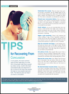
Click here to download the PDF.
Click here to download the PDF.

Are Women Athletes More Susceptible to Concussion?
BOSTON—Women athletes are 50% more likely to have a sports-related concussion than male athletes, according to research presented at the 69th Annual Meeting of the American Academy of Neurology.
“The findings from this study highlight the need for more research on the gender differences in concussion,” said James Noble, MD, Assistant Professor of Clinical Neurology at Columbia University in New York.

Little is known about the occurrence, severity, and recovery of sports-related concussion, especially among female athletes, since previous studies typically focused on male athletes. Gender-balanced studies have been limited by small sample size, incomplete or variable follow-up, or referral bias to tertiary concussion care centers. As a result, Dr. Noble and colleagues sought to determine gender differences in the incidence, symptomatology, neuropsychologic testing, and return-to-play length of sports-related concussion in collegiate varsity athletes.
For the study, Dr. Noble and colleagues followed 1,203 athletes at Columbia University from 2000 to 2014. In all, 822 of the participants were men, and 381 participants were women. All participants played sports such as soccer, basketball, and football.
Researchers assessed participants’ thinking skills and processing speed before and after a concussion. In addition, investigators tracked symptoms and when participants returned to play after a concussion.
A total of 228 athletes had at least one concussion, including 23% of the women (n = 88) and 17% of the men (n = 140). In addition, women who played soccer and basketball were more likely to have a concussion than their male counterparts. Finally, athletes who had had a previous concussion were three times more likely to have another concussion, compared with athletes who had never had a concussion.
The investigators also noted that women recovered from concussion about as quickly as men. Both men and women had a median return-to-play time of 10 days. Concussion symptoms were similar for men and women, although amnesia occurred more frequently in men (44% vs 31%), and insomnia occurred more frequently in women (42% vs 29%).
BOSTON—Women athletes are 50% more likely to have a sports-related concussion than male athletes, according to research presented at the 69th Annual Meeting of the American Academy of Neurology.
“The findings from this study highlight the need for more research on the gender differences in concussion,” said James Noble, MD, Assistant Professor of Clinical Neurology at Columbia University in New York.
Little is known about the occurrence, severity, and recovery of sports-related concussion, especially among female athletes, since previous studies typically focused on male athletes. Gender-balanced studies have been limited by small sample size, incomplete or variable follow-up, or referral bias to tertiary concussion care centers. As a result, Dr. Noble and colleagues sought to determine gender differences in the incidence, symptomatology, neuropsychologic testing, and return-to-play length of sports-related concussion in collegiate varsity athletes.
For the study, Dr. Noble and colleagues followed 1,203 athletes at Columbia University from 2000 to 2014. In all, 822 of the participants were men, and 381 participants were women. All participants played sports such as soccer, basketball, and football.
Researchers assessed participants’ thinking skills and processing speed before and after a concussion. In addition, investigators tracked symptoms and when participants returned to play after a concussion.
A total of 228 athletes had at least one concussion, including 23% of the women (n = 88) and 17% of the men (n = 140). In addition, women who played soccer and basketball were more likely to have a concussion than their male counterparts. Finally, athletes who had had a previous concussion were three times more likely to have another concussion, compared with athletes who had never had a concussion.
The investigators also noted that women recovered from concussion about as quickly as men. Both men and women had a median return-to-play time of 10 days. Concussion symptoms were similar for men and women, although amnesia occurred more frequently in men (44% vs 31%), and insomnia occurred more frequently in women (42% vs 29%).
How Can Neurologists Diagnose Traumatic Encephalopathy Syndrome?
BOSTON—Proposed diagnostic criteria for probable or possible chronic traumatic encephalopathy (CTE), a progressive neurodegenerative disease associated with repetitive brain trauma, include a history of head impacts and various core clinical and supportive features.

The preliminary criteria, which were presented by Andrew Budson, MD, Professor of Neurology at Boston University School of Medicine, at the 69th Annual Meeting of the American Academy of Neurology, primarily were designed for research purposes, but can serve as a guide for neurologists for the diagnosis of traumatic encephalopathy syndrome. CTE is a neuropathologic diagnosis, whereas traumatic encephalopathy syndrome is a clinical diagnosis. In addition to presenting the general criteria, Dr. Budson shared diagnostic subtypes, potential biomarkers, and treatment options.
General Criteria
There are five general criteria that patients must meet to receive a diagnosis of traumatic encephalopathy syndrome, said Dr. Budson. First, there must be a history of impacts to the head based on types of injuries (eg, mild or severe traumatic brain injury, concussions, or subconcussive trauma) and sources of exposure, such as military service or involvement in contact sports for a minimum of six years, including at least two years at the college level or higher.
Second, patients must not have another neurologic disorder that likely accounts for the clinical features. Third, clinical features must be present for at least 12 months. The fourth requirement is that at least one core clinical feature (ie, cognitive, behavioral, or mood features) must be present and considered a change from baseline. Finally, at least two of nine supportive features must be present.
Core Clinical and Supportive Features
Of the core clinical features, difficulties in cognition must be reported by the patient, an informant, or a clinician and substantiated by standardized tests. Core behavioral clinical features include emotionally explosive behavior or physical and verbal abuse. Core mood clinical features include feeling overly sad, depressed, or hopeless.
In addition to core clinical features, two of the following supportive features must be present: impulsivity, anxiety, apathy, paranoia, suicidality, headache, motor signs (eg, dysarthria, dysgraphia, or other features of parkinsonism), documented decline for at least a year, or delayed onset of clinical features after a significant head impact exposure (usually at least two years).
Syndrome Subtypes
Patients may have one of four possible traumatic encephalopathy syndrome diagnostic subtypes. A behavioral/mood variant is more common among younger patients, whereas a cognitive variant is more common in older populations, said Dr. Budson. Patients also may have a mixed variant or dementia. Patients with the dementia subtype must have a progressive course of cognitive core clinical features, with or without behavior or mood features. In addition, patients with dementia must have cognitive impairment that interferes with their ability to function independently during normal daily activities.
Biomarkers and Treatment
Cavum septum pellucidum, cavum vergae, or fenestrations on neuroimaging are potential CTE biomarkers, said Dr. Budson. Normal beta amyloid CSF levels, elevated CSF p-tau/tau ratio, negative amyloid imaging, as well as cortical atrophy beyond that expected for age could also be signs of CTE. Potential experimental biomarkers include positive tau imaging and cortical thinning based on MRI.
Once physicians have made a diagnosis of probable or possible CTE, there are several treatments that may benefit patients, although no medications are approved for the treatment of CTE. Cholinesterase inhibitors may help to treat memory impairment, said Dr. Budson. For patients with depression and anxiety, selective serotonin reuptake inhibitors may be helpful. For patients with violent or explosive behavior, atypical neuroleptics may be efficacious. Memantine may benefit patients with moderate or severe dementia. Finally, to manage agitation, a combination of dextromethorphan and quinidine may be a treatment option.
—Erica Tricarico
Suggested Reading
Montenigro PH, Baugh CM, Daneshvar DH, et al. Clinical subtypes of chronic traumatic encephalopathy: literature review and proposed research diagnostic criteria for traumatic encephalopathy syndrome. Alzheimers Res Ther. 2014;6(5):68.
BOSTON—Proposed diagnostic criteria for probable or possible chronic traumatic encephalopathy (CTE), a progressive neurodegenerative disease associated with repetitive brain trauma, include a history of head impacts and various core clinical and supportive features.
The preliminary criteria, which were presented by Andrew Budson, MD, Professor of Neurology at Boston University School of Medicine, at the 69th Annual Meeting of the American Academy of Neurology, primarily were designed for research purposes, but can serve as a guide for neurologists for the diagnosis of traumatic encephalopathy syndrome. CTE is a neuropathologic diagnosis, whereas traumatic encephalopathy syndrome is a clinical diagnosis. In addition to presenting the general criteria, Dr. Budson shared diagnostic subtypes, potential biomarkers, and treatment options.
General Criteria
There are five general criteria that patients must meet to receive a diagnosis of traumatic encephalopathy syndrome, said Dr. Budson. First, there must be a history of impacts to the head based on types of injuries (eg, mild or severe traumatic brain injury, concussions, or subconcussive trauma) and sources of exposure, such as military service or involvement in contact sports for a minimum of six years, including at least two years at the college level or higher.
Second, patients must not have another neurologic disorder that likely accounts for the clinical features. Third, clinical features must be present for at least 12 months. The fourth requirement is that at least one core clinical feature (ie, cognitive, behavioral, or mood features) must be present and considered a change from baseline. Finally, at least two of nine supportive features must be present.
Core Clinical and Supportive Features
Of the core clinical features, difficulties in cognition must be reported by the patient, an informant, or a clinician and substantiated by standardized tests. Core behavioral clinical features include emotionally explosive behavior or physical and verbal abuse. Core mood clinical features include feeling overly sad, depressed, or hopeless.
In addition to core clinical features, two of the following supportive features must be present: impulsivity, anxiety, apathy, paranoia, suicidality, headache, motor signs (eg, dysarthria, dysgraphia, or other features of parkinsonism), documented decline for at least a year, or delayed onset of clinical features after a significant head impact exposure (usually at least two years).
Syndrome Subtypes
Patients may have one of four possible traumatic encephalopathy syndrome diagnostic subtypes. A behavioral/mood variant is more common among younger patients, whereas a cognitive variant is more common in older populations, said Dr. Budson. Patients also may have a mixed variant or dementia. Patients with the dementia subtype must have a progressive course of cognitive core clinical features, with or without behavior or mood features. In addition, patients with dementia must have cognitive impairment that interferes with their ability to function independently during normal daily activities.
Biomarkers and Treatment
Cavum septum pellucidum, cavum vergae, or fenestrations on neuroimaging are potential CTE biomarkers, said Dr. Budson. Normal beta amyloid CSF levels, elevated CSF p-tau/tau ratio, negative amyloid imaging, as well as cortical atrophy beyond that expected for age could also be signs of CTE. Potential experimental biomarkers include positive tau imaging and cortical thinning based on MRI.
Once physicians have made a diagnosis of probable or possible CTE, there are several treatments that may benefit patients, although no medications are approved for the treatment of CTE. Cholinesterase inhibitors may help to treat memory impairment, said Dr. Budson. For patients with depression and anxiety, selective serotonin reuptake inhibitors may be helpful. For patients with violent or explosive behavior, atypical neuroleptics may be efficacious. Memantine may benefit patients with moderate or severe dementia. Finally, to manage agitation, a combination of dextromethorphan and quinidine may be a treatment option.
—Erica Tricarico
Suggested Reading
Montenigro PH, Baugh CM, Daneshvar DH, et al. Clinical subtypes of chronic traumatic encephalopathy: literature review and proposed research diagnostic criteria for traumatic encephalopathy syndrome. Alzheimers Res Ther. 2014;6(5):68.

Elders and Falls Lead TBI-Related ED Visits
Nearly 3 million emergency department (ED) visits, hospitalizations, and deaths were related to traumatic brain injury (TBI) in 2013, according to researchers from the National Center for Injury Prevention and Control. The age-adjusted rate of ED visits was higher in 2013 than in 2007 (787.1 vs 534.4), a change driven largely by people aged ≥ 75 years, who accounted for 18% of the increase in the number of TBI-related ED visits.
Related: Ideas for Helping TBI Patients
The most common mechanisms of injury in the study were falls, being struck by or against an object, and motor-vehicle crashes. Particular age groups were disproportionately affected by specific mechanisms. The researchers say about half of all fall-related TBI visits/hospitalizations/deaths were among babies, toddlers, and adults aged > 75 years.
Those data suggest an urgent need for more and stronger fall-prevention efforts, say the researchers. In older adults, TBIs are more likely to lead to hospitalizations that can be complicated by comorbidities. Moreover, the researchers say, older adults are more likely to use anticoagulants, which can increase the likelihood of intracranial hemorrhage.
Related: Making Fall Prevention “Routine”
Prevention strategies that have proved effective in randomized controlled trials include multicomponent exercise programs, tai chi, vitamin D supplements, cataract surgery, and making the home environment safer. The CDC also has developed the Stopping Elderly Accidents Deaths and Injuries (STEADI) program, which incorporates empirically supported clinical guidelines and scientifically tested interventions to help primary care providers address fall risk and use effective interventions.
Can Posttraumatic Headache Characteristics Inform Prognosis and Treatment?
OJAI, CA—Soldiers with posttraumatic headaches are “complicated patients,” said Alan G. Finkel, MD, Director of the Carolina Headache Institute in Chapel Hill, North Carolina. No drugs are approved for the treatment of posttraumatic complications, and persistent posttraumatic headaches may interfere with return to military service.

Characteristics of posttraumatic headaches—such as whether they are continuous, nummular, or holocephalic—may provide prognostic clues and suggest possible therapies, Dr. Finkel said at the 10th Annual Winter Conference of the Headache Cooperative of the Pacific. In addition, neurologists can address sleep, mood, and concussion symptoms when managing patients with posttraumatic headache.
Occupational Outcomes
Posttraumatic headaches most commonly are classified as migraine. Other classifications include tension-type headache and trigeminal autonomic cephalalgia. A patient may report multiple types of headache. Dr. Finkel and his research colleagues hypothesized that among patients with posttraumatic headache, the headache diagnosis may not be sufficient to predict occupational outcomes and that other headache characteristics might be more important.
To assess associations between headache characteristics and the outcome of leaving military service for medical reasons, Dr. Finkel and colleagues analyzed data from a retrospective cohort study. The cohort included 95 patients who were referred for headache evaluation at the Brain Injury Center at Womack Army Medical Center, Fort Bragg, North Carolina, between August 2008 and December 2009. The study was published online ahead of print February 27 in Headache.
About 14% of the patients had a history of headache, and about 40% had a prior history of concussion. The most common injury cited was blast injury (53.7%).
People were able to report as many as three headaches (ie, one continuous and two noncontinuous). The 95 patients reported 166 headaches. About 75% of the patients reported a continuous headache. Approximately 72% of patients reported a headache of a migraine type. The most clinically important headache was migraine for 61% of patients, tension-type headache for 4%, and trigeminal autonomic cephalalgias, including hemicrania continua, for 24%.
“The presence of a continuous headache was very likely to predict leaving service, and the headache diagnosis or the presence of a migraine diagnosis did not,” Dr. Finkel said.
Patients with continuous headache were approximately four times more likely to leave military service, compared with patients without continuous headache. Prior history of regular headache also appeared to predict the probability of discharge. Among patients with prior history of headache, continuous holocephalic headache, as well as the tendency to medicate and stay active with the most clinically important headache (as opposed to lying down or continuing activities without medication), also increased the likelihood of severance.
The study’s limitations included its retrospective design, the possibility of recall bias, and the lack of controls, Dr. Finkel noted.
Assessment Tools
When evaluating patients, instruments such as the Neurobehavioral Symptom Inventory and concussion checklists can be useful. “Get some tested baselines that you can then compare longitudinally,” he said.
The Balance Error Scoring System and the King–Devick test can assess concussion symptoms. “While you are making an assessment for persistent posttraumatic headache, make some comments in your chart about … whether or not they have concussive symptoms,” Dr. Finkel said. Neurologists also can assess problems with emotions and mood, which may be treatable. A combination of dextromethorphan hydrobromide and quinidine sulfate is approved for the treatment of emotional incontinence, which is associated with traumatic brain injury. Dr. Finkel uses the Pain Catastrophizing Scale and Posttraumatic Stress Disorder (PTSD) Checklist to evaluate pain-related anxiety. Neurologists also can ask patients about sleep, which may play an important role in patients’ recovery.
Treatment Options
In a clinic-based sample of 100 soldiers with chronic posttraumatic headache after mild head trauma, topiramate appeared to be an effective prophylactic.
Investigators plan to conduct a placebo-controlled trial of prazosin in patients with chronic postconcussive headache. Prazosin, an alpha one antagonist, may be prescribed to improve sleep and reduce nightmares. It may be a treatment option if a patient with chronic headache is hypervigilant and has insomnia, said Dr. Finkel. When prescribing prazosin, it is important to tell patients about the risk of fainting on the first night after taking the drug.
Defense Recommendation
The Department of Defense in February 2016 published a clinical recommendation for the primary care management of headache following concussion or mild traumatic brain injury. The recommendation describes red flags, establishes four categories into which symptoms might fall (ie, migraine, tension-type, cervicogenic, and neuropathic), and provides treatment guidance for each headache category.
If therapy alleviates holocephalic headaches, but focal pain persists, neurologists can try injecting onabotulinum toxin to treat the focal pain, Dr. Finkel said. In a case series of 64 patients with concussion-related headaches who were treated with onabotulinum toxin, 64% reported feeling better. The presence of PTSD did not appear to affect treatment outcomes, Dr. Finkel said.
Exercise and Expectation
Cardinal symptoms of concussion, including headache and PTSD, can improve with exercise, Dr. Finkel said. Evaluating patients on a treadmill can determine whether postconcussive symptoms recur at elevated heart rates. Patients can progressively increase the intensity of exercise until they are ready to resume activity.
When posttraumatic headache persists, neurologists should consider patients’ expectations. Research suggests that the language used to convey a diagnosis (eg, mild head injury, mild traumatic brain injury, or concussion) can affect what symptoms people anticipate. And patients’ perceptions of the illness may play a role in the persistence of postconcussion symptoms. Telling patients that they have a traumatic brain injury or expressing uncertainty about the diagnosis or prognosis is doing them a disservice, he said. “Tell them they are going to get better,” Dr. Finkel said.
—Jake Remaly
Suggested Reading
Erickson JC. Treatment outcomes of chronic post-traumatic headaches after mild head trauma in US soldiers: an observational study. Headache. 2011;51(6):932-944.
Finkel AG, Ivins BJ, Yerry JA, et al. Which matters more? A retrospective cohort study of headache characteristics and diagnosis type in soldiers with mTBI/concussion. Headache. 2017 Feb 27 [Epub ahead of print].
Finkel AG, Yerry JA, Klaric JS, et al. Headache in military service members with a history of mild traumatic brain injury: A cohort study of diagnosis and classification. Cephalalgia. 2016 May 20 [Epub ahead of print].
Whittaker R, Kemp S, House A. Illness perceptions and outcome in mild head injury: a longitudinal study. J Neurol Neurosurg Psychiatry. 2007;78(6):644-646.
Yerry JA, Kuehn D, Finkel AG. Onabotulinum toxin A for the treatment of headache in service members with a history of mild traumatic brain injury: a cohort study. Headache. 2015;55(3):395-406.
OJAI, CA—Soldiers with posttraumatic headaches are “complicated patients,” said Alan G. Finkel, MD, Director of the Carolina Headache Institute in Chapel Hill, North Carolina. No drugs are approved for the treatment of posttraumatic complications, and persistent posttraumatic headaches may interfere with return to military service.
Characteristics of posttraumatic headaches—such as whether they are continuous, nummular, or holocephalic—may provide prognostic clues and suggest possible therapies, Dr. Finkel said at the 10th Annual Winter Conference of the Headache Cooperative of the Pacific. In addition, neurologists can address sleep, mood, and concussion symptoms when managing patients with posttraumatic headache.
Occupational Outcomes
Posttraumatic headaches most commonly are classified as migraine. Other classifications include tension-type headache and trigeminal autonomic cephalalgia. A patient may report multiple types of headache. Dr. Finkel and his research colleagues hypothesized that among patients with posttraumatic headache, the headache diagnosis may not be sufficient to predict occupational outcomes and that other headache characteristics might be more important.
To assess associations between headache characteristics and the outcome of leaving military service for medical reasons, Dr. Finkel and colleagues analyzed data from a retrospective cohort study. The cohort included 95 patients who were referred for headache evaluation at the Brain Injury Center at Womack Army Medical Center, Fort Bragg, North Carolina, between August 2008 and December 2009. The study was published online ahead of print February 27 in Headache.
About 14% of the patients had a history of headache, and about 40% had a prior history of concussion. The most common injury cited was blast injury (53.7%).
People were able to report as many as three headaches (ie, one continuous and two noncontinuous). The 95 patients reported 166 headaches. About 75% of the patients reported a continuous headache. Approximately 72% of patients reported a headache of a migraine type. The most clinically important headache was migraine for 61% of patients, tension-type headache for 4%, and trigeminal autonomic cephalalgias, including hemicrania continua, for 24%.
“The presence of a continuous headache was very likely to predict leaving service, and the headache diagnosis or the presence of a migraine diagnosis did not,” Dr. Finkel said.
Patients with continuous headache were approximately four times more likely to leave military service, compared with patients without continuous headache. Prior history of regular headache also appeared to predict the probability of discharge. Among patients with prior history of headache, continuous holocephalic headache, as well as the tendency to medicate and stay active with the most clinically important headache (as opposed to lying down or continuing activities without medication), also increased the likelihood of severance.
The study’s limitations included its retrospective design, the possibility of recall bias, and the lack of controls, Dr. Finkel noted.
Assessment Tools
When evaluating patients, instruments such as the Neurobehavioral Symptom Inventory and concussion checklists can be useful. “Get some tested baselines that you can then compare longitudinally,” he said.
The Balance Error Scoring System and the King–Devick test can assess concussion symptoms. “While you are making an assessment for persistent posttraumatic headache, make some comments in your chart about … whether or not they have concussive symptoms,” Dr. Finkel said. Neurologists also can assess problems with emotions and mood, which may be treatable. A combination of dextromethorphan hydrobromide and quinidine sulfate is approved for the treatment of emotional incontinence, which is associated with traumatic brain injury. Dr. Finkel uses the Pain Catastrophizing Scale and Posttraumatic Stress Disorder (PTSD) Checklist to evaluate pain-related anxiety. Neurologists also can ask patients about sleep, which may play an important role in patients’ recovery.
Treatment Options
In a clinic-based sample of 100 soldiers with chronic posttraumatic headache after mild head trauma, topiramate appeared to be an effective prophylactic.
Investigators plan to conduct a placebo-controlled trial of prazosin in patients with chronic postconcussive headache. Prazosin, an alpha one antagonist, may be prescribed to improve sleep and reduce nightmares. It may be a treatment option if a patient with chronic headache is hypervigilant and has insomnia, said Dr. Finkel. When prescribing prazosin, it is important to tell patients about the risk of fainting on the first night after taking the drug.
Defense Recommendation
The Department of Defense in February 2016 published a clinical recommendation for the primary care management of headache following concussion or mild traumatic brain injury. The recommendation describes red flags, establishes four categories into which symptoms might fall (ie, migraine, tension-type, cervicogenic, and neuropathic), and provides treatment guidance for each headache category.
If therapy alleviates holocephalic headaches, but focal pain persists, neurologists can try injecting onabotulinum toxin to treat the focal pain, Dr. Finkel said. In a case series of 64 patients with concussion-related headaches who were treated with onabotulinum toxin, 64% reported feeling better. The presence of PTSD did not appear to affect treatment outcomes, Dr. Finkel said.
Exercise and Expectation
Cardinal symptoms of concussion, including headache and PTSD, can improve with exercise, Dr. Finkel said. Evaluating patients on a treadmill can determine whether postconcussive symptoms recur at elevated heart rates. Patients can progressively increase the intensity of exercise until they are ready to resume activity.
When posttraumatic headache persists, neurologists should consider patients’ expectations. Research suggests that the language used to convey a diagnosis (eg, mild head injury, mild traumatic brain injury, or concussion) can affect what symptoms people anticipate. And patients’ perceptions of the illness may play a role in the persistence of postconcussion symptoms. Telling patients that they have a traumatic brain injury or expressing uncertainty about the diagnosis or prognosis is doing them a disservice, he said. “Tell them they are going to get better,” Dr. Finkel said.
—Jake Remaly
Suggested Reading
Erickson JC. Treatment outcomes of chronic post-traumatic headaches after mild head trauma in US soldiers: an observational study. Headache. 2011;51(6):932-944.
Finkel AG, Ivins BJ, Yerry JA, et al. Which matters more? A retrospective cohort study of headache characteristics and diagnosis type in soldiers with mTBI/concussion. Headache. 2017 Feb 27 [Epub ahead of print].
Finkel AG, Yerry JA, Klaric JS, et al. Headache in military service members with a history of mild traumatic brain injury: A cohort study of diagnosis and classification. Cephalalgia. 2016 May 20 [Epub ahead of print].
Whittaker R, Kemp S, House A. Illness perceptions and outcome in mild head injury: a longitudinal study. J Neurol Neurosurg Psychiatry. 2007;78(6):644-646.
Yerry JA, Kuehn D, Finkel AG. Onabotulinum toxin A for the treatment of headache in service members with a history of mild traumatic brain injury: a cohort study. Headache. 2015;55(3):395-406.