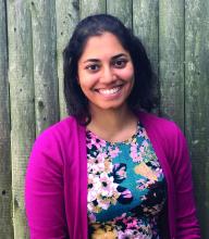
In 2015, Debasree Banerjee, MD, MS, received the CHEST Foundation Research Grant in Pulmonary Arterial Hypertension. She was also a 2016 NetWorks Challenge Travel Grantee as a member of the Women’s Health NetWork, allowing her to attend the 2016 CHEST Annual Meeting and network with peers and leaders in chest medicine. Read our follow-up interview with Dr. Banerjee on her research progress and how the grants she’s received have impacted her and the work she is doing.
What is the project you have been working on?
I have been researching the role of the specific sodium channel in the heart, how it affects the conductance in patients with pulmonary arterial hypertension, and how it might affect RV function. We know in some sources that about 25% of patients with PAH can die of sudden cardiac death, and sudden cardiac death is more common in patients with left-sided heart disease.
Instead of dying of sudden death or end stage heart failure, we wanted a way to see, just based on a physical exam, if there’s evidence of heart pump function not working well. With the funding, I’ve been able to more than double the sample size of the original pilot data and add in two more large objectives to complement my original aim.
What has receiving the grant meant to you?
One of the reasons I was able to stay at Brown was because of winning this grant from the CHEST Foundation. It was able to cement my interest in fully pursuing a physician scientist career, which is huge, because it is not what I had planned on doing. Because of this grant, I had an 80% protected research position in my first year. Winning the grant gave me a feeling of affirmation and validation, and that certainly motivates me to continue on this path.
Going into fellowship, if you had asked me what I had envisioned myself doing, I would have said I’d be a medical educator. I think I was surprised by my research year in fellowship when I was working on this project, because the grant created so much excitement. I felt like I could actually do this, and obtaining the grant uped the ante of investment and kept me excited. Plus, the grant allowed me to do everything, see the whole process, the full arc, and I’m not even done.
What barriers have you encountered with your research?
Dr. Debasree Banerjee
Not having all the control, like unplanned hospitalizations or advanced sickness in the patients. There are also things cost-wise that are needed for the research that I wouldn’t have had access to without the grant. I didn’t do much research in medical school and residency, since I was more focused on teaching, so I hadn’t been prepared for the administrative legwork. But, it’s something I’m learning.
Being able to follow up with the CHEST Foundation and attend the CHEST annual meeting are exciting ways to overcome any slumps or doubts, because you see the interest and encouragement for the work you’re doing. Receiving the travel grant and coming to the annual meeting as a new faculty member, it was the most high-yield conference I’ve ever attended. Every day, there is something new and interactive for development.
What advice would you give to someone who hasn’t received a grant before but is considering applying?
If they can get a good mentor, that’s invaluable. It takes perseverance, persistence, and passion, and if you believe your work is having an impact, it’s absolutely worth doing. Even if you apply and don’t get it the first time, try, try again. I have so much more faith in CHEST because of the positivity I see from the investment in my own mentor, who was a past foundation grant recipient and encouraged me to apply. CHEST gives ample opportunity to network and help to be steered in the right way. As a grant recipient and being folded into the CHEST community, you start to think, “I want this feeling again. Someone thinks this is important work.”
In keeping with the commitment of the American College of Chest Physicians (CHEST) to be the home of the clinician educator, and supporting CHEST’s strategic vision of advancing best patient outcomes through innovative chest medicine education, a new designation intended to provide national-level recognition of excellence in continuing medical education has been established—the innovation award-winning Distinguished CHEST Educator.
Distinguished CHEST Educators are within the top 5% of CHEST’s faculty and are recognized for their achievements in making significant and long-term contributions to the design and delivery of CHEST education. With more than 108 ways to educate, these faculty members have exceeded expectations by serving as CHEST committee chairs, vice-chairs, faculty, and peer reviewers for programs such as the CHEST Annual Meeting.
“The greatest achievement I can imagine is seen in the people we train—as that lives on. Real values in medicine live only by being handed down to others. Over the past decade, CHEST has afforded me the privilege to represent the organization on a national platform, and, in doing so, I have been able to refine my own skills and those of my peers, as well as adding both quality and detail to my understanding of how young physicians learn,” says Nader Kamangar, MD, FCCP, of UCLA, CHEST member since 2000, and Distinguished CHEST Educator.
This designation will be granted to select clinical educators each year. The inaugural class of Distinguished CHEST Educators was honored at the end of October at CHEST 2017 in Toronto, as will be the tradition for the classes that follow.
Distinguished CHEST Educator
Congratulations to the inaugural class of Distinguished CHEST Educators.
Sandra Adams, MD, MS, FCCP
Doreen Addrizzo-Harris, MD, FCCP
A. Christine Argento, MD, FCCP
Robert Arntfield, MD, FCCP
Anthony Asciutto, RRT
Olivier Axler, MD, PhD, FCCP
Meyer Balter, MD, FCCP
Gisela Banauch, MD, MS, FCCP
Robert Baughman, MD, FCCP
David Bell, MD, FCCP
Michel Boivin, MD, FCCP
Gabriel Bosslet, MD, FCCP
Jean Bourbeau, MD, MS, FCCP
David Bowton, MD, FCCP
Kevin Brown, MD, FCCP
Jack Buckley, MD, MPH, FCCP
Kristin Burkart, MD, MS, FCCP
Brian Carlin, MD, FCCP
Christopher Carroll, MD, FCCP
Roberto Casal, MD
Richard Castriotta, MD, FCCP
Kevin Chan, MD, FCCP
Alexander Chen, MD
Michael Christian, MD, FCCP
Nancy Collop, MD, FCCP
Clayton Cowl, MD, MS, FCCP
Angel Coz Yataco, MD, FCCP
Gerard Criner, MD, FCCP
Carolyn D’Ambrosio, MD, FCCP
Mauricio Danckers, MD, FCCP
Aneesa Das, MD, FCCP
John Davies, RRT, MA, FCCP
Frank Detterbeck, MD, FCCP
Emily Diederich, MD, FCCP
Kevin Doerschug, MD, MS, FCCP
Meagan Dubosky, RRT-ACCS
Kevin Dushay, MD, FCCP
Eric Edell, MD, FCCP
William Enfinger
Michael Ezzie, MD, FCCP
David Feller-Kopman, MD, FCCP
Kevin Felner, MD, FCCP
Neil Freedman, MD, FCCP
Thomas Fuhrman, MD, MS, FCCP
John Gaillard, MD, FCCP
Colin Gillespie, MD
Maritza Groth, MD, FCCP
Mark Hall, MD
Jesse Hall, MD, FCCP
Nicola Hanania, MD, MBBS, FCCP
D. Kyle Hogarth, MD, FCCP
Steven Hollenberg, MD, FCCP
Robert Hyzy, MD, FCCP
Richard Irwin, MD, Master FCCP
Nader Kamangar, MD, MS, FCCP
Carl Kaplan, MD, FCCP
Brian Kaufman, MD, FCCP
William Kelly, MD, FCCP
Seth Koenig, MD, FCCP
Anastassios Koumbourlis, MD, MPH, FCCP
Lindsey Kreisher, RRT
Karol Kremens, MD, FCCP
Sunita Kumar, MD, MBBS, FCCP
Viera Lakticova, MD
Carla Lamb, MD, FCCP
Hans Lee, MD, FCCP
Peter Lenz, MD, MEd, FCCP
Stephanie Levine, MD, FCCP
Deborah Levine, MD, MS, FCCP
Kenneth Lyn-Kew, MD
Joao Alberto de Andrade, MD, FCCP
Neil MacIntyre, MD, FCCP
Donald Mahler, MD, FCCP
Fabien Maldonado, MD, FCCP
Atul Malhotra, MD, FCCP
Haney Mallemat, MD
Darcy Marciniuk, MD, FCCP
Diego Maselli Caceres, MD, FCCP
Paul Mayo, MD, FCCP
Peter Mazzone, MD, MPH, FCCP
John McIlwaine, DO, MBA, FCCP
Mark Metersky, MD, FCCP
Scott Millington, MD
Taro Minami, MD, FCCP
Lisa Moores, MD, FCCP
Amy Morris, MD
John Mullon, MD, FCCP
Septimiu Murgu, MD, FCCP
Mangala Narasimhan, DO, FCCP
Michael Niederman, MD, FCCP
Alexander Niven, MD, FCCP
Anne O’Donnell, MD, FCCP
Erik Osborn, MD
David Ost, MD, MPH, FCCP
Ronald Oudiz, MD, FCCP
Daniel Ouellette, MD, MS, FCCP
Nicholas Pastis, MD, FCCP
Paru Patrawalla, MD, FCCP
Jay Peters, MD, FCCP
Barbara Phillips, MD, MSPH, FCCP
Margaret Pisani, MD, MS, FCCP
Janos Porszasz, MD, PhD
Whitney Prince, MD, FCCP
Suhail Raoof, MBBS, FCCP
Marcos Restrepo, MD, MSc, FCCP
Otis Rickman, DO, FCCP
Roy Ridgeway
Mary Ried, RN, CCRN
Antoni Rosell, MD
Mark Rosen, MD, Master FCCP
Bernard Roth, MD, FCCP
Anthony Saleh, MD, FCCP
Juan Sanchez, MD, FCCP
Pralay Sarkar, MBBS, FCCP
Lewis Satterwhite, MD, FCCP
Paul Scanlon, MD, FCCP
Gregory Schmidt, MD, FCCP
David Schulman, MD, MPH, FCCP
Brady Scott, RRT, MS
Bernardo Selim, MD, FCCP
Curtis Sessler, MD, FCCP
Rakesh Shah, MD, FCCP
Ray Wes Shepherd, MD, FCCP
John Sherner, MD, FCCP
Ariel Shiloh, MD
Samira Shojaee, MD, FCCP
Gerard Silvestri, MD, MS, FCCP
Steven Simpson, MD, FCCP
James Stoller, MD, MS, FCCP
Mary Strek, MD, FCCP
William Stringer, MD, FCCP
Eleanor Summerhill, MD, FCCP
Lynn Tanoue, MD, FCCP
Victor Test, MD, FCCP
Arthur Tokarczyk, MD, FCCP
Anil Vachani, MD, FCCP
Momen Wahidi, MD, MBA, FCCP
Keith Wille, MD, FCCP
Lisa Wolfe, MD, FCCP
Richard Wunderink, MD, FCCP
Lonny Yarmus, DO, FCCP
Kazuhiro Yasufuku, MD, PhD, FCCP
Gulrukh Zaidi, MD
In keeping with the commitment of the American College of Chest Physicians (CHEST) to be the home of the clinician educator, and supporting CHEST’s strategic vision of advancing best patient outcomes through innovative chest medicine education, a new designation intended to provide national-level recognition of excellence in continuing medical education has been established—the innovation award-winning Distinguished CHEST Educator.
Distinguished CHEST Educators are within the top 5% of CHEST’s faculty and are recognized for their achievements in making significant and long-term contributions to the design and delivery of CHEST education. With more than 108 ways to educate, these faculty members have exceeded expectations by serving as CHEST committee chairs, vice-chairs, faculty, and peer reviewers for programs such as the CHEST Annual Meeting.
“The greatest achievement I can imagine is seen in the people we train—as that lives on. Real values in medicine live only by being handed down to others. Over the past decade, CHEST has afforded me the privilege to represent the organization on a national platform, and, in doing so, I have been able to refine my own skills and those of my peers, as well as adding both quality and detail to my understanding of how young physicians learn,” says Nader Kamangar, MD, FCCP, of UCLA, CHEST member since 2000, and Distinguished CHEST Educator.
This designation will be granted to select clinical educators each year. The inaugural class of Distinguished CHEST Educators was honored at the end of October at CHEST 2017 in Toronto, as will be the tradition for the classes that follow.
Distinguished CHEST Educator
Congratulations to the inaugural class of Distinguished CHEST Educators.
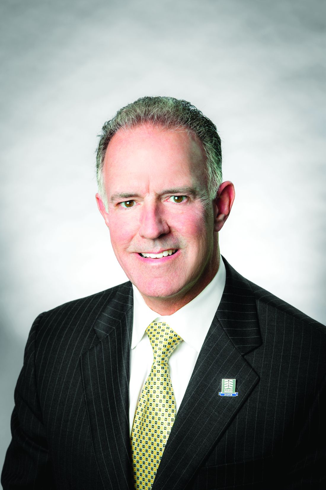
Stephen J. Welch was officially appointed Executive Vice President and CEO in April after serving as the interim for both positions since May 2016. Here’s a little “inside look” at what Steve is all about.
What is one major accomplishment you hope to achieve as Executive Vice President & Chief Executive Officer?
My goal as EVP/CEO is fairly simple and straightforward: to ensure the organization remains relevant and viable as a leader in providing clinically focused, innovative educational programs and content. I don’t really have one accomplishment that I’m focused on, but I do want to ensure that we achieve our annual organizational goals that support CHEST’s strategic plan. That may sound a little vague, but it’s true. We have so many outstanding programs and initiatives that I’d be doing a disservice to identify a single goal.
How does your previous experience with CHEST help you successfully lead the organization?
With CHEST being a not-for-profit organization, which relies on volunteer leadership and faculty, I think the relationships I’ve built over the past 23 years within the organization and the chest medicine community are invaluable. I personally know so many of our leadership because I’ve been part of the organization at the executive level working with them for those 23 years. They know me and how I approach opportunities, address issues, and handle challenges, which has helped build an immediate level of mutual respect, trust, and confidence between the staff and leadership. In addition, there was no disruption from having someone come in from the outside and have to get up to speed. It made the transition pretty seamless for the staff, as well.
During my time at CHEST, I’ve seen how the organization operates, from the journal, to the annual meeting and board reviews, to the simulation and hands-on skills training, to operational activities like the management of our finances and new global headquarters and training center. I’ve also had the opportunity to meet with many of our international members and sister societies. Those experiences have allowed me to work closely with many of our faculty, authors, and educators to understand their educational and professional needs, so we can ensure that we meet them.
CHEST is only as good as the education we provide, and it’s our subject matter experts who drive that content engine. In my previous role leading the Publishing Division and working on our journal CHEST® and programs like SEEK, I’ve had the honor and pleasure of working with some of the greatest minds in pulmonary, critical care, and sleep medicine. It’s humbling.
What will be some of the underlying themes as you work to outline the strategic plan for the next 5 years?
We are in the final stages of planning for 2018 and beyond, and although our proposed roadmap isn’t significantly different than what we have been doing, there’s some greater emphasis on a few key areas. For example, we’re looking at innovations in educational delivery. We’ve got some very forward thinking faculty educators and staff who are collaborating to develop innovations like gamification of educational and simulation programs, and augmented reality. Globalization and growth are also a key part of our strategic plan, and we are committed to the broad delivery of our educational programs and content both here and around the world. Finally, we have invested in a data analytics project that is maturing, and we’ll be leveraging that information to provide more personalized education plans – not just for the physician but for the entire health-care team. It’s important for us to stay relevant and viable.
Stephen J. Welch
Why has CHEST shifted to an interdisciplinary, team-focused approach?
I look at it as simply an evolution that reflects how health care is changing. It’s a team sport now, and our advanced practice providers (APPs) play a huge role in patient management and care. To be as effective and efficient as possible, and ensure the best patient outcomes, the whole team needs to be on the same page, and we believe that providing education for the interdisciplinary care team will help ensure that the best patient care is delivered.
There’s also a need for this education, and we want to fill it. Our APPs tell us that there is no formal pulmonary training or post-masters fellowship in pulmonary medicine for them. They are often left on their own to fill any gaps in knowledge and skills. That’s where our CHEST programs, such as our CHEST Annual Meeting, come in. We have an Interprofessional NetWork made up of APPs and physicians, and they were integral in working with the CHEST 2017 Program Committee to ensure plenty of relevant content was offered. Moving forward, we will continue to offer and build interdisciplinary programs designed for the entire team, as well as programs that address clinical issues across disciplines.
What are some of the critical skills CHEST physicians need to keep the population healthy during the ever-evolving field?
Educationally, we recognize that conferences like the annual CHEST meeting must provide more than just talking heads. We’ve invested heavily in high-fidelity medical simulation through small group, hands-on skill training in critical care techniques, airway management, EBUS, critical care ultrasound, bronchoscopy, and other chest medicine content. It’s like the old adage about fishing: instead of telling people how to fish, we teach them to fish.
Any final thoughts?
I always encourage our members to get involved with CHEST and experience the camaraderie and connectivity of the CHEST family. Ask any of our leadership, and you will surely hear their story of that special person who first introduced them to the College. Reach out and tell a colleague about CHEST. We are focused on clinically relevant education that our members can take back and put into action immediately. At the end of the day, it’s about providing state-of-the-art education via high fidelity medical simulation, hands-on skills training, clinically focused courses, case-based programming, and more—all intended to be immediately implemented to improve patient care and patient outcomes. That’s what the CHEST organization is all about.
Burden of Adult Community-Acquired, Health-care-Associated, Hospital-Acquired, and Ventilator-Associated Pneumonia: New York City, 2010 to 2014. By R. E. Corrado, et al.
Hyperbaric Oxygen Therapy Is Associated With Lower Short- and Long-Term Mortality in Patients With Carbon Monoxide Poisoning. By C-C Huang, et al.
Evidence-Based Medicine
Pharmacologic and Nonpharmacologic Treatment for Acute Cough Associated With the Common Cold: CHEST Expert Panel Report. By M. A. Malesker, et al, on behalf of the CHEST Expert Cough Panel.
Cough in Ambulatory Immunocompromised Adults: CHEST Expert Panel Report. By M. J. Rosen, et al, on behalf of the CHEST Expert Cough Panel.
Original Research
Burden of Adult Community-Acquired, Health-care-Associated, Hospital-Acquired, and Ventilator-Associated Pneumonia: New York City, 2010 to 2014. By R. E. Corrado, et al.
Hyperbaric Oxygen Therapy Is Associated With Lower Short- and Long-Term Mortality in Patients With Carbon Monoxide Poisoning. By C-C Huang, et al.
Evidence-Based Medicine
Pharmacologic and Nonpharmacologic Treatment for Acute Cough Associated With the Common Cold: CHEST Expert Panel Report. By M. A. Malesker, et al, on behalf of the CHEST Expert Cough Panel.
Cough in Ambulatory Immunocompromised Adults: CHEST Expert Panel Report. By M. J. Rosen, et al, on behalf of the CHEST Expert Cough Panel.
Don't forget that the end of the year is the time to keep up to date with your SVS membership dues. Invoices were emailed to all members earlier this month and are due by Dec. 31.
It's simple to pay your 2018 dues online -- and there's no need to write out a check or find a stamp! Just log on to vascular.org/payinvoice. (While you're at it, please make sure your record is up to date.) You also can make a donation to the SVS Foundation at the same time. For membership help, e-mail the SVS membership department, or call 312-334-2313
The SVS wants members to complete a short communications survey, hoping to get a better idea of what you read, what you find important, what you find interesting, plus your suggestions for other topics you would like to see.
Would you please take five to 10 minutes to answer the linked survey on our communications topics and vehicles: print publications, electronic newsletters and social media? The survey will close Nov. 10. For those willing to help, we are offering the chance to win a $100 Visa gift card -- so give us your thoughts for a chance to win!
The mobile app for the Vascular Education and Self-Assessment Program, fourth edition (VESAP4) is now available.
Owners of mobile Apple products (only) can download the app at the Apple App Store and take advantage of VESAP while off-line. Users can study and test themselves while in locations without Internet access, such as on an airplane, or in buildings where access too many sites are blocked. Then, when access is available again, the desktop and mobile versions will sync up without loss of any work or data.
Applications are due March 2, 2018, for the Wylie Scholar Award, co-sponsored by Vascular Cures and the SVS Foundation. The three-year, $150,000 grant is awarded to a promising vascular surgeon-scientist in North America and is designed to support outstanding surgeon-scientists conducting innovative academic research in the early stages of their careers.
This year's recipient, Dr. Sean English, is conducting research on AAA. Dr. Mohamed Zayed, MD, PhD, the 2015 recipient, is investigating why diabetics develop a unique lipid profile leading to PAD. For each $150,000 award, Wylie Scholars have received $3.3 million in subsequent national research funding, for a return on investment of nearly 22 to 1.
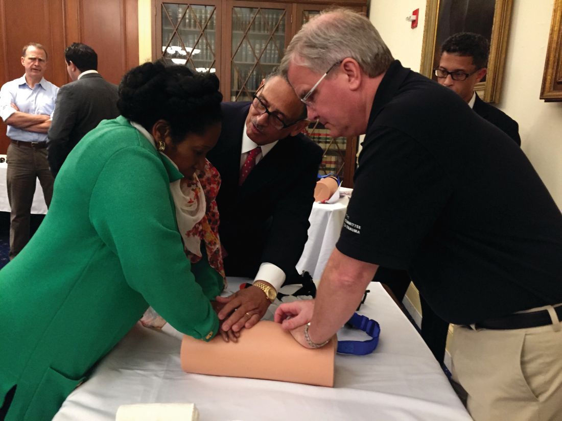
Leaders of the American College of Surgeons (ACS) hosted a Stop the Bleed® training program on Capitol Hill October 12 for members of Congress and their staffs. The congressional event focused on how early intervention from a Stop the Bleed-trained individual can save the life of someone suffering from a bleeding injury. Participants came to learn more about the ACS’ efforts with Stop the Bleed and engage in the hands-on training in how to control bleeding. The training was led by ACS Fellows, including Lenworth M. Jacobs, Jr., MD, MPH, FACS; Leonard J. Weireter, Jr., MD, FACS; Mark L. Gestring, MD, FACS; John H. Armstrong, MD, FACS; Joseph V. Sakran, MD, MPH, MPA, FACS; and Jack Sava, MD, FACS. Congressional guests included Reps. Ami Bera, MD (D-CA); Phil Roe, MD (R-TN); Raul Ruiz, MD (D-CA); and Brad Wenstrup, DPM (R-OH), who provided opening remarks.
U.S. Rep. Sheila Jackson Lee (D-TX) (left) learns how to apply pressure to stop bleeding from Dr. Jacobs (center), and Dr. Armstrong.
A poignant highlight of the training was the appearance of House Majority Whip Steve Scalise (R-LA), who recently returned to work while recovering from a gunshot injury in June. When Representative Scalise was injured, his wounds were immediately treated by Representative Wenstrup, who was at the scene at the time of the shooting and used bleeding control techniques.
Members of Congress and their staff left the program with a better understanding of how to become life-saving immediate responders and the value of Stop the Bleed training. In addition to promoting Stop the Bleed training, the College also is advocating for widespread access to bleeding control education before federal and state lawmakers.
For more information about ACS trauma advocacy, contact Justin Rosen, Congressional Lobbyist, at [email protected] or 202-672-1528. For more information about the Stop the Bleed program, visit BleedingControl.org.
The November issue of the Bulletin of the American College of Surgeons is now available online at bulletin.facs.org. This month’s Bulletin includes the following features, columns, and new stories, among others:
Features
• Should your health care system invest in an ambulatory surgery center? A decision-making framework
• Frank R. Lewis, Jr., MD, FACS: 15 years of visionary leadership at the American Board of Surgery
• A history of health information technology and the future of interoperability
Columns
• Looking forward: Health care reform
• What surgeons should know about...The New Medicare Card Project
• ACS NSQIP best practices case studies: Quality improvement in imaging strategies for pediatric appendicitis
News
• Barbara Lee Bass, MD, FACS, FRCS(Hon), installed as 98th ACS President
• Honorary Fellowship in the ACS awarded to 10 prominent surgeons
• Call for nominations for the ACS Board of Regents and ACS Officers-Elect
The Bulletin is available in a variety of digital formats to satisfy every reader’s preference, including an interactive version and a smartphone app. Go to the Bulletin website at bulletin.facs.org to connect to any of these versions or to read the articles directly online.