User login
New AHA statement on ischemia after cardiac surgery
The American Heart Association outlines “considerations” on the management of acute postoperative myocardial ischemia (PMI) after cardiac surgery in a scientific statement.
Although an infrequent event, acute PMI following cardiac surgery can rapidly evolve and become a potentially life-threatening complication, the writing group, led by Mario Gaudino, MD, PhD, with Weill Cornell Medicine, New York, points out.
The new statement was published online in Circulation.
Data show that the incidence of postoperative myocardial infarction after cardiac surgery ranges from 0.3% to 9.8% after isolated coronary artery bypass graft (CABG) surgery and 0.7% to 11.8% after concomitant valvular surgery. For isolated mitral valve surgery, incidence ranges from 1.7% to 2.2%.
Short-term mortality is elevated among patients with acute PMI, irrespective of the type of surgery. Reported mortality rates range from 5.1% to 24%; the evidence on long-term mortality has been mixed.
Graft-related factors are the most common cause of PMI after CABG, but other factors may contribute, including technical factors, competitive flow, suture entrapment, or coronary artery distortion, as well as non–graft related factors.
Prompt diagnosis and treatment important
Currently, there is no consensus definition of PMI. Elevations in cardiac biomarkers may not be reliable for diagnosis after surgery, and pain management regimens may mask symptoms of ischemia, the writing group notes.
because timely diagnosis and treatment are key to a good clinical outcome,” they write.
Delay in urgent angiography has been associated with higher mortality; thus, a low threshold for action is encouraged for patients with suspected acute PMI.
Indications for urgent angiography include new ECG changes, chest pain with ongoing signs of ischemia, cardiac imaging abnormalities, cardiac rhythm abnormalities, significant elevations in cardiac biomarkers, and low cardiac output syndrome despite postoperative pressor support.
Patients with acute PMI and low cardiac output syndrome may require mechanical support when first-line treatment fails.
The writing group says fast and effective reperfusion of the ischemic zone, which is generally achieved by percutaneous intervention and, less often, by repeat surgery, is the key to a good clinical outcome.
The statement was prepared by the volunteer writing group on behalf of the AHA Council on Cardiovascular Surgery and Anesthesia; Council on Clinical Cardiology; Council on Cardiovascular and Stroke Nursing; and Stroke Council.
The research had no commercial funding. Disclosures for the writing group are listed with the original article.
A version of this article originally appeared on Medscape.com.
FROM CIRCULATION
TAVI turmoil: Did an ANP perform transcatheter aortic valve replacement in the U.K.?
In the United Kingdom, John Steele, an advanced nurse practitioner (ANP) at Glenfield Hospital, part of the University Hospitals of Leicester NHS Trust (UHL), was congratulated on Twitter as “the first nurse-ANP who has performed the whole TAVI procedure as the first operator – true transformation addressing NHS needs.”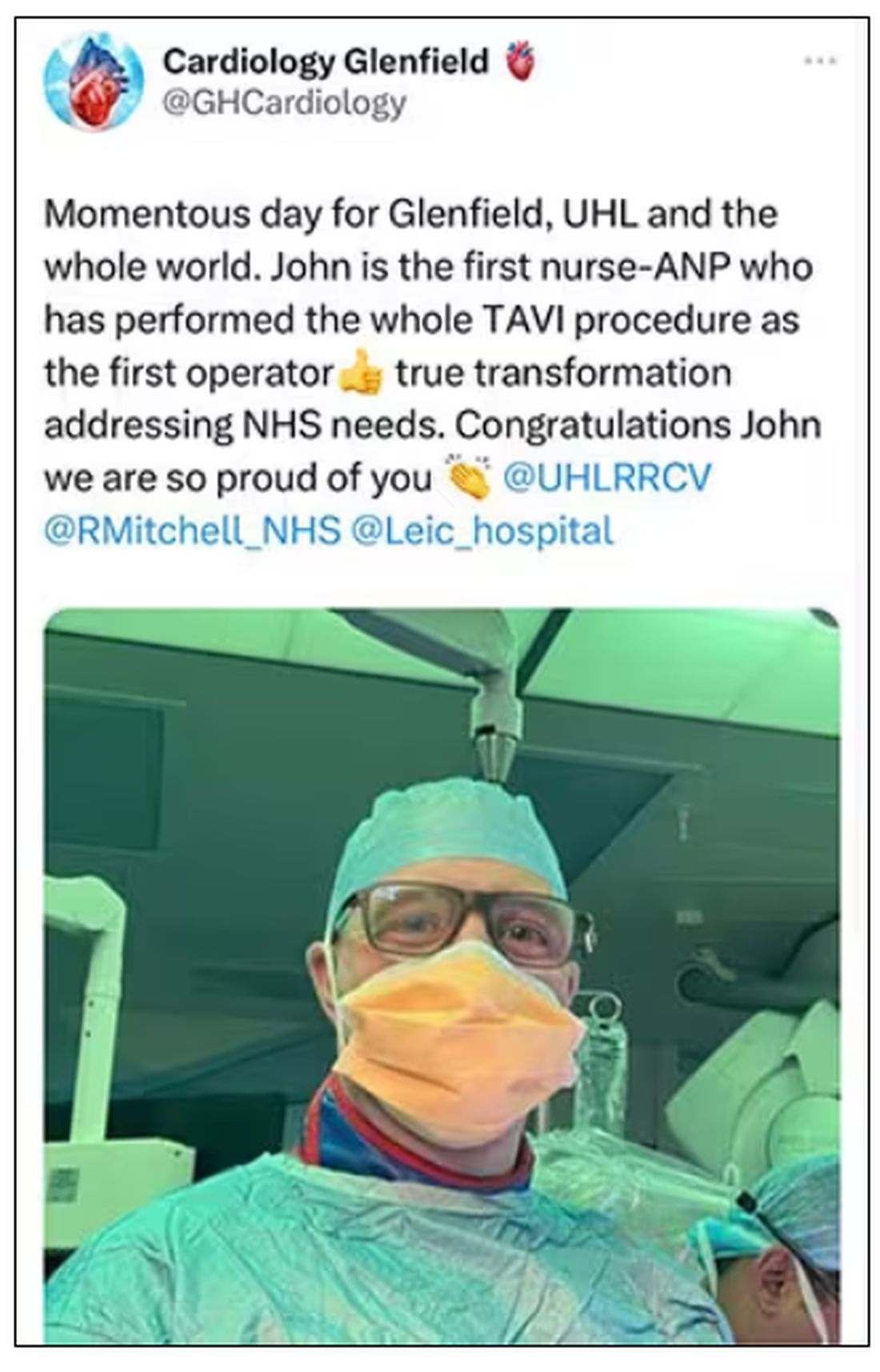
The now-deleted tweet from @GHCardiology is still visible in the Twitter thread of Mamas A. Mamas, a professor of interventional cardiology at Keele University, England. “This is so inappropriate on so many levels,” Dr. Mamas tweeted. “This is not safe for patients particularly given that there are numerous TAVI trained medically qualified operators in UK. You have also taken away training opportunities for medical / surgical trainees.”
Other followers also responded, largely negatively.
“This is crazy. Is this @TheOnion???” tweeted Martha Gulati, MD, director of preventive cardiology in the Smidt Heart Institute at Cedars-Sinai, Los Angeles, Calif., in response to Dr. Mamas, referring to the popular satirical news outlet. “Seriously I can’t see this as an actuality given the potential for so many other issues they wouldn’t know how to deal with.”
Could it happen in the U.S.?
Could a U.S.-based nurse practitioner perform TAVR? Possibly. Should they? No, says Andrew M. Goldsweig, MD, chair of the U.S. Society for Cardiovascular Angiography and Interventions Structural Heart Council. “Experienced nurse practitioners who have participated as secondary operators in many TAVR procedures and have observed the primary physician operators likely know the technical steps involved in an uncomplicated transfemoral TAVR procedure,” he told this news organization.
“However, a physician’s depth and breadth of training are absolutely required both to recognize and to address any periprocedural issues,” said Dr. Goldsweig, who is also director of the cardiac catheterization laboratory and director of cardiovascular clinical research at Baystate Medical Center in Springfield, Mass.
What it takes to do TAVR
Transcatheter aortic valves were first approved by the FDA in 2011 for use in patients with severe, inoperable, aortic stenosis. The procedure is now increasingly used as an alternative to surgical AVR in intermediate- and low-risk patients and has a longer history in Europe.
Dr. Goldsweig notes that “TAVR is a complex procedure with many potential challenges. Physicians are trained to diagnose and manage vascular access complications, heart failure and respiratory complications, rhythm disturbances, stroke, paravalvular leak, valve malpositioning/embolization, cardiogenic shock, and any other issues that may arise in the peri-TAVR period.
“Physicians can perform vascular imaging and interventions, transition to alternative access, manage intubation and ventilation, facilitate embolectomy, place a pacemaker, close a paravalvular leak, capture a misplaced valve, deploy mechanical circulatory support, and perform other diagnostic and interventional procedures as necessary that are required for TAVR operators and vastly exceed the training and scope of a nurse practitioner.”
The 2023 ACC/AHA/SCAI advanced training statement on interventional cardiology defines select competencies for interventional cardiologists who choose to focus their career on peripheral, vascular, or structural heart interventions.
In a recent article in Structural Heart, Dr. Goldsweig and colleagues write, “Training in SHD [structural heart disease] has historically been fragmented and informal. Current modes of SHD training include unaccredited fellowship training, industry-sponsored forums and device-specific training, and training through on-site proctorship.”
Such programs have grown “exponentially,” they write, “despite the conspicuous absence of formalized training requirements.”
In response to the John Steele uproar, the British Cardiovascular Intervention Society posted a statement on its website, noting, “As medicine has changed so there has increasingly been a role for allied health practitioners with advanced skills to take on responsibilities that were previously considered to be the domain of doctors ...
“TAVI procedures however carry a mortality risk, and the responsibility for undertaking a successful TAVI procedure will always lie with a Cardiologist who has had the breadth of training to manage the various complications that may occur during or after a procedure. This requires years of training, and there is no short-cut, or substitute.”
The BCIS promises a statement “later in the year [on] the expected training route for undertaking TAVI and other structural heart procedures.”
Why it matters: Scope creep
Despite the current upheaval, it’s not the first time that a nurse in the United Kingdom has performed a procedure normally performed by a medical doctor. A 3-year-old Reddit post on r/JuniorDoctorsUK points to a 2017 Guardian article titled, “Meet the nurse who will soon perform surgery on patients alone.” Although the “surgical care practitioner” seems to be performing within the scope of her practice, people responding to the post say it’s an example of “mid-level [scope] creep.”
More recently, a Reddit post in the same group points to a congratulatory post for a “nurse-led radial access.” One person commented, “Today they do the access. Tomorrow they do the full diagnostic. Day after they do the pressure wire. Next week they do the PCI [percutaneous coronary intervention].”
Broadly, “scope creep” refers to scope-of-practice expansions, but not turf wars, according to Rebekah Bernard, MD, a family physician in Fort Myers, Fla., who cowrote, “Patients at Risk: The Rise of the Nurse Practitioner and Physician Assistant in Healthcare,” with Niran Al-Agba, MD, a pediatrician in Silverdale, Wash.
The reasons behind U.K. scope creep aren’t clear. Some believe it’s money. Some say the system is broken and that doctors are being exploited.
In relation to the NP-TAVI case, the British Junior Cardiologist Association commented that it reflects a lack of support and advocacy for medical/surgical trainees who need the training opportunities that are going instead to allied health professionals.
In the United States, scope creep is being taken seriously (some may say too seriously) by the American Medical Association. The AMA is lobbying to stop “inappropriate scope expansions,” bolstered by its AMA Scope of Practice Partnership.
Pointing to a scope creep video produced by the AMA, one JuniorDoctorsUK Reddit post asks, “why isn’t the BMA doing anything similar?”
Time for a rethink?
Back to Glenfield Hospital. Not only has Cardiology Glenfield deleted the controversial tweet; it is now is backtracking on its congratulations to ANP Steele, tweeting, “We want to make clear that the lead operator for the procedure was a consultant structural interventionist. However, we are looking into the circumstances, including a review of clinical governance.” From the responses, few clinicians are buying that explanation.
In response to a request for a comment from Glenfield, Andrew Furlong, UHL medical director, reiterated to this news organization through communications manager Gareth Duggan, “We are investigating the circumstances of the procedure with our cardiology team and reviewing our governance processes.”
Dr. Goldsweig participated in a past speaking engagement for Edwards Lifesciences.
A version of this article originally appeared on Medscape.com.
Interventional cardiologists worldwide burned out: Survey
“What surprised me was the magnitude of the findings,” Emmanouil S. Brilakis, MD, PhD of the Minneapolis Heart Institute and Minneapolis Heart Institute Foundation, said in an interview.
“I was expecting that some interventionalists would feel burned out, but not that 78% would feel they are working too hard, 64% are emotionally exhausted, and 41% considered quitting their job during the past year.
The survey, conducted in January, also showed that while 69% of respondents were affected by burnout, many were either not seeking mental health support or not willing to share whether they were under treatment.
Overall, 28% of interventional cardiologists were not happy with their lives, similar to the 29% reported in the Medscape Cardiologist Lifestyle, Happiness & Burnout Report 2022.
“Many institutions have formed task forces to better understand burnout and recommend solutions, but progress has been slow,” Dr. Brilakis said. “Barriers include financial constraints, understaffing, lack of understanding of the root causes of burnout in each practice, and perhaps underappreciation of the consequences of burnout.”
The study was published online in JACC: Cardiovascular Interventions.
Too much paperwork
The investigators conducted an international, online survey of IC attending physicians and fellows to assess their psychological well-being. The 78 survey questions prepared by the coauthors were shown to perform similarly to the validated Maslach Burnout Inventory.
A total of 1,159 attendings and 192 fellows completed the survey, representing 12% of U.S. IC attendings and 19% of U.S. IC fellows.
Half of attending physicians were from the United States, followed by the European Union (16%). Overall, 37% were from academic institutions; the median age was 41-45 years; 91% were men; and mean clinical work hours per week were 63.
Most (86%) had a partner with whom they lived. Yet most (84%) also felt lonely; 41% considered leaving their jobs during the past year; and 32% said they were currently considering leaving.
Compared with the previous year, 12% had increased enthusiasm and 44% had decreased enthusiasm toward work. One-third (33%) felt overwhelmed and 20% doubted the significance of their work three or more times a week.
As noted, most (78%) felt they were “working too hard,” were emotionally exhausted (64%), and frustrated by work (58%). Almost one-third (30%) considered themselves physically unhealthy.
Unhappiness was highest (33%) among 51- to 60-year-olds, followed by 31- to 40-year-olds (31%); it was lowest (21%) among those over age 60.
Unhappiness was similar between men and women (27% vs. 30%) and was highest in North America (30%) and lower in Asia (26%).
Most (69%) respondents said that burnout impacted their life, with very little difference between men and women (68% vs. 73%).
Two-thirds (67%) said they had somebody they could share their mental health concerns with, yet only 37% reported having access to mental health support if needed through their hospital/practice.
For fellows, the median age was 31-35 years; 88% were men; 42% were from the United States and 22% from the European Union. Two-thirds were from academic institutions (67%) and the mean clinical work hours were 67 per week.
Two-thirds (67%) lived with a partner; half (48%) felt lonely, 29% considered leaving their jobs in the past year, and 15% were currently considering leaving.
Compared with the previous year, 27% had increased enthusiasm, and 32% had decreased enthusiasm toward work. More than one-quarter (29%) felt overwhelmed and 26% doubted the significance of their work three or more times per week.
Attendings rated excessive paperwork requirements, bureaucratic tasks, challenges in equipment acquisition, and excessive government regulations higher (in contributing to burnout) compared with fellows.
Non-U.S. attendings reported insufficient income and challenges with equipment acquisition as significant contributors to their burnout more than did their United States counterparts.
Fellows rated insufficient income as the most significant contributor to burnout.
Their main coping mechanisms were talking with family/friends (at 6.8 rated on a scale of 0-10), watching movies (6.4), and listening/playing music (6.0).
Attendings were more likely to use exercise as a coping skill, and fellows were more likely to cope by watching movies/series, sleeping, and eating junk food.
Asked what hospitals and practices can do to reduce burnout and improve well-being, attendings suggested removing rules/regulations that do not contribute to patient care, such as reforming prior authorization (mean rating, 8.1), better administrative support (8.0), and professional growth opportunities (7.9).
Non-U.S. attendings more often requested growth opportunities, increased compensation, availability of better hospital food, better hospital infrastructure, streamlined access to equipment, better on-call rooms, and access to mental health professionals to improve their well-being.
Overall, fellows were more likely than were attendings to request professional growth opportunities and were more likely to ask for availability of better food in the hospital, and better on-call rooms.
Reforms needed
Laxmi Mehta, MD, chief well-being leader, faculty director of the Gabbe Health and Well-Being Program, professor of medicine at The Ohio State University Wexner Medical Center in Columbus, and spokesperson for the American Heart Association, noted, “The burnout rates are much higher than our previously reported American College of Cardiology data, which found burnout rates at about 27%; however, that survey was conducted prepandemic,” she said. Dr. Mehta was the lead author of that 2019 report.
She said in an interview that she would have liked to see more breakdowns by gender, and whether there was an association between burnout and the number of procedures performed.
“Nevertheless,” she said, “the rates are very high for burnout, stress, and dissatisfaction, as well as mental health issues. Almost one half of the IC attendings considered leaving their job, which is also seen in other surveys, and is concerning given the projected shortages in the workforce.”
Changes need to be made in the profession of medicine as a whole, she said, though that is unlikely to happen any time soon. “Optimizing workflows and improving the work culture requires not only time, but also collaboration between administration and clinicians, along with an intent and strategic plan focused on well-being of the organization.”
With regard to prior authorization, she said, “medical organizations are advocating for reform at the state and national level. If meaningful reforms can occur, that can reduce some of the bureaucracy. However, there is much more [bureaucracy] in medicine.”
With respect to mental health, she added, “there is a lot that needs to be done to reduce the stigma of seeking help. Many physicians don’t seek help due to the shame, lack of time, and potential impact it can have on hospital credentialing and state medical licensing. Medical organizations and individuals are advocating for reforms in this space, as well, to normalize mental health.”
The Minneapolis Heart Institute Foundation’s Science Center for Coronary Artery Disease helped support this research project. Dr. Brilakis, study coauthors, and Dr. Mehta report no relevant financial relationships.
A version of this article originally appeared on Medscape.com.
FROM JACC: CARDIOVASCULAR INTERVENTIONS
Hospital patient catches on fire, highlighting need for prevention
On Thanksgiving Day 2022, Kathy Stark watched as her husband of 35 years, Bobby Ray Stark, caught fire at a Nashville hospital. According to Clint Kelly, Kathy Stark’s attorney, the hospital staff was performing cardioversion to restore Bobby Ray’s heart rhythm when a spark ignited the oxygen and set the patient aflame.
Mr. Stark, 64, died of “a combination of cardiovascular disease and thermal burns,” according to a local news report. In May, Kathy Stark filed a malpractice lawsuit in U.S. District Court. Mr. Kelly hopes that the lawsuit will help improve patient safety. Meanwhile, Kathy Stark “goes to bed at night and sees her husband on fire,” Mr. Kelly says. A similar incident occurred last December in the operating room at Oregon Health & Science University, resulting in minor injuries to a patient.
Underreported, but likely dropping
Reliable data on the incidence of surgical fires is lacking because incidents may go unreported over litigation fears, says Jeffrey Feldman, MD, MSE, anesthesiologist at Children’s Hospital of Philadelphia and chair of the Anesthesia Patient Safety Foundation’s Committee on Technology.
The Pennsylvania Patient Safety Authority has been tracking surgical fires for decades, however, and experts have used the agency’s data to extrapolate how often they occur in the United States.
In 2005, nationwide incidence was estimated to be somewhere in the neighborhood of 550-600 fires annually, says Barbara G. Malanga, acting director of health care incident investigation and technology consulting at ECRI (formerly the Emergency Care Research Institute). By 2011, that number appeared to have dropped to 200-240 incidents per year.
A similar analysis in 2018 found the incidence may now be as low as 88-105 a year. The drop is likely a result of increased awareness because of educational efforts on the part of the ECRI and the APSF, including a widely disseminated video on fire safety.
The decline of surgical fires “sounds great,” says Dr. Feldman, “except that it’s a 100% preventable complication, and they’re still happening.”
Accidents waiting to happen
How do these fires happen? It comes down to the ‘fire triangle’ often taught in grade school. Fire requires three things: an ignition source, fuel, and oxygen or an oxidizing agent. Ignition sources are plentiful in a surgical suite, including any of a variety of electrical devices commonly used in surgical procedures, including defibrillators. Gowns, gauze, drapes, sponges, oxygen masks, nasal cannulae, a patient’s hair or their clothing – all provide the necessary fuel.
But the key factor for surgical fire risk is the presence of high concentrations of oxygen.
Safety protocols
The best and most obvious way to mitigate risk is to reduce the amount of supplemental oxygen, explains Dr. Feldman.
“Many patients do not require a high concentration of oxygen during sedation,” he says.
When a patient does require a higher concentration for their safety, the APSF and ECRI recommend placing an endotracheal tube or supraglottic airway rather than using an oxygen mask or a nasal cannula. “You want to deliver the oxygen in such a way that high concentration doesn’t exist in the surgical field,” Dr. Feldman says. In cases where supplemental oxygen is necessary, ECRI and APSF recommend reducing the oxygen concentration to less than 30%.
In addition, safety protocols include giving flammable prep solutions time to dry before applying towels or drapes and beginning the procedure. These precautions to ensure the safety of patients take just a moment, says Chester H. Lake Jr, MD, MS, of the department of anesthesiology at the University of Mississippi Medical Center, Jackson.
Making fire safety part of the preop routine
These safety protocols are straightforward but not always observed, experts say. Part of the reason is a matter of culture. Both anesthesiologists and surgeons have absorbed the attitude that placing an airway escalates the procedure beyond what the patient needs, says Dr. Feldman. And indeed, according to a 2013 analysis of the American Society of Anesthesiologists closed claims database, 85% of surgical fires occur in outpatient settings where airways are less likely to be placed, and 81% of those claims were for procedures that used monitored anesthesia care.
In an article on prevention of surgical fires, Dr. Lake and colleagues recommend in-house education on preventing and responding to fires at least once a year. But it shouldn’t stop there. Because these fires – horrific as they are – are fairly rare, it’s important to maintain awareness. Making fire safety a regular part of the surgical “time-out” can help further reduce incidents, he says. ECRI and the APSF have teamed up to create a poster that can help surgical teams make fire safety a regular part of their routines.
Although the national decline in surgical fires is encouraging, the problem remains serious. “You can classify these incidents as low, but it’s not low if it happens to you or a family member,” says Dr. Lake. “One is too many.”
ECRI’s Ms. Malanga agrees. “I do like to emphasize that it’s rare,” she says. “But I’d like to see us reduce this until it’s zero.”
A version of this article originally appeared on Medscape.com.
Support for minimally invasive mitral valve repair: Mini Mitral published
The trial, which was first presented earlier this year at the American College of Cardiology meeting, showed that minimally invasive mitral valve repair does not improve physical function at 12 weeks, compared with sternotomy, but outcomes at 1 year show minimally invasive repair is as safe and effective as sternotomy for degenerative mitral regurgitation.
The full results are now published online in JAMA.
The authors, led by Enoch Akowuah, MD, South Tees Hospitals NHS Foundation Trust, Middlesbrough, United Kingdom, explain that mitral valve repair surgery is the preferred treatment for patients with degenerative mitral regurgitation and is routinely performed via full sternotomy, enabling easy access to the heart, flexibility in myocardial protection strategies, and multiple ways of accessing the mitral valve and easing de-airing to prevent air emboli, which cause cerebrovascular accidents.
However, the invasiveness of sternotomy is associated with delayed return to presurgery physical function levels and an increase in postoperative complications.
An alternative new video-guided minimally invasive approach involving a 4- to 7-cm lateral thoracotomy, completely avoiding sternotomy, has been developed, with the hope that it should speed physical recovery function after surgery and reduce postoperative complications and costs by reducing hospital stay.
Dr. Akowuah et al. note that uptake of minithoracotomy is variable, with low rates in the United States and the United Kingdom but high rates in Germany. They say that this variation is attributable to the absence of high-quality evidence from randomized trials demonstrating equivalent or superior benefits, compared with sternotomy, and there are also concerns that the increased technical complexity of minithoracotomy may impair the ability to repair complex valve lesions or increase perioperative complications, particularly vascular injuries and stroke.
The U.K. Mini Mitral trial was therefore conducted to compare the effectiveness and safety of minithoracotomy versus sternotomy mitral valve repair.
For the trial, 330 patients with degenerative mitral regurgitation were randomized to receive either minithoracotomy or sternotomy mitral valve repair performed by an expert surgeon.
The primary outcome was physical functioning and associated return to usual activities measured by change from baseline in the 36-Item Short Form Health Survey (SF-36) physical functioning scale 12 weeks after the surgery.
This failed to show superiority of minithoracotomy, with a mean difference of 0.68 (95% confidence interval, −1.89 to 3.26) between the two groups.
Analysis of secondary outcomes demonstrated that time spent undertaking moderate to vigorous physical activity was higher among participants receiving minithoracotomy at 6 weeks, although the treatment effect was small at an average of 9 minutes and was not different at 12 weeks.
Postoperative length of hospital stay was reduced after minithoracotomy by 1 day, with a median of 5 days, compared with 6 days after sternotomy.
Although repair techniques were at the discretion of the surgeons and differed between the two procedures, high rates of valve repair and low rates of recurrent mitral regurgitation were observed in both groups. Cardiopulmonary bypass times were longer with minithoracotomy, but postoperative complications and adverse events were similar.
There was no difference between the two groups with respect to the prespecified safety outcome of death, repeat mitral valve surgery, or heart failure hospitalization up to 1 year, which occurred in 5.4% of patients undergoing minithoracotomy and 6.1% of those undergoing sternotomy.
“These findings can inform shared decision-making and treatment guidelines,” the authors conclude.
Approach ‘may appeal to patients’
In an editorial accompanying the publication of the study in JAMA, Maurice Enriquez-Sarano, MD, Minneapolis Heart Institute, Minnesota, says the results should be integrated into patient management.
“Mini-thoracotomy mitral repair carried low risk and was highly effective compared with sternotomy. It can thus be applied successfully by surgeons who achieve the necessary expertise,” he notes.
“Mini-thoracotomy may appeal to patients because the procedure is less disfiguring than sternotomy. The early (6-week) benefit, albeit small and transient, is important to patients,” he adds.
The study was funded by the United Kingdom’s National Institute for Health and Care Research. Dr. Akowuah reports no relevant financial relationships with industry. Dr. Enriquez-Sarano reports receiving consulting fees from Edwards Lifesciences, Artivion, ChemImage, HighLife, and Corcym.
A version of this article first appeared on Medscape.com.
Survival similar with hearts donated after circulatory or brain death
in the first randomized trial comparing the two approaches.
“This randomized trial showing recipient survival with DCD to be similar to DBD should lead to DCD becoming the standard of care alongside DBD,” lead author Jacob Schroder, MD, surgical director, heart transplantation program, Duke University Medical Center, Durham, N.C., said in an interview.
“This should enable many more heart transplants to take place and for us to be able to cast the net further and wider for donors,” he said.
The trial was published online in the New England Journal of Medicine.
Dr. Schroder estimated that only around one-fifth of the 120 U.S. heart transplant centers currently carry out DCD transplants, but he is hopeful that the publication of this study will encourage more transplant centers to do these DCD procedures.
“The problem is there are many low-volume heart transplant centers, which may not be keen to do DCD transplants as they are a bit more complicated and expensive than DBD heart transplants,” he said. “But we need to look at the big picture of how many lives can be saved by increasing the number of heart transplant procedures and the money saved by getting more patients off the waiting list.”
The authors explain that heart transplantation has traditionally been limited to the use of hearts obtained from donors after brain death, which allows in situ assessment of cardiac function and of the suitability for transplantation of the donor allograft before surgical procurement.
But because the need for heart transplants far exceeds the availability of suitable donors, the use of DCD hearts has been investigated and this approach is now being pursued in many countries. In the DCD approach, the heart will have stopped beating in the donor, and perfusion techniques are used to restart the organ.
There are two different approaches to restarting the heart in DCD. The first approach involves the heart being removed from the donor and reanimated, preserved, assessed, and transported with the use of a portable extracorporeal perfusion and preservation system (Organ Care System, TransMedics). The second involves restarting the heart in the donor’s body for evaluation before removal and transportation under the traditional cold storage method used for donations after brain death.
The current trial was designed to compare clinical outcomes in patients who had received a heart from a circulatory death donor using the portable extracorporeal perfusion method for DCD transplantation, with outcomes from the traditional method of heart transplantation using organs donated after brain death.
For the randomized, noninferiority trial, adult candidates for heart transplantation were assigned to receive a heart after the circulatory death of the donor or a heart from a donor after brain death if that heart was available first (circulatory-death group) or to receive only a heart that had been preserved with the use of traditional cold storage after the brain death of the donor (brain-death group).
The primary end point was the risk-adjusted survival at 6 months in the as-treated circulatory-death group, as compared with the brain-death group. The primary safety end point was serious adverse events associated with the heart graft at 30 days after transplantation.
A total of 180 patients underwent transplantation, 90 of whom received a heart donated after circulatory death and 90 who received a heart donated after brain death. A total of 166 transplant recipients were included in the as-treated primary analysis (80 who received a heart from a circulatory-death donor and 86 who received a heart from a brain-death donor).
The risk-adjusted 6-month survival in the as-treated population was 94% among recipients of a heart from a circulatory-death donor, as compared with 90% among recipients of a heart from a brain-death donor (P < .001 for noninferiority).
There were no substantial between-group differences in the mean per-patient number of serious adverse events associated with the heart graft at 30 days after transplantation.
Of 101 hearts from circulatory-death donors that were preserved with the use of the perfusion system, 90 were successfully transplanted according to the criteria for lactate trend and overall contractility of the donor heart, which resulted in overall utilization percentage of 89%.
More patients who received a heart from a circulatory-death donor had moderate or severe primary graft dysfunction (22%) than those who received a heart from a brain-death donor (10%). However, graft failure that resulted in retransplantation occurred in two (2.3%) patients who received a heart from a brain-death donor versus zero patients who received a heart from a circulatory-death donor.
The researchers note that the higher incidence of primary graft dysfunction in the circulatory-death group is expected, given the period of warm ischemia that occurs in this approach. But they point out that this did not affect patient or graft survival at 30 days or 1 year.
“Primary graft dysfunction is when the heart doesn’t fully work immediately after transplant and some mechanical support is needed,” Dr. Schroder commented to this news organization. “This occurred more often in the DCD group, but this mechanical support is only temporary, and generally only needed for a day or two.
“It looks like it might take the heart a little longer to start fully functioning after DCD, but our results show this doesn’t seem to affect recipient survival.”
He added: “We’ve started to become more comfortable with DCD. Sometimes it may take a little longer to get the heart working properly on its own, but the rate of mechanical support is now much lower than when we first started doing these procedures. And cardiac MRI on the recipient patients before discharge have shown that the DCD hearts are not more damaged than those from DBD donors.”
The authors also report that there were six donor hearts in the DCD group for which there were protocol deviations of functional warm ischemic time greater than 30 minutes or continuously rising lactate levels and these hearts did not show primary graft dysfunction.
On this observation, Dr. Schroder said: “I think we need to do more work on understanding the ischemic time limits. The current 30 minutes time limit was estimated in animal studies. We need to look more closely at data from actual DCD transplants. While 30 minutes may be too long for a heart from an older donor, the heart from a younger donor may be fine for a longer period of ischemic time as it will be healthier.”
“Exciting” results
In an editorial, Nancy K. Sweitzer, MD, PhD, vice chair of clinical research, department of medicine, and director of clinical research, division of cardiology, Washington University in St. Louis, describes the results of the current study as “exciting,” adding that, “They clearly show the feasibility and safety of transplantation of hearts from circulatory-death donors.”
However, Dr. Sweitzer points out that the sickest patients in the study – those who were United Network for Organ Sharing (UNOS) status 1 and 2 – were more likely to receive a DBD heart and the more stable patients (UNOS 3-6) were more likely to receive a DCD heart.
“This imbalance undoubtedly contributed to the success of the trial in meeting its noninferiority end point. Whether transplantation of hearts from circulatory-death donors is truly safe in our sickest patients with heart failure is not clear,” she says.
However, she concludes, “Although caution and continuous evaluation of data are warranted, the increased use of hearts from circulatory-death donors appears to be safe in the hands of experienced transplantation teams and will launch an exciting phase of learning and improvement.”
“A safely expanded pool of heart donors has the potential to increase fairness and equity in heart transplantation, allowing more persons with heart failure to have access to this lifesaving therapy,” she adds. “Organ donors and transplantation teams will save increasing numbers of lives with this most precious gift.”
The current study was supported by TransMedics. Dr. Schroder reports no relevant financial relationships.
A version of this article first appeared on Medscape.com.
FROM THE NEW ENGLAND JOURNAL OF MEDICINE
FDA class 1 recall for some Abiomed Impella heart pumps
“If a purge leak occurs, the system will experience low purge pressures, prompting alarms and requiring evaluation,” the U.S. Food and Drug Administration says in an advisory posted on its website.

“If the leak issue is not resolved, persistent low purge pressure and purge flow may lead to pump stop and loss of therapy. In patients who are critical, failure of the pump’s support can lead to further deterioration and worsening of their already critical condition and may even lead to serious injury or death,” the FDA says.
The FDA has identified this as a class I recall, the most serious type, because of the potential for serious injury or death.
To date, Abiomed says it has received 179 complaints; there have been three injuries and no deaths related to this problem.
The Impella 5.5 with SmartAssist System is used for up to 14 days to support the ventricles in the setting of ongoing cardiogenic shock that occurs less than 48 hours after acute myocardial infarction, open-heart surgery, or when the heart is not functioning well owing to cardiomyopathy.
All the devices that are being recalled were distributed from September 2021 to March 2023. Detailed product information is available on the FDA website.
Abiomed has sent an urgent medical device recall letter to customers asking them to review their inventory to check for any recalled product and to contact Abiomed customer support to coordinate return of the product.
Customers are advised not to use affected products unless no other product is available. The letter includes “best practices” for situations in which no other option is available and the device must be used until a replacement is available.
Customers with questions about this recall should contact Abiomed’s clinical support center at 1-800-422-8666.
A version of this article was first published on Medscape.com.
“If a purge leak occurs, the system will experience low purge pressures, prompting alarms and requiring evaluation,” the U.S. Food and Drug Administration says in an advisory posted on its website.
“If the leak issue is not resolved, persistent low purge pressure and purge flow may lead to pump stop and loss of therapy. In patients who are critical, failure of the pump’s support can lead to further deterioration and worsening of their already critical condition and may even lead to serious injury or death,” the FDA says.
The FDA has identified this as a class I recall, the most serious type, because of the potential for serious injury or death.
To date, Abiomed says it has received 179 complaints; there have been three injuries and no deaths related to this problem.
The Impella 5.5 with SmartAssist System is used for up to 14 days to support the ventricles in the setting of ongoing cardiogenic shock that occurs less than 48 hours after acute myocardial infarction, open-heart surgery, or when the heart is not functioning well owing to cardiomyopathy.
All the devices that are being recalled were distributed from September 2021 to March 2023. Detailed product information is available on the FDA website.
Abiomed has sent an urgent medical device recall letter to customers asking them to review their inventory to check for any recalled product and to contact Abiomed customer support to coordinate return of the product.
Customers are advised not to use affected products unless no other product is available. The letter includes “best practices” for situations in which no other option is available and the device must be used until a replacement is available.
Customers with questions about this recall should contact Abiomed’s clinical support center at 1-800-422-8666.
A version of this article was first published on Medscape.com.
Noncardiac mortality is not increased by revascularization in a meta-analysis: New data refute recent study
In response to a randomized trial that associated elective revascularization for ischemia with an increase in noncardiac mortality versus medical therapy alone, a meta-analysis with a far larger dataset challenges this assertion, suggesting the initial conclusion is due to a type 1 error.
, reports William Wijns, MD, PhD, professor of interventional cardiology, National University of Ireland, Galway.
The larger pool of data from the meta-analysis was considered compelling by several experts at the annual meeting of the European Association of Percutaneous Cardiovascular Interventions, where it was presented.
“I think these data will close once and forever this controversy,” said Davide Capodanno, MD, PhD, a professor of cardiology and interventional cardiologist at the University of Catania (Italy).
Evidence for an unexpected increased risk of noncardiac mortality was drawn from the ISCHEMIA-EXTEND study, which was published earlier this year. Numerous prior studies comparing percutaneous intervention (PCI) to medical therapy for relief of ischemia had shown no such safety signal.
The ISCHEMIA-EXTEND study provided long-term follow up of patients enrolled in ISCHEMIA, a study that randomized patients with stable coronary disease and moderate or severe ischemia to PCI or a conservative approach. After 3.2 years of follow up, there was no reduction in risk of cardiovascular events or all-cause death. While this lack of benefit was a disappointing result from the perspective of interventional cardiology, there was also no increase in these risks.
In ISCHEMIA-EXTEND, the more than 5,000 patients originally randomized were followed for an additional 2.5 years (total 5.7 years). During this extended period, the estimated 7-year risk of cardiovascular mortality was 22% lower in the group randomized to PCI (hazard ratio, 0.78; 95% confidence interval, 0.63-0.96) but the noncardiac mortality was increased by 44% (HR, 1.44; 95% CI, 1.08-1.91). Because of the counterbalancing effects on survival, all-cause mortality was similar in the two groups.
The newly completed meta-analysis was undertaken to address this surprising result not least because the increased rates of noncardiac death did not have a plausible explanation, according to Dr. Wijns.
When the patients from the 18 randomized trials were compared, noncardiac death occurred in 4.68% of the 8,665 patients assigned to elective revascularization and in 4.17% of the 8,243 patients assigned to medical therapy alone at an average follow up of 5.7 years.
This difference was not significant overall (HR, 1.09; 95% CI, 0.94-1.26; P = .26) or after sensitivity analyses. For example, there was no difference (P = .52) between an invasive or conservative approach after controlling for length of follow up.
There was also no heterogeneity (I2 = 0%) among the studies when ISCHEMIA-EXTEND was excluded.
Absence of negative effect ‘is confirmed’
On the basis of a Bayesian meta-analysis designed to account for residual uncertainty (relative risk, 1.08, 95% CI, 0.90-1.30) and the consistency of results among all studies with the exception of ISCHEMIA-EXTEND (RR, 1.0; 95% CI, 0.84-1;18; P = .7), “the absence of a negative effect of revascularization on noncardiac death was confirmed,” Dr. Wijns reported.
Based on the preponderance of evidence assembled in this meta-analysis, the “noncardiac mortality excess risk observed following revascularization relative to medical therapy was confined to a single large trial and is likely due to a type 1 error,” Dr. Wijns reported. He noted that this study is “the first large-scale meta-analysis study designed to systematically evaluate potential differences in noncardiac mortality between treatment strategies for chronic coronary syndromes.”
Eliano P. Navarese, MD, PhD, an associate professor of interventional cardiology at Nicolaus Copernicus University, Bydgoszcz, Poland, was the lead author of this study and Dr. Wijns was a coinvestigator. The study was published simultaneously in the Journal of the American College of Cardiology at the time of the EuroPCR meeting.
In the late-breaking session where these data were presented, there was a general consensus among invited panelists that the data are convincing. For example, Michael Joner, MD, PhD, director of early clinical trials, German Heart Centre, Munich, agreed that these data “resolve the issue.”
Bernard de Bruyne, MD, PhD, an interventional cardiologist associated with the Cardiovascular Center Aalst, Kraainem, Belgium, also agreed that these data argue convincingly against the concern raised by publication of ISCHEMIA-EXTEND, but he added that this controversy has raised an important issue.
“We should always be reporting all-cause mortality, not just cardiovascular mortality, in our clinical trials,” he said, emphasizing that extending all-cause survival, not just preventing cardiovascular-related events, should be recognized as the goal of invasive strategies.
In an editorial accompanying the publication, Dr. Harvey D. White, MD, Te Whatu Ora-Health New Zealand, Auckland, writes similarly that the current findings, “alert us to the importance of adjudicating causes of death in clinical trials.
“The current trial-level meta-analysis may seem to dispel concerns about increases in noncardiac and cardiovascular deaths seen in some revascularization trials, but paradoxically, it has raised the need for more and careful analysis of causes of death,” Dr. White notes. He feels the signal of increased noncardiac or noncardiovascular death in ISCHEMIA EXTEND and the REVIVED trials is something “that we should pay attention to and explore the possibility that increased radiation doses with PCI may cause increased rates of cancer.”
Further study, including longer follow-up, other datasets, and quality of life data including cognitive function and “patient-focused outcomes such as day alive out of hospital,” is needed, he concludes.
Dr. Navarese has received research grants from Abbott and Amgen and lecture fees/honoraria from Amgen, AstraZeneca, Bayer, Pfizer, and Sanofi-Regeneron. Dr. Wijns reports financial relationships with Argonauts, Corrib Core Laboratory, and Rede Optimus Research. Dr. Capodanno reports financial relationships with Amgen, Daiichi Sankyo, and Sanofi. Dr. de Bruyne and Dr. Joner report financial relationships with multiple pharmaceutical and device manufacturers. Prof. White, as the John Neutze scholar, is supported by the Green Lane Research and Educational Fund. Prof. White has received grant support paid to the institution and fees for serving on steering committees of multiple trials sponsored by various companies.
FROM EUROPCR 2023
Ticagrelor, DAPT equal in preventing repeat revascularization
PHOENIX – Post hoc analysis of the randomized TWILIGHT trial comparing ticagrelor alone with ticagrelor plus aspirin in high-risk patients after percutaneous coronary intervention (PCI) shows both regimens were similarly effective in preventing repeat revascularization after 1 year.
In TWILIGHT, the main findings of which were previously published in the New England Journal of Medicine, 7,119 high-risk PCI patients on standard dual antiplatelet therapy (DAPT) of ticagrelor plus aspirin for 3 months were randomized to continuation of DAPT or to ticagrelor plus placebo for 12 months.
The new post hoc analysis included 6,759 patients and shows the rates of clinically driven revascularization were similar between the two groups: 7.1% and 6.6% for the ticagrelor monotherapy and ticagrelor-based DAPT groups, respectively (P = .363).
The findings were presented at the Society for Cardiovascular Angiography & Interventions annual scientific sessions.
Three key findings come from the post hoc analysis, Usman Baber, MD, director of the cardiac catheterization lab and associate professor at the University of Oklahoma Health Sciences Center, Oklahoma City, who presented the findings, said in an interview.
“The first is that, over the 1-year follow-up of our trial, we found that a repeat revascularization event occurred in 6.7% of patients,” he said. “We found that a slight majority of these repeat revascularization events were due to events at the target lesion or target vessel; and we found that most of the repeat revascularization events actually occurred in patients without a concomitant acute coronary syndrome. In other words, these were essentially stable patients when they were getting their repeat revascularization.”
The second major finding was that these high-risk patients who had repeat revascularization were at three times greater risk for major adverse cardiac and cerebrovascular events (MACCE), based on a multivariable adjusted model, Dr. Baber said.
“And then third is that he said.
Repeat revascularization
The goal of the analysis was to focus on clinically driven repeat revascularization as an outcome, Dr. Baber said. The analysis also aimed to understand the association between repeat revascularization and subsequent risk.
Secondary endpoints included target lesion revascularization (TLR); target vessel revascularization (TVR); MACCE, including clinically driven revascularization; and net adverse clinical events (NACE), a composite of MACCE or Bleeding Academic Research Consortium (BARC) 2, 3, or 5 bleeding.
The outcomes of all those endpoints, except for NACE, were similar, Dr. Baber said. “Overall, ticagrelor monotherapy, as expected, reduced rates of bleeding as compared with ticagrelor plus aspirin,” he said. The rates of NACE were 12.2% versus 14.6%, respectively (P = .004). For BARC 2,3, or 5 bleeding, the rates were 3.4% versus 7.1% (P < .001).
The findings validated repeat revascularization as a meaningful endpoint, Dr. Baber said. “Certainly, we don’t elevate repeat revascularization as an endpoint to the same level as death or stroke, but certainly this analysis and some others prior to it highlight the fact that when these patients come back for repeat revascularization, even if they’re stable, they clearly are at elevated risk for future ischemic events,” he said.
One limitation of the analysis is that the data are from a clinical trial, “which renders the findings not as generalizable to the broader patients in a clinical practice,” he said. However, the TWILIGHT data are validated and adjusted for multiple risk factors.
“When patients come in and they have a repeat revascularization, should there be a consideration to placing them on more intensive antithrombotic therapy?” he asked. “Right now, if patients have a repeat revascularization event and they’re stable, guidelines and clinical practice usually calls for continuing clopidogrel, but again our study and others like it indicate these patients are at a higher thrombotic risk, so maybe there’s a rationale for at least a short course of a more potent antiplatelet agent in such patients.”
The post hoc findings confirm those of the primary TWILIGHT trial, Lorenzo Azzalini, MD, PhD, MSc, director of interventional cardiology research at the University of Washington Medical Center, Seattle, said in an interview.
“It’s not surprising to find no difference between the two therapies with regard to unplanned revascularization,” he said. “It’s considered that only stent thrombosis can only actually be mitigated by the drugs being investigated in the trial; all the other ischemic endpoints reflect more chronic ischemia—TLR or known TVR—upon which ticagrelor and aspirin do not play any role.”
However, he added, “I still think this study provides useful information to the community in a period of intense scrutiny on the relative benefits and merits of PCI versus CABG [coronary artery bypass graft], and this study confirms that shortening DAPT to 3 months and then continuing with just ticagrelor does not bring any penalty in terms of ischemic events or repeat revascularization.”
TWILIGHT enrolled high-risk patients, but not “very-high-risk” patients, Dr. Azzalini noted. The enrollment criteria excluded patients on chronic anticoagulation, who had a prior stroke or liver sclerosis, or were on dialysis.
“Future trials should focus more on very-high-risk patients because these are the patients that we deal with on a daily basis in our clinical practice and we need data to inform our decisions,” he said. “I’m not sure I could use the science contained in this study and extrapolate them to patients on dialysis because these patients really have a high risk of restenosis on follow-up.”
Dr. Baber disclosed relationships with Amgen and Abbott. Dr. Azzalini had no relevant disclosures.
AT SCAI 2023
ECMO signals benefit for cardiogenic shock after MI in halted trial
Data support new randomized trial
At the time that it was halted, a multicenter randomized trial was associating venoarterial extracorporeal membrane oxygenation (VA-ECMO) with an intriguing signal of benefit for patients in cardiogenic shock undergoing percutaneous intervention (PCI) for acute myocardial infarction.
Stopped early because of the pandemic, the EURO SHOCK trial has data on only 35 patients, but all-cause mortality at 30 days was nearly 30% lower in the VA-ECMO arm than in the standard-therapy arm, reported Manel Sabate, MD, PhD, chief of the interventional cardiology unit, Clinic University Hospital, Barcelona.
When patients were followed out to 12 months, the numerical survival advantage appeared to persist.
Yet, because of the early trial termination, “there really are no definite conclusions to be drawn from these results,” acknowledged Dr. Sabate, who noted that less than 10% of the planned enrollment had been reached. In addition, the survival benefit in the VA-ECMO arm was achieved at the cost of a higher rate of complications.
Despite the small numbers, results from the halted trial were presented as a late-breaker at the annual meeting of the European Association of Percutaneous Cardiovascular Interventions. They were also simultaneously published in EuroIntervention.
The interest is based on an important unmet need, said Dr. Sabate. Cardiogenic shock occurs in about 10% of acute MI patients. Of those that continue on to revascularization, the 30-day mortality can approach 50%.
Meanwhile, the potential of mechanical circulatory support to maintain perfusion during cardiogenic shock makes it one of the most attractive, if unproven, approaches for improving outcomes.
Major multicenter trial terminated
The EURO SHOCK trial had a planned enrollment of 428 patients when it was initiated; 15 centers in six European countries participated. Recruitment and the trial were brought to a halt by the COVID-19 pandemic.
When trial recruitment was stopped, 18 patients had been assigned to standard supportive care and 17 patients to VA-ECMO. The primary endpoint of the trial was all-cause mortality at 30 days. Mortality at 12 months along with bleeding complications, cerebrovascular events, and readmission for heart failure, were among secondary endpoints.
At 30 days, the mortality rate was 61.1% among patients randomized to standard care, versus 43.8% among patients randomized to VA ECMO (hazard ratio, 0.56; 95% confidence interval, 0.21-01.45; P = 0.22).
At 12 months, the numerical advantage of VA-ECMO persisted (81.5% vs. 51.8%) with a similar nonsignificant signal for potential benefit despite the small sample size (HR, 0.52; 95% CI, 0.21-1.26; P = 0.14).
There were also numerically lower rates of cardiovascular death, ischemic stroke, recurrent MI, and acute kidney injury among patients in the VA-ECMO group relative to those in the standard-care group, Dr. Sabate reported.
However, VA-ECMO was associated with more vascular complications (21.4% vs. 0%) and bleeding events (35.7% vs. 5.6%).
Furthermore, although quality of life data were limited, Dr. Sabate noted that about half of patients in the VA-ECMO group reported problems with mobility, self-care, or usual activities on the basis of the EQ-5D-3L questionnaire at 30 days. None of the patients in the standard-care group reported any such difficulties.
When standard care was compared with VA-ECMO, rates of readmission for heart failure over 12 months (8.0% vs. 6.9%) were not different.
To be enrolled in this study, patients being treated for MI had to be in cardiogenic shock for at least 30 minutes following primary PCI. The median time from onset of cardiogenic shock to VA-ECMO in the active treatment arm was 4.8 hours.
Patient enrollment was challenging
Even independent of the COVID-19 pandemic, enrolling patients proved to be difficult. The 35 patients enrolled represented about 10% of the 333 patients screened at the participating centers. Unwitnessed out-of-hospital cardiac arrest, cardiogenic shock from a cause other than MI, and recovery from cardiogenic shock after the PCI was performed were among reasons for the high rate of exclusions.
The difficulty of identifying and engaging appropriate candidates for VA-ECMO, along with a substantial crossover rate, should be among lessons for investigators planning the next trial, said Dr. Sabate, who pointed out that 5 of the 17 patients assigned to VA-ECMO were never treated due to complications or patient refusal.
Dr. Sabate said.
Davide Capodanno, MD, PhD, a professor of cardiology and interventional cardiologist at the University of Catania (Italy), agreed.
“It was a good decision to publish these results,” he said. Noting that there were challenges in conducting the trial unrelated to COVID-19, Dr. Capodanno acknowledged the promise of mechanical ventilatory support for a relatively common and life-threatening complication.
“This study must be read for the lessons it will provide for future trials,” he said.
Dr. Sabate reported he has no potential conflicts of interest. Dr. Capodanno reported financial relationships with Amgen, Daiichi Sankyo, and Sanofi.
Data support new randomized trial
Data support new randomized trial
At the time that it was halted, a multicenter randomized trial was associating venoarterial extracorporeal membrane oxygenation (VA-ECMO) with an intriguing signal of benefit for patients in cardiogenic shock undergoing percutaneous intervention (PCI) for acute myocardial infarction.
Stopped early because of the pandemic, the EURO SHOCK trial has data on only 35 patients, but all-cause mortality at 30 days was nearly 30% lower in the VA-ECMO arm than in the standard-therapy arm, reported Manel Sabate, MD, PhD, chief of the interventional cardiology unit, Clinic University Hospital, Barcelona.
When patients were followed out to 12 months, the numerical survival advantage appeared to persist.
Yet, because of the early trial termination, “there really are no definite conclusions to be drawn from these results,” acknowledged Dr. Sabate, who noted that less than 10% of the planned enrollment had been reached. In addition, the survival benefit in the VA-ECMO arm was achieved at the cost of a higher rate of complications.
Despite the small numbers, results from the halted trial were presented as a late-breaker at the annual meeting of the European Association of Percutaneous Cardiovascular Interventions. They were also simultaneously published in EuroIntervention.
The interest is based on an important unmet need, said Dr. Sabate. Cardiogenic shock occurs in about 10% of acute MI patients. Of those that continue on to revascularization, the 30-day mortality can approach 50%.
Meanwhile, the potential of mechanical circulatory support to maintain perfusion during cardiogenic shock makes it one of the most attractive, if unproven, approaches for improving outcomes.
Major multicenter trial terminated
The EURO SHOCK trial had a planned enrollment of 428 patients when it was initiated; 15 centers in six European countries participated. Recruitment and the trial were brought to a halt by the COVID-19 pandemic.
When trial recruitment was stopped, 18 patients had been assigned to standard supportive care and 17 patients to VA-ECMO. The primary endpoint of the trial was all-cause mortality at 30 days. Mortality at 12 months along with bleeding complications, cerebrovascular events, and readmission for heart failure, were among secondary endpoints.
At 30 days, the mortality rate was 61.1% among patients randomized to standard care, versus 43.8% among patients randomized to VA ECMO (hazard ratio, 0.56; 95% confidence interval, 0.21-01.45; P = 0.22).
At 12 months, the numerical advantage of VA-ECMO persisted (81.5% vs. 51.8%) with a similar nonsignificant signal for potential benefit despite the small sample size (HR, 0.52; 95% CI, 0.21-1.26; P = 0.14).
There were also numerically lower rates of cardiovascular death, ischemic stroke, recurrent MI, and acute kidney injury among patients in the VA-ECMO group relative to those in the standard-care group, Dr. Sabate reported.
However, VA-ECMO was associated with more vascular complications (21.4% vs. 0%) and bleeding events (35.7% vs. 5.6%).
Furthermore, although quality of life data were limited, Dr. Sabate noted that about half of patients in the VA-ECMO group reported problems with mobility, self-care, or usual activities on the basis of the EQ-5D-3L questionnaire at 30 days. None of the patients in the standard-care group reported any such difficulties.
When standard care was compared with VA-ECMO, rates of readmission for heart failure over 12 months (8.0% vs. 6.9%) were not different.
To be enrolled in this study, patients being treated for MI had to be in cardiogenic shock for at least 30 minutes following primary PCI. The median time from onset of cardiogenic shock to VA-ECMO in the active treatment arm was 4.8 hours.
Patient enrollment was challenging
Even independent of the COVID-19 pandemic, enrolling patients proved to be difficult. The 35 patients enrolled represented about 10% of the 333 patients screened at the participating centers. Unwitnessed out-of-hospital cardiac arrest, cardiogenic shock from a cause other than MI, and recovery from cardiogenic shock after the PCI was performed were among reasons for the high rate of exclusions.
The difficulty of identifying and engaging appropriate candidates for VA-ECMO, along with a substantial crossover rate, should be among lessons for investigators planning the next trial, said Dr. Sabate, who pointed out that 5 of the 17 patients assigned to VA-ECMO were never treated due to complications or patient refusal.
Dr. Sabate said.
Davide Capodanno, MD, PhD, a professor of cardiology and interventional cardiologist at the University of Catania (Italy), agreed.
“It was a good decision to publish these results,” he said. Noting that there were challenges in conducting the trial unrelated to COVID-19, Dr. Capodanno acknowledged the promise of mechanical ventilatory support for a relatively common and life-threatening complication.
“This study must be read for the lessons it will provide for future trials,” he said.
Dr. Sabate reported he has no potential conflicts of interest. Dr. Capodanno reported financial relationships with Amgen, Daiichi Sankyo, and Sanofi.
FROM EUROPCR 2023