User login
Time to scrap LMWH for prevention of placenta-mediated pregnancy complications?
SAN DIEGO – Low molecular weight heparin does not appear to reduce the risk of recurrent placenta-mediated pregnancy complications in women with prior such complications, according to Marc Rodger, MD.
“It’s time to put the needles away for pregnant patients,” he said at the biennial summit of the Thrombosis & Hemostasis Societies of North America.
The pathophysiology of placenta-mediated pregnancy complications includes placental thrombosis. Thrombophilias predispose to the development of thrombosis in slow-flow circulation of the placenta. “It’s possible that the etiology mix of placental-mediated pregnancy complications includes thrombophilias, and by extension, that anticoagulants would prevent these complications,” said Dr. Rodger, a senior scientist at the hospital and professor at the University of Ottawa.
In a study from 1999, researchers demonstrated that patients with pregnancy-mediated placental complications were 8.2 times more likely to develop thrombophilia, compared with controls (N Engl J Med. 1999;340:9-13). “But as with positive initial case-control studies, subsequent work downplayed this association,” Dr. Rodger said. “Now, we’re at a point where we recognize that thrombophilias are weakly associated with recurrent early loss, late pregnancy loss, and severe preeclampsia ([odds ratio] of about 1.5-2.0 for all associations), while thrombophilias are not associated with nonsevere preeclampsia and small for gestational age.”
Currently, low-molecular-weight heparin (LMWH) is the preferred pharmacoprophylaxis in pregnancy. Unfractionated heparin, meanwhile, requires b.i.d. or t.i.d. injections, and has a 10-fold higher risk of heparin-induced thrombocytopenia and a greater than 10-fold higher risk of osteoporotic fracture. Warfarin is teratogenic antepartum and inconvenient postpartum, while direct oral anticoagulants cross the placenta and enter breast milk.
Downsides of LMWH include the burden of self-injections and costs of over $10,000 per antepartum period, Dr. Rodger said. Common side effects include minor bleeding and elevated liver function tests, and it complicates regional anesthetic options at term. Uncommon side effects include major bleeding, skin reactions, and postpartum wound complications, while rare but serious complications include heparin-induced thrombocytopenia and osteoporotic fractures.
He offered a hypothetical case. A 32-year-old woman with prior severe preeclampsia who delivered at 32 weeks asks you, “Should I be treated with LMWH in my next pregnancy?” What should you tell her? To answer this question, Dr. Rodger and his associates conducted a study-level meta-analysis of six randomized controlled trials that included 848 pregnant women with prior placenta-mediated pregnancy complications (Blood. 2014;123[6]:822-8). The primary objective was to determine the effect of LMWH in preventing placenta-mediated pregnancy complications in women with prior late placenta-mediated pregnancy complications. This included patients with or without thrombophilia who were treated with or without LMWH. The primary outcome was a composite of preeclampsia, birth of an SGA newborn, placental abruption, or pregnancy loss greater than 20 weeks. Overall, 67 (18.7%) of 358 of women being given prophylactic LMWH had recurrent severe placenta-mediated pregnancy complications, compared with 127 (42.9%) of 296 women with no LMWH (relative risk reduction, 0.52; P = .01, indicating moderate heterogeneity). They identified similar relative risk reductions with LMWH for individual outcomes, including any preeclampsia, severe preeclampsia, SGA below the 10th percentile, SGA below the 5th percentile, preterm delivery less than 37 weeks, and preterm delivery less than 34 weeks with minimal heterogeneity. They concluded that LMWH “may be a promising therapy for recurrent, especially severe, placenta-mediated pregnancy complications, but further research is required.”
At the meeting, Dr. Rodger noted that the positive studies in the analysis were single-center trials, “which are generally acknowledged to be of a lesser methodologic quality, and the majority of patients in these single-center trials are from a small area in the south of France. Multicenter trials don’t show an effect, so is it single-centeredness or is it something else? The other feature that’s distinct is that the positive trials recruited patients with prior severe complications only, while the negative trials included patients with nonsevere complications. So maybe LMWH works in patients who have a very strong phenotype that have had very bad prior complications. We can’t tease that out with a study-level meta-analysis because we’re getting average effects over heterogeneous groups of patients.”
To expand on the study-level meta-analysis, Dr. Rodger and his associates conducted a systematic review and individual patient data meta-analysis of eight randomized trials of 963 patients conducted between 2000 and 2013 of LMWH to prevent recurrent placenta-mediated pregnancy complications (Lancet. 2016;388:2629-41). “In this approach you get individual patient data from the trials, and you create a new randomized, controlled data set,” he explained. “That way we could tease out the patients who have had the prior severe complications and whether their mild or severe outcomes are being prevented or not.”
The study’s composite primary outcome was one or more of the following: early-onset or severe preeclampsia, SGA newborn below the 5th percentile, late pregnancy loss (over 20 weeks), or placental abruption. Dr. Rodger and his associates found that LMWH did not significantly reduce the risk of recurrent placenta-mediated pregnancy complications, compared with patients who did not receive LMWH (14% vs. 22%, respectively; P = .09). In subgroup analyses, however, LMWH in multicenter trials reduced the primary outcome in women with previous abruption (P = .006) but not in any of the other subgroups of previous complications. “There were small numbers of patients in this subgroup, though, so I would use caution,” Dr. Rodger said. Two recent randomized, controlled trials from separate investigators further support the overall null findings of the individual patient data meta-analysis (Obstet Gynecol. 2016;128[5]:1053-63 and Am J Obstet Gynecol. 2017 Mar;216[3]:296.e1-296.e14).
Revisiting the hypothetical case of a 32-year-old woman with prior severe preeclampsia who delivered at 32 weeks, Dr. Rodger said that he would “definitely not” recommend LMWH during her next pregnancy.
He acknowledged limitations of the systematic review, including the limited numbers of patients in subgroups and the large differences between single-center and multicenter trials. “We still can’t explain this, and it remains an open question that bugs me,” he said. “This has been seen in many disease areas. Empirically, single-centeredness leans toward positivity.”
He called for more research in women with recurrent pregnancy loss. Dr. Rodger reported having no financial disclosures.
REPORTING FROM THSNA 2018
Experts offer guidance on use of emicizumab
SAN DIEGO – Emicizumab is a safe and effective new therapy for individuals with hemophilia A and inhibitor antibodies that will likely provide a paradigm shift for managing this patient population, according to Michael U. Callaghan, MD.
“It’s a safe drug, but you do have to be cautious about treating breakthrough bleeds with activated prothrombin complex concentrate (aPCC) resistance in particular,” Dr. Callaghan, a pediatric hematologist/oncologist at Children’s Hospital of Michigan, Detroit, said in an interview at the biennial summit of the Thrombosis & Hemostasis Societies of North America. “Patients require laboratory monitoring, and you need to educate anyone who’s going to see the patient about how the drug affects laboratory tests.”
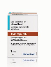
Approved in November 2017, emicizumab (Hemlibra) is a recombinant, humanized bispecific immunoglobulin G4 monoclonal antibody that mimics the cofactor function of activated factor VIII (FVIIIa) by bridging activated factor IX and factor X. After 4 weeks of a loading dose of 3 mg/kg, subcutaneous once weekly dosing at 1.5 mg/kg demonstrated significant reduction in annualized bleeding rates in patients of all ages with congenital hemophilia A and inhibitors. But treatment-related adverse events occurred during the pivotal trials.
In an effort to provide recommendations on use of emicizumab beyond information contained in the agent’s package insert, Dr. Callaghan and his associates reviewed published literature, meeting abstracts, and expert experience with emicizumab on clinical trials.
Since emicizumab is highly selective for human FIXa and FX, only chromogenic FVIII assays using human reagents can assess emicizumab activity but those assays are not widely available, the researchers noted in their abstract. “In contrast, emicizumab does not affect chromogenic assays that contain bovine reagents and thus both native and infused factor FVIII levels as well as inhibitor titers (modified Bethesda assay) can be measured using this platform.”
In a phase 3 trial of emicizumab known as HAVEN 1, serious adverse events included three cases of thrombotic microangiopathy (TMA) and two thrombotic events (TE) (N Engl J Med 2017;377:809-18).* To prevent breakthrough bleeding, aPCC should be avoided unless there are no effective alternatives to control bleeding, Dr. Callaghan said. Treatment options for bleeding include bypassing agents such as human or recombinant porcine FVIII.
To prevent, monitor, and treat TMA and TE, prior to starting emicizumab, patients should be informed that baseline hemostasis is increased with the agent and that there is an increased risk of pathologic thrombosis with bypassing agents.
Patients should also be informed about the risk of TE/TMA and the signs and symptoms of TE/TMA. “If repeated dosing of bypass agents is needed, particularly aPCC, patients should contact their hemophilia treatment center,” the researchers wrote. “If TE/TMA is suspected, platelet count, creatinine, d-dimer, and fibrinogen should be monitored. If TE/TMA occur, emicizumab should be held and aPCC discontinued until resolution. Upon resolution of TE/TMA, consideration should be given to restarting emicizumab on a case-by-case basis.”
As for laboratory considerations, the researchers noted that results of activated partial prothrombin time (aPTT) will be shortened in patients on emicizumab, often into the normal range even at low concentrations. In addition, one-stage aPTT based factor VIII activity assays will yield high factor VIII activities, even at low concentrations of the drug. “Health care providers including dentists, surgeons, and emergency room staff need to be informed of the effects of emicizumab on laboratory tests,” they wrote in a poster at THSNA 2018.
HAVEN 1 and HAVEN 2 showed that 22 patients underwent 29 surgical procedures: tooth extractions (6), CVAD procedures (9), and other procedures (14). Of the 29 surgical procedures, 9 (31%) were managed with prophylactic bypassing agents, and one treated bleed occurred. At the same time, 20 procedures (69%) were managed without prophylactic bypassing agents, and two treated bleeds occurred.
The researchers concluded that additional studies are needed to inform the use of emicizumab in people with hemophilia A, with and without inhibitor antibodies.
Dr. Callaghan reported having no financial disclosures.
*Correction, 4/26/2018: An earlier version of this story misstated the number of cases of thrombotic microangiopathy.
SAN DIEGO – Emicizumab is a safe and effective new therapy for individuals with hemophilia A and inhibitor antibodies that will likely provide a paradigm shift for managing this patient population, according to Michael U. Callaghan, MD.
“It’s a safe drug, but you do have to be cautious about treating breakthrough bleeds with activated prothrombin complex concentrate (aPCC) resistance in particular,” Dr. Callaghan, a pediatric hematologist/oncologist at Children’s Hospital of Michigan, Detroit, said in an interview at the biennial summit of the Thrombosis & Hemostasis Societies of North America. “Patients require laboratory monitoring, and you need to educate anyone who’s going to see the patient about how the drug affects laboratory tests.”
Approved in November 2017, emicizumab (Hemlibra) is a recombinant, humanized bispecific immunoglobulin G4 monoclonal antibody that mimics the cofactor function of activated factor VIII (FVIIIa) by bridging activated factor IX and factor X. After 4 weeks of a loading dose of 3 mg/kg, subcutaneous once weekly dosing at 1.5 mg/kg demonstrated significant reduction in annualized bleeding rates in patients of all ages with congenital hemophilia A and inhibitors. But treatment-related adverse events occurred during the pivotal trials.
In an effort to provide recommendations on use of emicizumab beyond information contained in the agent’s package insert, Dr. Callaghan and his associates reviewed published literature, meeting abstracts, and expert experience with emicizumab on clinical trials.
Since emicizumab is highly selective for human FIXa and FX, only chromogenic FVIII assays using human reagents can assess emicizumab activity but those assays are not widely available, the researchers noted in their abstract. “In contrast, emicizumab does not affect chromogenic assays that contain bovine reagents and thus both native and infused factor FVIII levels as well as inhibitor titers (modified Bethesda assay) can be measured using this platform.”
In a phase 3 trial of emicizumab known as HAVEN 1, serious adverse events included three cases of thrombotic microangiopathy (TMA) and two thrombotic events (TE) (N Engl J Med 2017;377:809-18).* To prevent breakthrough bleeding, aPCC should be avoided unless there are no effective alternatives to control bleeding, Dr. Callaghan said. Treatment options for bleeding include bypassing agents such as human or recombinant porcine FVIII.
To prevent, monitor, and treat TMA and TE, prior to starting emicizumab, patients should be informed that baseline hemostasis is increased with the agent and that there is an increased risk of pathologic thrombosis with bypassing agents.
Patients should also be informed about the risk of TE/TMA and the signs and symptoms of TE/TMA. “If repeated dosing of bypass agents is needed, particularly aPCC, patients should contact their hemophilia treatment center,” the researchers wrote. “If TE/TMA is suspected, platelet count, creatinine, d-dimer, and fibrinogen should be monitored. If TE/TMA occur, emicizumab should be held and aPCC discontinued until resolution. Upon resolution of TE/TMA, consideration should be given to restarting emicizumab on a case-by-case basis.”
As for laboratory considerations, the researchers noted that results of activated partial prothrombin time (aPTT) will be shortened in patients on emicizumab, often into the normal range even at low concentrations. In addition, one-stage aPTT based factor VIII activity assays will yield high factor VIII activities, even at low concentrations of the drug. “Health care providers including dentists, surgeons, and emergency room staff need to be informed of the effects of emicizumab on laboratory tests,” they wrote in a poster at THSNA 2018.
HAVEN 1 and HAVEN 2 showed that 22 patients underwent 29 surgical procedures: tooth extractions (6), CVAD procedures (9), and other procedures (14). Of the 29 surgical procedures, 9 (31%) were managed with prophylactic bypassing agents, and one treated bleed occurred. At the same time, 20 procedures (69%) were managed without prophylactic bypassing agents, and two treated bleeds occurred.
The researchers concluded that additional studies are needed to inform the use of emicizumab in people with hemophilia A, with and without inhibitor antibodies.
Dr. Callaghan reported having no financial disclosures.
*Correction, 4/26/2018: An earlier version of this story misstated the number of cases of thrombotic microangiopathy.
EXPERT ANALYSIS FROM THSNA 2018
FDA approves new drug for thrombocytopenia
with chronic immune thrombocytopenia (ITP).
The Food and Drug Administration approved the oral spleen tyrosine kinase (SYK) inhibitor, which works by impeding platelet destruction, on April 17. Its approval was based on data from FIT clinical program, including two randomized placebo-controlled phase 3 trials.
Physicians are advised to monitor blood pressure and liver function with the drug. Fostamatinib may interact with CYP3A4 inhibitors and inducers. Concomitant use of fostamatinib with CYP3A4 inhibitors increases exposure to R406 – the drug’s major metabolite – and may increase the risk of adverse reactions. Use with strong CYP3A4 inducers is not recommended because it reduces exposure to R406.
Women of reproductive potential should be advised to use appropriate contraception while using fostamatinib and for a month after stopping the drug; pregnant women should be told of potential risk to the fetus. Advise women not to breastfeed during treatment and for at least 1 month after the last dose, according to a press statement.
Rigel Pharmaceuticals plans to release the drug in the United States in late May 2018.
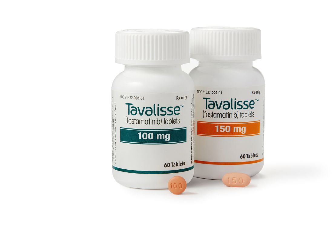
Fostamatinib approved to treat adults with chronic ITP
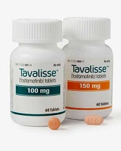
The US Food and Drug Administration (FDA) has granted approval for the oral SYK inhibitor fostamatinib disodium hexahydrate (Tavalisse™).
Fostamatinib is now approved for the treatment of thrombocytopenia in adults with chronic immune thrombocytopenia (ITP) who have had an insufficient response to a previous treatment.
Rigel Pharmaceuticals, Inc., plans to launch fostamatinib in the US in late May 2018.
The FDA’s approval of fostamatinib was supported by data from the FIT clinical program, which included 2 randomized, placebo-controlled, phase 3 trials—FIT-1 (NCT02076399) and FIT-2 (NCT02076412)—and an open-label extension study—FIT-3 (NCT02077192)—as well as an initial proof-of-concept study.
FIT-1 and FIT-2
FIT-1 and FIT-2 included 150 patients with persistent or chronic ITP who had an insufficient response to previous treatment. The patients had a median age of 54 (range, 20 to 88), 61% were female, and 93% were white.
Prior ITP treatments included corticosteroids (94%), immunoglobulins (53%), and thrombopoietin receptor agonists (48%). Thirty-five percent of patients had undergone splenectomy.
The patients’ median platelet count at baseline was 16 x 109/L, and 47% were on stable ITP therapy.
In each study, patients were randomized 2:1 to receive fostamatinib or placebo for 24 weeks. In FIT-1, 76 patients were randomized—51 to fostamatinib and 25 to placebo. In FIT-2, 74 patients were randomized—50 to fostamatinib and 24 to placebo.
All patients initially received fostamatinib at 100 mg twice daily. Most (88%) were escalated to 150 mg twice daily at week 4 or later. Patients could also receive stable concurrent ITP therapy—glucocorticoids (< 20 mg prednisone equivalent per day), azathioprine, or danazol—and rescue therapy if needed.
The efficacy of fostamatinib was based on stable platelet response, defined as a platelet count of at least 50 x 109/L on at least 4 of the 6 visits between weeks 14 to 24.
In FIT-1, 18% of patients in the fostamatinib arm and 0% of those in the placebo arm achieved a stable platelet response (P=0.03).
In FIT-2, 16% of patients in the fostamatinib arm and 4% in the placebo arm achieved a stable platelet response. In this trial, the between-arm difference was not statistically significant.
For both studies, the incidence of bleeding was 29% in the fostamatinib arm and 37% in the placebo arm. The rate of severe bleeding-related events was 1% and 6%, respectively, and the rate of serious bleeding-related events was 4% and 10%, respectively.
FIT-3
Patients from FIT-1 and FIT-2 could enroll in FIT-3 if they completed 24 weeks of treatment or did not respond to treatment any time after 12 weeks.
Patients who were in response at the time of roll over (those who had achieved a platelet count of at least 50 x 109/L) continued in FIT-3 at their current dose and regimen.
Non-responders received fostamatinib at 100 mg twice daily regardless of their dose and regimen in the prior study.
In all, 123 patients were enrolled—44 previously randomized to placebo and 79 previously randomized to fostamatinib. Half of patients (n=61) discontinued the study early.
Of the 44 patients with prior placebo, 10 (23%) had a stable platelet response to fostamatinib. This included a patient who was a responder to placebo in the prior study.
In FIT-3, stable platelet response was defined as having no 2 visits, at least 4 weeks apart, with a platelet count less than 50 x 109/L without an intervening visit with a platelet count of at least 50 x 109/L (unrelated to rescue therapy) within a period of 12 weeks after the patient’s initial achievement of the target platelet count.
Of all the responders in the FIT trials, there were 18 who maintained a platelet count of at least 50 x 109/L for 12 months or longer.
For more details on these trials and fostamatinib, see the full prescribing information, which is available at www.TAVALISSE.com.
FDA expands approved use of product for VWD
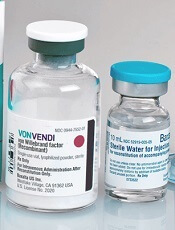
The US Food and Drug Administration (FDA) has expanded the approved use of Vonvendi, a recombinant von Willebrand factor product.
Vonvendi is now approved for perioperative management of bleeding in adults (age 18 and older) with von Willebrand disease (VWD).
Vonvendi was previously FDA-approved for on-demand treatment and control of bleeding episodes in adults with VWD.
“Persons with von Willebrand disease face a heightened risk of bleeding during surgery and may require factor treatment before, during, or after surgery,” said Michael Tarantino, MD, of the Bleeding & Clotting Disorders Institute in Peoria, Illinois.
“For surgeries requiring repeated, frequent infusions with combined von Willebrand factor and factor VIII concentrates, an excessive rise in factor VIII levels may increase the risk of thromboembolic complications, such as blood clots. The expanded use for Vonvendi in surgical settings gives healthcare professionals flexibility in treating von Willebrand disease with an appropriate dose of von Willebrand factor, with or without recombinant factor VIII, based on each patient’s unique needs.”
The FDA’s approval of Vonvendi in surgical settings was based on results from a phase 3 trial. The study enrolled 15 adults with severe VWD who were undergoing elective surgical procedures (10 of them major procedures).
Patients received Vonvendi at 40 to 60 IU per kg of body weight 12 to 24 hours before surgery. Within 3 hours of surgery, each patient’s factor VIII level (FVIII:C) was assessed, with a target of 30 IU/dL for minor surgeries and 60 IU/dL for major surgeries.
Within an hour of surgery, patients received a dose of Vonvendi, with or without recombinant factor VIII, depending on the target FVIII:C levels at the 3-hour assessment.
The study’s primary endpoint was met. Vonvendi demonstrated overall hemostatic efficacy, as assessed 24 hours after the last perioperative infusion or the completion of the study visit, whichever occurred earlier.
Intra- and post-operative hemostasis was rated as “excellent” (as good as or better than expected) in 60% of patients and “good” (probably as good as expected) in 40% of patients.
One patient developed deep vein thrombosis 3 days after undergoing hip replacement surgery.
One patient tested positive for binding antibodies to von Willebrand factor. None of the patients developed binding antibodies against potential impurities such as rFurin, CHO-protein, or mouse IgG.
More details on this study are available in the full prescribing information for Vonvendi.
Emicizumab receives breakthrough designation
The US Food and Drug Administration (FDA) has granted breakthrough therapy designation to emicizumab (Hemlibra®) for patients who have hemophilia A without factor VIII inhibitors.
Emicizumab is a bispecific factor IXa- and factor X-directed antibody approved by the FDA for routine prophylaxis to prevent or reduce the frequency of bleeding episodes in adults and children who have hemophilia A with factor VIII inhibitors.
The FDA has granted emicizumab breakthrough designation for patients without inhibitors based on data from the phase 3 HAVEN 3 trial.
The trial enrolled 152 patients age 12 and older who had hemophilia A without inhibitors and were previously treated with factor VIII therapy either on-demand or for prophylaxis.
Patients received either no prophylaxis or emicizumab prophylaxis, dosed subcutaneously every week or every 2 weeks.
Detailed results from HAVEN 3 have not been released.
However, according to Genentech, prophylaxis with emicizumab produced a statistically significant and clinically meaningful reduction in treated bleeds, when compared to no prophylaxis.
In an intra-patient comparison, once-weekly emicizumab prophylaxis was superior to prior factor VIII prophylaxis, as demonstrated by a statistically significant and clinically meaningful reduction in treated bleeds.
The most common adverse events with emicizumab were injection site reactions, and no new safety signals were observed in this trial. No thrombotic microangiopathy or thrombotic events occurred.
About breakthrough designation
The FDA’s breakthrough designation is intended to expedite the development and review of new treatments for serious or life-threatening conditions.
The designation entitles the company developing a therapy to more intensive FDA guidance on an efficient and accelerated development program, as well as eligibility for other actions to expedite FDA review, such as rolling submission and priority review.
To earn breakthrough designation, a treatment must show encouraging early clinical results demonstrating substantial improvement over available therapies with regard to a clinically significant endpoint, or it must fulfill an unmet need.
Team modifies platelets to improve coagulation

Researchers say they have found a way to improve the coagulability of platelets—by loading them with thrombin encapsulated in liposomes.
Platelets modified in this way decreased clotting time and enhanced clot strength in blood samples from healthy subjects.
The platelets also reduced clotting time in samples from patients with hemophilia A and decreased thrombin generation time in samples from patients with trauma-induced coagulopathy (TIC).
“Coagulation, which depends on a series of complex biochemical reactions, works great for scrapes and paper cuts,” said study author Christian Kastrup, PhD, of the University of British Columbia in Vancouver, Canada.
“But trauma often overwhelms this intricate, delicate process. We wanted to make it more resilient.”
Dr Kastrup and his colleagues described this endeavor in the Journal of Thrombosis and Haemostasis.
The researchers inserted thrombin into nanoliposomes and mixed them with platelets. After the platelets absorbed the nanoparticles, the team immersed the cells in blood samples for testing.
In platelet-rich plasma (PRP), the clot initiation time was about 26% faster with the modified platelets than with normal platelets—8.8 ± 1.7 min and 11.9 ± 2.5 min, respectively. And the clot strength was about 16% greater—13.4 ± 1.7 kdyne cm-1 and 11.5 ± 1.6 kdyne cm-1, respectively.
In whole blood, the clot initiation time was about 13% faster with modified platelets than with normal platelets—11.2 ± 1.2 min and 12.9 ± 1.2 min, respectively. And the clot strength was about 22% greater—5.0 ± 0.6 kdyne cm-1 and 4.1 ± 0.4 kdyne cm-1, respectively.
The researchers also found the modified platelets offset the effect of acidosis. Acidifying PRP increased thrombin generation time by 1.0 (± 0.4) min, but the modified platelets decreased thrombin generation time by 1.6 (± 0.2) min.
The modified platelets lessened the effects of antiplatelet agents as well. Aspirin or naproxen slowed thrombin generation time by 1.3 (± 0.5) min.
However, the modified platelets generated thrombin 0.4 (± 0.4) min faster in the presence of aspirin and 0.6 (± 0.4) min faster in the presence of naproxen, compared to normal platelets.
The researchers also tested the modified platelets in samples from patients with hemophilia A. Normal PRP had a clot initiation time of 5.3 (± 0.5) min, and clotting time for hemophilia A PRP was 33% slower.
In the presence of the modified platelets, clotting time was 17% slower in hemophilia A PRP than in normal PRP.
Finally, the researchers tested the modified platelets in plasma from 2 patients with TIC. With normal platelets, thrombin generation times were 5.0 (± 0.1) min and 6.7 (± 0.2) min.
With the modified platelets, thrombin generation times decreased to 3.9 (± 0.1) min and 5.9 (± 0.1) min, respectively.
If these modified platelets prove effective in subsequent preclinical and clinical testing, Dr Kastrup envisions that trauma centers could have bags of plasma containing modified platelets on hand for patients with severe bleeding. The modification could conceivably be done at the time donated blood is processed.
“Trauma is the leading killer of people under 45 years old, and blood loss is the second most common cause of such deaths,” Dr Kastrup noted. “By tweaking the body’s own clotting mechanism, we might be able to make trauma more survivable.”
Two scoring systems helpful in diagnosing heparin-induced thrombocytopenia
SAN DIEGO – Both the 4Ts Score and the HIT Expert Probability (HEP) Score are useful in clinical practice for the diagnosis of heparin-induced thrombocytopenia, but the HEP score may have better operative characteristics in ICU patients, results from a “real world” analysis showed.
“The diagnosis of heparin-induced thrombocytopenia (HIT) is challenging,” Allyson M. Pishko, MD, one of the study authors, said at the biennial summit of the Thrombosis & Hemostasis Societies of North America. “The 4Ts Score is commonly used, but limitations include its low positive predictive value and significant interobserver variability.”
One external prospective study showed operating characteristics similar to those of 4Ts scores (Thromb Haemost 2015;113[3]:633-40).
The aim of the current study was to validate the HEP Score in a “real world” setting and to compare the performance of the HEP Score versus the 4Ts Score. The researchers enrolled 292 adults with suspected acute HIT who were hospitalized at the University of Pennsylvania or affiliated community hospitals, and who had HIT laboratory testing ordered.
The HEP Score and the 4Ts Score were calculated by a member of the clinical team and were completed prior to return of the HIT lab test result. The majority of scorers (62%) were hematology fellows, followed by attendings (35%), and residents/students (3%). All patients underwent testing with an HIT ELISA and serotonin-release assay (SRA). Patients in whom the optical density of the ELISA was less than 0.4 units were classified as not having HIT. The researchers used the Wilcoxon rank-sum test to compare HEP and 4Ts Scores in patients with and without HIT.
Of the 292 patients, 209 were HIT negative and 83 had their data reviewed by an expert panel. Of these 83 patients, 40 were found to be HIT negative and 43 were HIT positive, and their mean ages were 65 years and 63 years, respectively. Among the cases found to be positive for HIT, 93% had HIT ELISA optical density of 1 or greater and 69.7% were SRA positive. The median HEP Score in patients with and without HIT was 8 versus 5 (P less than .0001).
At the prespecified screening cut-off of 2 or more points, the HEP Score was 97.7% sensitive and 21.9% specific, with a positive predictive value of 17.7% and a negative predictive value of 98.2%. A cut-off of 5 or greater provided 90.7% sensitivity and 47.8% specificity with a positive predictive value of 23.1% and a negative predictive value of 96.8%. The mean time to calculate the HEP Score was 4.1 minutes.
The median 4Ts Score in patients with and without HIT was 5 versus 4 (P less than .0001), Dr. Pishko reported. A 4Ts Score of 4 or greater had a sensitivity of 97.7% and specificity of 32.9%, with a positive predictive value of 20.1% and a negative predictive value of 98.8%.
The area under the ROC curves for the HEP Score and 4Ts Score were similar (0.81 vs. 0.76; P = .121). Subset analysis revealed that compared with the 4Ts Score, the HEP Score had better operating characteristics in ICU patients (AUC 0.87 vs. 0.79; P= .029) and with trainee scorers (AUC 0.79 vs. 0.73; P = .032).
“Our data suggest that either the HEP Score or the 4Ts Score could be used in clinical practice,” Dr. Pishko said.
The National Institutes of Health funded the study. Dr. Pishko reported having no financial disclosures.
SOURCE: Pishko A et al. THSNA 2018.
REPORTING FROM THSNA 2018
Key clinical point:
Major finding: The area under the ROC curves for the HEP Score and 4Ts Score were similar (0.81 vs. 0.76; P = .121).
Study details: A prospective study of 292 adults with suspected acute HIT who were hospitalized at the University of Pennsylvania or affiliated community hospitals.
Disclosures: The National Institutes of Health funded the study. Dr. Pishko reported having no financial disclosures.
Source: Pishko A et al. THSNA 2018.
Researchers seek better understanding of von Willebrand disease
SAN DIEGO – Several groups of researchers are examining cohorts of von Willebrand disease (VWD), looking at its pathogenesis and molecular causes, as well as ways to improve treatment strategies.
At the biennial summit of the Thrombosis & Hemostasis Societies of North America, Robert F. Sidonio Jr., MD, MSc, highlighted the efforts underway, including the Zimmerman Program for the Molecular and Clinical Biology of VWD study (ZPMCB-VWD), a large grant project funded by the National Institutes of Health.
At the time of publication, there were more than 700 index cases and more than 2,200 families in the study. It includes data from 8 primary centers and 23 secondary centers. The goals are to characterize the molecular causes of VWD and the examine the fidelity of diagnosis, “which is a critical component of the study,” said Dr. Sidonio, clinical director of the hemostasis/thrombosis program at Children’s Healthcare of Atlanta.

When the ZPMCB-VWD investigators examined the correlation of bleeding phenotype and the genotype, the found that as the level of VW factor goes down, the bleeding score generally goes up, but it becomes a little bit flat in the 20-30 IU/dL range.
“Where we spend a lot of our time is with patients who have levels of 30%-50%, which can be quite heterogeneous,” Dr. Sidonio said.
Another finding made out of the ZPMCB-VWD project was the discovery of the single nucleotide polymorphism p.D1472H, which was noted to be more common in African American patients and leads to low Von Willebrand Ristocetin Cofactor (VWF:RCo) and VWF:RCo/VWF:Ag ratio, but does not increase the bleeding score.
The fidelity of diagnosis was another key finding to come out of ZPMCB-VWD. Most type 1 VWD cases were identified by low VWF:RCo. There was poor correlation between historical and current assays (r2 = 0.22), and diagnostic labs improved after central lab testing.
Next, Dr. Sidonio discussed findings from RENAWI 1 and 2, which are Italian registries of about 1,000 VWF patients that were organized by 12 centers in 2002. The goals are to evaluate the natural history of VWD in Italy and to characterize treatment strategies. According to preliminary findings from the researchers, the biological response to desmopressin (DDAVP) was 69% in those with VWD1, 26% in those with VWD2A, 20% in those with VWD2B, 33% in those with VWD2M, 71% in those with VWD2N, and 0% in those with VWD3 (Blood 2014;123:4037-44).
These researchers also found that a mean bleeding score of 3.5 corresponds to a VWF:RCo score of 30 U/dL or greater. “This indicates that there is something slightly different about patients that are above and below that threshold,” Dr. Sidonio said. “I think that’s something we’ve all been struggling with: trying to understand where the differences are and how aggressively we should be treating our patients with mild VWD.”
Another effort, The Willebrand in the Netherlands’ study (WiN), is a prospective cohort trial of about 700 patients with types 1, 2, and 3 VWD from 12 centers in that country (Blood 2008;112:4510). It was the first large study to use VWF propeptide (pp) to discriminate between severe type 1 and type 3 VWD. It also found that type 2 VWD is more characterized by increased clearance in VWF in contrast to type 1 VWD, leading to higher VWFpp/VWF:Ag ratio. In addition, in type 1 VWD, antigen rates increased about 3.5 U/dL per decade, RCo increased about 9.5 U/dL per decade, and Factor VIII: C increased about 7.1 U/dL per decade (J Thromb Haemost. 2014; 12[7]:1066-75).
“I don’t think we have a study to be able to follow patients for 20 years or so, knowing that we rely on the assays that we were using 20 years ago,” Dr. Sidonio said. “That’s a challenge at a lot of our centers, but we know that VWF generally rises with age in mild VWD patients.”
In patients with definitively diagnosed type 2 VWD, no age-related VWF or FVIII changes were observed. The researchers also observed an increase in surgical bleeding and GI bleeding in elderly VWD patients.
In the meantime, the Canadian Type 1 VWD study was one of the first to elucidate the complexity of the pathogenesis of type 1 VWD. It identified CLEC4M as playing a role in VWF clearance, with polymorphisms contributing to the variability of VWF.
More data collection is underway through a partnership between the Centers for Disease Control and Prevention and the American Thrombosis and Hemostasis Network (ATHN).
The CDC Universal Data Collection Project gathered surveillance data on bleeding disorders on patients from 1998-2011. The goals were to characterize bleeding complications, monitor safety of blood-based products to manage bleeding, identify health issues in need of additional research, evaluate bleeding disorders over the lifespan, and evaluate quality of life. In 2006, the ATHN was formed to provide stewardship of the secured national database housed at the CDC. To date, at least 34,000 patients have opted in to the data set, which includes demographic and clinical data used for research.
In a separate, phase 4 study of about 130 patients funded by Shire and led by Dr. Sidonio and Angela C. Weyand, MD, researchers will conduct a “real-world” safety and efficacy study of prophylaxis for severe VWD. Known as ATHN 9, the study includes patients currently enrolled in the ATHN data set. Treatment regimen is at the discretion of patients’ providers, and patients will be followed for up to 2 years from the start of enrollment. The study’s primary aim is to collect data on effectiveness and safety, including adverse events of various VWF regimens in adult and pediatric patients with severe congenital VWD.
Another effort is the Medical and Scientific Advisory Council (MASAC) Working Group, of which Dr. Sidonio is a member. The first meeting took place in July of 2016. The goals include making improvements to diagnostic testing and laboratory standards, assessing existing standards of care and clinical practice guidelines, developing educational programming, conducting research to better understand and develop effective treatments for VWD, and collaborating with partner organizations.
Dr. Sidonio reported that he has participated in advisory boards for Shire, CSL Behring, Biogen/Bioverativ, Pfizer, Emergent Solutions, Roche/Genentech, Aptevo, Novo Nordisk, Hema Biologics, and Octapharma. He also has received investigator-initiated grant funding from Bioverativ, Grifols, Kedrion, and Shire.
SAN DIEGO – Several groups of researchers are examining cohorts of von Willebrand disease (VWD), looking at its pathogenesis and molecular causes, as well as ways to improve treatment strategies.
At the biennial summit of the Thrombosis & Hemostasis Societies of North America, Robert F. Sidonio Jr., MD, MSc, highlighted the efforts underway, including the Zimmerman Program for the Molecular and Clinical Biology of VWD study (ZPMCB-VWD), a large grant project funded by the National Institutes of Health.
At the time of publication, there were more than 700 index cases and more than 2,200 families in the study. It includes data from 8 primary centers and 23 secondary centers. The goals are to characterize the molecular causes of VWD and the examine the fidelity of diagnosis, “which is a critical component of the study,” said Dr. Sidonio, clinical director of the hemostasis/thrombosis program at Children’s Healthcare of Atlanta.
When the ZPMCB-VWD investigators examined the correlation of bleeding phenotype and the genotype, the found that as the level of VW factor goes down, the bleeding score generally goes up, but it becomes a little bit flat in the 20-30 IU/dL range.
“Where we spend a lot of our time is with patients who have levels of 30%-50%, which can be quite heterogeneous,” Dr. Sidonio said.
Another finding made out of the ZPMCB-VWD project was the discovery of the single nucleotide polymorphism p.D1472H, which was noted to be more common in African American patients and leads to low Von Willebrand Ristocetin Cofactor (VWF:RCo) and VWF:RCo/VWF:Ag ratio, but does not increase the bleeding score.
The fidelity of diagnosis was another key finding to come out of ZPMCB-VWD. Most type 1 VWD cases were identified by low VWF:RCo. There was poor correlation between historical and current assays (r2 = 0.22), and diagnostic labs improved after central lab testing.
Next, Dr. Sidonio discussed findings from RENAWI 1 and 2, which are Italian registries of about 1,000 VWF patients that were organized by 12 centers in 2002. The goals are to evaluate the natural history of VWD in Italy and to characterize treatment strategies. According to preliminary findings from the researchers, the biological response to desmopressin (DDAVP) was 69% in those with VWD1, 26% in those with VWD2A, 20% in those with VWD2B, 33% in those with VWD2M, 71% in those with VWD2N, and 0% in those with VWD3 (Blood 2014;123:4037-44).
These researchers also found that a mean bleeding score of 3.5 corresponds to a VWF:RCo score of 30 U/dL or greater. “This indicates that there is something slightly different about patients that are above and below that threshold,” Dr. Sidonio said. “I think that’s something we’ve all been struggling with: trying to understand where the differences are and how aggressively we should be treating our patients with mild VWD.”
Another effort, The Willebrand in the Netherlands’ study (WiN), is a prospective cohort trial of about 700 patients with types 1, 2, and 3 VWD from 12 centers in that country (Blood 2008;112:4510). It was the first large study to use VWF propeptide (pp) to discriminate between severe type 1 and type 3 VWD. It also found that type 2 VWD is more characterized by increased clearance in VWF in contrast to type 1 VWD, leading to higher VWFpp/VWF:Ag ratio. In addition, in type 1 VWD, antigen rates increased about 3.5 U/dL per decade, RCo increased about 9.5 U/dL per decade, and Factor VIII: C increased about 7.1 U/dL per decade (J Thromb Haemost. 2014; 12[7]:1066-75).
“I don’t think we have a study to be able to follow patients for 20 years or so, knowing that we rely on the assays that we were using 20 years ago,” Dr. Sidonio said. “That’s a challenge at a lot of our centers, but we know that VWF generally rises with age in mild VWD patients.”
In patients with definitively diagnosed type 2 VWD, no age-related VWF or FVIII changes were observed. The researchers also observed an increase in surgical bleeding and GI bleeding in elderly VWD patients.
In the meantime, the Canadian Type 1 VWD study was one of the first to elucidate the complexity of the pathogenesis of type 1 VWD. It identified CLEC4M as playing a role in VWF clearance, with polymorphisms contributing to the variability of VWF.
More data collection is underway through a partnership between the Centers for Disease Control and Prevention and the American Thrombosis and Hemostasis Network (ATHN).
The CDC Universal Data Collection Project gathered surveillance data on bleeding disorders on patients from 1998-2011. The goals were to characterize bleeding complications, monitor safety of blood-based products to manage bleeding, identify health issues in need of additional research, evaluate bleeding disorders over the lifespan, and evaluate quality of life. In 2006, the ATHN was formed to provide stewardship of the secured national database housed at the CDC. To date, at least 34,000 patients have opted in to the data set, which includes demographic and clinical data used for research.
In a separate, phase 4 study of about 130 patients funded by Shire and led by Dr. Sidonio and Angela C. Weyand, MD, researchers will conduct a “real-world” safety and efficacy study of prophylaxis for severe VWD. Known as ATHN 9, the study includes patients currently enrolled in the ATHN data set. Treatment regimen is at the discretion of patients’ providers, and patients will be followed for up to 2 years from the start of enrollment. The study’s primary aim is to collect data on effectiveness and safety, including adverse events of various VWF regimens in adult and pediatric patients with severe congenital VWD.
Another effort is the Medical and Scientific Advisory Council (MASAC) Working Group, of which Dr. Sidonio is a member. The first meeting took place in July of 2016. The goals include making improvements to diagnostic testing and laboratory standards, assessing existing standards of care and clinical practice guidelines, developing educational programming, conducting research to better understand and develop effective treatments for VWD, and collaborating with partner organizations.
Dr. Sidonio reported that he has participated in advisory boards for Shire, CSL Behring, Biogen/Bioverativ, Pfizer, Emergent Solutions, Roche/Genentech, Aptevo, Novo Nordisk, Hema Biologics, and Octapharma. He also has received investigator-initiated grant funding from Bioverativ, Grifols, Kedrion, and Shire.
EXPERT ANALYSIS FROM THSNA 2018
Why is gene therapy for hemophilia taking so long?
SAN DIEGO – The goal of gene therapy for hemophilia and other genetic diseases is to achieve long-term expression and levels adequate to improve the phenotype of disease, according to Katherine A. High, MD.
“Sometimes people ask me, ‘Why is it taking so long to develop these therapeutics?’ ” Dr. High said at the biennial summit of the Thrombosis & Hemostasis Societies of North America. “The answer is that gene therapy vectors are arguably one of the most complex therapeutics yet developed.”

Spark Therapeutics is currently developing gene therapies for hemophilia A (SPK-8011) and hemophilia B (SPK-9001).
Hemostasis and thrombosis targets in gene therapy trials include hemophilia, as well as peripheral artery disease/claudication and congestive heart failure. In the latter, a prior phase 2b trial of adeno-associated virus (AAV) expressing SERCA2a did not support efficacy (Lancet 2016;387:1178-86), while a current trial of adenovirus 5–vector expressing adenylyl cyclase–type 6 is entering phase 3 study (NCT03360448).
To get a sense of how long it may take for a new class of therapeutics to become established, Dr. High noted that the first monoclonal antibody to be licensed was OKT3 (muromonab-CD3) in 1986, followed by abciximab in 1994, rituximab and daclizumab in 1997, and four additional products in 1998. By 2007, 8 of the top 20 biotech drugs were monoclonal antibodies.
Hemophilia has long been a favored gene therapy target because biology is in its favor. “It has a wide therapeutic window, it does not require tissue-specific expression of transgene, small and large animal models exist, and endpoints are well validated and easy to measure,” she said. “Thus, early gene-therapy clinical investigation since 1998 explored many strategies.”
There are several current investigational efforts in AAV-mediated gene transfer in hemophilia, including:
- A single-arm study to evaluate the efficacy and safety of valoctocogene roxaparvovec in hemophilia A patients at a dose of 4×10¹³ vector genome per kilogram (NCT03392974).
- A dose-ranging study of recombinant AAV2/6 human factor 8 gene therapy SB-525 in subjects with severe hemophilia A (NCT03061201).
- A safety and dose-escalation study of an adeno-associated viral vector for gene transfer in hemophilia A subjects (NCT03370172).
Other approaches in preclinical investigation include lentiviral transduction of hematopoietic stem cells with megakaryocyte-restricted expression, lentiviral transduction of liver cells and endothelial cells, and genome editing using zinc finger nucleases.
“AAV vectors are one of the smallest of all naturally occurring viruses,” said Dr. High, who is also emeritus professor of pediatrics at the University of Pennsylvania, Philadelphia. “The recombinant AAV consists of a highly ordered set of proteins [vector capsid] containing DNA [the active agent].”
Overall goals for a hemophilia gene therapy include long-term expression and levels adequate to prevent bleeds in someone with a normal active lifestyle. “We’d like to see consistency of results from one person to the next, and we’d like to use the lowest possible dose,” she said. “In the setting of gene transfer, the lower the dose, the lower the likelihood of immune responses that need to be managed. Theoretically, the lower the dose, the lower the risk of insertional mutagenesis, and the shorter-term duration of vector shedding in body fluids, including in semen.”
Going forward, a key question for researchers relates to the long-term effect of gene therapy. “How long is long enough?” Dr. High asked. “The longest reported durability is 8 years, with observation ongoing, from studies initially reported in men with severe hemophilia B. The durability in large animal models exceeds 10 years.”
Another unanswered question is what level of factor VIII to aim for in treatment. “Some data suggest that FVIII levels greater than 100 IU/dL are associated with a greater level of thrombosis,” Dr. High said. “So I think somewhere between 12% and 100% is probably the ideal level.”
SAN DIEGO – The goal of gene therapy for hemophilia and other genetic diseases is to achieve long-term expression and levels adequate to improve the phenotype of disease, according to Katherine A. High, MD.
“Sometimes people ask me, ‘Why is it taking so long to develop these therapeutics?’ ” Dr. High said at the biennial summit of the Thrombosis & Hemostasis Societies of North America. “The answer is that gene therapy vectors are arguably one of the most complex therapeutics yet developed.”
Spark Therapeutics is currently developing gene therapies for hemophilia A (SPK-8011) and hemophilia B (SPK-9001).
Hemostasis and thrombosis targets in gene therapy trials include hemophilia, as well as peripheral artery disease/claudication and congestive heart failure. In the latter, a prior phase 2b trial of adeno-associated virus (AAV) expressing SERCA2a did not support efficacy (Lancet 2016;387:1178-86), while a current trial of adenovirus 5–vector expressing adenylyl cyclase–type 6 is entering phase 3 study (NCT03360448).
To get a sense of how long it may take for a new class of therapeutics to become established, Dr. High noted that the first monoclonal antibody to be licensed was OKT3 (muromonab-CD3) in 1986, followed by abciximab in 1994, rituximab and daclizumab in 1997, and four additional products in 1998. By 2007, 8 of the top 20 biotech drugs were monoclonal antibodies.
Hemophilia has long been a favored gene therapy target because biology is in its favor. “It has a wide therapeutic window, it does not require tissue-specific expression of transgene, small and large animal models exist, and endpoints are well validated and easy to measure,” she said. “Thus, early gene-therapy clinical investigation since 1998 explored many strategies.”
There are several current investigational efforts in AAV-mediated gene transfer in hemophilia, including:
- A single-arm study to evaluate the efficacy and safety of valoctocogene roxaparvovec in hemophilia A patients at a dose of 4×10¹³ vector genome per kilogram (NCT03392974).
- A dose-ranging study of recombinant AAV2/6 human factor 8 gene therapy SB-525 in subjects with severe hemophilia A (NCT03061201).
- A safety and dose-escalation study of an adeno-associated viral vector for gene transfer in hemophilia A subjects (NCT03370172).
Other approaches in preclinical investigation include lentiviral transduction of hematopoietic stem cells with megakaryocyte-restricted expression, lentiviral transduction of liver cells and endothelial cells, and genome editing using zinc finger nucleases.
“AAV vectors are one of the smallest of all naturally occurring viruses,” said Dr. High, who is also emeritus professor of pediatrics at the University of Pennsylvania, Philadelphia. “The recombinant AAV consists of a highly ordered set of proteins [vector capsid] containing DNA [the active agent].”
Overall goals for a hemophilia gene therapy include long-term expression and levels adequate to prevent bleeds in someone with a normal active lifestyle. “We’d like to see consistency of results from one person to the next, and we’d like to use the lowest possible dose,” she said. “In the setting of gene transfer, the lower the dose, the lower the likelihood of immune responses that need to be managed. Theoretically, the lower the dose, the lower the risk of insertional mutagenesis, and the shorter-term duration of vector shedding in body fluids, including in semen.”
Going forward, a key question for researchers relates to the long-term effect of gene therapy. “How long is long enough?” Dr. High asked. “The longest reported durability is 8 years, with observation ongoing, from studies initially reported in men with severe hemophilia B. The durability in large animal models exceeds 10 years.”
Another unanswered question is what level of factor VIII to aim for in treatment. “Some data suggest that FVIII levels greater than 100 IU/dL are associated with a greater level of thrombosis,” Dr. High said. “So I think somewhere between 12% and 100% is probably the ideal level.”
EXPERT ANALYSIS FROM THSNA 2018