User login
Gene editing aims to recreate beneficial mutation in SCD, beta-thalassemia
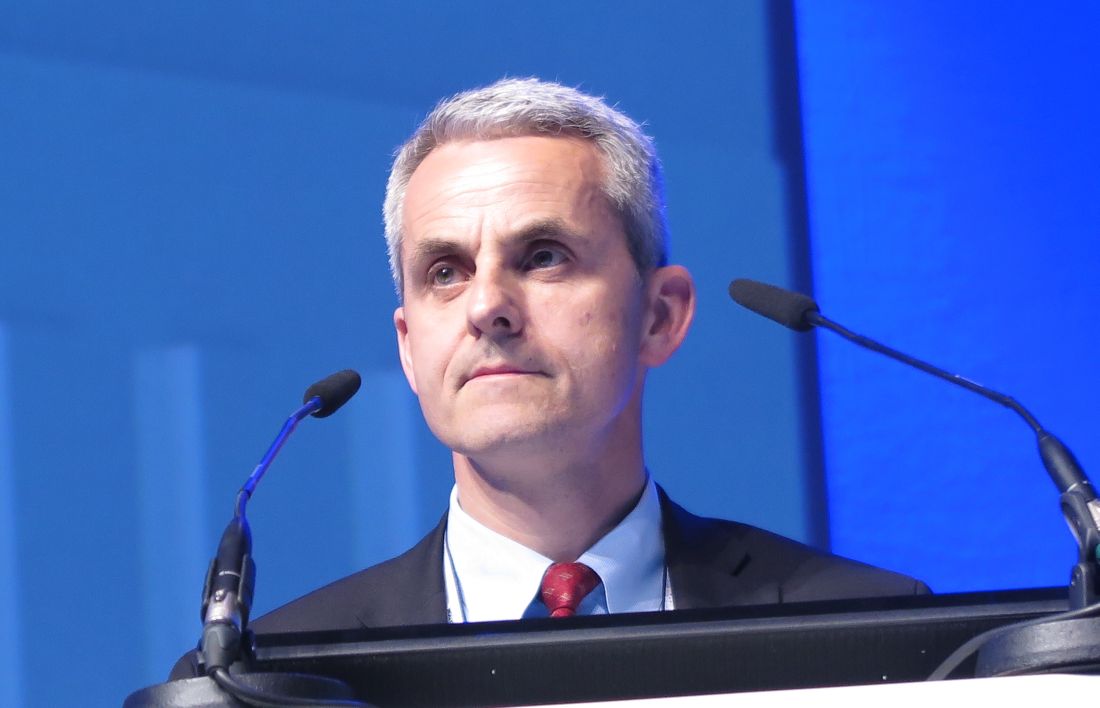
MADRID – With some genetic sleight-of-hand, investigators hope to mimic a rare, naturally occurring mutation that protects some patients with beta-thalassemia or sickle-cell disease (SCD) from becoming symptomatic.
Although human studies have yet to begin, investigators in a biotech company report that they can use CRISPR/Cas9 gene editing to recreate the rare condition known as hereditary persistence of fetal hemoglobin, or HPFH, which causes some patients with SCD or beta-thalassemia to continue to produce the fully functional fetal rather than adult form of hemoglobin into adulthood.
To date, there have been no detectable undesirable off-target genetic modifications seen by targeted deep sequencing for the two leading guide RNAs (gRNA) in development, he said at the annual congress of the European Hematology Association.
“Patients with sickle-cell disease and beta-thalassemia are not sick when they are first born, but symptoms develop shortly after birth as the fetal hemoglobin levels decline, and as adult hemoglobin rises,” Dr. Lundberg said.
Fetal hemoglobin has been shown to reduce the risk of sickle events in SCD, and reduce symptoms and morbidity of beta-thalassemia.
“Modest amounts of fetal hemoglobin are beneficial, and more appears to be better,” he said.
In rare instances, the genetic switch to turn off fetal hemoglobin fails, leading to HPFH. It is this rare circumstance that the investigators hope to copy.
CRISPR/Cas9 technology copies a bacterial defense mechanism using clustered regularly interspaced short palindromic (CRISPR) repeats of DNA as a template for adaptive immunity against viruses and plasmids. The system allows bacteria to prevent the activation of invading nucleic acids by effectively snipping out target sequences of foreign DNA.
The investigators plan to harvest hematopoietic stem cells from patients with SCD and beta-thalassemia and use CRISPR/Cas9 technology to modify the cells to express the fetal form of hemoglobin. The reintroduced stem cells would, ideally, proliferate and help protect patients against painful crises and debilitating symptoms of the hemoglobinopathies.
Dr. Lundberg said that to date they have been able to edit blood stem cells with more than 80% on-target efficiency, and that the editing leads to “clinically meaningful” levels of protective fetal hemoglobin, in the range of 30%.
As noted, the investigators have to date seen no evidence of off-target editing that could lead to undesirable or dangerous complications. When introduced into an immunodeficient mouse model (NOD scid gamma, or NSG mice), the edited human cells persisted for long periods.
Finally, the investigators have been able to demonstrate that the modified stem cells are able to differentiate and reconstitute different types of cells in blood.
The company plans to submit a clinical trial application in 2017, and start clinical trials in 2018, Dr. Lundberg said.
At a media briefing where he discussed his research prior to his presentation of data in a symposium, Dr. Lundberg said that to date they have worked only with mouse models and with cells from healthy donors.

He pointed out, however, that there are many thousands of patients with inherited hemoglobin disorders worldwide and beta-thalassemia, and asked “how do you proceed to implement this very expensive treatment?”
Dr. Lundberg acknowledged that the diseases are common in both the developing world and in wealthy countries. He said that the technique relies as much as possible on existing technologies, and pointed out that if the treatment is successful, it’s costs could be at least partially offset by reducing the costs of other forms of therapy.
The work is supported by CRISPR Therapeutics. Dr. Lundberg is an employee and shareholder in the company. Dr. Hagenbeek reported having no relevant conflicts of interest.
MADRID – With some genetic sleight-of-hand, investigators hope to mimic a rare, naturally occurring mutation that protects some patients with beta-thalassemia or sickle-cell disease (SCD) from becoming symptomatic.
Although human studies have yet to begin, investigators in a biotech company report that they can use CRISPR/Cas9 gene editing to recreate the rare condition known as hereditary persistence of fetal hemoglobin, or HPFH, which causes some patients with SCD or beta-thalassemia to continue to produce the fully functional fetal rather than adult form of hemoglobin into adulthood.
To date, there have been no detectable undesirable off-target genetic modifications seen by targeted deep sequencing for the two leading guide RNAs (gRNA) in development, he said at the annual congress of the European Hematology Association.
“Patients with sickle-cell disease and beta-thalassemia are not sick when they are first born, but symptoms develop shortly after birth as the fetal hemoglobin levels decline, and as adult hemoglobin rises,” Dr. Lundberg said.
Fetal hemoglobin has been shown to reduce the risk of sickle events in SCD, and reduce symptoms and morbidity of beta-thalassemia.
“Modest amounts of fetal hemoglobin are beneficial, and more appears to be better,” he said.
In rare instances, the genetic switch to turn off fetal hemoglobin fails, leading to HPFH. It is this rare circumstance that the investigators hope to copy.
CRISPR/Cas9 technology copies a bacterial defense mechanism using clustered regularly interspaced short palindromic (CRISPR) repeats of DNA as a template for adaptive immunity against viruses and plasmids. The system allows bacteria to prevent the activation of invading nucleic acids by effectively snipping out target sequences of foreign DNA.
The investigators plan to harvest hematopoietic stem cells from patients with SCD and beta-thalassemia and use CRISPR/Cas9 technology to modify the cells to express the fetal form of hemoglobin. The reintroduced stem cells would, ideally, proliferate and help protect patients against painful crises and debilitating symptoms of the hemoglobinopathies.
Dr. Lundberg said that to date they have been able to edit blood stem cells with more than 80% on-target efficiency, and that the editing leads to “clinically meaningful” levels of protective fetal hemoglobin, in the range of 30%.
As noted, the investigators have to date seen no evidence of off-target editing that could lead to undesirable or dangerous complications. When introduced into an immunodeficient mouse model (NOD scid gamma, or NSG mice), the edited human cells persisted for long periods.
Finally, the investigators have been able to demonstrate that the modified stem cells are able to differentiate and reconstitute different types of cells in blood.
The company plans to submit a clinical trial application in 2017, and start clinical trials in 2018, Dr. Lundberg said.
At a media briefing where he discussed his research prior to his presentation of data in a symposium, Dr. Lundberg said that to date they have worked only with mouse models and with cells from healthy donors.
He pointed out, however, that there are many thousands of patients with inherited hemoglobin disorders worldwide and beta-thalassemia, and asked “how do you proceed to implement this very expensive treatment?”
Dr. Lundberg acknowledged that the diseases are common in both the developing world and in wealthy countries. He said that the technique relies as much as possible on existing technologies, and pointed out that if the treatment is successful, it’s costs could be at least partially offset by reducing the costs of other forms of therapy.
The work is supported by CRISPR Therapeutics. Dr. Lundberg is an employee and shareholder in the company. Dr. Hagenbeek reported having no relevant conflicts of interest.
AT EHA 2017
Key clinical point: The fetal form of hemoglobin is protective against symptoms of sickle-cell disease (SCD) and beta-thalassemia.
Major finding: Human blood stem cells modified by CRISPR/Cas9 gene editing produced fetal hemoglobin without observable off-target effects.
Data source: Summary of preclinical studies with blood from healthy human volunteers and mouse models.
Disclosures: The work is supported by CRISPR Therapeutics. Dr. Lundberg is an employee and shareholder in the company. Dr. Hagenbeek reported having no relevant conflicts of interest.
Fostamatinib under review by FDA for chronic ITP

The US Food and Drug Administration (FDA) has accepted a new drug application (NDA) for the use of fostamatinib disodium (Tavalisse™) in the treatment of patients with chronic/persistent immune thrombocytopenia (ITP).
Fostamatinib is an oral spleen tyrosine kinase (SYK) inhibitor that investigators believe may address an underlying autoimmune cause of ITP by impeding platelet destruction.
Rigel Pharmaceuticals, the drug’s developer, anticipates an action date by the FDA of April 17, 2018.
The FDA previously granted fostamatinib orphan drug designation.
Data from 3 clinical studies support the new drug application. Study 047 and 048 were randomized placebo-controlled trials, and study 049 was an open-label extension study.
Together with an initial proof of concept study, the application included data on 163 ITP patients. Fostamatinib has been evaluated in over 4,600 individuals across all indications.
The patients enrolled in studies 047 and 048 had been diagnosed with persistent or chronic ITP, had failed at least 1 prior therapy for ITP, and had platelet counts consistently below 30,000/uL.
In both studies, patients were randomized in a 2:1 ratio to receive either fostamatinib or placebo orally twice a day for up to 24 weeks.
Study 047
In this study, 51 patients were randomized to fostamatinib and 25 to placebo.
The median platelet counts at baseline were 15,000/uL and 16,000/uL in the fostamatinib and placebo arms, respectively.
The median age was 57 in both treatment arms. The duration of ITP was 7.5 years (range, 0.6-53) in the fostamatinib arm and 5.5 years (range, 0.4-45) in the placebo arm.
The primary efficacy endpoint in both study 047 and 048 was a stable platelet response, defined as achieving platelet counts greater than 50,000/uL for at least 4 of the 6 scheduled clinic visits between weeks 14 and 24 of treatment.
In study 047, the rate of stable platelet response was significantly higher in the fostamatinib arm than the placebo arm—18% (n=9) and 0%, respectively (P=0.026).
Study 048
Fifty patients were randomized to fostamatinib and 24 to placebo.
Patients’ median platelet counts at baseline were 16,000/uL and 21,000/uL in the fostamatinib and placebo arms, respectively.
The median age was 50 in both treatment arms. The duration of ITP was 8.8 years (range, 0.3-50) in the fostamatinib arm and 10.8 years (range, 0.9-29) in the placebo arm.
In this study, however, the difference in stable platelet response between the 2 arms was not significant—18% (n=9) and 4% (n=1), respectively (P=0.152).
However, when the data from study 047 and 048 were combined, the response rate was significantly higher in the fostamatinib arm than the placebo arm—18% (18/101) and 2% (1/49), respectively (P=0.007).
"The FDA acceptance for filing of our NDA is an exciting milestone for Rigel," Raul Rodriguez, Rigel's president and chief executive officer, said.
The approval of fostamatinib “will provide a new treatment option for patients with chronic or persistent ITP,” he affirmed. “We look forward to working closely with the FDA as they review our submission." ![]()
Ferrous sulfate bests iron complex in treating IDA in infants, young kids

A trial comparing ferrous sulfate with iron polysaccharide complex to treat infants and young children with nutritional iron-deficiency anemia (IDA) has shown ferrous sulfate to be more effective at raising hemoglobin levels in this population, according to researchers.
Dozens of oral iron supplements exist for IDA treatment, ferrous sulfate being the most commonly used. Iron polysaccharide complex, however, may be better tolerated.
Investigators undertook the BESTIRON study (NCT01904864) to determine whether the iron complex was more efficacious than ferrous sulfate in increasing hemoglobin concentrations in infants and young children aged 9 to 48 months.
Up to 3% of children aged 1 to 2 years in the United States have IDA, as do millions worldwide. IDA is associated with impaired neurodevelopment in the young.
Inadequate dietary iron intake in this group is the most common cause of IDA. It most often results from excessive cow milk consumption and/or prolonged breastfeeding without appropriate iron supplementation.
For this study, investigators randomized 80 infants and young children with nutritional IDA to receive 3 mg/kg of ferrous sulfate (n=40) or iron complex (n=40) drops once daily for 12 weeks.
Patients had to have hemoglobin concentrations of 10 g/dL or less, mean corpuscular volumes of 70 fL or less, reticulocyte hemoglobin equivalents of 25 pg or less, and either serum ferritin level of 15 ng/mL or less or total iron-binding capacity of 425 μg/dL or greater.
And they could have no clinical or laboratory evidence of other causes of anemia.
All 80 patients were included in the primary analysis evaluating change in hemoglobin concentration during the 12 weeks after starting oral iron therapy.
Patient characteristics
Patient characteristics were similar between the groups. The mean age was 23 months and 55% were male.
Most patients (61%) were Hispanic white, 9% were non-Hispanic white, and 11% were black.
Ten patients in the ferrous sulfate group and 8 in the iron complex group had received a packed red blood cell transfusion prior to study enrollment.
Results
Fifty-nine patients completed all study visits, 28 in the ferrous sulfate group and 31 in the iron complex group.
Patients’ mean hemoglobin level in the ferrous sulfate group increased from 7.9 g/dL to 11.9 g/dL over the 12 weeks. In the iron complex group, the patients’ hemoglobin level increased from 7.7 g/dL to 11.1 g/dL.
Using a linear mixed model, the primary outcome demonstrated a significant difference in the change in hemoglobin concentration of 1.0 g/dL (95% CI, 0.4-1.6; P < .001) between the groups, favoring ferrous sulfate.
IDA completely resolved in 8 of 28 (29%) patients in the ferrous sulfate group and 2 of 31 (6%) in the iron complex group (P=0.04).
However, successful administration of the supplement—meaning he child did not spit out the medication—was higher in the iron complex group (94%) than the iron sulfate group (82%), P=0.009.
The median serum ferritin level increased from 3.0 ng/mL to 15.6 ng/mL in the ferrous sulfate arm, which was significantly better than in the iron complex arm, which increased from 2.0 ng/mL to 7.5 ng/mL, P<0.001.
And the mean total iron binding capacity significantly increased in the ferrous sulfate group compared with the iron oxide group (P<0.001).
Safety
The investigators reported that patients treated with iron complex had significantly more diahrrea, while patients treated with ferrous sulfate had more vomiting, although the latter was not statistically significant.
A gastrointestinal adverse effect profile created at the end of the study showed no significant differences between the groups.
The investigators noted a few limitations of the study.
First, it was conducted in a single tertiary-care children’s hospital, the Children’s Medical Center in Dallas, Texas.
Second, a disproportionate number of patients were from lower income and minority families and frequently had severe anemia, with approximately 23% requiring blood transfusion prior to study start.
And third, the trial had a high lost-to-follow-up rate of 25% at the final visit.
So the results may not be generalizable to the general pediatric population.
Nevertheless, the investigators concluded, “Once daily, low-dose ferrous sulfate should be considered for children with nutritional iron-deficiency anemia.”
The team reported their findings in JAMA.
The study was an investigator-initiated trial with sponsorship from Gensavis Pharmaceuticals LLC, the manufacturer of the iron polysaccharide complex used in the trial. The company provided funding for both trial drugs.
The study received additional grant support from the National Center for Advancing Translational Sciences and the National Heart, Lung, and Blood Institute. ![]()
BM-MSCs may be an option for AA patients refractory to IST
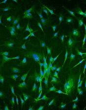
Researchers report that infusions of bone marrow-derived mesenchymal stromal cells (BM-MSCs) may be a treatment option for patients with aplastic anemia (AA) who are refractory to immunosuppressive therapy (IST).
They conducted a phase 2, non-comparative multicenter study to assess the safety and efficacy of this approach and found that after 12 months, 28.4% of patients responded, with 6.8% achieving a complete response (CR) and 21.6% a partial response (PR).
The trial involved 74 patients at 7 centers in China. The research team reported its findings in Stem Cells Translational Medicine.
About 30% to 40% of patients with severe AA (sAA) don’t respond well to IST and continue to have abnormally low levels of red blood cells, white blood cells, and platelets.
The benefit of treatment with BM-MSCs is they support hematopoiesis, express low levels of major histocompatibility (MHC)-I, and lack expression of MHC-II surface molecules.
And BM-MSCs have been reported to cure diseases, including graft-versus-host disease, arthritis, lupus, and other immune and non-immune disorders.
The current study, led by Yan Pang, MD, and Yang Xiao, MD, PhD, of Guangzhou General Hospital of Guangzhou Military Command in China, is based on their previous data evaluating intravenous administration of MSCs from a related donor in 18 patients with refractory AA.
This earlier data showed that 33% of patients with AA refractory to IST achieved a CR or PR to BM-MSC treatment.
So the team undertook to further investigate the use of BM-MSCs in AA.
Study design
Each of the 74 patients received 4 doses of BM-MSCs over a period of 4 weeks. If patients responded after the first month, they continued to receive 4 doses.
Investigators obtained the BM-MSCs from 74 healthy donors—48 males and 26 females. Forty were related donors, 27 haploidentical donors, and 7 unrelated donors.
Patients with AA by standard criteria had to be 16 years or older, had an incomplete response to antithymocyte globulin (ATG) and cyclosporine for at leas 6 months or cyclosporine alone for at least 12 months, did not have a donor available for bone marrow transplantation, and had at least one of the following: hemoglobin <70 g/L, neutrophilic granulocytes <1 × 10⁹/L, or platelet count <30 × 109/L.
Almost half the patients (47%) were between 20 and 40 years, 54% were male, and 32% had severe aplastic anemia.
Patients’ previous therapy for aplastic anemia included cyclosporine and andriol (65%) cyclosporine and ATG (19%), and cyclosporine (16%).
Patients could continue cyclosporine, but no immunosuppressive agents were permitted.
Fifty-three patients (71.6%) completed 1 course of therapy, and 21 patients (18.4%) completed 2 courses.
Response
At 1 year, the overall response rate was 28.4% (n=21) and the PR rate was 21.6% (n=16).
The median time to a leukocyte response was 19 days (range, 11 – 29), to an erythrocyte response 17 days (range, 12 – 25), and to a megakaryocyte response 31 days (range, 26 – 84).
Ten of the patients with hematologic response had normalization of cellularity for more than 1 year.
The median follow-up among survivors was 17 months (range, 3 – 24). The 2-year overall survival was 87.8%.
Three patients progressed to myelodysplasia, 1 with refractory anemia with excess blasts (RAEB)-I and 2 with RAEB-II.
The median time to progression was 11 months (range, 8 – 12).
Nine patients died, all of whom had severe AA. One patient with RAEB-II died of disease progression, two patients died of intracranial hemorrhage, and six patients died of serious infection.
Safety
Adverse events included grade 1 (n=5) and grade 2 (n=2) fever. Two of these patients also had grade 1 headache.
The investigators observed no other adverse events.
Response predictors
The investigators determined that 2 factors predicted response in patients: prior treatment with antithymocyte globulin (ATG) and absence of infection throughout the treatment.
The odds ratio for patients treated with ATG was 1.41 (95% CI: -0.50, 3.31) and for patients without infection, 2.19 (95% CI: 0.50, 3.87).
“Our study strongly indicates that MSC infusion is a promising therapy for severe AA," Dr Pang said, “but improved MSC cultures in vitro and the MSC doses need further study to maximize their therapeutic potential." ![]()
Azacitidine alone comparable to AZA combos for most MDS patients

A 3-arm phase 2 study of azacitidine alone or in combination with lenalidomide or vorinostat in patients with higher-risk myelodysplastic syndromes (MDS) or chronic myelomonocytic leukemia (CMML) has shown the combination therapies to have similar overall response rates (ORR) to azacitidine monotherapy. Based on these findings, investigators did not choose either combination arm for phase 3 testing of overall survival.
However, patients with CMML treated with the azacitidine-lenalidomide combination had twice the ORR compared with azacitidine monotherapy, they reported.
And patients with certain mutations, such as DNMT3A, BCOR, and NRAS, had higher overall response rates, although only those with the DNMT3A mutation were significant.
Mikkael A. Sekeres, MD, of the Cleveland Clinic in Cleveland, Ohio, and colleagues reported these findings in the Journal of Clinical Oncology on behalf of the North American Intergroup Study SWOG S117.
Doses of azacitidine were the same for monotherapy and combination arms: 75 mg/m2/day intravenously or subcutaneously on days 1 to 7 of a 28-day cycle.
Patients in the lenalidomide arm received 10 mg/day orally of that drug on days 1 to 21, and patients in the vorinostat arm received 300 mg twice daily orally on days 3 to 9.
Patient characteristics
Patients had MDS of IPSS Intermediate-2 or higher or bone marrow blasts 5% or greater. Patients with CMML had fewer than 20% blasts.
The investigators randomized 277 patients to receive either azacitidine alone (n=92), azacitidine plus lenalidomide (n=93), or azacitidine plus vorinostat (n=92).
Patients were a median age of 70 years (range, 28 to 93). Eighty-five patients (31%) were female, 53 (19%) had CMML, and 18 (6%) had treatment-related MDS. More than half the patients were transfusion-dependent at baseline.
Baseline characteristics were similar across the 3 arms. The investigators noted that the baseline characteristics were also similar across the 90 centers participating in the study, whether they were an MDS Center of Excellence or a high-volume center.
Adverse events
For the most part, therapy-related adverse events were similar across the arms.
Rates of grade 3 or higher febrile neutropenia and infection and infestations were similar for all 3 cohorts: 89% for azaciditine monotherapy, 91% for the lenalidomide combination, and 91% for the vorinostat combination.
However, the vorinostat arm had more grade 3 or higher gastrointestinal toxicities (14 patients, 15%) compared with the monotherapy arm (4 patients, 4%), P=0.02.
And patients receiving lenalidomide experienced more grade 3 or higher rash (14 patients, 16%) compared with patients receiving monotherapy (3 patients, 3%), P=0.005.
Patients in the combination arms stopped therapy at significantly higher rates than the monotherapy arm. Eight percent of patients receiving monotherapy stopped treatment compared with 20% in the lenalidomide arm and 21% in the vorinostat arm.
Patients in the combination arms also had more dose modifications not specified in the protocol than those in the monotherapy arm. Twenty-four percent receiving azacitidine monotherapy had non-protocol defined dose modifications, compared with 43% in the lenalidomide arm and 42% in the vorinostat arm.
Responses
The ORR for the entire study population was 38%.
Patients in the monotherapy arm had an ORR of 38%, those in the lenalidomide arm, 49%, and those in the vorinostate arm, 27%. Neither arm achieved significance compared with the monotherapy arm.
Patients who were treatment-naïve in the lenalidomide arm had a somewhat improved ORR compared with monotherapy, P=0.08.
The median duration of response for all cohorts was 15 months: 10 months for monotherapy, 14 months for lenalidomide, and 18 months for vorinostat.
Patients who were able to remain on therapy for 6 months or more in the lenalidomide arm achieved a higher ORR of 87% compared with monotherapy (62%, P=0.01). However, there was no difference in response duration with longer therapy.
The median overall survival (OS) was 17 months for all patients, 15 months for patients in the monotherapy group, 19 months for those in the lenalidomide arm, and 17 months for those in the vorinostat group.
CMML patients had similar OS across treatment arms, with the median not yet reached for patients in the monotherapy arm.
Subgroup responses
Patients with CMML in the lenalidomide arm had a significantly higher ORR than CMML patients in the monotherapy arm, 68% and 28%, respectively (P=0.02).
Median duration of response for CMML patients was 19 months, with no differences between the arms.
The investigators observed no differences in ORR for therapy-related MDS, IPSS subgroups, transfusion-dependent patients, or allogeneic transplant rates.
However, they noted ORR was better for patients with chromosome 5 abnormality regardless of treatment arm than for those without the abnormality (odds ratio, 2.17, P=0.008).
One hundred thirteen patients had mutational data available. They had a median number of 2 mutations (range, 0 to 7), with the most common being ASXL1 (n = 31), TET2 (n = 26), SRSF2 (n = 23), TP53 (n = 22), RUNX1 (n = 21), and U2AF1 (n = 19).
Patients with DNMT3A mutation had a significantly higher ORR than for patients without mutations, 67% and 34%, respectively P=0.025).
Patients with BCOR and NRAS mutations had numerically higher, but non-significant, ORR than non-mutated patients. Patients with BCOR mutation had a 57% ORR compared with 34% for non-mutated patients (P=0.23). Patients with NRAS mutation had a 60% ORR compared with 36% for non-mutated patients (P=0.28).
Patients with mutations in TET2 (P = .046) and TP53 (P = .003) had a worse response duration than those without mutations.
Response duration was significantly better with fewer mutations. For 2 or more mutations, the hazard ration was 6.86 versus no mutations (P=0.01).
The investigators believed under-dosing may have compromised response and survival in the combination arms. They suggested that studies focused on the subgroups that seemed to benefit from the combinations should be conducted. ![]()
Gene plays key role in iron homeostasis

A gene known to prevent autoimmune diseases is a key regulator in iron uptake, according to research published in Cell Reports.
“We found previously that, when mice lack the gene Regnase-1, they suffer from severe autoimmune diseases and anemia,” said study author Masanori Yoshinaga, MD, of Kyoto University in Japan.
“At first, we assumed that anemia was a secondary effect, but, after detailed analysis, we found that the 2 symptoms develop independently.”
Continued study of mice with a Regnase-1 mutation revealed a functional defect in the principal site for iron absorption in the body, the duodenum.
“The next step was to find the role of Regnase-1 in iron-uptake maintenance,” Dr Yoshinaga said. “We started by looking at the most important iron-uptake gene, Transferrin Receptor 1, or TfR1.”
“Our results showed that Regnase-1 degrades the mRNA of TfR1, thereby inhibiting the synthesis of the TfR1 protein and, additionally, that it likely regulates other important iron-controlling genes.”
“Further analysis of Regnase-1 in iron-related homeostasis may provide insight into the mechanisms causing anemia and other iron-related disorders, perhaps eventually leading to new methods of treatment,” said study author Osamu Takeuchi, MD, PhD, of Kyoto University. ![]()
Authority on hematologic malignancies dies

Physician, researcher, and educator H. Jean Khoury, MD, recently passed away.
He died on Monday, May 22, at the age of 50, after a year-long battle with esophageal cancer.
Dr Khoury led the division of hematology at Winship Cancer Institute of Emory University in Atlanta, Georgia.
He was considered an authority on hematologic malignancies, particularly chronic myeloid leukemia (CML), acute leukemia, and myelodysplastic syndromes (MDS).
Dr Khoury joined Winship Cancer Institute in 2004 as director of the Leukemia Service and associate professor in the Emory School of Medicine.
In 2009, he was promoted to professor and director of the Division of Hematology in the Department of Hematology and Medical Oncology, and he was later named to the R. Randall Rollins Chair in Oncology.
“We are all deeply grieving the loss of this remarkable man who gave so much to Winship,” said Walter J. Curran, Jr, MD, Winship Cancer Institute’s executive director.
“His enthusiasm and love for his patients and his commitment to lessening the burden of cancer for all has been unwavering throughout his life.”
A native of Beirut, Lebanon, Dr Khoury came to the Winship Cancer Institute from Washington University in St Louis, Missouri, where he served on the faculty after completing a fellowship in hematology-oncology.
He earned his medical degree from the Université Catholique de Louvain in Brussels, Belgium, and completed a residency in internal medicine at Memorial Medical Center in Savannah, Georgia.
Dr Khoury was recruited to Winship Cancer Institute by Fadlo R. Khuri, MD, former deputy director of the institute and now president of the American University of Beirut. What he first saw in Dr Khoury was someone who was “in the best sense, a disruptive presence.”
“What you always want in a leader is someone who’s not afraid to be wrong, to take risks,” Dr Khuri said. “Being wrong disrupts the pattern, and Jean was very brave. He didn’t like business as usual, and that showed in the way he took about redeveloping the hematology division, the leukemia program, and his interactions with the transplant division, with faculty, and all across Winship.”
According to his colleagues, Dr Khoury’s guiding principle was how to improve his patients’ lives, whether through research discoveries or through compassionate care.
Even after being diagnosed with cancer himself, Dr Khoury continued to see patients and carry on his work in the clinic and his research.
Dr Khoury pioneered the development of personalized treatment for CML patients and better approaches to improve quality of life for survivors. His research focused on drug development in leukemia and MDS, genomic abnormalities in leukemia, development of cost-effective practice models, and outcome analysis of bone marrow transplant.
He conducted several leukemia and transplant clinical trials, including trials that led to the approval of drugs such as imatinib, dasatinib, and nilotinib.
Dr Khoury received the Georgia Cancer Coalition Distinguished Cancer Scholarship, which allowed for establishment of the Hematological Disorders Tissue Bank at Emory, which now contains annotated germline and somatic samples from more than 800 patients with various hematologic disorders.
Dr Khoury died at home with his family by his side. He is survived by his wife, Angela, and 3 children, Mikhail, Iman, and Alya.
In lieu of flowers, the family requests that contributions be made to a new fund at Winship Cancer Institute that will memorialize the life and work of Dr Khoury by supporting a fellowship program that was so meaningful to him.
Contributions, marked in Memory of Dr H. Jean Khoury, can be sent to Winship Cancer Institute of Emory University, Office of Gift Records, Emory University, 1762 Clifton Rd. NE, Suite 1400, Atlanta, GA 30322. Gifts can also be made online.
There will be a memorial service for Dr Khoury on Wednesday, May 31, at 4:30 pm at Glenn Memorial Church, 1652 North Decatur Road in Atlanta, Georgia. ![]()
FDA panel backs licensure for epoetin alfa biosimilar
A Food and Drug Administration advisory committee voted overwhelmingly in support of licensure for Epoetin Hospira as a biosimilar product to epoetin alfa (Epogen/Procrit) for all approved Epogen/Procrit indications.
Most committee members who voted “yes” said they were hesitant to do so, however, because of safety concerns, particularly in HIV and oncology patients.
“I voted yes. I came in with concerns about the HIV and oncology patients. However, I do believe ... after hearing the justification, that it meets the [FDA regulatory guideline],” said temporary voting member and patient representative Karen E. Arscott, DO, of the Commonwealth Medical College, Scranton, Penn. “I would like to see extensive follow-up in these two population groups,” she said.
The concerns about HIV and oncology patients are related to the potential for immunogenicity-related events. Some members noted that the study populations were likely too small to detect these rare events.
Scott A. Waldman, MD, of Thomas Jefferson University, Philadelphia, also a temporary member, said he voted “yes” because there was “no substantial difference analytically, biologically, or clinically in what was tested.”
“I think the residual uncertainty of immunogenicity and hypersensitivity and the extrapolation across different patient populations will emerge in postmarketing surveillance. I think that’s when we’ll get the clearest picture of whether there really is any uncertainty in how these drugs perform,” he said.
For one member, however, the concerns were enough for a “no” vote.
“The analytical, clinical, and preclinical data support biosimilarity, and I strongly support approval for indications 1 and 4 based on the clinical data ... but I have residual concerns about lack of data, immunogenicity, basic safety data in patients with HIV and cancer and, for that reason, voted no for the broader indication,” said Thomas S. Uldrick, MD, of the HIV & AIDS Malignancy Branch at the Center for Cancer Research, National Cancer Institute, Bethesda, Md.
Gregory J. Riely, MD, of Memorial Sloan-Kettering Cancer Center in New York expressed similar concerns but said he found the data compelling.
“I understand the concerns around immunogenicity for HIV and cancer patients. I was somewhat reassured by the nonclinical data showing an absence of increased immunogenicity for this biosimilar,” he said.
To support its BLA for Epoetin Hospira, Hospira submitted data from four studies comparing it with the U.S.-licensed Epogen/Procrit and presented an analytical biosimilarity assessment and a nonclinical, clinical pharmacology and clinical biosimilarity assessment. The FDA analysis of the data considered chemistry, manufacturing, and controls, as well as pharmacology/toxicology, immunogenicity, clinical pharmacology, and clinical efficacy and safety.
Sumant Ramachandra, MD, of Pfizer Essential Health noted that the company is not currently seeking an interchangeability designation and that, while this is the first application for a biosimilar product to Amgen’s Epogen/Procrit product, which was approved in 1989, a “highly related epoetin product” from Pfizer (Retacrit) has been available in Europe for 9 years, with more than 363,000 patient years of treatment administered.
The demonstration of biosimilarity in the Epoetin Hospira data presented to the FDA, coupled with well-characterized nature of the reference product, “together support extrapolation across all conditions of use for the reference product,” he said, noting that the data demonstrate that the mechanism of action for both the biosimilar and reference product is the same, and that the immunogenicity profiles are consistent.
The FDA assessment of the data led to a similar conclusion that no clinically significant differences were found between the biosimilar and reference product.
Among other concerns expressed by advisory committee members and/or the public were in regard to the extrapolation of data from patients with chronic kidney disease on hemodialysis to other indications (although this is considered acceptable, according to FDA regulations) and to populations that weren’t studied and about the possibility that Epoetin Hospira would be forced on patients inappropriately, despite the fact that Hospira is not seeking an interchangeability designation. Patients and others speaking on behalf of various patient groups and advocacy organizations called for safeguards against such inappropriate substitution.
Acting committee chair, Brian I. Rini, MD, of the Cleveland Clinic Taussig Cancer Clinic, Ohio, said he voted “yes” because the product met all regulatory requirements but agreed that the “need for vigilance is exceedingly important not only for this drug but for all drugs in this circumstance.”
The FDA, which usually follows the recommendations of its advisory committees, will now consider the BLA for Epoetin Hospira.
In a statement released after the vote, Diem Nguyen, Pfizer Essential Health global president, Americas, said the committee’s recommendation for approval “reinforces the potential value of biosimilars in expanding access to additional high-quality treatment options for the patients in the U.S. who need them.”
The advisory committee members were screened and found to have no relevant conflicts of interest.
Key clinical point:
Major finding: The data demonstrate that the mechanism of action for both the biosimilar and reference product is the same, and that the immunogenicity profiles are consistent.
Data source: An FDA advisory committee review of epoetin alfa biosimilar Epoetin Hospira.
Disclosures: The advisory committee members were screened and found to have no relevant conflicts of interest.
FDA advisory committee supports L-glutamine for SCD
The available data on the use of L-glutamine powder for treating sickle cell disease is favorable in terms of the agent’s overall benefit-risk profile, a majority of the Food and Drug Administration’s Oncologic Drugs Advisory Committee agreed during a meeting May 24.
L-glutamine powder, which is used as an oral solution for treating sickle cell disease, showed moderate benefit in a phase III study and a smaller phase II study, and if approved by the FDA – which usually follows the recommendations of its advisory committees – would be only the second treatment approved for the debilitating and sometimes deadly disease. The first, hydroxyurea, was approved for use in adults in 1998.
The committee voted 10-3 in favor of L-glutamate, after hearing from representatives of the new drug marketing applicant, Emmaus Medical, about the efficacy and safety data, as well as from FDA representatives who analyzed the data, physicians who treat sickle cell patients, patient advocates, and patients and their family members who gave emotional testimony in favor of approving this treatment.
“This is clearly a bad disease. It’s worse than cancer in many ways. I think probably mostly from a stigma standpoint it has a desperate need [for treatments],” said acting committee chair Brian I. Rini, MD, who voted in favor of the agent.
While there were some concerns about the data, including questions about methodologies, differential dropout rates between study arms, baseline characteristics that may have affected outcomes, and discrepancies between the Emmaus Medical data and the FDA’s analyses of the data, “all seemed to come down in favor of the agent,” Dr. Rini said, citing its “modest but consistent benefit” and low risk.
“I think one thing that’s strikingly clear is that even any sort of modest benefit in this community, given the sequelae of crises, is significant; it doesn’t take much to produce a clinical impact, and that should be motivation to study more drugs in this disease,” he said.
Another focus among those who voted “yes” was on the overwhelming need for treatment for sickle cell patients, who spend “a hugely disproportionate part of their life in the health care system, and who have had a tremendous burden imposed on them by their disease,” according to Harold J. Burstein, MD, PhD, of Dana-Farber Cancer Institute in Boston, who said he was swayed by the low risk of toxicity and the corroborating evidence in the phase II and III trials.
“What I took away was that one fewer hospital visit per year was a clinically compelling benefit for any individual or family or hospital that might be caring for patients with sickle cell disease,” he said, referencing a finding that treated patients had three visits, compared with four visits among patients in the placebo group, in the phase III study.
The data presented to the committee during the meeting by Yutaka Niihara, MD, of Emmaus Medical, included findings from a phase III randomized, placebo-controlled multicenter study (GLUSCC09-01) involving patients aged 5 years and older with sickle cell disease or beta-0 thalassemia who had at least two episodes of painful crises within the 12 months prior to screening. A total of 152 patients were randomized to receive oral L-glutamine at a dose of 0.3 mg/kg per day for 48 weeks followed by a 3-week tapering period, and 78 received placebo.
The Emmaus Medical analysis demonstrated a significant decrease in crisis events (median of 3 vs. 4) among treatment vs. placebo group patients, and the time to second crisis was delayed by 79 days in the treatment group (hazard ratio 0.68). The analysis, however, was complicated by the differential dropout rates (36% vs. 24% in the treatment and placebo arms, respectively), necessitating the use of imputation methods. Various methods were used to handle the missing data, and the findings with each of them favored L-glutamine, but each had important limitations, and while the FDA’s analysis of the various approaches showed that each favored L-glutamine over placebo with a range of reduction in the rates of crises from 0.4 to 0.9, this contributed to the decision by some panel member to vote against the agent.
The phase II study (Study 10478), which had a similar design, failed to meet its specified significance level for primary efficacy analysis, but showed a trend in favor of L-glutamine vs. placebo, Dr. Niihara said.
As for safety, Dr. Niihara reported that a safety population of 187 patients treated with L-glutamine and 111 treated with placebo in the phase II and III studies showed that most patients experienced a treatment-emergent adverse event – most often sickle cell anemia with crisis (66% and 72% in the groups, respectively) and acute chest syndrome (7% and 19%, respectively). Treatment-emergent adverse events led to withdrawal in 2.7% and 0.9% of patients, respectively. The most common adverse reactions were constipation, nausea, headache, cough, pain in the extremities, back pain, chest pain, and abdominal pain.
Bernard F. Cole, PhD, who was among the “no” votes, said he had concerns about “the limitations resulting from differential dropout” rates, which may have artificially shifted the risk profile.
“As a result of those limitations, it’s not clear whether patients at higher risk of a [sickle cell crisis] event might have disproportionately dropped out of the L-glutamine arm,” he explained. “My hope is that the sponsors can more thoroughly address the limitations of this pivotal trial with the FDA,” said Dr. Cole, a professor in the department of mathematics and statistics at the University of Vermont, Burlington.
Dr. Rini, despite his “yes” vote, agreed with the need for more data, noting that additional data analysis could help when it comes to clinical application of L-glutamine.
Specifically, more data regarding duration of therapy and quality data collection that “borrows from the cancer world ... in terms of rigor or data collection,” is needed, he said.
The committee members had no relevant conflicts of interests to disclose.
Key clinical point:
Major finding: In a phase III study, there was a significant decrease in crisis events (median of 3 vs. 4) among treatment vs. placebo group patients.
Data source: A total of 152 patients were randomized to receive oral L-glutamine at a dose of 0.3 mg/kg per day for 48 weeks followed by a 3-week tapering period, and 78 received placebo.
Disclosures: The committee members had no relevant conflicts of interests to disclose.
FDA approves new formulation of deferasirox
The US Food and Drug Administration (FDA) has approved a new formulation of deferasirox known as Jadenu Sprinkle granules.
The granules are approved for use in the same population as Jadenu film-coated tablets.
Both formulations of Jadenu have accelerated approval from the FDA for the treatment of chronic iron overload due to blood transfusions in patients age 2 and older.
The formulations also have accelerated FDA approval for the treatment of chronic iron overload in patients age 10 and older with non-transfusion-dependent-thalassemia and a liver iron concentration of at least 5 mg Fe per gram of dry weight and a serum ferritin greater than 300 mcg/L.
Continued FDA approval for Jadenu in these indications may be contingent upon verification and description of clinical benefit in confirmatory trials.
Jadenu Sprinkle granules are intended for patients who have difficulty swallowing whole tablets. The granules can be sprinkled over soft foods (eg, yogurt or applesauce) prior to consumption.
Like Jadenu film-coated tablets, Jadenu Sprinkle granules are available in 3 strengths—90 mg, 180 mg, and 360 mg.
Both formulations of Jadenu are products of Novartis. For more details on Jadenu, see the prescribing information. ![]()
The US Food and Drug Administration (FDA) has approved a new formulation of deferasirox known as Jadenu Sprinkle granules.
The granules are approved for use in the same population as Jadenu film-coated tablets.
Both formulations of Jadenu have accelerated approval from the FDA for the treatment of chronic iron overload due to blood transfusions in patients age 2 and older.
The formulations also have accelerated FDA approval for the treatment of chronic iron overload in patients age 10 and older with non-transfusion-dependent-thalassemia and a liver iron concentration of at least 5 mg Fe per gram of dry weight and a serum ferritin greater than 300 mcg/L.
Continued FDA approval for Jadenu in these indications may be contingent upon verification and description of clinical benefit in confirmatory trials.
Jadenu Sprinkle granules are intended for patients who have difficulty swallowing whole tablets. The granules can be sprinkled over soft foods (eg, yogurt or applesauce) prior to consumption.
Like Jadenu film-coated tablets, Jadenu Sprinkle granules are available in 3 strengths—90 mg, 180 mg, and 360 mg.
Both formulations of Jadenu are products of Novartis. For more details on Jadenu, see the prescribing information.