User login
Harmony pulmonary valve update: Regurgitation resolved 1 year out
The 1-year results of the Harmony transcatheter pulmonary valve to treat severe pulmonary regurgitation have shown a high rate of eliminating or reducing the degree of symptoms as well as freedom from endocarditis, sustained ventricular tachycardia, and the need for further interventions.
“Simply put, the good news is no endocarditis,” said Daniel S. Levi, MD, in presenting results from three different studies with 108 patients who received three different iterations of the device at the Society for Cardiovascular Angiography & Interventions annual scientific sessions.

“Endocarditis has been an issue for us in the pulmonary position; we have yet to have an endocarditis in these patients in 1 year,” he stressed.
The studies evaluated three different versions of the Harmony valve: TPV22 (42 patients), the first version with a 22-mm diameter; the Clinical TPV25 (17 patients), the first iteration of a 25 mm–wide device that has since been discontinued; and the modified TPV25 (45 patients), the second version of the 25-mm valve. The three studies are the early feasibility study of the TPV22, the continued-access study of the TPV22 and the mTPV25, and the pivotal study that included all three versions.
At baseline, 89% of patients had severe and 11% had moderate pulmonary regurgitation (PR). At 1 year, 92% had none or trace PR, 3% had mild PR, and 4% moderate disease.
Dr. Levi said the device “speaks for itself” in the results he presented. They include no deaths, no heart attacks, and no pulmonary thromboembolism. Other key outcomes include:
- One major stent fracture in one of the early feasibility study patients at 1-month follow-up.
- Four explants, with two in the discontinued cTPV25 and two with the TPV22 in the early-feasibility study.
- Four reinterventions, two with the discontinued cTPV25 and two valve-in-valve procedures with the mTPV25 in the continued-access study, one with stent placement in the right ventricular outflow tract.
Dr. Levi and coinvestigators also performed a breakdown of 1-year outcomes – freedom from PR, stenosis, and interventions – by device: 95.1% for TPV22; 89.7% for mTPV25; and 73.3% for the discontinued cTPV25.
Although the valve is indicated for adolescents and adults, most of the patients in the three studies were adults, with an average weight of 165 pounds (75 kg) who have had PR for decades, said Dr. Levi, an interventional pediatric cardiologist at the University of California, Los Angeles. “With a device like this we are hopefully shifting to treating that a little bit earlier, but fortunately we don’t usually need to treat it before puberty.” The 25-mm TPV gives “a really nice landing zone” for future valve placement. “The goal is to keep patients out of the operating room for at least a few decades if not their whole lives,” he said.
Dr. Levi said the Harmony investigators will follow outcomes with the 22- and modified 25-mm Harmony valves, both of which remain commercially available, out to 10 years.

The study represents the first collective cohort evaluating the Harmony device across the early feasibility, continued access and pivotal studies, said Brian Morray, MD. “It’s important that people understand that evolution and how that impacts the way we look at outcomes, because when you aggregate the data, particularly for the TPV25, some of the procedural outcomes and the adverse events are no longer really reflective in the current time frame.”
These Harmony results “represent another big step in the evolution of interventional cardiology and will be up there with development of the Melody valve and the utility and the use of the Sapien valve in the pulmonary position,” said Dr. Morray, an associate professor of pediatrics at the University of Washington, Seattle, and an interventional cardiologist at Seattle Children’s Hospital.
Dr. Levi disclosed he is a consultant to Medtronic and Edwards Lifesciences. Dr. Morray disclosed he is a clinical proctor for Abbott and a consultant to Medtronic, but not for the Harmony device.
The 1-year results of the Harmony transcatheter pulmonary valve to treat severe pulmonary regurgitation have shown a high rate of eliminating or reducing the degree of symptoms as well as freedom from endocarditis, sustained ventricular tachycardia, and the need for further interventions.
“Simply put, the good news is no endocarditis,” said Daniel S. Levi, MD, in presenting results from three different studies with 108 patients who received three different iterations of the device at the Society for Cardiovascular Angiography & Interventions annual scientific sessions.
“Endocarditis has been an issue for us in the pulmonary position; we have yet to have an endocarditis in these patients in 1 year,” he stressed.
The studies evaluated three different versions of the Harmony valve: TPV22 (42 patients), the first version with a 22-mm diameter; the Clinical TPV25 (17 patients), the first iteration of a 25 mm–wide device that has since been discontinued; and the modified TPV25 (45 patients), the second version of the 25-mm valve. The three studies are the early feasibility study of the TPV22, the continued-access study of the TPV22 and the mTPV25, and the pivotal study that included all three versions.
At baseline, 89% of patients had severe and 11% had moderate pulmonary regurgitation (PR). At 1 year, 92% had none or trace PR, 3% had mild PR, and 4% moderate disease.
Dr. Levi said the device “speaks for itself” in the results he presented. They include no deaths, no heart attacks, and no pulmonary thromboembolism. Other key outcomes include:
- One major stent fracture in one of the early feasibility study patients at 1-month follow-up.
- Four explants, with two in the discontinued cTPV25 and two with the TPV22 in the early-feasibility study.
- Four reinterventions, two with the discontinued cTPV25 and two valve-in-valve procedures with the mTPV25 in the continued-access study, one with stent placement in the right ventricular outflow tract.
Dr. Levi and coinvestigators also performed a breakdown of 1-year outcomes – freedom from PR, stenosis, and interventions – by device: 95.1% for TPV22; 89.7% for mTPV25; and 73.3% for the discontinued cTPV25.
Although the valve is indicated for adolescents and adults, most of the patients in the three studies were adults, with an average weight of 165 pounds (75 kg) who have had PR for decades, said Dr. Levi, an interventional pediatric cardiologist at the University of California, Los Angeles. “With a device like this we are hopefully shifting to treating that a little bit earlier, but fortunately we don’t usually need to treat it before puberty.” The 25-mm TPV gives “a really nice landing zone” for future valve placement. “The goal is to keep patients out of the operating room for at least a few decades if not their whole lives,” he said.
Dr. Levi said the Harmony investigators will follow outcomes with the 22- and modified 25-mm Harmony valves, both of which remain commercially available, out to 10 years.
The study represents the first collective cohort evaluating the Harmony device across the early feasibility, continued access and pivotal studies, said Brian Morray, MD. “It’s important that people understand that evolution and how that impacts the way we look at outcomes, because when you aggregate the data, particularly for the TPV25, some of the procedural outcomes and the adverse events are no longer really reflective in the current time frame.”
These Harmony results “represent another big step in the evolution of interventional cardiology and will be up there with development of the Melody valve and the utility and the use of the Sapien valve in the pulmonary position,” said Dr. Morray, an associate professor of pediatrics at the University of Washington, Seattle, and an interventional cardiologist at Seattle Children’s Hospital.
Dr. Levi disclosed he is a consultant to Medtronic and Edwards Lifesciences. Dr. Morray disclosed he is a clinical proctor for Abbott and a consultant to Medtronic, but not for the Harmony device.
The 1-year results of the Harmony transcatheter pulmonary valve to treat severe pulmonary regurgitation have shown a high rate of eliminating or reducing the degree of symptoms as well as freedom from endocarditis, sustained ventricular tachycardia, and the need for further interventions.
“Simply put, the good news is no endocarditis,” said Daniel S. Levi, MD, in presenting results from three different studies with 108 patients who received three different iterations of the device at the Society for Cardiovascular Angiography & Interventions annual scientific sessions.
“Endocarditis has been an issue for us in the pulmonary position; we have yet to have an endocarditis in these patients in 1 year,” he stressed.
The studies evaluated three different versions of the Harmony valve: TPV22 (42 patients), the first version with a 22-mm diameter; the Clinical TPV25 (17 patients), the first iteration of a 25 mm–wide device that has since been discontinued; and the modified TPV25 (45 patients), the second version of the 25-mm valve. The three studies are the early feasibility study of the TPV22, the continued-access study of the TPV22 and the mTPV25, and the pivotal study that included all three versions.
At baseline, 89% of patients had severe and 11% had moderate pulmonary regurgitation (PR). At 1 year, 92% had none or trace PR, 3% had mild PR, and 4% moderate disease.
Dr. Levi said the device “speaks for itself” in the results he presented. They include no deaths, no heart attacks, and no pulmonary thromboembolism. Other key outcomes include:
- One major stent fracture in one of the early feasibility study patients at 1-month follow-up.
- Four explants, with two in the discontinued cTPV25 and two with the TPV22 in the early-feasibility study.
- Four reinterventions, two with the discontinued cTPV25 and two valve-in-valve procedures with the mTPV25 in the continued-access study, one with stent placement in the right ventricular outflow tract.
Dr. Levi and coinvestigators also performed a breakdown of 1-year outcomes – freedom from PR, stenosis, and interventions – by device: 95.1% for TPV22; 89.7% for mTPV25; and 73.3% for the discontinued cTPV25.
Although the valve is indicated for adolescents and adults, most of the patients in the three studies were adults, with an average weight of 165 pounds (75 kg) who have had PR for decades, said Dr. Levi, an interventional pediatric cardiologist at the University of California, Los Angeles. “With a device like this we are hopefully shifting to treating that a little bit earlier, but fortunately we don’t usually need to treat it before puberty.” The 25-mm TPV gives “a really nice landing zone” for future valve placement. “The goal is to keep patients out of the operating room for at least a few decades if not their whole lives,” he said.
Dr. Levi said the Harmony investigators will follow outcomes with the 22- and modified 25-mm Harmony valves, both of which remain commercially available, out to 10 years.
The study represents the first collective cohort evaluating the Harmony device across the early feasibility, continued access and pivotal studies, said Brian Morray, MD. “It’s important that people understand that evolution and how that impacts the way we look at outcomes, because when you aggregate the data, particularly for the TPV25, some of the procedural outcomes and the adverse events are no longer really reflective in the current time frame.”
These Harmony results “represent another big step in the evolution of interventional cardiology and will be up there with development of the Melody valve and the utility and the use of the Sapien valve in the pulmonary position,” said Dr. Morray, an associate professor of pediatrics at the University of Washington, Seattle, and an interventional cardiologist at Seattle Children’s Hospital.
Dr. Levi disclosed he is a consultant to Medtronic and Edwards Lifesciences. Dr. Morray disclosed he is a clinical proctor for Abbott and a consultant to Medtronic, but not for the Harmony device.
FROM SCAI 2022
FDA warning released for Volara respiratory system
The Food and Drug Administration published a warning from the medical device company Baxter International, citing problems with their device used for at-home respiratory therapy. The release cautions Volara system users that using certain therapies from the device may cause a change in lung pressure and a decrease in oxygen level. This cautionary warning was issued following a single reported case of oxygen loss while using the device.
The Volara system is meant to help patients with persistent pulmonary problems who are transitioning from the hospital to the outpatient setting. It can connect to three pieces commonly used in treating the respiratory conditions – a tracheostomy tube, a mask, and an in-line ventilator. The device offers three therapies – one to expand lungs (OLE), one to shake mucus from the lungs (CHFO), and a nebulizer to deliver medication.
This particular warning is relevant only to patients who use the system with an in-line ventilator or to patients who use OLE and CHFO therapies. The concern is that a rapid change in lung pressure (barotrauma), could damage the tissue by overextending the surface of the organ. Additionally, as noted in the reported case, Volara users may be at risk for a decrease in the level of oxygen while using the device (oxygen desaturation).
If patients have been directed to use Volara by a physician, the FDA recommends they continue to use it as prescribed. But they should look out for signs of respiratory distress. These include changes in alertness, the appearance of a blue tint around the mouth, increased breathing rate, and wheezing. If a patient or caregiver sees these signs, the patient should stop using Volara immediately and should seek help if their symptoms don’t improve.
In response to these precautions, Baxter says it will update the instructions for the use of its device and will add additional warnings. The company says it will dispatch a trainer to patients’ homes to help them understand the newest guidelines.
Both the FDA and Baxter urge patients who have experienced any problems with the device to report it to the hotlines listed at the bottom of their release.
A version of this article first appeared on Medscape.com.
The Food and Drug Administration published a warning from the medical device company Baxter International, citing problems with their device used for at-home respiratory therapy. The release cautions Volara system users that using certain therapies from the device may cause a change in lung pressure and a decrease in oxygen level. This cautionary warning was issued following a single reported case of oxygen loss while using the device.
The Volara system is meant to help patients with persistent pulmonary problems who are transitioning from the hospital to the outpatient setting. It can connect to three pieces commonly used in treating the respiratory conditions – a tracheostomy tube, a mask, and an in-line ventilator. The device offers three therapies – one to expand lungs (OLE), one to shake mucus from the lungs (CHFO), and a nebulizer to deliver medication.
This particular warning is relevant only to patients who use the system with an in-line ventilator or to patients who use OLE and CHFO therapies. The concern is that a rapid change in lung pressure (barotrauma), could damage the tissue by overextending the surface of the organ. Additionally, as noted in the reported case, Volara users may be at risk for a decrease in the level of oxygen while using the device (oxygen desaturation).
If patients have been directed to use Volara by a physician, the FDA recommends they continue to use it as prescribed. But they should look out for signs of respiratory distress. These include changes in alertness, the appearance of a blue tint around the mouth, increased breathing rate, and wheezing. If a patient or caregiver sees these signs, the patient should stop using Volara immediately and should seek help if their symptoms don’t improve.
In response to these precautions, Baxter says it will update the instructions for the use of its device and will add additional warnings. The company says it will dispatch a trainer to patients’ homes to help them understand the newest guidelines.
Both the FDA and Baxter urge patients who have experienced any problems with the device to report it to the hotlines listed at the bottom of their release.
A version of this article first appeared on Medscape.com.
The Food and Drug Administration published a warning from the medical device company Baxter International, citing problems with their device used for at-home respiratory therapy. The release cautions Volara system users that using certain therapies from the device may cause a change in lung pressure and a decrease in oxygen level. This cautionary warning was issued following a single reported case of oxygen loss while using the device.
The Volara system is meant to help patients with persistent pulmonary problems who are transitioning from the hospital to the outpatient setting. It can connect to three pieces commonly used in treating the respiratory conditions – a tracheostomy tube, a mask, and an in-line ventilator. The device offers three therapies – one to expand lungs (OLE), one to shake mucus from the lungs (CHFO), and a nebulizer to deliver medication.
This particular warning is relevant only to patients who use the system with an in-line ventilator or to patients who use OLE and CHFO therapies. The concern is that a rapid change in lung pressure (barotrauma), could damage the tissue by overextending the surface of the organ. Additionally, as noted in the reported case, Volara users may be at risk for a decrease in the level of oxygen while using the device (oxygen desaturation).
If patients have been directed to use Volara by a physician, the FDA recommends they continue to use it as prescribed. But they should look out for signs of respiratory distress. These include changes in alertness, the appearance of a blue tint around the mouth, increased breathing rate, and wheezing. If a patient or caregiver sees these signs, the patient should stop using Volara immediately and should seek help if their symptoms don’t improve.
In response to these precautions, Baxter says it will update the instructions for the use of its device and will add additional warnings. The company says it will dispatch a trainer to patients’ homes to help them understand the newest guidelines.
Both the FDA and Baxter urge patients who have experienced any problems with the device to report it to the hotlines listed at the bottom of their release.
A version of this article first appeared on Medscape.com.
FDA: Urgent device correction, recall for Philips ventilator
The U.S. Food and Drug Administration has announced a Class I recall for Philips Respironics V60 and V60 Plus ventilators, citing a power failure leading to potential oxygen deprivation. Class I recalls, the most severe, are reserved for devices that may cause serious injury or death, as noted in the FDA’s announcement. As of April 14, one death and four injuries have been associated with this device failure.
These ventilators are commonly used in hospitals or under medical supervision for patients who have difficulty regulating breathing on their own. Normally, if oxygen flow is interrupted, the device sounds alarms, alerting supervisors. The failure comes when a power fluctuation causes the device to randomly shut down, which forces the alarm system to reboot. This internal disruption is the reason for the recall.
When the device shuts down out of the blue, it may or may not sound the requisite alarm that would allow providers to intervene. If the device does not sound the alarm, patients may lose oxygen for an extended period, without a provider even knowing.
Philips was notified of these problems and began the recall process on March 10. Currently, it is estimated that 56,671 devices have been distributed throughout the United States. The FDA and Philips Respironics advise that if providers are already using these ventilators, they may continue to do so in accordance with extra set of instructions.
First, customers should connect the device to an external alarm or nurse call system. Second, they should use an external oxygen monitor and a pulse oximeter to keep track of air flow. Finally, if one is available, there should be a backup ventilator on the premises. That way, if there is an interruption in oxygen flow, someone will be alerted and can quickly intervene.
If there is a problem, the patient should be removed from the Philips ventilator and immediately placed on an alternate device. The FDA instructs customers who have experienced problems to report them to its MedWatch database.
A version of this article first appeared on Medscape.com.
The U.S. Food and Drug Administration has announced a Class I recall for Philips Respironics V60 and V60 Plus ventilators, citing a power failure leading to potential oxygen deprivation. Class I recalls, the most severe, are reserved for devices that may cause serious injury or death, as noted in the FDA’s announcement. As of April 14, one death and four injuries have been associated with this device failure.
These ventilators are commonly used in hospitals or under medical supervision for patients who have difficulty regulating breathing on their own. Normally, if oxygen flow is interrupted, the device sounds alarms, alerting supervisors. The failure comes when a power fluctuation causes the device to randomly shut down, which forces the alarm system to reboot. This internal disruption is the reason for the recall.
When the device shuts down out of the blue, it may or may not sound the requisite alarm that would allow providers to intervene. If the device does not sound the alarm, patients may lose oxygen for an extended period, without a provider even knowing.
Philips was notified of these problems and began the recall process on March 10. Currently, it is estimated that 56,671 devices have been distributed throughout the United States. The FDA and Philips Respironics advise that if providers are already using these ventilators, they may continue to do so in accordance with extra set of instructions.
First, customers should connect the device to an external alarm or nurse call system. Second, they should use an external oxygen monitor and a pulse oximeter to keep track of air flow. Finally, if one is available, there should be a backup ventilator on the premises. That way, if there is an interruption in oxygen flow, someone will be alerted and can quickly intervene.
If there is a problem, the patient should be removed from the Philips ventilator and immediately placed on an alternate device. The FDA instructs customers who have experienced problems to report them to its MedWatch database.
A version of this article first appeared on Medscape.com.
The U.S. Food and Drug Administration has announced a Class I recall for Philips Respironics V60 and V60 Plus ventilators, citing a power failure leading to potential oxygen deprivation. Class I recalls, the most severe, are reserved for devices that may cause serious injury or death, as noted in the FDA’s announcement. As of April 14, one death and four injuries have been associated with this device failure.
These ventilators are commonly used in hospitals or under medical supervision for patients who have difficulty regulating breathing on their own. Normally, if oxygen flow is interrupted, the device sounds alarms, alerting supervisors. The failure comes when a power fluctuation causes the device to randomly shut down, which forces the alarm system to reboot. This internal disruption is the reason for the recall.
When the device shuts down out of the blue, it may or may not sound the requisite alarm that would allow providers to intervene. If the device does not sound the alarm, patients may lose oxygen for an extended period, without a provider even knowing.
Philips was notified of these problems and began the recall process on March 10. Currently, it is estimated that 56,671 devices have been distributed throughout the United States. The FDA and Philips Respironics advise that if providers are already using these ventilators, they may continue to do so in accordance with extra set of instructions.
First, customers should connect the device to an external alarm or nurse call system. Second, they should use an external oxygen monitor and a pulse oximeter to keep track of air flow. Finally, if one is available, there should be a backup ventilator on the premises. That way, if there is an interruption in oxygen flow, someone will be alerted and can quickly intervene.
If there is a problem, the patient should be removed from the Philips ventilator and immediately placed on an alternate device. The FDA instructs customers who have experienced problems to report them to its MedWatch database.
A version of this article first appeared on Medscape.com.
Think it’s ILD? Tell it to the machines
SAN FRANCISCO – Interstitial lung disease is a difficult diagnosis to make, but a combination of artificial intelligence (AI) techniques and automated language processing could help clinicians identify the early signs of ILD and start patients on therapy, investigators say.
For example, applying an AI algorithm to spirometry readings taken from patients whose data were registered in the UK Biobank identified 27% as having ILD, and of this group, 66% had ostensibly normal lung function on spirometry but were later diagnosed with ILD, reported Marko Topalovic, PhD, from the AI company ArtiQ in Leuven, Belgium, at the American Thoracic Society’s international conference.
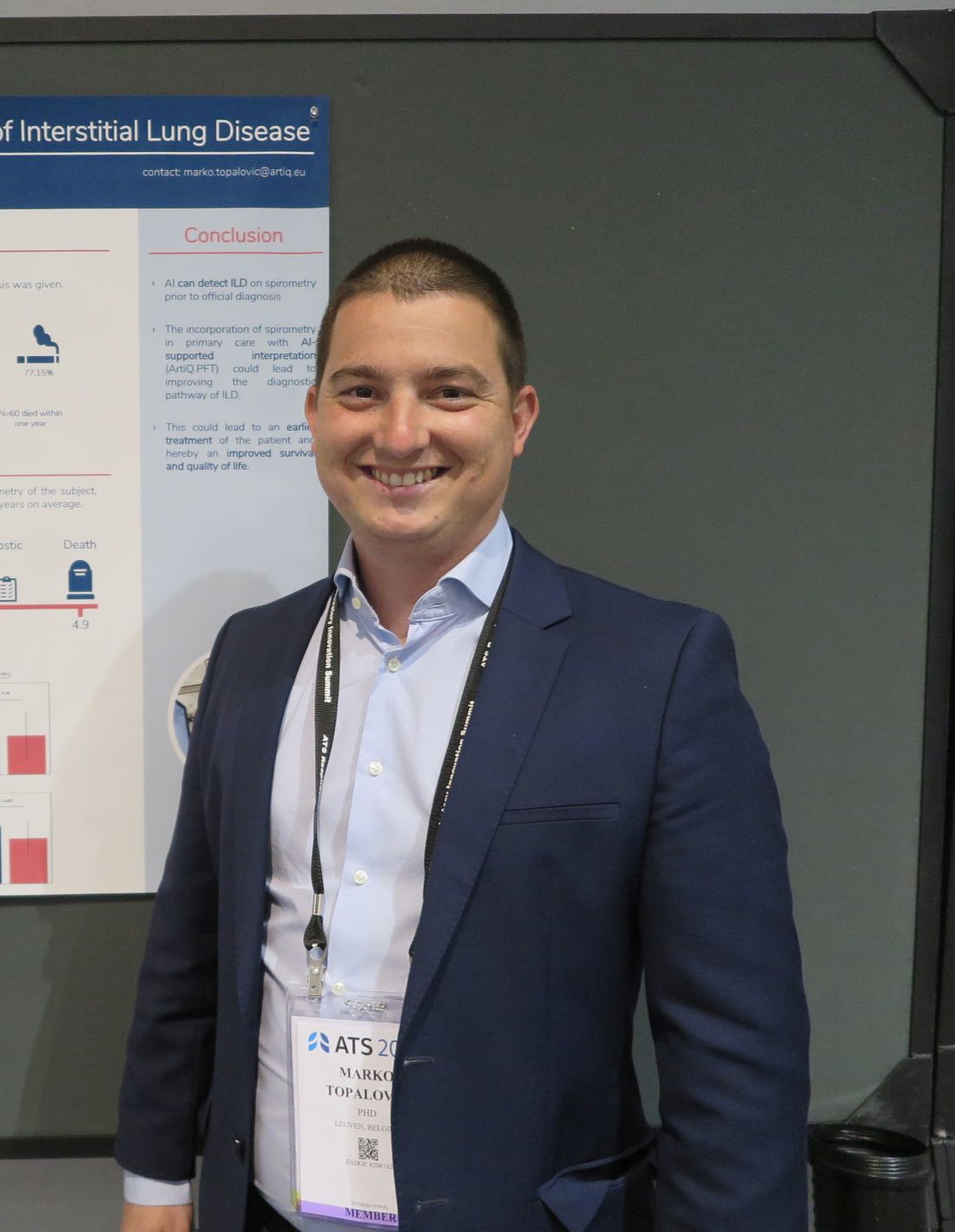
“A diagnosis of ILD is very challenging, so you have patients who are going to be misdiagnosed or have a very late diagnosis, so we aimed to apply our AI algorithm on spirometry to see whether we could detect ILD much earlier,” he said in an interview conducted during a poster discussion session.
AI detected ILD up to 6.8 years before a clinician’s diagnosis, Dr. Topalovic said.
Reading between the lines
In a separate study, investigators at the University of California, Davis, used language analysis software to scour electronic health records for words indicative of early ILD, and found that the technique dramatically shortened the median time to a pulmonary referral, compared with historical controls.
“This is a language processing program that can essentially look through the radiology reports and look for the key words that often describe interstitial lung disease, like traction, honeycomb, fibrotic, etc. With those studies being flagged, an actual pulmonologist will then further review the scan, and see whether it meets criteria for one of the interstitial lung diseases,” lead author William Leon, MD, a resident in the department of internal medicine at the University of California, Davis, said in an interview.
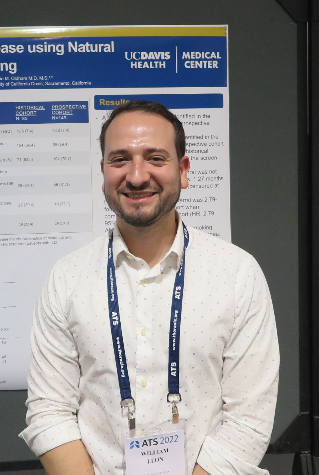
“We then sent the primary care doctor a message to say: ‘Hey, this patient has ILD. You need to send them to a pulmonologist,’ ” he added.
Putting it together
Philip L. Molyneaux, MRCP (UK), MBBS, BS (Hons), from Imperial College London, who comoderated the session but was not involved in the studies, speculated that combining these and other, nontechnical interventions also discussed could help to improve diagnosis of ILD and allow clinicians to prescribe therapy earlier in the disease course.
“What’s going to give you the biggest impact for patients? Everyone working individually is coming up with great advances, and if you put them all together it’s going to provide much greater benefit for our patients,” he said in an interview.
AI Spirometry details
In collaboration with colleagues at the Laboratory of Respiratory Disease at University Hospital in Leuven, Dr. Topalovic applied AI to results of spirometry performed prior to diagnosis of ILD among 109 patients registered in the UK Biobank, a repository of information on more than 500,000 volunteers.
The patients selected had ILD listed as their cause of death, had spirometry performed up to 7 years before their deaths, and did not receive a diagnosis of ILD on the day of the index spirometry.
In all 73% of patients were men, 27% women, with an average age of 64.6 years. A large majority of the sample (77.15%) had a history of smoking, and 60 of the patients (55%) died within one year of an ILD diagnosis.
The investigators plugged the spirometry data and each patients demographic information – including gender, age, height, weight, race, and smoking status – into the AI clinical decision support program, which yielded a statistical probability for each subject of having normal lung function, asthma, COPD, ILD, another obstructive disease, or another unidentifiable respiratory disease.
In 29 patients (27%) the software listed ILD as the highest probability, and of this group 19 patients (66%) had normal lung function according to standard interpretation guidelines.
Spirometry parameters among patients identified as having probable ILD were different from those where ILD was not detected. For example, forced vital capacity (FVC) was 76% of predicted among patients with likely ILD versus 87% of predicted in those who had a diagnosis later (P = .003). Similar differences were seen in the forced expiratory volume in 1 second to FVC ratio, at 0.82 vs. 0.75, respectively (P = .007).
There were no differences in mortality or in median time between spirometry and clinician diagnosis between the groups.
Language processing details
Dr. Leon and colleagues used a language analysis software package to review CT chest reports. Reports were flagged if they contained the words traction, honeycomb, fibrotic, fibrosis, reticular, or reticulation.
The CT scan accompanying each flagged reported was reviewed by a pulmonologist for the presence of ILD, and scans with ILD identified were referred to pulmonary specialists. The results of 2,198 prospective scans followed by prospective screening were compared with those of 1,690 historical controls seen in 2015 and 2016.
The investigators found that 85 incident cases of ILD were identified in the historical controls, compared with 143 in the prospective cohort, leading to 38 and 120 pulmonary referrals, respectively.
For the primary outcome of median time from CT to pulmonary referral, the authors found that it was 1.27 months for the prospective cohort, compared with not reached (censored after 18 months) in historical controls.
The hazard ratio for a pulmonary referral in the prospective versus historical cohort was 2.79, an association that was strengthened after adjusting for sex, age, race, smoking pack-years, cough, crackles, and dyspnea (HR, 4.54; both comparisons significant according to confidence intervals).
The studies were internally funded. Dr. Topalovic is CEO and cofounder of ArtiQ. Dr. Leon and Dr. Molyneaux reported no relevant conflicts of interest.
SAN FRANCISCO – Interstitial lung disease is a difficult diagnosis to make, but a combination of artificial intelligence (AI) techniques and automated language processing could help clinicians identify the early signs of ILD and start patients on therapy, investigators say.
For example, applying an AI algorithm to spirometry readings taken from patients whose data were registered in the UK Biobank identified 27% as having ILD, and of this group, 66% had ostensibly normal lung function on spirometry but were later diagnosed with ILD, reported Marko Topalovic, PhD, from the AI company ArtiQ in Leuven, Belgium, at the American Thoracic Society’s international conference.
“A diagnosis of ILD is very challenging, so you have patients who are going to be misdiagnosed or have a very late diagnosis, so we aimed to apply our AI algorithm on spirometry to see whether we could detect ILD much earlier,” he said in an interview conducted during a poster discussion session.
AI detected ILD up to 6.8 years before a clinician’s diagnosis, Dr. Topalovic said.
Reading between the lines
In a separate study, investigators at the University of California, Davis, used language analysis software to scour electronic health records for words indicative of early ILD, and found that the technique dramatically shortened the median time to a pulmonary referral, compared with historical controls.
“This is a language processing program that can essentially look through the radiology reports and look for the key words that often describe interstitial lung disease, like traction, honeycomb, fibrotic, etc. With those studies being flagged, an actual pulmonologist will then further review the scan, and see whether it meets criteria for one of the interstitial lung diseases,” lead author William Leon, MD, a resident in the department of internal medicine at the University of California, Davis, said in an interview.
“We then sent the primary care doctor a message to say: ‘Hey, this patient has ILD. You need to send them to a pulmonologist,’ ” he added.
Putting it together
Philip L. Molyneaux, MRCP (UK), MBBS, BS (Hons), from Imperial College London, who comoderated the session but was not involved in the studies, speculated that combining these and other, nontechnical interventions also discussed could help to improve diagnosis of ILD and allow clinicians to prescribe therapy earlier in the disease course.
“What’s going to give you the biggest impact for patients? Everyone working individually is coming up with great advances, and if you put them all together it’s going to provide much greater benefit for our patients,” he said in an interview.
AI Spirometry details
In collaboration with colleagues at the Laboratory of Respiratory Disease at University Hospital in Leuven, Dr. Topalovic applied AI to results of spirometry performed prior to diagnosis of ILD among 109 patients registered in the UK Biobank, a repository of information on more than 500,000 volunteers.
The patients selected had ILD listed as their cause of death, had spirometry performed up to 7 years before their deaths, and did not receive a diagnosis of ILD on the day of the index spirometry.
In all 73% of patients were men, 27% women, with an average age of 64.6 years. A large majority of the sample (77.15%) had a history of smoking, and 60 of the patients (55%) died within one year of an ILD diagnosis.
The investigators plugged the spirometry data and each patients demographic information – including gender, age, height, weight, race, and smoking status – into the AI clinical decision support program, which yielded a statistical probability for each subject of having normal lung function, asthma, COPD, ILD, another obstructive disease, or another unidentifiable respiratory disease.
In 29 patients (27%) the software listed ILD as the highest probability, and of this group 19 patients (66%) had normal lung function according to standard interpretation guidelines.
Spirometry parameters among patients identified as having probable ILD were different from those where ILD was not detected. For example, forced vital capacity (FVC) was 76% of predicted among patients with likely ILD versus 87% of predicted in those who had a diagnosis later (P = .003). Similar differences were seen in the forced expiratory volume in 1 second to FVC ratio, at 0.82 vs. 0.75, respectively (P = .007).
There were no differences in mortality or in median time between spirometry and clinician diagnosis between the groups.
Language processing details
Dr. Leon and colleagues used a language analysis software package to review CT chest reports. Reports were flagged if they contained the words traction, honeycomb, fibrotic, fibrosis, reticular, or reticulation.
The CT scan accompanying each flagged reported was reviewed by a pulmonologist for the presence of ILD, and scans with ILD identified were referred to pulmonary specialists. The results of 2,198 prospective scans followed by prospective screening were compared with those of 1,690 historical controls seen in 2015 and 2016.
The investigators found that 85 incident cases of ILD were identified in the historical controls, compared with 143 in the prospective cohort, leading to 38 and 120 pulmonary referrals, respectively.
For the primary outcome of median time from CT to pulmonary referral, the authors found that it was 1.27 months for the prospective cohort, compared with not reached (censored after 18 months) in historical controls.
The hazard ratio for a pulmonary referral in the prospective versus historical cohort was 2.79, an association that was strengthened after adjusting for sex, age, race, smoking pack-years, cough, crackles, and dyspnea (HR, 4.54; both comparisons significant according to confidence intervals).
The studies were internally funded. Dr. Topalovic is CEO and cofounder of ArtiQ. Dr. Leon and Dr. Molyneaux reported no relevant conflicts of interest.
SAN FRANCISCO – Interstitial lung disease is a difficult diagnosis to make, but a combination of artificial intelligence (AI) techniques and automated language processing could help clinicians identify the early signs of ILD and start patients on therapy, investigators say.
For example, applying an AI algorithm to spirometry readings taken from patients whose data were registered in the UK Biobank identified 27% as having ILD, and of this group, 66% had ostensibly normal lung function on spirometry but were later diagnosed with ILD, reported Marko Topalovic, PhD, from the AI company ArtiQ in Leuven, Belgium, at the American Thoracic Society’s international conference.
“A diagnosis of ILD is very challenging, so you have patients who are going to be misdiagnosed or have a very late diagnosis, so we aimed to apply our AI algorithm on spirometry to see whether we could detect ILD much earlier,” he said in an interview conducted during a poster discussion session.
AI detected ILD up to 6.8 years before a clinician’s diagnosis, Dr. Topalovic said.
Reading between the lines
In a separate study, investigators at the University of California, Davis, used language analysis software to scour electronic health records for words indicative of early ILD, and found that the technique dramatically shortened the median time to a pulmonary referral, compared with historical controls.
“This is a language processing program that can essentially look through the radiology reports and look for the key words that often describe interstitial lung disease, like traction, honeycomb, fibrotic, etc. With those studies being flagged, an actual pulmonologist will then further review the scan, and see whether it meets criteria for one of the interstitial lung diseases,” lead author William Leon, MD, a resident in the department of internal medicine at the University of California, Davis, said in an interview.
“We then sent the primary care doctor a message to say: ‘Hey, this patient has ILD. You need to send them to a pulmonologist,’ ” he added.
Putting it together
Philip L. Molyneaux, MRCP (UK), MBBS, BS (Hons), from Imperial College London, who comoderated the session but was not involved in the studies, speculated that combining these and other, nontechnical interventions also discussed could help to improve diagnosis of ILD and allow clinicians to prescribe therapy earlier in the disease course.
“What’s going to give you the biggest impact for patients? Everyone working individually is coming up with great advances, and if you put them all together it’s going to provide much greater benefit for our patients,” he said in an interview.
AI Spirometry details
In collaboration with colleagues at the Laboratory of Respiratory Disease at University Hospital in Leuven, Dr. Topalovic applied AI to results of spirometry performed prior to diagnosis of ILD among 109 patients registered in the UK Biobank, a repository of information on more than 500,000 volunteers.
The patients selected had ILD listed as their cause of death, had spirometry performed up to 7 years before their deaths, and did not receive a diagnosis of ILD on the day of the index spirometry.
In all 73% of patients were men, 27% women, with an average age of 64.6 years. A large majority of the sample (77.15%) had a history of smoking, and 60 of the patients (55%) died within one year of an ILD diagnosis.
The investigators plugged the spirometry data and each patients demographic information – including gender, age, height, weight, race, and smoking status – into the AI clinical decision support program, which yielded a statistical probability for each subject of having normal lung function, asthma, COPD, ILD, another obstructive disease, or another unidentifiable respiratory disease.
In 29 patients (27%) the software listed ILD as the highest probability, and of this group 19 patients (66%) had normal lung function according to standard interpretation guidelines.
Spirometry parameters among patients identified as having probable ILD were different from those where ILD was not detected. For example, forced vital capacity (FVC) was 76% of predicted among patients with likely ILD versus 87% of predicted in those who had a diagnosis later (P = .003). Similar differences were seen in the forced expiratory volume in 1 second to FVC ratio, at 0.82 vs. 0.75, respectively (P = .007).
There were no differences in mortality or in median time between spirometry and clinician diagnosis between the groups.
Language processing details
Dr. Leon and colleagues used a language analysis software package to review CT chest reports. Reports were flagged if they contained the words traction, honeycomb, fibrotic, fibrosis, reticular, or reticulation.
The CT scan accompanying each flagged reported was reviewed by a pulmonologist for the presence of ILD, and scans with ILD identified were referred to pulmonary specialists. The results of 2,198 prospective scans followed by prospective screening were compared with those of 1,690 historical controls seen in 2015 and 2016.
The investigators found that 85 incident cases of ILD were identified in the historical controls, compared with 143 in the prospective cohort, leading to 38 and 120 pulmonary referrals, respectively.
For the primary outcome of median time from CT to pulmonary referral, the authors found that it was 1.27 months for the prospective cohort, compared with not reached (censored after 18 months) in historical controls.
The hazard ratio for a pulmonary referral in the prospective versus historical cohort was 2.79, an association that was strengthened after adjusting for sex, age, race, smoking pack-years, cough, crackles, and dyspnea (HR, 4.54; both comparisons significant according to confidence intervals).
The studies were internally funded. Dr. Topalovic is CEO and cofounder of ArtiQ. Dr. Leon and Dr. Molyneaux reported no relevant conflicts of interest.
AT ATS 2022
‘Smart inhalers’ may help diagnose and treat asthma – if used
After years going on and off medications for occasional asthma symptoms, things went downhill for Brian Blome in November 2020. The retired carpenter started feeling short of breath and wheezing during bike rides. At home, he struggled with chores.
“I was having a hard time climbing a flight of stairs, just doing laundry,” said Mr. Blome, who lives in the Chicago suburb of Palatine.
To get things under control, he saw an allergist and started regular medications – two tablets, two nasal sprays, and inhaled corticosteroids each day, plus an albuterol inhaler for flare-ups.
The inhalers have an extra feature: an electronic monitor that attaches to the device and automatically tracks where and when the medication is used. Bluetooth sends this information to an app on the patient’s mobile phone and to a dashboard where the medical team can see, at a glance, when symptoms are popping up and how regularly medications are taken – leading to the devices often being called “smart inhalers.”
At the 2022 American Academy of Allergy, Asthma, and Immunology conference in Phoenix, researchers explained how digital monitoring devices can help diagnose and treat hard-to-control asthma, potentially reducing the need for oral steroids or biologic therapies.
Even though electric monitors have been on the market for years, their use has been slow to catch on because of uncertainties around insurance coverage, liability, and how to manage and best use the data. One recent study said these devices cost $100-$500, but that price depends on many things, such as insurance.
About 17% of adult asthma patients have “difficult-to-control” asthma, meaning they limit their activity because of breathing symptoms and use reliever medications multiple times a week.
But research suggests that correcting inhaling technique and sticking to the use of the medications can cut that 17% down to just 3.7%, said Mr. Blome’s allergist, Giselle Mosnaim, MD, of NorthShore University HealthSystem in Glenview, Ill. Dr. Mosnaim spoke about digital monitoring at a conference session on digital technologies for asthma management.
A study of more than 5,000 asthma patients “showed that, if you have critical errors in inhaler technique, this leads to worse asthma outcomes and increased asthma exacerbations,” she said. It also shows that, despite new devices and new technologies, “we still have poor inhaler technique.”
Yet adherence is poorly gauged by doctors and patient self-reporting. “The ideal measure of adherence should be objective, accurate, and unobtrusive to minimize impact on patient behavior and allow reliable data collection in real-world settings,” Dr. Mosnaim said. “So electronic medication monitors are the gold standard.”
Improving use
Patients not following instructions or guidelines “is something we saw nonstop with kids,” said Caroline Moassessi, founder of the allergy and asthma blog Gratefulfoodie.com who formerly served on a regional board of the American Lung Association. She’s also the mother of two asthmatic children, now in college, who years ago used electronic medication monitors as part of a research trial.
They were “unimpressed – mostly since I think they thought their asthma was controlled,” she said. “When patients are not in crisis, they don’t manage their asthma well.”
Even in research studies such as the one Rachelle Ramsey, PhD, presented at the conference, it’s not only hard to determine if better adherence leads to improved health, but when.
“For example, does your adherence this week impact your asthma control this week, or does it impact your asthma control next week? Or is it even further out? Do you need to have some level of adherence over the course of a month in order to have better outcomes at the end of that month?” said Dr. Ramsey, a pediatric research psychologist at Cincinnati Children’s Hospital Medical Center. “I think it’s a little complicated.”
That said, results from several small studies do show a connection between remote monitoring and better clinical outcomes. One study enrolled asthma patients in the United Kingdom, and another was done by Dr. Mosnaim with Chicago-area patients.
In the U.K. quality improvement project, nurses asked patients with difficult-to-control asthma if they knew how to use their inhalers and were following treatment guidelines.
Those who said “yes” were invited to swap their steroid/inhalers for a controller fitted with a device that tracks use and measures acoustics to test inhaler technique. After 28 days of monitoring, many people in the study had better clinical outcomes.
And after 3 months of digital monitoring, patients didn’t use their rescue medication quite as often.
Mr. Blome has seen a marked improvement in his asthma since starting regular appointments and getting back on daily medications a year and a half ago. He says that now and then, he has wheezing and shortness of breath, usually while biking or exercising. But those symptoms aren’t as severe or frequent as before.
From a doctor’s perspective, “digital inhaler systems allow me to discern patterns in order to determine what triggers his asthma symptoms and to adjust medications at different times of the year,” Dr. Mosnaim said.
Electronic systems can monitor pollen counts and air quality as well as how often a patient uses a quick reliever medication. Thus, she said, tracking these measures year-round could raise attention to impending asthma attacks and suggest when to increase the dose of controller medications or add other treatments.
A version of this article first appeared on WebMD.com.
After years going on and off medications for occasional asthma symptoms, things went downhill for Brian Blome in November 2020. The retired carpenter started feeling short of breath and wheezing during bike rides. At home, he struggled with chores.
“I was having a hard time climbing a flight of stairs, just doing laundry,” said Mr. Blome, who lives in the Chicago suburb of Palatine.
To get things under control, he saw an allergist and started regular medications – two tablets, two nasal sprays, and inhaled corticosteroids each day, plus an albuterol inhaler for flare-ups.
The inhalers have an extra feature: an electronic monitor that attaches to the device and automatically tracks where and when the medication is used. Bluetooth sends this information to an app on the patient’s mobile phone and to a dashboard where the medical team can see, at a glance, when symptoms are popping up and how regularly medications are taken – leading to the devices often being called “smart inhalers.”
At the 2022 American Academy of Allergy, Asthma, and Immunology conference in Phoenix, researchers explained how digital monitoring devices can help diagnose and treat hard-to-control asthma, potentially reducing the need for oral steroids or biologic therapies.
Even though electric monitors have been on the market for years, their use has been slow to catch on because of uncertainties around insurance coverage, liability, and how to manage and best use the data. One recent study said these devices cost $100-$500, but that price depends on many things, such as insurance.
About 17% of adult asthma patients have “difficult-to-control” asthma, meaning they limit their activity because of breathing symptoms and use reliever medications multiple times a week.
But research suggests that correcting inhaling technique and sticking to the use of the medications can cut that 17% down to just 3.7%, said Mr. Blome’s allergist, Giselle Mosnaim, MD, of NorthShore University HealthSystem in Glenview, Ill. Dr. Mosnaim spoke about digital monitoring at a conference session on digital technologies for asthma management.
A study of more than 5,000 asthma patients “showed that, if you have critical errors in inhaler technique, this leads to worse asthma outcomes and increased asthma exacerbations,” she said. It also shows that, despite new devices and new technologies, “we still have poor inhaler technique.”
Yet adherence is poorly gauged by doctors and patient self-reporting. “The ideal measure of adherence should be objective, accurate, and unobtrusive to minimize impact on patient behavior and allow reliable data collection in real-world settings,” Dr. Mosnaim said. “So electronic medication monitors are the gold standard.”
Improving use
Patients not following instructions or guidelines “is something we saw nonstop with kids,” said Caroline Moassessi, founder of the allergy and asthma blog Gratefulfoodie.com who formerly served on a regional board of the American Lung Association. She’s also the mother of two asthmatic children, now in college, who years ago used electronic medication monitors as part of a research trial.
They were “unimpressed – mostly since I think they thought their asthma was controlled,” she said. “When patients are not in crisis, they don’t manage their asthma well.”
Even in research studies such as the one Rachelle Ramsey, PhD, presented at the conference, it’s not only hard to determine if better adherence leads to improved health, but when.
“For example, does your adherence this week impact your asthma control this week, or does it impact your asthma control next week? Or is it even further out? Do you need to have some level of adherence over the course of a month in order to have better outcomes at the end of that month?” said Dr. Ramsey, a pediatric research psychologist at Cincinnati Children’s Hospital Medical Center. “I think it’s a little complicated.”
That said, results from several small studies do show a connection between remote monitoring and better clinical outcomes. One study enrolled asthma patients in the United Kingdom, and another was done by Dr. Mosnaim with Chicago-area patients.
In the U.K. quality improvement project, nurses asked patients with difficult-to-control asthma if they knew how to use their inhalers and were following treatment guidelines.
Those who said “yes” were invited to swap their steroid/inhalers for a controller fitted with a device that tracks use and measures acoustics to test inhaler technique. After 28 days of monitoring, many people in the study had better clinical outcomes.
And after 3 months of digital monitoring, patients didn’t use their rescue medication quite as often.
Mr. Blome has seen a marked improvement in his asthma since starting regular appointments and getting back on daily medications a year and a half ago. He says that now and then, he has wheezing and shortness of breath, usually while biking or exercising. But those symptoms aren’t as severe or frequent as before.
From a doctor’s perspective, “digital inhaler systems allow me to discern patterns in order to determine what triggers his asthma symptoms and to adjust medications at different times of the year,” Dr. Mosnaim said.
Electronic systems can monitor pollen counts and air quality as well as how often a patient uses a quick reliever medication. Thus, she said, tracking these measures year-round could raise attention to impending asthma attacks and suggest when to increase the dose of controller medications or add other treatments.
A version of this article first appeared on WebMD.com.
After years going on and off medications for occasional asthma symptoms, things went downhill for Brian Blome in November 2020. The retired carpenter started feeling short of breath and wheezing during bike rides. At home, he struggled with chores.
“I was having a hard time climbing a flight of stairs, just doing laundry,” said Mr. Blome, who lives in the Chicago suburb of Palatine.
To get things under control, he saw an allergist and started regular medications – two tablets, two nasal sprays, and inhaled corticosteroids each day, plus an albuterol inhaler for flare-ups.
The inhalers have an extra feature: an electronic monitor that attaches to the device and automatically tracks where and when the medication is used. Bluetooth sends this information to an app on the patient’s mobile phone and to a dashboard where the medical team can see, at a glance, when symptoms are popping up and how regularly medications are taken – leading to the devices often being called “smart inhalers.”
At the 2022 American Academy of Allergy, Asthma, and Immunology conference in Phoenix, researchers explained how digital monitoring devices can help diagnose and treat hard-to-control asthma, potentially reducing the need for oral steroids or biologic therapies.
Even though electric monitors have been on the market for years, their use has been slow to catch on because of uncertainties around insurance coverage, liability, and how to manage and best use the data. One recent study said these devices cost $100-$500, but that price depends on many things, such as insurance.
About 17% of adult asthma patients have “difficult-to-control” asthma, meaning they limit their activity because of breathing symptoms and use reliever medications multiple times a week.
But research suggests that correcting inhaling technique and sticking to the use of the medications can cut that 17% down to just 3.7%, said Mr. Blome’s allergist, Giselle Mosnaim, MD, of NorthShore University HealthSystem in Glenview, Ill. Dr. Mosnaim spoke about digital monitoring at a conference session on digital technologies for asthma management.
A study of more than 5,000 asthma patients “showed that, if you have critical errors in inhaler technique, this leads to worse asthma outcomes and increased asthma exacerbations,” she said. It also shows that, despite new devices and new technologies, “we still have poor inhaler technique.”
Yet adherence is poorly gauged by doctors and patient self-reporting. “The ideal measure of adherence should be objective, accurate, and unobtrusive to minimize impact on patient behavior and allow reliable data collection in real-world settings,” Dr. Mosnaim said. “So electronic medication monitors are the gold standard.”
Improving use
Patients not following instructions or guidelines “is something we saw nonstop with kids,” said Caroline Moassessi, founder of the allergy and asthma blog Gratefulfoodie.com who formerly served on a regional board of the American Lung Association. She’s also the mother of two asthmatic children, now in college, who years ago used electronic medication monitors as part of a research trial.
They were “unimpressed – mostly since I think they thought their asthma was controlled,” she said. “When patients are not in crisis, they don’t manage their asthma well.”
Even in research studies such as the one Rachelle Ramsey, PhD, presented at the conference, it’s not only hard to determine if better adherence leads to improved health, but when.
“For example, does your adherence this week impact your asthma control this week, or does it impact your asthma control next week? Or is it even further out? Do you need to have some level of adherence over the course of a month in order to have better outcomes at the end of that month?” said Dr. Ramsey, a pediatric research psychologist at Cincinnati Children’s Hospital Medical Center. “I think it’s a little complicated.”
That said, results from several small studies do show a connection between remote monitoring and better clinical outcomes. One study enrolled asthma patients in the United Kingdom, and another was done by Dr. Mosnaim with Chicago-area patients.
In the U.K. quality improvement project, nurses asked patients with difficult-to-control asthma if they knew how to use their inhalers and were following treatment guidelines.
Those who said “yes” were invited to swap their steroid/inhalers for a controller fitted with a device that tracks use and measures acoustics to test inhaler technique. After 28 days of monitoring, many people in the study had better clinical outcomes.
And after 3 months of digital monitoring, patients didn’t use their rescue medication quite as often.
Mr. Blome has seen a marked improvement in his asthma since starting regular appointments and getting back on daily medications a year and a half ago. He says that now and then, he has wheezing and shortness of breath, usually while biking or exercising. But those symptoms aren’t as severe or frequent as before.
From a doctor’s perspective, “digital inhaler systems allow me to discern patterns in order to determine what triggers his asthma symptoms and to adjust medications at different times of the year,” Dr. Mosnaim said.
Electronic systems can monitor pollen counts and air quality as well as how often a patient uses a quick reliever medication. Thus, she said, tracking these measures year-round could raise attention to impending asthma attacks and suggest when to increase the dose of controller medications or add other treatments.
A version of this article first appeared on WebMD.com.
Exercise response divides COPD patients into four groups
Not all patients with chronic obstructive pulmonary disease (COPD) respond equally well to pulmonary rehabilitation (PR).
Now, physicians can better categorize which patients will do well with PR and which ones less well or not well at all based on a new system of clustering of COPD patients according to their response to exercise therapy.
“We identified four clusters of COPD patients and their response to PR in the aim to better understand PR outcome and [adapt] it to patients’ profiles and needs,” lead author Yara Al Chikhanie, MD, of the cardiopulmonary rehabilitation center Dieulefit Sante (France), and colleagues observed.
“Identification of patients likely to show smaller responses to PR may help to target patients benefiting the most and to adapt PR settings for nonresponders to standard PR,” they suggested.
The study was published online in Respiratory Medicine.
Single-center cohort
The cohort consisted of 835 patients from a single center who had been admitted to a cardiopulmonary rehabilitation center over a 6-year period from 2021 to 2017. “The PR program used in the center was the same over the 6-year period,” the authors note – consisting of a 3- to 4-week, inpatient program with activities 5 days a week.
Each day, patients attended a 25-minute aerobic training session on a cycling ergometer or a treadmill; a 30-minute low-intensity gym session; a 30-minute group walk outdoors, and 30 minutes of strength training. “We aimed to cluster patients with COPD admitted to PR based on patients’ clinical characteristics and 6-meter walk test results (6MWT), pulse oxygen saturation (SPO2), heart rate (HR), and dyspnea,” the authors explained.
They then evaluated patient response to PR in each of these clusters based on the amount of improvement in the 6-meter walk distance (6MWD), lung function, and quality of life observed, they added.
The population consisted of seniors, equally men and women, mostly GOLD II and III patients (a measure of lung function) with a limited walking capacity, some 84% of the cohort having a 6MWD <80% predicted. The characteristics of the four identified clusters were as follows:
- Cluster 1: Consisted of younger men, GOLD I to II, average walkers, obese. The average 6MWD was 430 meters and patients had a large exercise HR response to PR. This cluster had a 76 meter improvement in their 6MWD, although 16% of the same cluster still did not respond to PR.
- Cluster 2: Consisted of older women, GOLD II-III, who were slow walkers. This cluster had a reduced 6MWD of 362 meters, but they also had a significant 97-meter improvement in their 6MWD following PR. Some 18% were still nonresponders to PR.
- Cluster 3: Consisted of older men, GOLD II to III, dyspneic, slow walkers, some 32% of whom responded to PR. This cluster also had a reduced 6MWD at 388 meters, but again, they also had a significant improvement of 79 meters in their 6MWD following the introduction of PR. Some 11% were nonresponders to PR.
- Cluster 4: Consisted of older men, GOLD III to IV, very slow walkers, oxygen-dependent, very dyspneic. This cluster had a severely reduced 6MWD of only 290 meters with severe exercise desaturation and dyspnea, and almost all of them were on long-term oxygen therapy. Nevertheless, this cluster also had a significant, 66-meter improvement in their 6MWD. Twenty-eight percent of them were nonresponders to PR.
Clinical practice
“The highly heterogeneous nature of the enrolled patient population reflects clinical practice,” the authors point out. For example, cluster 1 included patients with the best lung function, compared with those in clusters 2, 3, and 4 – which may be due, at least in part, to the aggravation in disease severity with age given that patients in cluster 1 were the youngest overall.
The fact that those in cluster 4 had the worst performance may also have been because of age and disease severity, the authors note, as those in cluster 4 had the highest proportion of patients on long-term oxygen therapy, again suggestive of disease severity. “Of note, these patients show the most impaired 6MWT responses despite the use of oxygen supplementation during walking,” the researchers added.
The authors also suggest that patients such as those in cluster 4 may require specific PR modalities in order to optimize their functional benefits. In contrast, those in cluster 1 had a significantly higher body mass index, compared with those in the other 3 clusters, which, interestingly enough, was not associated with more severe functional exercise impairment. The fact that older age participants, such as those in cluster 3 as well as those with high BMI in cluster 1, were both able to improve their 6MWD post-PR to the same extent as younger patients without obesity suggests that most older or overweight/obese patients can still show clinically significant improvement in 6MWD post PR, as the authors suggest.
Notably, the 6MWT was the only test available both pre-and post PR, making this an important limitation of the study, because only one aspect of the effect of PR was evaluated, omitting other physical and psychosocial benefits of PR, investigators suggest.
Adds to the literature
Asked to comment on the findings, Sachin Gupta, MD, attending physician, pulmonary & critical care medicine, Alameda Health System, Highland Hospital, Oakland, Calif., felt that these data add to the literature in defining COPD patient profiles, helping to categorize those in whom to expect greater walk distance improvements with PR versus those who will respond less well.
“Because 6MWD is a surrogate marker for quality of life (QOL) and mortality, further analysis in the form of a randomized controlled trial to determine long-term outcomes among the four clusters with adjustment for baseline characteristics would help determine the extent to which certain patient clusters may respond to PR,” Dr. Gupta told this news organization in an email.
At the same time, he suggested that while patients may not experience much net benefit in their 6MWD, their QOL or mortality risk may still improve with PR. “I cannot recall a patient ever describing their experience with PR as anything other than positive,” Dr. Gupta stressed.
“And as the authors [themselves] note, because PR serves to benefit patients beyond the 6MWD, I would not recommend limiting PR referrals based on the patient clusters identified,” he said.
The authors had no conflicts of interest to declare. Dr. Gupta declared that he is an employee and shareholder at Genentech.
Not all patients with chronic obstructive pulmonary disease (COPD) respond equally well to pulmonary rehabilitation (PR).
Now, physicians can better categorize which patients will do well with PR and which ones less well or not well at all based on a new system of clustering of COPD patients according to their response to exercise therapy.
“We identified four clusters of COPD patients and their response to PR in the aim to better understand PR outcome and [adapt] it to patients’ profiles and needs,” lead author Yara Al Chikhanie, MD, of the cardiopulmonary rehabilitation center Dieulefit Sante (France), and colleagues observed.
“Identification of patients likely to show smaller responses to PR may help to target patients benefiting the most and to adapt PR settings for nonresponders to standard PR,” they suggested.
The study was published online in Respiratory Medicine.
Single-center cohort
The cohort consisted of 835 patients from a single center who had been admitted to a cardiopulmonary rehabilitation center over a 6-year period from 2021 to 2017. “The PR program used in the center was the same over the 6-year period,” the authors note – consisting of a 3- to 4-week, inpatient program with activities 5 days a week.
Each day, patients attended a 25-minute aerobic training session on a cycling ergometer or a treadmill; a 30-minute low-intensity gym session; a 30-minute group walk outdoors, and 30 minutes of strength training. “We aimed to cluster patients with COPD admitted to PR based on patients’ clinical characteristics and 6-meter walk test results (6MWT), pulse oxygen saturation (SPO2), heart rate (HR), and dyspnea,” the authors explained.
They then evaluated patient response to PR in each of these clusters based on the amount of improvement in the 6-meter walk distance (6MWD), lung function, and quality of life observed, they added.
The population consisted of seniors, equally men and women, mostly GOLD II and III patients (a measure of lung function) with a limited walking capacity, some 84% of the cohort having a 6MWD <80% predicted. The characteristics of the four identified clusters were as follows:
- Cluster 1: Consisted of younger men, GOLD I to II, average walkers, obese. The average 6MWD was 430 meters and patients had a large exercise HR response to PR. This cluster had a 76 meter improvement in their 6MWD, although 16% of the same cluster still did not respond to PR.
- Cluster 2: Consisted of older women, GOLD II-III, who were slow walkers. This cluster had a reduced 6MWD of 362 meters, but they also had a significant 97-meter improvement in their 6MWD following PR. Some 18% were still nonresponders to PR.
- Cluster 3: Consisted of older men, GOLD II to III, dyspneic, slow walkers, some 32% of whom responded to PR. This cluster also had a reduced 6MWD at 388 meters, but again, they also had a significant improvement of 79 meters in their 6MWD following the introduction of PR. Some 11% were nonresponders to PR.
- Cluster 4: Consisted of older men, GOLD III to IV, very slow walkers, oxygen-dependent, very dyspneic. This cluster had a severely reduced 6MWD of only 290 meters with severe exercise desaturation and dyspnea, and almost all of them were on long-term oxygen therapy. Nevertheless, this cluster also had a significant, 66-meter improvement in their 6MWD. Twenty-eight percent of them were nonresponders to PR.
Clinical practice
“The highly heterogeneous nature of the enrolled patient population reflects clinical practice,” the authors point out. For example, cluster 1 included patients with the best lung function, compared with those in clusters 2, 3, and 4 – which may be due, at least in part, to the aggravation in disease severity with age given that patients in cluster 1 were the youngest overall.
The fact that those in cluster 4 had the worst performance may also have been because of age and disease severity, the authors note, as those in cluster 4 had the highest proportion of patients on long-term oxygen therapy, again suggestive of disease severity. “Of note, these patients show the most impaired 6MWT responses despite the use of oxygen supplementation during walking,” the researchers added.
The authors also suggest that patients such as those in cluster 4 may require specific PR modalities in order to optimize their functional benefits. In contrast, those in cluster 1 had a significantly higher body mass index, compared with those in the other 3 clusters, which, interestingly enough, was not associated with more severe functional exercise impairment. The fact that older age participants, such as those in cluster 3 as well as those with high BMI in cluster 1, were both able to improve their 6MWD post-PR to the same extent as younger patients without obesity suggests that most older or overweight/obese patients can still show clinically significant improvement in 6MWD post PR, as the authors suggest.
Notably, the 6MWT was the only test available both pre-and post PR, making this an important limitation of the study, because only one aspect of the effect of PR was evaluated, omitting other physical and psychosocial benefits of PR, investigators suggest.
Adds to the literature
Asked to comment on the findings, Sachin Gupta, MD, attending physician, pulmonary & critical care medicine, Alameda Health System, Highland Hospital, Oakland, Calif., felt that these data add to the literature in defining COPD patient profiles, helping to categorize those in whom to expect greater walk distance improvements with PR versus those who will respond less well.
“Because 6MWD is a surrogate marker for quality of life (QOL) and mortality, further analysis in the form of a randomized controlled trial to determine long-term outcomes among the four clusters with adjustment for baseline characteristics would help determine the extent to which certain patient clusters may respond to PR,” Dr. Gupta told this news organization in an email.
At the same time, he suggested that while patients may not experience much net benefit in their 6MWD, their QOL or mortality risk may still improve with PR. “I cannot recall a patient ever describing their experience with PR as anything other than positive,” Dr. Gupta stressed.
“And as the authors [themselves] note, because PR serves to benefit patients beyond the 6MWD, I would not recommend limiting PR referrals based on the patient clusters identified,” he said.
The authors had no conflicts of interest to declare. Dr. Gupta declared that he is an employee and shareholder at Genentech.
Not all patients with chronic obstructive pulmonary disease (COPD) respond equally well to pulmonary rehabilitation (PR).
Now, physicians can better categorize which patients will do well with PR and which ones less well or not well at all based on a new system of clustering of COPD patients according to their response to exercise therapy.
“We identified four clusters of COPD patients and their response to PR in the aim to better understand PR outcome and [adapt] it to patients’ profiles and needs,” lead author Yara Al Chikhanie, MD, of the cardiopulmonary rehabilitation center Dieulefit Sante (France), and colleagues observed.
“Identification of patients likely to show smaller responses to PR may help to target patients benefiting the most and to adapt PR settings for nonresponders to standard PR,” they suggested.
The study was published online in Respiratory Medicine.
Single-center cohort
The cohort consisted of 835 patients from a single center who had been admitted to a cardiopulmonary rehabilitation center over a 6-year period from 2021 to 2017. “The PR program used in the center was the same over the 6-year period,” the authors note – consisting of a 3- to 4-week, inpatient program with activities 5 days a week.
Each day, patients attended a 25-minute aerobic training session on a cycling ergometer or a treadmill; a 30-minute low-intensity gym session; a 30-minute group walk outdoors, and 30 minutes of strength training. “We aimed to cluster patients with COPD admitted to PR based on patients’ clinical characteristics and 6-meter walk test results (6MWT), pulse oxygen saturation (SPO2), heart rate (HR), and dyspnea,” the authors explained.
They then evaluated patient response to PR in each of these clusters based on the amount of improvement in the 6-meter walk distance (6MWD), lung function, and quality of life observed, they added.
The population consisted of seniors, equally men and women, mostly GOLD II and III patients (a measure of lung function) with a limited walking capacity, some 84% of the cohort having a 6MWD <80% predicted. The characteristics of the four identified clusters were as follows:
- Cluster 1: Consisted of younger men, GOLD I to II, average walkers, obese. The average 6MWD was 430 meters and patients had a large exercise HR response to PR. This cluster had a 76 meter improvement in their 6MWD, although 16% of the same cluster still did not respond to PR.
- Cluster 2: Consisted of older women, GOLD II-III, who were slow walkers. This cluster had a reduced 6MWD of 362 meters, but they also had a significant 97-meter improvement in their 6MWD following PR. Some 18% were still nonresponders to PR.
- Cluster 3: Consisted of older men, GOLD II to III, dyspneic, slow walkers, some 32% of whom responded to PR. This cluster also had a reduced 6MWD at 388 meters, but again, they also had a significant improvement of 79 meters in their 6MWD following the introduction of PR. Some 11% were nonresponders to PR.
- Cluster 4: Consisted of older men, GOLD III to IV, very slow walkers, oxygen-dependent, very dyspneic. This cluster had a severely reduced 6MWD of only 290 meters with severe exercise desaturation and dyspnea, and almost all of them were on long-term oxygen therapy. Nevertheless, this cluster also had a significant, 66-meter improvement in their 6MWD. Twenty-eight percent of them were nonresponders to PR.
Clinical practice
“The highly heterogeneous nature of the enrolled patient population reflects clinical practice,” the authors point out. For example, cluster 1 included patients with the best lung function, compared with those in clusters 2, 3, and 4 – which may be due, at least in part, to the aggravation in disease severity with age given that patients in cluster 1 were the youngest overall.
The fact that those in cluster 4 had the worst performance may also have been because of age and disease severity, the authors note, as those in cluster 4 had the highest proportion of patients on long-term oxygen therapy, again suggestive of disease severity. “Of note, these patients show the most impaired 6MWT responses despite the use of oxygen supplementation during walking,” the researchers added.
The authors also suggest that patients such as those in cluster 4 may require specific PR modalities in order to optimize their functional benefits. In contrast, those in cluster 1 had a significantly higher body mass index, compared with those in the other 3 clusters, which, interestingly enough, was not associated with more severe functional exercise impairment. The fact that older age participants, such as those in cluster 3 as well as those with high BMI in cluster 1, were both able to improve their 6MWD post-PR to the same extent as younger patients without obesity suggests that most older or overweight/obese patients can still show clinically significant improvement in 6MWD post PR, as the authors suggest.
Notably, the 6MWT was the only test available both pre-and post PR, making this an important limitation of the study, because only one aspect of the effect of PR was evaluated, omitting other physical and psychosocial benefits of PR, investigators suggest.
Adds to the literature
Asked to comment on the findings, Sachin Gupta, MD, attending physician, pulmonary & critical care medicine, Alameda Health System, Highland Hospital, Oakland, Calif., felt that these data add to the literature in defining COPD patient profiles, helping to categorize those in whom to expect greater walk distance improvements with PR versus those who will respond less well.
“Because 6MWD is a surrogate marker for quality of life (QOL) and mortality, further analysis in the form of a randomized controlled trial to determine long-term outcomes among the four clusters with adjustment for baseline characteristics would help determine the extent to which certain patient clusters may respond to PR,” Dr. Gupta told this news organization in an email.
At the same time, he suggested that while patients may not experience much net benefit in their 6MWD, their QOL or mortality risk may still improve with PR. “I cannot recall a patient ever describing their experience with PR as anything other than positive,” Dr. Gupta stressed.
“And as the authors [themselves] note, because PR serves to benefit patients beyond the 6MWD, I would not recommend limiting PR referrals based on the patient clusters identified,” he said.
The authors had no conflicts of interest to declare. Dr. Gupta declared that he is an employee and shareholder at Genentech.
FROM RESPIRATORY MEDICINE
Paradigm-challenging heart failure treatment strategy hopeful in early trial
A small group of patients with heart failure (HF) who underwent a novel transcatheter nerve-ablation procedure seemed to benefit with improved hemodynamics, symptoms, and quality of life in an admittedly limited observational series.
All had HF with preserved ejection fraction (HFpEF) and remained on guideline-directed medical therapy during the study.
The open-label experience has launched a randomized trial, featuring a sham control group, that could ultimately challenge dogma about volume overload in patients with chronic and acute HF and the perceived essential role of diuretics.
Researchers see transvenous ablation of the right greater splanchnic nerve (GSN) as potentially appropriate for patients with HF, regardless of ventricular function or acuity. But the ongoing REBALANCE-HF trial aims to enroll up to 80 patients with chronic HFpEF.
Meanwhile, the current 18 patients with elevated resting or exertional pulmonary capillary wedge pressure (PCWP), given the procedure as part of the main trial’s “roll-in” phase, showed declines in exercise PCWP after 1 month (P = .007) and improved quality-of-life scores at both 1 and 3 months (P < .01). Also at 1 month, a third of the patients improved by at least one step in NYHA functional class.
The procedure, called splanchnic ablation for volume management (SAVM), could potentially be used “across the spectrum of acute and chronic heart failure, maybe even with reduced ejection fraction (HFrEF) and preserved ejection fraction,” Marat Fudim, MD, MHS, Duke University Medical Center, Durham, N.C., told this news organization.
However, “for outcomes, we’ve really only looked in the ambulatory setting,” and only at symptomatic and functional responses. To that extent, based on the current experience and a few small previous studies, Dr. Fudim said, SAVM seems to benefit patients with HF in general who have dyspnea at exercise. Beyond that, the kind of patient who may be most suitable for it “is something I hope we will be able answer once the randomized dataset is in.”
Dr. Fudim reported the REBALANCE-HF roll-in results at the Heart Failure Association of the European Society of Cardiology (HFA-ESC) 2022 sessions, held virtually and live in Madrid. He is also lead author on the same-day publication in the European Journal of Heart Failure.
A different treatment paradigm
Splanchnic-nerve blockade as a possible HF treatment is based on growing evidence that volume overload in patients with HF is not always the cause, at least not a main cause, of congestion and dyspnea. Rather, those classic HF signs and symptoms may often be triggered by adverse redistribution of stable fluid volume from primarily the splanchnic vascular compartment to the intrathoracic space.
In other words, what might seem like classic volume overload calling for diuresis often might actually be euvolemic redistribution of fluid from the abdomen to the chest, raising intracardiac pressures and causing dyspnea.
In that scenario, loop diuretics might only dehydrate the patient and potentially put the kidneys at risk, Dr. Fudim proposed. His recent experience with HF patients implanted with a pulmonary-artery pressure monitor, he said, suggests many who received standard volume-overload therapy had actually been normo- or hypovolemic.
More then half the patients “did not have high volume, they just had high pressures,” he said. “So there is a significant portion of the population that has pathological processes leading to high pressures, but it’s not volume overload. Diuresing those patients would probably not be the right decision.”
The unilateral SAVM procedure appears to attenuate sympathetically mediated splanchnic volume redistribution to the heart and lungs, but as it doesn’t affect the left GSN, preserves some normal sympathetic response.
Sometimes in studies of surgical or catheter-based SAVM, Dr. Fudim said, “we have observationally seen that people discontinued diuretics or decreased doses in the treatment arm.”
‘Beyond our classical thinking’
It’s “impressive” that such right-GSN ablation seemed to reduce exercise-filling pressures, but one should be circumspect because “it’s way beyond our classical thinking,” Wilfried Mullens, MD, PhD, Hospital Oost-Limburg, Genk, Belgium, said as a panelist after Dr. Fudim’s presentation.
“These are invasive procedures,” he noted, “and our physiological understanding does not always match up with what we’re doing in real life, if you look at other interventional procedures, like renal denervation, which showed neutral effects, or if you look at even interatrial shunt devices, which might even be dangerous.”
The field should be “very prudent” before using SAVM in practice, which shouldn’t be “before we have sufficient data to support the efficacy and safety,” Dr. Mullens said. “It remains to be seen how treatment success will be defined. Is it during exercise? How long does the treatment last? What is the effect of the treatment over time; is it not harmful? These are things that we don’t know yet.”
The procedure was considered successful in all 18 patients, 14 of whom were women and 16 of whom were in NYHA class 3. Their average age was 75, and their mean left ventricular ejection fraction (LVEF) at baseline was 61%. The primary efficacy endpoints were a reduction in PCWP at rest, with legs raised, and at 20W exercise at 1 month. Their baseline invasively measured peak exercise PCWP was at least 25 mm Hg.
At 1 month, mean PCWP at 20W exercise fell from 36.4 mm Hg to 28.9 mm Hg (P = .007) and peak PCWP declined from 39.5 mm Hg to 31.9 mm Hg (P = .013); resting PCWP wasn’t significantly affected. Twelve patients improved by at least one NYHA functional class (P = .02).
Scores on the Kansas City Cardiomyopathy Questionnaire (KCCQ), which assesses quality of life, improved by 22 points at 1 month and 18.3 points at 3 months (P < .01 for both differences).
No significant effects on 6-minute walk distance or natriuretic peptide levels were observed, nor were any observed on LVEF or echocardiographic measures of diastolic function, left ventricular (LV) atrial volume, or LV mass at 3 months.
Three “nonserious” device-related adverse events were observed, including one case of acute decompensation early in the experience, ostensibly due to excessive saline administration, Dr. Fudim reported. There was also one case of transient periprocedural hypertension and one instance of postprocedure back pain.
The SAVM procedure is performed transvenously and in general is technically “really not that challenging,” Dr. Fudim said. In most cases, the necessary skills would be accessible not only to interventional cardiologists but also heart failure specialists. “I have performed this procedure myself, and I’m a heart failure guy.”
The REBALANCE-HF roll-in phase and main trial are supported by Axon Therapies. Dr. Fudim discloses receiving support from Bayer, Bodyport, and BTG Specialty Pharmaceuticals; and consulting fees from Abbott, Audicor, Axon Therapies, Bodyguide, Bodyport, Boston Scientific, CVRx, Daxor, Edwards LifeSciences, Feldschuh Foundation, Fire1, Gradient, Intershunt, NXT Biomedical, Pharmacosmos, PreHealth, Splendo, Vironix, Viscardia, and Zoll. Dr. Mullens discloses receiving fees for speaking from Medtronic, Abbott, Novartis, Boston Scientific, AstraZeneca, and Boehringer Ingelheim.
A version of this article first appeared on Medscape.com.
A small group of patients with heart failure (HF) who underwent a novel transcatheter nerve-ablation procedure seemed to benefit with improved hemodynamics, symptoms, and quality of life in an admittedly limited observational series.
All had HF with preserved ejection fraction (HFpEF) and remained on guideline-directed medical therapy during the study.
The open-label experience has launched a randomized trial, featuring a sham control group, that could ultimately challenge dogma about volume overload in patients with chronic and acute HF and the perceived essential role of diuretics.
Researchers see transvenous ablation of the right greater splanchnic nerve (GSN) as potentially appropriate for patients with HF, regardless of ventricular function or acuity. But the ongoing REBALANCE-HF trial aims to enroll up to 80 patients with chronic HFpEF.
Meanwhile, the current 18 patients with elevated resting or exertional pulmonary capillary wedge pressure (PCWP), given the procedure as part of the main trial’s “roll-in” phase, showed declines in exercise PCWP after 1 month (P = .007) and improved quality-of-life scores at both 1 and 3 months (P < .01). Also at 1 month, a third of the patients improved by at least one step in NYHA functional class.
The procedure, called splanchnic ablation for volume management (SAVM), could potentially be used “across the spectrum of acute and chronic heart failure, maybe even with reduced ejection fraction (HFrEF) and preserved ejection fraction,” Marat Fudim, MD, MHS, Duke University Medical Center, Durham, N.C., told this news organization.
However, “for outcomes, we’ve really only looked in the ambulatory setting,” and only at symptomatic and functional responses. To that extent, based on the current experience and a few small previous studies, Dr. Fudim said, SAVM seems to benefit patients with HF in general who have dyspnea at exercise. Beyond that, the kind of patient who may be most suitable for it “is something I hope we will be able answer once the randomized dataset is in.”
Dr. Fudim reported the REBALANCE-HF roll-in results at the Heart Failure Association of the European Society of Cardiology (HFA-ESC) 2022 sessions, held virtually and live in Madrid. He is also lead author on the same-day publication in the European Journal of Heart Failure.
A different treatment paradigm
Splanchnic-nerve blockade as a possible HF treatment is based on growing evidence that volume overload in patients with HF is not always the cause, at least not a main cause, of congestion and dyspnea. Rather, those classic HF signs and symptoms may often be triggered by adverse redistribution of stable fluid volume from primarily the splanchnic vascular compartment to the intrathoracic space.
In other words, what might seem like classic volume overload calling for diuresis often might actually be euvolemic redistribution of fluid from the abdomen to the chest, raising intracardiac pressures and causing dyspnea.
In that scenario, loop diuretics might only dehydrate the patient and potentially put the kidneys at risk, Dr. Fudim proposed. His recent experience with HF patients implanted with a pulmonary-artery pressure monitor, he said, suggests many who received standard volume-overload therapy had actually been normo- or hypovolemic.
More then half the patients “did not have high volume, they just had high pressures,” he said. “So there is a significant portion of the population that has pathological processes leading to high pressures, but it’s not volume overload. Diuresing those patients would probably not be the right decision.”
The unilateral SAVM procedure appears to attenuate sympathetically mediated splanchnic volume redistribution to the heart and lungs, but as it doesn’t affect the left GSN, preserves some normal sympathetic response.
Sometimes in studies of surgical or catheter-based SAVM, Dr. Fudim said, “we have observationally seen that people discontinued diuretics or decreased doses in the treatment arm.”
‘Beyond our classical thinking’
It’s “impressive” that such right-GSN ablation seemed to reduce exercise-filling pressures, but one should be circumspect because “it’s way beyond our classical thinking,” Wilfried Mullens, MD, PhD, Hospital Oost-Limburg, Genk, Belgium, said as a panelist after Dr. Fudim’s presentation.
“These are invasive procedures,” he noted, “and our physiological understanding does not always match up with what we’re doing in real life, if you look at other interventional procedures, like renal denervation, which showed neutral effects, or if you look at even interatrial shunt devices, which might even be dangerous.”
The field should be “very prudent” before using SAVM in practice, which shouldn’t be “before we have sufficient data to support the efficacy and safety,” Dr. Mullens said. “It remains to be seen how treatment success will be defined. Is it during exercise? How long does the treatment last? What is the effect of the treatment over time; is it not harmful? These are things that we don’t know yet.”
The procedure was considered successful in all 18 patients, 14 of whom were women and 16 of whom were in NYHA class 3. Their average age was 75, and their mean left ventricular ejection fraction (LVEF) at baseline was 61%. The primary efficacy endpoints were a reduction in PCWP at rest, with legs raised, and at 20W exercise at 1 month. Their baseline invasively measured peak exercise PCWP was at least 25 mm Hg.
At 1 month, mean PCWP at 20W exercise fell from 36.4 mm Hg to 28.9 mm Hg (P = .007) and peak PCWP declined from 39.5 mm Hg to 31.9 mm Hg (P = .013); resting PCWP wasn’t significantly affected. Twelve patients improved by at least one NYHA functional class (P = .02).
Scores on the Kansas City Cardiomyopathy Questionnaire (KCCQ), which assesses quality of life, improved by 22 points at 1 month and 18.3 points at 3 months (P < .01 for both differences).
No significant effects on 6-minute walk distance or natriuretic peptide levels were observed, nor were any observed on LVEF or echocardiographic measures of diastolic function, left ventricular (LV) atrial volume, or LV mass at 3 months.
Three “nonserious” device-related adverse events were observed, including one case of acute decompensation early in the experience, ostensibly due to excessive saline administration, Dr. Fudim reported. There was also one case of transient periprocedural hypertension and one instance of postprocedure back pain.
The SAVM procedure is performed transvenously and in general is technically “really not that challenging,” Dr. Fudim said. In most cases, the necessary skills would be accessible not only to interventional cardiologists but also heart failure specialists. “I have performed this procedure myself, and I’m a heart failure guy.”
The REBALANCE-HF roll-in phase and main trial are supported by Axon Therapies. Dr. Fudim discloses receiving support from Bayer, Bodyport, and BTG Specialty Pharmaceuticals; and consulting fees from Abbott, Audicor, Axon Therapies, Bodyguide, Bodyport, Boston Scientific, CVRx, Daxor, Edwards LifeSciences, Feldschuh Foundation, Fire1, Gradient, Intershunt, NXT Biomedical, Pharmacosmos, PreHealth, Splendo, Vironix, Viscardia, and Zoll. Dr. Mullens discloses receiving fees for speaking from Medtronic, Abbott, Novartis, Boston Scientific, AstraZeneca, and Boehringer Ingelheim.
A version of this article first appeared on Medscape.com.
A small group of patients with heart failure (HF) who underwent a novel transcatheter nerve-ablation procedure seemed to benefit with improved hemodynamics, symptoms, and quality of life in an admittedly limited observational series.
All had HF with preserved ejection fraction (HFpEF) and remained on guideline-directed medical therapy during the study.
The open-label experience has launched a randomized trial, featuring a sham control group, that could ultimately challenge dogma about volume overload in patients with chronic and acute HF and the perceived essential role of diuretics.
Researchers see transvenous ablation of the right greater splanchnic nerve (GSN) as potentially appropriate for patients with HF, regardless of ventricular function or acuity. But the ongoing REBALANCE-HF trial aims to enroll up to 80 patients with chronic HFpEF.
Meanwhile, the current 18 patients with elevated resting or exertional pulmonary capillary wedge pressure (PCWP), given the procedure as part of the main trial’s “roll-in” phase, showed declines in exercise PCWP after 1 month (P = .007) and improved quality-of-life scores at both 1 and 3 months (P < .01). Also at 1 month, a third of the patients improved by at least one step in NYHA functional class.
The procedure, called splanchnic ablation for volume management (SAVM), could potentially be used “across the spectrum of acute and chronic heart failure, maybe even with reduced ejection fraction (HFrEF) and preserved ejection fraction,” Marat Fudim, MD, MHS, Duke University Medical Center, Durham, N.C., told this news organization.
However, “for outcomes, we’ve really only looked in the ambulatory setting,” and only at symptomatic and functional responses. To that extent, based on the current experience and a few small previous studies, Dr. Fudim said, SAVM seems to benefit patients with HF in general who have dyspnea at exercise. Beyond that, the kind of patient who may be most suitable for it “is something I hope we will be able answer once the randomized dataset is in.”
Dr. Fudim reported the REBALANCE-HF roll-in results at the Heart Failure Association of the European Society of Cardiology (HFA-ESC) 2022 sessions, held virtually and live in Madrid. He is also lead author on the same-day publication in the European Journal of Heart Failure.
A different treatment paradigm
Splanchnic-nerve blockade as a possible HF treatment is based on growing evidence that volume overload in patients with HF is not always the cause, at least not a main cause, of congestion and dyspnea. Rather, those classic HF signs and symptoms may often be triggered by adverse redistribution of stable fluid volume from primarily the splanchnic vascular compartment to the intrathoracic space.
In other words, what might seem like classic volume overload calling for diuresis often might actually be euvolemic redistribution of fluid from the abdomen to the chest, raising intracardiac pressures and causing dyspnea.
In that scenario, loop diuretics might only dehydrate the patient and potentially put the kidneys at risk, Dr. Fudim proposed. His recent experience with HF patients implanted with a pulmonary-artery pressure monitor, he said, suggests many who received standard volume-overload therapy had actually been normo- or hypovolemic.
More then half the patients “did not have high volume, they just had high pressures,” he said. “So there is a significant portion of the population that has pathological processes leading to high pressures, but it’s not volume overload. Diuresing those patients would probably not be the right decision.”
The unilateral SAVM procedure appears to attenuate sympathetically mediated splanchnic volume redistribution to the heart and lungs, but as it doesn’t affect the left GSN, preserves some normal sympathetic response.
Sometimes in studies of surgical or catheter-based SAVM, Dr. Fudim said, “we have observationally seen that people discontinued diuretics or decreased doses in the treatment arm.”
‘Beyond our classical thinking’
It’s “impressive” that such right-GSN ablation seemed to reduce exercise-filling pressures, but one should be circumspect because “it’s way beyond our classical thinking,” Wilfried Mullens, MD, PhD, Hospital Oost-Limburg, Genk, Belgium, said as a panelist after Dr. Fudim’s presentation.
“These are invasive procedures,” he noted, “and our physiological understanding does not always match up with what we’re doing in real life, if you look at other interventional procedures, like renal denervation, which showed neutral effects, or if you look at even interatrial shunt devices, which might even be dangerous.”
The field should be “very prudent” before using SAVM in practice, which shouldn’t be “before we have sufficient data to support the efficacy and safety,” Dr. Mullens said. “It remains to be seen how treatment success will be defined. Is it during exercise? How long does the treatment last? What is the effect of the treatment over time; is it not harmful? These are things that we don’t know yet.”
The procedure was considered successful in all 18 patients, 14 of whom were women and 16 of whom were in NYHA class 3. Their average age was 75, and their mean left ventricular ejection fraction (LVEF) at baseline was 61%. The primary efficacy endpoints were a reduction in PCWP at rest, with legs raised, and at 20W exercise at 1 month. Their baseline invasively measured peak exercise PCWP was at least 25 mm Hg.
At 1 month, mean PCWP at 20W exercise fell from 36.4 mm Hg to 28.9 mm Hg (P = .007) and peak PCWP declined from 39.5 mm Hg to 31.9 mm Hg (P = .013); resting PCWP wasn’t significantly affected. Twelve patients improved by at least one NYHA functional class (P = .02).
Scores on the Kansas City Cardiomyopathy Questionnaire (KCCQ), which assesses quality of life, improved by 22 points at 1 month and 18.3 points at 3 months (P < .01 for both differences).
No significant effects on 6-minute walk distance or natriuretic peptide levels were observed, nor were any observed on LVEF or echocardiographic measures of diastolic function, left ventricular (LV) atrial volume, or LV mass at 3 months.
Three “nonserious” device-related adverse events were observed, including one case of acute decompensation early in the experience, ostensibly due to excessive saline administration, Dr. Fudim reported. There was also one case of transient periprocedural hypertension and one instance of postprocedure back pain.
The SAVM procedure is performed transvenously and in general is technically “really not that challenging,” Dr. Fudim said. In most cases, the necessary skills would be accessible not only to interventional cardiologists but also heart failure specialists. “I have performed this procedure myself, and I’m a heart failure guy.”
The REBALANCE-HF roll-in phase and main trial are supported by Axon Therapies. Dr. Fudim discloses receiving support from Bayer, Bodyport, and BTG Specialty Pharmaceuticals; and consulting fees from Abbott, Audicor, Axon Therapies, Bodyguide, Bodyport, Boston Scientific, CVRx, Daxor, Edwards LifeSciences, Feldschuh Foundation, Fire1, Gradient, Intershunt, NXT Biomedical, Pharmacosmos, PreHealth, Splendo, Vironix, Viscardia, and Zoll. Dr. Mullens discloses receiving fees for speaking from Medtronic, Abbott, Novartis, Boston Scientific, AstraZeneca, and Boehringer Ingelheim.
A version of this article first appeared on Medscape.com.
FROM ESC HEART FAILURE 2022
Most COVID long-haulers suffer long-term debilitating neurologic symptoms
Most COVID-19 long-haulers continue to have brain fog, fatigue, and compromised quality of life more than a year after the initial infection, results from the most extensive follow-up to date of a group of long COVID patients show.
Most patients continue to experience debilitating neurologic symptoms an average of 15 months from symptom onset, Igor Koralnik, MD, who oversees the Neuro COVID-19 Clinic at Northwestern Medicine in Chicago, said during a press briefing.
Surprisingly, in some cases, new symptoms appear that didn’t exist before, including variation of heart rate and blood pressure, and gastrointestinal symptoms, indicating there may be a late appearance in dysfunction of the autonomic nervous system in those patients, Dr. Koralnik said.
The study was published online in Annals of Clinical and Translational Neurology.
Evolving symptoms
The investigators evaluated the evolution of neurologic symptoms in 52 adults who had mild COVID-19 symptoms and were not admitted to the hospital.
Their mean age was 43 years, 73% were women and 77% had received a COVID-19 vaccine. These patients have now been followed for between 11 and 18 months since their initial infection.
Overall, between first and follow-up evaluations, there was no significant change in the frequency of most neurologic symptoms, including brain fog (81% vs. 71%), numbness/tingling (69% vs. 65%), headache (67% vs. 54%), dizziness (50% vs. 54%), blurred vision (34% vs. 44%), tinnitus (33% vs. 42%), and fatigue (87% vs. 81%).
The only neurologic symptoms that decreased over time were loss of taste (63% vs. 27%) and smell (58% vs. 21%).
Conversely, heart rate and blood pressure variation (35% vs. 56%) and gastrointestinal symptoms (27% vs. 48%; P = .04) increased at follow-up evaluations.
Patients reported subjective improvements in their recovery, cognitive function and fatigue, but quality of life measures remained lower than the average population of the United States.
There was a neutral effect of COVID vaccination on long COVID symptoms – it didn’t cure long COVID or make long COVID worse, which is a reason given by some long-haulers for not getting vaccinated, Dr. Koralnik told the briefing.
Therefore, “we continue to encourage our patients to get vaccinated and boosted according to the Centers for Disease Control and Prevention recommendation,” he said.
Escape from the ‘pit of despair’
To date, the Northwestern Medicine Neuro COVID-19 Clinic has treated nearly 1,400 COVID long-haulers from across the United States.
Emily Caffee, a physical therapist from Wheaton, Ill., is one of them.
Speaking at the briefing, the 36-year-old described her saga and roller coaster of recovering from long COVID in three acts: her initial infection, followed by a descent into a pit of physical and emotional despair, followed by her eventual escape from that pit more than two years later.
Following a fairly mild case of COVID, Ms. Caffee said worsening neurologic symptoms forced her to take medical leave from her very physical and cognitively demanding job.
Ms. Caffee said she experienced crushing fatigue and brain fog, as well as rapid heart rate and blood pressure changes going from sitting to standing position.
She went from being a competitive athlete to someone who could barely get off the couch or empty the dishwasher.
With the ongoing help of her medical team, she slowly returned to daily activities and eventually to work on a limited basis.
Today, Ms. Caffee says she’s 90%-95% better but still she has some lingering symptoms and does not yet feel like her pre-COVID self.
It’s been a very slow climb out of the pit, Ms. Caffee said.
This study has no specific funding. The authors disclosed no relevant conflicts of interest.
A version of this article first appeared on Medscape.com.
Most COVID-19 long-haulers continue to have brain fog, fatigue, and compromised quality of life more than a year after the initial infection, results from the most extensive follow-up to date of a group of long COVID patients show.
Most patients continue to experience debilitating neurologic symptoms an average of 15 months from symptom onset, Igor Koralnik, MD, who oversees the Neuro COVID-19 Clinic at Northwestern Medicine in Chicago, said during a press briefing.
Surprisingly, in some cases, new symptoms appear that didn’t exist before, including variation of heart rate and blood pressure, and gastrointestinal symptoms, indicating there may be a late appearance in dysfunction of the autonomic nervous system in those patients, Dr. Koralnik said.
The study was published online in Annals of Clinical and Translational Neurology.
Evolving symptoms
The investigators evaluated the evolution of neurologic symptoms in 52 adults who had mild COVID-19 symptoms and were not admitted to the hospital.
Their mean age was 43 years, 73% were women and 77% had received a COVID-19 vaccine. These patients have now been followed for between 11 and 18 months since their initial infection.
Overall, between first and follow-up evaluations, there was no significant change in the frequency of most neurologic symptoms, including brain fog (81% vs. 71%), numbness/tingling (69% vs. 65%), headache (67% vs. 54%), dizziness (50% vs. 54%), blurred vision (34% vs. 44%), tinnitus (33% vs. 42%), and fatigue (87% vs. 81%).
The only neurologic symptoms that decreased over time were loss of taste (63% vs. 27%) and smell (58% vs. 21%).
Conversely, heart rate and blood pressure variation (35% vs. 56%) and gastrointestinal symptoms (27% vs. 48%; P = .04) increased at follow-up evaluations.
Patients reported subjective improvements in their recovery, cognitive function and fatigue, but quality of life measures remained lower than the average population of the United States.
There was a neutral effect of COVID vaccination on long COVID symptoms – it didn’t cure long COVID or make long COVID worse, which is a reason given by some long-haulers for not getting vaccinated, Dr. Koralnik told the briefing.
Therefore, “we continue to encourage our patients to get vaccinated and boosted according to the Centers for Disease Control and Prevention recommendation,” he said.
Escape from the ‘pit of despair’
To date, the Northwestern Medicine Neuro COVID-19 Clinic has treated nearly 1,400 COVID long-haulers from across the United States.
Emily Caffee, a physical therapist from Wheaton, Ill., is one of them.
Speaking at the briefing, the 36-year-old described her saga and roller coaster of recovering from long COVID in three acts: her initial infection, followed by a descent into a pit of physical and emotional despair, followed by her eventual escape from that pit more than two years later.
Following a fairly mild case of COVID, Ms. Caffee said worsening neurologic symptoms forced her to take medical leave from her very physical and cognitively demanding job.
Ms. Caffee said she experienced crushing fatigue and brain fog, as well as rapid heart rate and blood pressure changes going from sitting to standing position.
She went from being a competitive athlete to someone who could barely get off the couch or empty the dishwasher.
With the ongoing help of her medical team, she slowly returned to daily activities and eventually to work on a limited basis.
Today, Ms. Caffee says she’s 90%-95% better but still she has some lingering symptoms and does not yet feel like her pre-COVID self.
It’s been a very slow climb out of the pit, Ms. Caffee said.
This study has no specific funding. The authors disclosed no relevant conflicts of interest.
A version of this article first appeared on Medscape.com.
Most COVID-19 long-haulers continue to have brain fog, fatigue, and compromised quality of life more than a year after the initial infection, results from the most extensive follow-up to date of a group of long COVID patients show.
Most patients continue to experience debilitating neurologic symptoms an average of 15 months from symptom onset, Igor Koralnik, MD, who oversees the Neuro COVID-19 Clinic at Northwestern Medicine in Chicago, said during a press briefing.
Surprisingly, in some cases, new symptoms appear that didn’t exist before, including variation of heart rate and blood pressure, and gastrointestinal symptoms, indicating there may be a late appearance in dysfunction of the autonomic nervous system in those patients, Dr. Koralnik said.
The study was published online in Annals of Clinical and Translational Neurology.
Evolving symptoms
The investigators evaluated the evolution of neurologic symptoms in 52 adults who had mild COVID-19 symptoms and were not admitted to the hospital.
Their mean age was 43 years, 73% were women and 77% had received a COVID-19 vaccine. These patients have now been followed for between 11 and 18 months since their initial infection.
Overall, between first and follow-up evaluations, there was no significant change in the frequency of most neurologic symptoms, including brain fog (81% vs. 71%), numbness/tingling (69% vs. 65%), headache (67% vs. 54%), dizziness (50% vs. 54%), blurred vision (34% vs. 44%), tinnitus (33% vs. 42%), and fatigue (87% vs. 81%).
The only neurologic symptoms that decreased over time were loss of taste (63% vs. 27%) and smell (58% vs. 21%).
Conversely, heart rate and blood pressure variation (35% vs. 56%) and gastrointestinal symptoms (27% vs. 48%; P = .04) increased at follow-up evaluations.
Patients reported subjective improvements in their recovery, cognitive function and fatigue, but quality of life measures remained lower than the average population of the United States.
There was a neutral effect of COVID vaccination on long COVID symptoms – it didn’t cure long COVID or make long COVID worse, which is a reason given by some long-haulers for not getting vaccinated, Dr. Koralnik told the briefing.
Therefore, “we continue to encourage our patients to get vaccinated and boosted according to the Centers for Disease Control and Prevention recommendation,” he said.
Escape from the ‘pit of despair’
To date, the Northwestern Medicine Neuro COVID-19 Clinic has treated nearly 1,400 COVID long-haulers from across the United States.
Emily Caffee, a physical therapist from Wheaton, Ill., is one of them.
Speaking at the briefing, the 36-year-old described her saga and roller coaster of recovering from long COVID in three acts: her initial infection, followed by a descent into a pit of physical and emotional despair, followed by her eventual escape from that pit more than two years later.
Following a fairly mild case of COVID, Ms. Caffee said worsening neurologic symptoms forced her to take medical leave from her very physical and cognitively demanding job.
Ms. Caffee said she experienced crushing fatigue and brain fog, as well as rapid heart rate and blood pressure changes going from sitting to standing position.
She went from being a competitive athlete to someone who could barely get off the couch or empty the dishwasher.
With the ongoing help of her medical team, she slowly returned to daily activities and eventually to work on a limited basis.
Today, Ms. Caffee says she’s 90%-95% better but still she has some lingering symptoms and does not yet feel like her pre-COVID self.
It’s been a very slow climb out of the pit, Ms. Caffee said.
This study has no specific funding. The authors disclosed no relevant conflicts of interest.
A version of this article first appeared on Medscape.com.
FROM ANNALS OF CLINICAL AND TRANSLATIONAL NEUROLOGY
Does Viagra reduce mortality in pulmonary fibrosis?
Sildenafil (Viagra, Pfizer), a phosphodieterase-5 (PDE-5) inhibitor and a pulmonary-selective vasodilator, may reduce mortality in patients with idiopathic pulmonary fibrosis (IPF), compared with placebo or standard of care but it does not reduce hospitalizations or acute exacerbations from the disorder, a small meta-analysis suggests.
“There have only been four trials investigating sildenafil [in IPF] and the results were very close to being statistically significant so the addition of a few events would cause that to be true,” Tyler Pitre, MD, McMaster University, Hamilton, Ont., and Dena Zeraatkar, PhD, Harvard Medical School, Boston, told this news organization in a joint email.
“So lack of statistical significance does not preclude benefit,” they added, “and we think these results warrant additional trials and, if results remain consistent, [we] suspect the next update of the analysis may demonstrate statistical significance.”
The study was published online in Pulmonary Pharmacology & Therapeutics.
Reanalysis necessary
As the investigators pointed out, the most recent international guidelines have a conditional recommendation against the use of sildenafil in IPF patients so reanalysis of the data was felt to be necessary in order to inform upcoming guidelines. The purpose of the review was to provide an update of the evidence as to whether sildenafil not only provides mortality benefit in this patient population but also whether it improves overall lung function, reduces exacerbations and hospitalizations along with adverse events (AEs) leading to drug discontinuation.
The four studies included in the meta-analysis were all randomized, controlled trials in which either standalone PDE-5 inhibitors were compared with placebo or with standard IPF care with either pirfenidone (Esbroef ) or nintedanib (Ofev). The age of participants across the trials ranged from 68.6 years to 70.4 years and participants were predominantly male.
Follow-up ranged from just 12 weeks to 52 weeks. “Four trials including 659 patients and 88 deaths, reported on mortality,” the investigators noted. At a relative risk reduction of 0.73 (95% confidence interval, 0.51-1.04), the investigators concluded with moderate certainty that sildenafil probably reduces mortality in IPF patients.
Four trials including 659 patients reported on acute exacerbations and hospitalizations. At a RR of 1.10 (95% CI, 0.61-1.67), pooled results showed sildenafil may not reduce hospitalizations or acute exacerbations, compared with controls, although this conclusion was reached with low certainty. Four trials containing slightly more patients at 661 participants reported on AEs leading to drug discontinuation.
Again with moderate certainty, the authors concluded there is probably no difference in drug discontinuation rates because of AEs when comparing sildenafil to controls, at a RR of 0.79 (95% CI, 0.56-1.10). Four trials including 602 patients reported on lung function changes while diffusion capacity of carbon monoxide (DLCO) results were available for 487 patients. Based on these four trials, sildenafil may not change the decline of forced vital capacity (FVC) at a mean difference of 0.61% (95% CI, –0.29 to 1.59), compared with standard of care or placebo.
Nor may it change the rate of DLCO decline at a MD of 0.97 (95% CI, 0.04-1.90), both outcomes again being rated with moderate certainty. Asked if the combination of either nintedanib plus sildenafil or pirfenidone plus sildenafil led to a mortality benefit in IPF patients, Dr. Pitre and Dr. Zeraatkar noted that there was no mortality benefit in either the INSTAGE trial or in another recent study published in Lancet Respiratory Medicine.
“However, both of these trials were quite small and therefore unlikely to detect a mortality benefit,” Dr. Pitre and Dr. Zeraatkar noted. Indeed, the benefit of doing a systematic review is the ability to pool event rates across trials to see if a benefit emerges as well as to evaluate the consistency of the direction of these effects.
“Our review presented the most up-to-date and comprehensive summary of the evidence on sildenafil therapy for IPF patients,” the authors stated.
While they did acknowledge that the mortality benefit seen with sildenafil over placebo or standard of care did not reach statistical significance, this was likely because of too few patients and events. For example, in a systematic review published in the New England Journal of Medicine in 1988, the authors were able to show a statistically significant benefit on 5-year mortality risk with the combination of tamoxifen and cytotoxic therapy whereas none of the individual trials analyzed were able to detect a mortality benefit because they were underpowered.
“Similarly, we suggest that something like this is possible with sildenafil, as the three major trials addressing sildenafil show the same direction toward benefit with little inconsistency,” Dr. Pitre and Dr. Zeraatkar noted.
“We should not exclude benefits based on P values alone,” they said, adding: “Clearly out systematic review is not going to change clinical practice given the uncertainty of the results but I do think that in a disease such as IPF, further research is warranted in targeted patient populations [and] for clinicians, we suggest they keep an open mind to sildenafil.”
Commentary
Asked to comment on the findings, Krishna Thavarajah, MD, director of the interstitial lung disease program at Henry Ford Hospital and clinical assistant professor at Wayne State University, both in Detroit, agreed with the authors that the lack of a statistically significant mortality benefit seen in the meta-analysis does not necessarily translate into a lack of benefit from the use of sildenafil in IPF patients. “As the authors point out, there are simply not enough data available to know if there is a mortality benefit, limited by the variable follow-up times and IPF patients targeted with or without pulmonary hypertension.”
Indeed, Dr. Thavarajah felt that a mortality benefit might be difficult to show in IPF patients, especially those on antifibrotics, given the duration of the studies analyzed and the number of patients needed to be able to show a statistically significant difference. “I myself have not prescribed sildenafil for IPF patients given the lack of clear data,” Dr. Thavarajah acknowledged.
“[But] the meta-analysis shows that sildenafil could have a mortality benefit in IPF patients without evidence of a benefit in FVC, DLCO, or acute exacerbations,” she confirmed, agreeing that further study would be helpful in assessing the potential for sildenafil to provide a mortality benefit in IPF patients.
No funding for the study was reported. Neither the authors nor Dr. Thavarajah had any conflicts of interest to declare.
Sildenafil (Viagra, Pfizer), a phosphodieterase-5 (PDE-5) inhibitor and a pulmonary-selective vasodilator, may reduce mortality in patients with idiopathic pulmonary fibrosis (IPF), compared with placebo or standard of care but it does not reduce hospitalizations or acute exacerbations from the disorder, a small meta-analysis suggests.
“There have only been four trials investigating sildenafil [in IPF] and the results were very close to being statistically significant so the addition of a few events would cause that to be true,” Tyler Pitre, MD, McMaster University, Hamilton, Ont., and Dena Zeraatkar, PhD, Harvard Medical School, Boston, told this news organization in a joint email.
“So lack of statistical significance does not preclude benefit,” they added, “and we think these results warrant additional trials and, if results remain consistent, [we] suspect the next update of the analysis may demonstrate statistical significance.”
The study was published online in Pulmonary Pharmacology & Therapeutics.
Reanalysis necessary
As the investigators pointed out, the most recent international guidelines have a conditional recommendation against the use of sildenafil in IPF patients so reanalysis of the data was felt to be necessary in order to inform upcoming guidelines. The purpose of the review was to provide an update of the evidence as to whether sildenafil not only provides mortality benefit in this patient population but also whether it improves overall lung function, reduces exacerbations and hospitalizations along with adverse events (AEs) leading to drug discontinuation.
The four studies included in the meta-analysis were all randomized, controlled trials in which either standalone PDE-5 inhibitors were compared with placebo or with standard IPF care with either pirfenidone (Esbroef ) or nintedanib (Ofev). The age of participants across the trials ranged from 68.6 years to 70.4 years and participants were predominantly male.
Follow-up ranged from just 12 weeks to 52 weeks. “Four trials including 659 patients and 88 deaths, reported on mortality,” the investigators noted. At a relative risk reduction of 0.73 (95% confidence interval, 0.51-1.04), the investigators concluded with moderate certainty that sildenafil probably reduces mortality in IPF patients.
Four trials including 659 patients reported on acute exacerbations and hospitalizations. At a RR of 1.10 (95% CI, 0.61-1.67), pooled results showed sildenafil may not reduce hospitalizations or acute exacerbations, compared with controls, although this conclusion was reached with low certainty. Four trials containing slightly more patients at 661 participants reported on AEs leading to drug discontinuation.
Again with moderate certainty, the authors concluded there is probably no difference in drug discontinuation rates because of AEs when comparing sildenafil to controls, at a RR of 0.79 (95% CI, 0.56-1.10). Four trials including 602 patients reported on lung function changes while diffusion capacity of carbon monoxide (DLCO) results were available for 487 patients. Based on these four trials, sildenafil may not change the decline of forced vital capacity (FVC) at a mean difference of 0.61% (95% CI, –0.29 to 1.59), compared with standard of care or placebo.
Nor may it change the rate of DLCO decline at a MD of 0.97 (95% CI, 0.04-1.90), both outcomes again being rated with moderate certainty. Asked if the combination of either nintedanib plus sildenafil or pirfenidone plus sildenafil led to a mortality benefit in IPF patients, Dr. Pitre and Dr. Zeraatkar noted that there was no mortality benefit in either the INSTAGE trial or in another recent study published in Lancet Respiratory Medicine.
“However, both of these trials were quite small and therefore unlikely to detect a mortality benefit,” Dr. Pitre and Dr. Zeraatkar noted. Indeed, the benefit of doing a systematic review is the ability to pool event rates across trials to see if a benefit emerges as well as to evaluate the consistency of the direction of these effects.
“Our review presented the most up-to-date and comprehensive summary of the evidence on sildenafil therapy for IPF patients,” the authors stated.
While they did acknowledge that the mortality benefit seen with sildenafil over placebo or standard of care did not reach statistical significance, this was likely because of too few patients and events. For example, in a systematic review published in the New England Journal of Medicine in 1988, the authors were able to show a statistically significant benefit on 5-year mortality risk with the combination of tamoxifen and cytotoxic therapy whereas none of the individual trials analyzed were able to detect a mortality benefit because they were underpowered.
“Similarly, we suggest that something like this is possible with sildenafil, as the three major trials addressing sildenafil show the same direction toward benefit with little inconsistency,” Dr. Pitre and Dr. Zeraatkar noted.
“We should not exclude benefits based on P values alone,” they said, adding: “Clearly out systematic review is not going to change clinical practice given the uncertainty of the results but I do think that in a disease such as IPF, further research is warranted in targeted patient populations [and] for clinicians, we suggest they keep an open mind to sildenafil.”
Commentary
Asked to comment on the findings, Krishna Thavarajah, MD, director of the interstitial lung disease program at Henry Ford Hospital and clinical assistant professor at Wayne State University, both in Detroit, agreed with the authors that the lack of a statistically significant mortality benefit seen in the meta-analysis does not necessarily translate into a lack of benefit from the use of sildenafil in IPF patients. “As the authors point out, there are simply not enough data available to know if there is a mortality benefit, limited by the variable follow-up times and IPF patients targeted with or without pulmonary hypertension.”
Indeed, Dr. Thavarajah felt that a mortality benefit might be difficult to show in IPF patients, especially those on antifibrotics, given the duration of the studies analyzed and the number of patients needed to be able to show a statistically significant difference. “I myself have not prescribed sildenafil for IPF patients given the lack of clear data,” Dr. Thavarajah acknowledged.
“[But] the meta-analysis shows that sildenafil could have a mortality benefit in IPF patients without evidence of a benefit in FVC, DLCO, or acute exacerbations,” she confirmed, agreeing that further study would be helpful in assessing the potential for sildenafil to provide a mortality benefit in IPF patients.
No funding for the study was reported. Neither the authors nor Dr. Thavarajah had any conflicts of interest to declare.
Sildenafil (Viagra, Pfizer), a phosphodieterase-5 (PDE-5) inhibitor and a pulmonary-selective vasodilator, may reduce mortality in patients with idiopathic pulmonary fibrosis (IPF), compared with placebo or standard of care but it does not reduce hospitalizations or acute exacerbations from the disorder, a small meta-analysis suggests.
“There have only been four trials investigating sildenafil [in IPF] and the results were very close to being statistically significant so the addition of a few events would cause that to be true,” Tyler Pitre, MD, McMaster University, Hamilton, Ont., and Dena Zeraatkar, PhD, Harvard Medical School, Boston, told this news organization in a joint email.
“So lack of statistical significance does not preclude benefit,” they added, “and we think these results warrant additional trials and, if results remain consistent, [we] suspect the next update of the analysis may demonstrate statistical significance.”
The study was published online in Pulmonary Pharmacology & Therapeutics.
Reanalysis necessary
As the investigators pointed out, the most recent international guidelines have a conditional recommendation against the use of sildenafil in IPF patients so reanalysis of the data was felt to be necessary in order to inform upcoming guidelines. The purpose of the review was to provide an update of the evidence as to whether sildenafil not only provides mortality benefit in this patient population but also whether it improves overall lung function, reduces exacerbations and hospitalizations along with adverse events (AEs) leading to drug discontinuation.
The four studies included in the meta-analysis were all randomized, controlled trials in which either standalone PDE-5 inhibitors were compared with placebo or with standard IPF care with either pirfenidone (Esbroef ) or nintedanib (Ofev). The age of participants across the trials ranged from 68.6 years to 70.4 years and participants were predominantly male.
Follow-up ranged from just 12 weeks to 52 weeks. “Four trials including 659 patients and 88 deaths, reported on mortality,” the investigators noted. At a relative risk reduction of 0.73 (95% confidence interval, 0.51-1.04), the investigators concluded with moderate certainty that sildenafil probably reduces mortality in IPF patients.
Four trials including 659 patients reported on acute exacerbations and hospitalizations. At a RR of 1.10 (95% CI, 0.61-1.67), pooled results showed sildenafil may not reduce hospitalizations or acute exacerbations, compared with controls, although this conclusion was reached with low certainty. Four trials containing slightly more patients at 661 participants reported on AEs leading to drug discontinuation.
Again with moderate certainty, the authors concluded there is probably no difference in drug discontinuation rates because of AEs when comparing sildenafil to controls, at a RR of 0.79 (95% CI, 0.56-1.10). Four trials including 602 patients reported on lung function changes while diffusion capacity of carbon monoxide (DLCO) results were available for 487 patients. Based on these four trials, sildenafil may not change the decline of forced vital capacity (FVC) at a mean difference of 0.61% (95% CI, –0.29 to 1.59), compared with standard of care or placebo.
Nor may it change the rate of DLCO decline at a MD of 0.97 (95% CI, 0.04-1.90), both outcomes again being rated with moderate certainty. Asked if the combination of either nintedanib plus sildenafil or pirfenidone plus sildenafil led to a mortality benefit in IPF patients, Dr. Pitre and Dr. Zeraatkar noted that there was no mortality benefit in either the INSTAGE trial or in another recent study published in Lancet Respiratory Medicine.
“However, both of these trials were quite small and therefore unlikely to detect a mortality benefit,” Dr. Pitre and Dr. Zeraatkar noted. Indeed, the benefit of doing a systematic review is the ability to pool event rates across trials to see if a benefit emerges as well as to evaluate the consistency of the direction of these effects.
“Our review presented the most up-to-date and comprehensive summary of the evidence on sildenafil therapy for IPF patients,” the authors stated.
While they did acknowledge that the mortality benefit seen with sildenafil over placebo or standard of care did not reach statistical significance, this was likely because of too few patients and events. For example, in a systematic review published in the New England Journal of Medicine in 1988, the authors were able to show a statistically significant benefit on 5-year mortality risk with the combination of tamoxifen and cytotoxic therapy whereas none of the individual trials analyzed were able to detect a mortality benefit because they were underpowered.
“Similarly, we suggest that something like this is possible with sildenafil, as the three major trials addressing sildenafil show the same direction toward benefit with little inconsistency,” Dr. Pitre and Dr. Zeraatkar noted.
“We should not exclude benefits based on P values alone,” they said, adding: “Clearly out systematic review is not going to change clinical practice given the uncertainty of the results but I do think that in a disease such as IPF, further research is warranted in targeted patient populations [and] for clinicians, we suggest they keep an open mind to sildenafil.”
Commentary
Asked to comment on the findings, Krishna Thavarajah, MD, director of the interstitial lung disease program at Henry Ford Hospital and clinical assistant professor at Wayne State University, both in Detroit, agreed with the authors that the lack of a statistically significant mortality benefit seen in the meta-analysis does not necessarily translate into a lack of benefit from the use of sildenafil in IPF patients. “As the authors point out, there are simply not enough data available to know if there is a mortality benefit, limited by the variable follow-up times and IPF patients targeted with or without pulmonary hypertension.”
Indeed, Dr. Thavarajah felt that a mortality benefit might be difficult to show in IPF patients, especially those on antifibrotics, given the duration of the studies analyzed and the number of patients needed to be able to show a statistically significant difference. “I myself have not prescribed sildenafil for IPF patients given the lack of clear data,” Dr. Thavarajah acknowledged.
“[But] the meta-analysis shows that sildenafil could have a mortality benefit in IPF patients without evidence of a benefit in FVC, DLCO, or acute exacerbations,” she confirmed, agreeing that further study would be helpful in assessing the potential for sildenafil to provide a mortality benefit in IPF patients.
No funding for the study was reported. Neither the authors nor Dr. Thavarajah had any conflicts of interest to declare.
FROM PULMONARY PHARMACOLOGY & THERAPEUTICS
Exploding e-cigarettes cause traumatic injuries in teens
A study shows that, over a 4-year period, 15 teenagers were injured from exploding e-cigarettes, according to surgeons who have treated young people at nine hospitals in the United States.
“It definitely was an injury we were seeing frequently,” Shannon Acker, MD, an assistant professor of pediatric surgery at the University of Colorado at Denver, Aurora, and a pediatric surgeon at Children’s Hospital Colorado, said in a statement.
Reporting in the Journal of Surgical Research, doctors detail injuries from e-cigarette explosions from January 2016 through December 2019. Ten teens were hospitalized, including three who were admitted to ICUs.
“When we think about e-cigarettes, vaping, and the problems of marketing cigarettes to teenagers, it usually has to do with addiction and lung injury,” said Dr. Acker, a coauthor of the new study. “Whereas we, as trauma surgeons, were seeing these other traumatic injuries.”
Six of the teens had facial burns, five of them lost multiple teeth, five had burns around the thighs and groin, four burned their hands, and four burned their eyes. One teen injured their radial nerve, which runs through the arm. Another cut their face, and one fractured their jaw.
Overall, six teens needed surgery, including one who needed multiple operations for a severe hand injury.
Three of the teenagers had never used e-cigarettes before the day they were hurt.
Vaping has become far more common than smoking traditional cigarettes among U.S. teens in recent years. More than 2 million of them currently use e-cigarettes, according to the Food and Drug Administration, including more than 11% of high school students and almost 3% of middle schoolers.
Most e-cigarettes contain nicotine, which is highly addictive and can impair healthy brain development in adolescents, according to the CDC. Other chemicals and flavorings in the liquids that are heated during vaping can also damage the lungs. Fires and explosions, while rare, are also a risk that’s been previously documented by the FDA, the Centers for Disease Control and Prevention, and the Federal Emergency Management Agency.
Nationwide, there were 195 reported explosions and fires involving e-cigarettes in all ages between 2009 and 2016, according to a FEMA report. While no deaths were reported, 29% of these cases involved severe injuries.
“The shape and construction of electronic cigarettes” can make them behave like “flaming rockets when a battery fails,” according to FEMA.
Vaping devices typically use a rechargeable lithium-ion battery that vaporizes the liquid nicotine solution, Dr. Acker said. “They are not highly regulated, and the batteries may be of inferior quality and prone to explosion.”
A version of this article first appeared on WebMD.com.
A study shows that, over a 4-year period, 15 teenagers were injured from exploding e-cigarettes, according to surgeons who have treated young people at nine hospitals in the United States.
“It definitely was an injury we were seeing frequently,” Shannon Acker, MD, an assistant professor of pediatric surgery at the University of Colorado at Denver, Aurora, and a pediatric surgeon at Children’s Hospital Colorado, said in a statement.
Reporting in the Journal of Surgical Research, doctors detail injuries from e-cigarette explosions from January 2016 through December 2019. Ten teens were hospitalized, including three who were admitted to ICUs.
“When we think about e-cigarettes, vaping, and the problems of marketing cigarettes to teenagers, it usually has to do with addiction and lung injury,” said Dr. Acker, a coauthor of the new study. “Whereas we, as trauma surgeons, were seeing these other traumatic injuries.”
Six of the teens had facial burns, five of them lost multiple teeth, five had burns around the thighs and groin, four burned their hands, and four burned their eyes. One teen injured their radial nerve, which runs through the arm. Another cut their face, and one fractured their jaw.
Overall, six teens needed surgery, including one who needed multiple operations for a severe hand injury.
Three of the teenagers had never used e-cigarettes before the day they were hurt.
Vaping has become far more common than smoking traditional cigarettes among U.S. teens in recent years. More than 2 million of them currently use e-cigarettes, according to the Food and Drug Administration, including more than 11% of high school students and almost 3% of middle schoolers.
Most e-cigarettes contain nicotine, which is highly addictive and can impair healthy brain development in adolescents, according to the CDC. Other chemicals and flavorings in the liquids that are heated during vaping can also damage the lungs. Fires and explosions, while rare, are also a risk that’s been previously documented by the FDA, the Centers for Disease Control and Prevention, and the Federal Emergency Management Agency.
Nationwide, there were 195 reported explosions and fires involving e-cigarettes in all ages between 2009 and 2016, according to a FEMA report. While no deaths were reported, 29% of these cases involved severe injuries.
“The shape and construction of electronic cigarettes” can make them behave like “flaming rockets when a battery fails,” according to FEMA.
Vaping devices typically use a rechargeable lithium-ion battery that vaporizes the liquid nicotine solution, Dr. Acker said. “They are not highly regulated, and the batteries may be of inferior quality and prone to explosion.”
A version of this article first appeared on WebMD.com.
A study shows that, over a 4-year period, 15 teenagers were injured from exploding e-cigarettes, according to surgeons who have treated young people at nine hospitals in the United States.
“It definitely was an injury we were seeing frequently,” Shannon Acker, MD, an assistant professor of pediatric surgery at the University of Colorado at Denver, Aurora, and a pediatric surgeon at Children’s Hospital Colorado, said in a statement.
Reporting in the Journal of Surgical Research, doctors detail injuries from e-cigarette explosions from January 2016 through December 2019. Ten teens were hospitalized, including three who were admitted to ICUs.
“When we think about e-cigarettes, vaping, and the problems of marketing cigarettes to teenagers, it usually has to do with addiction and lung injury,” said Dr. Acker, a coauthor of the new study. “Whereas we, as trauma surgeons, were seeing these other traumatic injuries.”
Six of the teens had facial burns, five of them lost multiple teeth, five had burns around the thighs and groin, four burned their hands, and four burned their eyes. One teen injured their radial nerve, which runs through the arm. Another cut their face, and one fractured their jaw.
Overall, six teens needed surgery, including one who needed multiple operations for a severe hand injury.
Three of the teenagers had never used e-cigarettes before the day they were hurt.
Vaping has become far more common than smoking traditional cigarettes among U.S. teens in recent years. More than 2 million of them currently use e-cigarettes, according to the Food and Drug Administration, including more than 11% of high school students and almost 3% of middle schoolers.
Most e-cigarettes contain nicotine, which is highly addictive and can impair healthy brain development in adolescents, according to the CDC. Other chemicals and flavorings in the liquids that are heated during vaping can also damage the lungs. Fires and explosions, while rare, are also a risk that’s been previously documented by the FDA, the Centers for Disease Control and Prevention, and the Federal Emergency Management Agency.
Nationwide, there were 195 reported explosions and fires involving e-cigarettes in all ages between 2009 and 2016, according to a FEMA report. While no deaths were reported, 29% of these cases involved severe injuries.
“The shape and construction of electronic cigarettes” can make them behave like “flaming rockets when a battery fails,” according to FEMA.
Vaping devices typically use a rechargeable lithium-ion battery that vaporizes the liquid nicotine solution, Dr. Acker said. “They are not highly regulated, and the batteries may be of inferior quality and prone to explosion.”
A version of this article first appeared on WebMD.com.
FROM THE JOURNAL OF SURGICAL RESEARCH