User login
For MD-IQ use only
How Long Should it Take to Get a Pathology Diagnosis?
Justification: A diagnosis of malignancy is of great relevance to the patient and sets in motion numerous activities. How long is it reasonable to wait for a pathologic diagnosis on a biopsy obtained for suspected cancer?
Methods: To address this question, we analyzed our turn-around-time (TAT) for biopsies and cytologies obtained for initial diagnosis of malignancy and compared it to relevant literature. Another goal was to evaluate the influence of special stains on TAT. We obtained from VISTA TAT on surgical pathology and cytopathology specimens in which an initial diagnosis of malignancy was made (excluding non-melanoma skin cancer, GYN, and urine cytologies) between January 2016 and August 2018. We analyzed the impact of histochemical and immunohistochemical stains performed on TAT.
Results and Discussion: During this period, 2014 new malignancies were diagnosed among 31,407 biopsies (6.41%). Average TAT for all biopsies was 1.48 days; average TAT for biopsies with initial diagnosis of malignancy was 2.2 days. 149 new diagnoses of malignancy were made by cytology, with an average TAT of 1.49 days, compared with 1.63 days TAT for all cytologies. Performance of special stains had no statistical impact on TAT when compared with cases with no special stains.
Remarkably, no guidelines have been promulgated by institutions or accrediting bodies for TAT on specimens obtained for initial diagnosis of malignancy. Likewise, such data is not available in the literature; it is unclear how many institutions monitor this. The College of American Pathologists indicates that 90% of routine biopsies should be nalized within 2 working days; the Association of Directors of Anatomic and Surgical Pathology indicates that at least 80% of routine biopsies should be nalized in 3 days. However, guidelines for specimens obtained for initial diagnosis of malignancy, which frequently require special handling/ancillary testing (deeper sections, histo/immunohistochemistry, molecular studies, consultation) are not available.
Recommendations: Institutions should develop practices that prioritize study of specimens obtained to rule out malignancy and should monitor their TAT. All institutions and accrediting bodies (CAP, Commission on Cancer, etc.) should develop guidelines for TAT for initial diagnosis of malignancy and audit this information.
Justification: A diagnosis of malignancy is of great relevance to the patient and sets in motion numerous activities. How long is it reasonable to wait for a pathologic diagnosis on a biopsy obtained for suspected cancer?
Methods: To address this question, we analyzed our turn-around-time (TAT) for biopsies and cytologies obtained for initial diagnosis of malignancy and compared it to relevant literature. Another goal was to evaluate the influence of special stains on TAT. We obtained from VISTA TAT on surgical pathology and cytopathology specimens in which an initial diagnosis of malignancy was made (excluding non-melanoma skin cancer, GYN, and urine cytologies) between January 2016 and August 2018. We analyzed the impact of histochemical and immunohistochemical stains performed on TAT.
Results and Discussion: During this period, 2014 new malignancies were diagnosed among 31,407 biopsies (6.41%). Average TAT for all biopsies was 1.48 days; average TAT for biopsies with initial diagnosis of malignancy was 2.2 days. 149 new diagnoses of malignancy were made by cytology, with an average TAT of 1.49 days, compared with 1.63 days TAT for all cytologies. Performance of special stains had no statistical impact on TAT when compared with cases with no special stains.
Remarkably, no guidelines have been promulgated by institutions or accrediting bodies for TAT on specimens obtained for initial diagnosis of malignancy. Likewise, such data is not available in the literature; it is unclear how many institutions monitor this. The College of American Pathologists indicates that 90% of routine biopsies should be nalized within 2 working days; the Association of Directors of Anatomic and Surgical Pathology indicates that at least 80% of routine biopsies should be nalized in 3 days. However, guidelines for specimens obtained for initial diagnosis of malignancy, which frequently require special handling/ancillary testing (deeper sections, histo/immunohistochemistry, molecular studies, consultation) are not available.
Recommendations: Institutions should develop practices that prioritize study of specimens obtained to rule out malignancy and should monitor their TAT. All institutions and accrediting bodies (CAP, Commission on Cancer, etc.) should develop guidelines for TAT for initial diagnosis of malignancy and audit this information.
Justification: A diagnosis of malignancy is of great relevance to the patient and sets in motion numerous activities. How long is it reasonable to wait for a pathologic diagnosis on a biopsy obtained for suspected cancer?
Methods: To address this question, we analyzed our turn-around-time (TAT) for biopsies and cytologies obtained for initial diagnosis of malignancy and compared it to relevant literature. Another goal was to evaluate the influence of special stains on TAT. We obtained from VISTA TAT on surgical pathology and cytopathology specimens in which an initial diagnosis of malignancy was made (excluding non-melanoma skin cancer, GYN, and urine cytologies) between January 2016 and August 2018. We analyzed the impact of histochemical and immunohistochemical stains performed on TAT.
Results and Discussion: During this period, 2014 new malignancies were diagnosed among 31,407 biopsies (6.41%). Average TAT for all biopsies was 1.48 days; average TAT for biopsies with initial diagnosis of malignancy was 2.2 days. 149 new diagnoses of malignancy were made by cytology, with an average TAT of 1.49 days, compared with 1.63 days TAT for all cytologies. Performance of special stains had no statistical impact on TAT when compared with cases with no special stains.
Remarkably, no guidelines have been promulgated by institutions or accrediting bodies for TAT on specimens obtained for initial diagnosis of malignancy. Likewise, such data is not available in the literature; it is unclear how many institutions monitor this. The College of American Pathologists indicates that 90% of routine biopsies should be nalized within 2 working days; the Association of Directors of Anatomic and Surgical Pathology indicates that at least 80% of routine biopsies should be nalized in 3 days. However, guidelines for specimens obtained for initial diagnosis of malignancy, which frequently require special handling/ancillary testing (deeper sections, histo/immunohistochemistry, molecular studies, consultation) are not available.
Recommendations: Institutions should develop practices that prioritize study of specimens obtained to rule out malignancy and should monitor their TAT. All institutions and accrediting bodies (CAP, Commission on Cancer, etc.) should develop guidelines for TAT for initial diagnosis of malignancy and audit this information.
Nivolumab-Induced Hypothyoidism With Consequent Hypothyroid Related Myopathy
Purpose: Nivolumab is a fully human IgG4 programmed death 1 immune checkpoint inhibitor (ICI) antibody that has anti-tumor activity by selectively blocking the interaction of the programmed death 1 receptor with its two known programmed death ligands PD-L1 and PD-L2. In doing so, this immune checkpoint inhibitor removes the negative signal stifling T cell activation and proliferation within the tumor microenvironment and demonstrates favorable antitumor activity.
Case Report: We report an interesting case of immune checkpoint inhibitor-induced primary hypothyroidism with associated hypothyroid myopathy in a young patient with surgically resected stage IIIB melanoma receiving adjuvant nivolumab. He presented 12 weeks into therapy with severe myalgias, arthralgias, and intermittent disequilibrium of unclear etiology. Laboratory evaluation demonstrated a significant elevation in thyroid stimulating hormone and creatine kinase with an undetectable free T4 with standard laboratory measurement. With thyroid hormone replacement therapy alone, he had rapid improvement in his musculoskeletal symptoms and laboratory parameters over a threeweek period.
Discussion: This case emphasizes the serious nature of endocrine immune-related adverse events in patients receiving immune checkpoint inhibitors. Additionally, it highlights that unlike most other immunerelated adverse events, endocrine immune-related adverse events can generally be managed with adequate hormone replacement alone with swift improvements in symptoms. This allows patients to continue immune checkpoint inhibitors safely without immunosuppression which may dampen the anti-tumor activity of these agents.
Conclusion: This case highlights the importance of early recognition and the appropriate management of endocrine immune-related adverse events to maximize patient safety and good outcomes.
Purpose: Nivolumab is a fully human IgG4 programmed death 1 immune checkpoint inhibitor (ICI) antibody that has anti-tumor activity by selectively blocking the interaction of the programmed death 1 receptor with its two known programmed death ligands PD-L1 and PD-L2. In doing so, this immune checkpoint inhibitor removes the negative signal stifling T cell activation and proliferation within the tumor microenvironment and demonstrates favorable antitumor activity.
Case Report: We report an interesting case of immune checkpoint inhibitor-induced primary hypothyroidism with associated hypothyroid myopathy in a young patient with surgically resected stage IIIB melanoma receiving adjuvant nivolumab. He presented 12 weeks into therapy with severe myalgias, arthralgias, and intermittent disequilibrium of unclear etiology. Laboratory evaluation demonstrated a significant elevation in thyroid stimulating hormone and creatine kinase with an undetectable free T4 with standard laboratory measurement. With thyroid hormone replacement therapy alone, he had rapid improvement in his musculoskeletal symptoms and laboratory parameters over a threeweek period.
Discussion: This case emphasizes the serious nature of endocrine immune-related adverse events in patients receiving immune checkpoint inhibitors. Additionally, it highlights that unlike most other immunerelated adverse events, endocrine immune-related adverse events can generally be managed with adequate hormone replacement alone with swift improvements in symptoms. This allows patients to continue immune checkpoint inhibitors safely without immunosuppression which may dampen the anti-tumor activity of these agents.
Conclusion: This case highlights the importance of early recognition and the appropriate management of endocrine immune-related adverse events to maximize patient safety and good outcomes.
Purpose: Nivolumab is a fully human IgG4 programmed death 1 immune checkpoint inhibitor (ICI) antibody that has anti-tumor activity by selectively blocking the interaction of the programmed death 1 receptor with its two known programmed death ligands PD-L1 and PD-L2. In doing so, this immune checkpoint inhibitor removes the negative signal stifling T cell activation and proliferation within the tumor microenvironment and demonstrates favorable antitumor activity.
Case Report: We report an interesting case of immune checkpoint inhibitor-induced primary hypothyroidism with associated hypothyroid myopathy in a young patient with surgically resected stage IIIB melanoma receiving adjuvant nivolumab. He presented 12 weeks into therapy with severe myalgias, arthralgias, and intermittent disequilibrium of unclear etiology. Laboratory evaluation demonstrated a significant elevation in thyroid stimulating hormone and creatine kinase with an undetectable free T4 with standard laboratory measurement. With thyroid hormone replacement therapy alone, he had rapid improvement in his musculoskeletal symptoms and laboratory parameters over a threeweek period.
Discussion: This case emphasizes the serious nature of endocrine immune-related adverse events in patients receiving immune checkpoint inhibitors. Additionally, it highlights that unlike most other immunerelated adverse events, endocrine immune-related adverse events can generally be managed with adequate hormone replacement alone with swift improvements in symptoms. This allows patients to continue immune checkpoint inhibitors safely without immunosuppression which may dampen the anti-tumor activity of these agents.
Conclusion: This case highlights the importance of early recognition and the appropriate management of endocrine immune-related adverse events to maximize patient safety and good outcomes.
The month of new beginnings is here
This month’s Letter from the Editor is guest authored by Dr. Megan A. Adams, GI & Hepatology News Associate Editor
September is a month of new beginnings, as summer transitions to fall, kids go back to school, and we return to more consistent work routines, refreshed and reinvigorated after some well-deserved time off with family and friends. Among our cover stories this month is a study showing a novel application of deep learning to inform clinical care of patients with pancreatic cysts. We also feature several high-impact studies from AGA’s journals, including a large randomized controlled trial by Dr. Paul Moayyedi and colleagues, demonstrating that PPI therapy may be unnecessary in the majority of patients on oral anticoagulants, despite current guideline recommendations. This study has the potential to substantially change clinical practice, particularly in the context of the current discussion regarding PPI benefits and harms, and our transition to value-based care.
We also highlight a proof-of-concept study demonstrating a potential role for probiotics (specifically Bifidobacteria) in reducing the risk of NSAID-related gastrointestinal bleeding, and another study showing a possible role for clopidogrel in chemoprevention of colorectal cancer. Both articles are accompanied by expert commentaries highlighting their potential effect on clinical practice.

Our September issue also emphasizes the importance of professional advocacy by chronicling the participation of four AGA leaders (Dr. Carr, Dr. Kaufman, Dr. Ketwaroo, and Dr. Mathews) in the 2019 Alliance of Specialty Medicine Fly In, a multisociety effort to lobby legislators on key issues such as reducing prior authorization burdens and minimizing the strict constraints of step-therapy protocols. We also are pleased to acknowledge the future leaders of gastroenterology by recognizing the 17 exceptional fellows who demonstrated their passion for advancing GI clinical care by presenting their institutional quality improvement projects at a special session at DDW® 2019. We hope you find these stories to be thought provoking, inspiring, and directly relevant to your clinical practice – thank you for reading!
Megan A. Adams, MD, JD, MSc
Associate Editor
This month’s Letter from the Editor is guest authored by Dr. Megan A. Adams, GI & Hepatology News Associate Editor
September is a month of new beginnings, as summer transitions to fall, kids go back to school, and we return to more consistent work routines, refreshed and reinvigorated after some well-deserved time off with family and friends. Among our cover stories this month is a study showing a novel application of deep learning to inform clinical care of patients with pancreatic cysts. We also feature several high-impact studies from AGA’s journals, including a large randomized controlled trial by Dr. Paul Moayyedi and colleagues, demonstrating that PPI therapy may be unnecessary in the majority of patients on oral anticoagulants, despite current guideline recommendations. This study has the potential to substantially change clinical practice, particularly in the context of the current discussion regarding PPI benefits and harms, and our transition to value-based care.
We also highlight a proof-of-concept study demonstrating a potential role for probiotics (specifically Bifidobacteria) in reducing the risk of NSAID-related gastrointestinal bleeding, and another study showing a possible role for clopidogrel in chemoprevention of colorectal cancer. Both articles are accompanied by expert commentaries highlighting their potential effect on clinical practice.
Our September issue also emphasizes the importance of professional advocacy by chronicling the participation of four AGA leaders (Dr. Carr, Dr. Kaufman, Dr. Ketwaroo, and Dr. Mathews) in the 2019 Alliance of Specialty Medicine Fly In, a multisociety effort to lobby legislators on key issues such as reducing prior authorization burdens and minimizing the strict constraints of step-therapy protocols. We also are pleased to acknowledge the future leaders of gastroenterology by recognizing the 17 exceptional fellows who demonstrated their passion for advancing GI clinical care by presenting their institutional quality improvement projects at a special session at DDW® 2019. We hope you find these stories to be thought provoking, inspiring, and directly relevant to your clinical practice – thank you for reading!
Megan A. Adams, MD, JD, MSc
Associate Editor
This month’s Letter from the Editor is guest authored by Dr. Megan A. Adams, GI & Hepatology News Associate Editor
September is a month of new beginnings, as summer transitions to fall, kids go back to school, and we return to more consistent work routines, refreshed and reinvigorated after some well-deserved time off with family and friends. Among our cover stories this month is a study showing a novel application of deep learning to inform clinical care of patients with pancreatic cysts. We also feature several high-impact studies from AGA’s journals, including a large randomized controlled trial by Dr. Paul Moayyedi and colleagues, demonstrating that PPI therapy may be unnecessary in the majority of patients on oral anticoagulants, despite current guideline recommendations. This study has the potential to substantially change clinical practice, particularly in the context of the current discussion regarding PPI benefits and harms, and our transition to value-based care.
We also highlight a proof-of-concept study demonstrating a potential role for probiotics (specifically Bifidobacteria) in reducing the risk of NSAID-related gastrointestinal bleeding, and another study showing a possible role for clopidogrel in chemoprevention of colorectal cancer. Both articles are accompanied by expert commentaries highlighting their potential effect on clinical practice.
Our September issue also emphasizes the importance of professional advocacy by chronicling the participation of four AGA leaders (Dr. Carr, Dr. Kaufman, Dr. Ketwaroo, and Dr. Mathews) in the 2019 Alliance of Specialty Medicine Fly In, a multisociety effort to lobby legislators on key issues such as reducing prior authorization burdens and minimizing the strict constraints of step-therapy protocols. We also are pleased to acknowledge the future leaders of gastroenterology by recognizing the 17 exceptional fellows who demonstrated their passion for advancing GI clinical care by presenting their institutional quality improvement projects at a special session at DDW® 2019. We hope you find these stories to be thought provoking, inspiring, and directly relevant to your clinical practice – thank you for reading!
Megan A. Adams, MD, JD, MSc
Associate Editor
Productivity-based salary structure not associated with value-based culture
Background: Although new payment models have been implemented by the Centers for Medicare & Medicaid Services (CMS) for hospital reimbursement, little is known about the effects of reimbursement models on the culture of providing value-based care among individual hospitalists. The concern is that productivity-based models increase pressure on hospitalists to maximize volume and billing, as opposed to focusing on value.

Study design: Observational, cross-sectional, survey-based study.
Setting: A total of 12 hospitals in California, which represented university, community, and safety-net settings.
Synopsis: Hospitalists were asked to complete the High-Value Care Culture Survey (HVCCS), a validated tool that assesses value-based decision making. Components of the survey assessed leadership and health system messaging, data transparency and access, comfort with cost conversations, and blame-free environments. Hospitalists were also asked to self-report their reimbursement structure: salary alone, salary plus productivity, or salary plus value-based adjustments.
A total of 255 hospitalists completed the survey. The mean HVCCS score was 50.2 on a 0-100 scale. Hospitalists who reported reimbursement with salary plus productivity adjustments had a lower mean HVCCS score (beta = –6.2; 95% confidence interval, –9.9 to –2.5) when compared with hospitalists paid with salary alone. An association was not found between HVCCS score and reimbursement with salary plus value-based adjustments when compared with salary alone, though this finding may have been limited by sample size.
Bottom line: A hospitalist reimbursement model of salary plus productivity was associated with lower measures of value-based care culture.
Citation: Gupta R et al. Association between hospitalist productivity payments and high-value care culture. J Hosp Med. 2019;14(1):16-21.
Dr. Huang is a physician adviser and associate clinical professor in the division of hospital medicine at the University of California, San Diego.
Background: Although new payment models have been implemented by the Centers for Medicare & Medicaid Services (CMS) for hospital reimbursement, little is known about the effects of reimbursement models on the culture of providing value-based care among individual hospitalists. The concern is that productivity-based models increase pressure on hospitalists to maximize volume and billing, as opposed to focusing on value.
Study design: Observational, cross-sectional, survey-based study.
Setting: A total of 12 hospitals in California, which represented university, community, and safety-net settings.
Synopsis: Hospitalists were asked to complete the High-Value Care Culture Survey (HVCCS), a validated tool that assesses value-based decision making. Components of the survey assessed leadership and health system messaging, data transparency and access, comfort with cost conversations, and blame-free environments. Hospitalists were also asked to self-report their reimbursement structure: salary alone, salary plus productivity, or salary plus value-based adjustments.
A total of 255 hospitalists completed the survey. The mean HVCCS score was 50.2 on a 0-100 scale. Hospitalists who reported reimbursement with salary plus productivity adjustments had a lower mean HVCCS score (beta = –6.2; 95% confidence interval, –9.9 to –2.5) when compared with hospitalists paid with salary alone. An association was not found between HVCCS score and reimbursement with salary plus value-based adjustments when compared with salary alone, though this finding may have been limited by sample size.
Bottom line: A hospitalist reimbursement model of salary plus productivity was associated with lower measures of value-based care culture.
Citation: Gupta R et al. Association between hospitalist productivity payments and high-value care culture. J Hosp Med. 2019;14(1):16-21.
Dr. Huang is a physician adviser and associate clinical professor in the division of hospital medicine at the University of California, San Diego.
Background: Although new payment models have been implemented by the Centers for Medicare & Medicaid Services (CMS) for hospital reimbursement, little is known about the effects of reimbursement models on the culture of providing value-based care among individual hospitalists. The concern is that productivity-based models increase pressure on hospitalists to maximize volume and billing, as opposed to focusing on value.
Study design: Observational, cross-sectional, survey-based study.
Setting: A total of 12 hospitals in California, which represented university, community, and safety-net settings.
Synopsis: Hospitalists were asked to complete the High-Value Care Culture Survey (HVCCS), a validated tool that assesses value-based decision making. Components of the survey assessed leadership and health system messaging, data transparency and access, comfort with cost conversations, and blame-free environments. Hospitalists were also asked to self-report their reimbursement structure: salary alone, salary plus productivity, or salary plus value-based adjustments.
A total of 255 hospitalists completed the survey. The mean HVCCS score was 50.2 on a 0-100 scale. Hospitalists who reported reimbursement with salary plus productivity adjustments had a lower mean HVCCS score (beta = –6.2; 95% confidence interval, –9.9 to –2.5) when compared with hospitalists paid with salary alone. An association was not found between HVCCS score and reimbursement with salary plus value-based adjustments when compared with salary alone, though this finding may have been limited by sample size.
Bottom line: A hospitalist reimbursement model of salary plus productivity was associated with lower measures of value-based care culture.
Citation: Gupta R et al. Association between hospitalist productivity payments and high-value care culture. J Hosp Med. 2019;14(1):16-21.
Dr. Huang is a physician adviser and associate clinical professor in the division of hospital medicine at the University of California, San Diego.
IHS Launches Pilot to Redress Racial Misclassification in Records
American Indians and Alaska Natives (AI/AN) are often misidentified as other races in public health administrative records. In the Northwest, for instance, the Northwest Tribal Epidemiology Center (NTEC) has found that about 10% of AI/AN birth and death records and up to 60% of hospitalization records are misclassified.
Racial misclassification makes it difficult to accurately assess the health of Native people: The numbers affected by a disease may appear lower or higher than they actually are. It can muddle and misrepresent information in birth certificates, cancer registries, death certificates, emergency department records, hospitalization records, injury reports. Without accurate health data, says Lisa Neel, director of the Indian Health Service (IHS) Tribal Epidemiology Center Program, tribes cannot make informed decisions about how best to serve their people.
That is why the IHS and the Center recently signed an agreement supporting a new information-sharing project. The agreement will allow the IHS to provide the NTEC with a list of people who have received health services at IHS, tribal, and urban Indian health programs in the Portland Area. The list will include no information about patients’ medical histories and will not be shared outside the NTEC. The center will then compare the list with outside information sources, such as state cancer registries, to check for racial misclassification.
The NTEC is 1 of 13 national “EpiCenters” charged with collecting tribal health status data, evaluating data monitoring and delivery systems, and helping tribes identify local priorities for health care delivery and health education. NTEC serves the 43 federally recognized tribes in Idaho, Oregon, and Washington. The center is housed in the Northwest Portland Area Indian Health Board (NPAIHB), whose delegates, representing the member tribes, direct and oversee activities, including health promotion, disease prevention, training and technical assistance.
The IHS plans for this to be a pilot project, possibly pointing the way for other tribal EpiCenters to launch similar projects.
American Indians and Alaska Natives (AI/AN) are often misidentified as other races in public health administrative records. In the Northwest, for instance, the Northwest Tribal Epidemiology Center (NTEC) has found that about 10% of AI/AN birth and death records and up to 60% of hospitalization records are misclassified.
Racial misclassification makes it difficult to accurately assess the health of Native people: The numbers affected by a disease may appear lower or higher than they actually are. It can muddle and misrepresent information in birth certificates, cancer registries, death certificates, emergency department records, hospitalization records, injury reports. Without accurate health data, says Lisa Neel, director of the Indian Health Service (IHS) Tribal Epidemiology Center Program, tribes cannot make informed decisions about how best to serve their people.
That is why the IHS and the Center recently signed an agreement supporting a new information-sharing project. The agreement will allow the IHS to provide the NTEC with a list of people who have received health services at IHS, tribal, and urban Indian health programs in the Portland Area. The list will include no information about patients’ medical histories and will not be shared outside the NTEC. The center will then compare the list with outside information sources, such as state cancer registries, to check for racial misclassification.
The NTEC is 1 of 13 national “EpiCenters” charged with collecting tribal health status data, evaluating data monitoring and delivery systems, and helping tribes identify local priorities for health care delivery and health education. NTEC serves the 43 federally recognized tribes in Idaho, Oregon, and Washington. The center is housed in the Northwest Portland Area Indian Health Board (NPAIHB), whose delegates, representing the member tribes, direct and oversee activities, including health promotion, disease prevention, training and technical assistance.
The IHS plans for this to be a pilot project, possibly pointing the way for other tribal EpiCenters to launch similar projects.
American Indians and Alaska Natives (AI/AN) are often misidentified as other races in public health administrative records. In the Northwest, for instance, the Northwest Tribal Epidemiology Center (NTEC) has found that about 10% of AI/AN birth and death records and up to 60% of hospitalization records are misclassified.
Racial misclassification makes it difficult to accurately assess the health of Native people: The numbers affected by a disease may appear lower or higher than they actually are. It can muddle and misrepresent information in birth certificates, cancer registries, death certificates, emergency department records, hospitalization records, injury reports. Without accurate health data, says Lisa Neel, director of the Indian Health Service (IHS) Tribal Epidemiology Center Program, tribes cannot make informed decisions about how best to serve their people.
That is why the IHS and the Center recently signed an agreement supporting a new information-sharing project. The agreement will allow the IHS to provide the NTEC with a list of people who have received health services at IHS, tribal, and urban Indian health programs in the Portland Area. The list will include no information about patients’ medical histories and will not be shared outside the NTEC. The center will then compare the list with outside information sources, such as state cancer registries, to check for racial misclassification.
The NTEC is 1 of 13 national “EpiCenters” charged with collecting tribal health status data, evaluating data monitoring and delivery systems, and helping tribes identify local priorities for health care delivery and health education. NTEC serves the 43 federally recognized tribes in Idaho, Oregon, and Washington. The center is housed in the Northwest Portland Area Indian Health Board (NPAIHB), whose delegates, representing the member tribes, direct and oversee activities, including health promotion, disease prevention, training and technical assistance.
The IHS plans for this to be a pilot project, possibly pointing the way for other tribal EpiCenters to launch similar projects.
Five genetic variations associated with same-sex sexual behavior
There is no single “gay gene.”
There are, however, signals that nonheterosexual behavior has at least some genetic component, according to Andrea Ganna, PhD, and colleagues.
Five candidate genes found in a half-million subject genetic study each account for less than 1% of the variance in same-sex sexual behavior, the scientists found. Over the entire genome, genetic variants accounted for less than a quarter of such behavior.
None of the genetic signals can reliably predict sexual behavior, Benjamin Neale, PhD, director of genetics in the Stanley Center for Psychiatric Research at the Broad Institute of the Massachusetts Institute of Technology and Harvard Medical School, both in Boston, said in a telebriefing that included Dr. Ganna, a postdoctoral researcher in his lab. Instead, the variability of human sexuality is an entirely natural continuum of human behavior.
“Whether we are attracted exclusively to the opposite sex, the same sex, or both sexes falls along a spectrum that is an integral, and entirely normal, part of the human experience,” said Dr. Neale. “The choice of a sexual partner and the fraction of partners that are of the same sex are all consistent with this diversity being a key feature of our sexual behavior as a species and this diversity is a natural part of being human.”
The study, published in Science, clarifies findings of smaller, previous studies, which determined that sexual behavior is a combination of genetics and environment – although environment is a much more difficult association to assess.
“[Environment] can range from anything in utero, all the way all the way through who you happen to stand next to on the tube in the morning, right? That’s all potentially environmental factors that can have some influence on complex traits and so we don’t really understand,” Dr. Neale said.
However, he added, the concept of individual choice in sexual behaviors was beyond the scope of the study, which strictly centered on genetic associations with sexual behavior.
The study combined genetic information from three extant databases (the U.K. Biobank, National Longitudinal Study of Adolescent to Adult Health, the Molecular Genetic Study of Sexual Orientation, and the Child and Adolescent Twin Study in Sweden) with newly collected data from 23andMe, the technology company that provides at-home genetics tests largely used to determine ethnic origin.
The primary phenotype investigated was a binary measure: self-reported sexual behavior with someone of the same sex (nonheterosexuality) or someone of the opposite sex (heterosexuality).
“The binary variable also collapses rich and multifaceted diversity among nonheterosexuality individuals, wrote Dr. Ganna and his coauthors. Therefore, “we explored finer-scaled measurements and some of the complexities of the phenotype, although intricacies of the social and cultural influences on sexuality made it impossible to fully explore this complexity.”
The team also performed replication analyses on three smaller datasets: the Molecular Genetic Study of Sexual Orientation (2,308 U.S. adult males), Add Health (4,755 U.S. young adults), and the Child and Adolescent Twin Study in Sweden (8,093 Swedish adolescents). Data were available for 188,825 males and 220,170 females overall. When broken down by sexual behavior, data were available for 1,766 homosexual and 180,431 heterosexual males, and 693 homosexual and 214,062 heterosexual females.
The team identified two genes that significantly predicted same-sex sexual behavior (rs11114975-12q21.31 and rs10261857-7q31.2). Two more genes predicted same-sex sexual behavior in males only (rs28371400-15q21.3 and rs34730029-11q12.1), and one additional gene predicted the behavior in females only (rs13135637-4p14).
Three of the single nucleotide polymorphisms (SNPs) nominally replicated those in some of the other datasets, despite the much smaller sample sizes.
“The SNPs that reached genome-wide significance had very small effects (odds ratio, 1.1),” the authors wrote. “For example, in the U.K. Biobank, males with a GT genotype at the rs34730029 locus had 0.4% higher prevalence of same-sex sexual behavior than those with a TT genotype. Nevertheless, the contribution of all measured common SNPs in aggregate was estimated to [account for] 8%-25% of variation in female and male same-sex sexual behavior. … The discrepancy between the variance captured by the significant SNPs and all common SNPs suggests that same-sex sexual behavior, like most complex human traits, is influenced by the small, additive effects of very many genetic variants, most of which cannot be detected at the current sample size.”
The Child and Adolescent Twin Study in Sweden contained the youngest subjects. The polygenic scores in this dataset were significantly associated with sexual attraction at age 15 years, “suggesting that at least some of the genetic influences on same-sex sexual behavior manifest early in sexual development.”
The team also investigated the biological pathways associated with the SNPs. Among the male variants, rs34730029-11q12.1 contains numerous olfactory receptor genes.
“Second, rs28371400-15q21.3 had several indications of being involved in sex hormone regulation. The allele positively associated with same-sex sexual behavior is associated with higher rate of male pattern balding, in which sex-hormone sensitivity is implicated,” they wrote.
This is located near the TCF12 gene, related to a normal gonadal development in mice.
Among women, there were inverse associations with the level of sex hormone–binding globulin, which regulates the balance between testosterone and estrogen.
There were significant associations with some mental health traits, including loneliness, openness to experience, and risky behaviors such as smoking and using cannabis. There were also associations with depression and schizophrenia. The genetic correlations for bipolar disorder, cannabis use, and number of sexual partners were significantly higher in females than in males.
“We emphasize that the causal processes underlying these genetic correlations are unclear and could be generated by environmental factors relating to prejudice against individuals engaging in same-sex sexual behavior,” the authors wrote.
In an interview, Jack Drescher, MD, said he was not surprised by the findings and cited the results a twin study by J. Michael Bailey, PhD, and Richard C. Pillard, MD, as evidence of the complexities surrounding sexual orientation. The study examined the likelihood of one twin having a gay twin (Arch Gen Psychiatry. 1991 Dec;48[12]:1089-96).
“If you were a gay identical twin, you had a 52% chance of having a gay twin, he said. “If you were a gay fraternal twin, you only had a 22% chance of having a gay twin. The chance of an adoptive brother being gay was 11%. If homosexuality were simply a result of simple genetic transmission, then one would expect closer to 100% gay identical twins, since they both have the same genes.”
Dr. Drescher, clinical professor of psychiatry at the Center for Psychoanalytic Training and Research at Columbia University, New York, has written extensively about human sexuality, gender-conversion therapies, and gender.
The study by Dr. Ganna and associates as a whole invalidates several commonly used sexual behavior scales, including the Kinsey Scale, which is solely predicated upon self-reported attraction. The Klein Sexual Orientation Grid, which measures sexual behavior, fantasies, and sexual identification, is similarly problematic, the authors noted.
“Overall, our findings suggest that the most popular measures are based on a misconception of the underlying structure of sexual orientation and may need to be rethought. In particular, using separate measures of attraction to the opposite sex and attraction to the same sex, such as in the Sell Assessment of Sexual Orientation, would remove the assumption that these variables are perfectly inversely related and would enable more nuanced exploration of the full diversity of sexual orientation, including bisexuality and asexuality,” they wrote.
During the telebriefing discussion, Dr. Neale said the study supports the nuances in sexuality espoused by self-identified sexual orientation communities. “I think those things that we’ve learned include the idea that there is more diversity out there in the world. We see that diversity in the genetic analysis. And we reinforce that sort of message that the expanding acronyms in the LGBTQIA+ [lesbian, gay, bisexual, transgender, queer, intersex, asexual, and other] is justified.”
The study “underscores that there is an element of biology and it underscores that there’s an element of the environment,” he said. “And it underscores that this is a natural part of our species and so these are the things that both matter and there’s no way to get away from that idea.”
Several entities funded the study, including the Eunice Kennedy Shriver National Institute of Child Health and Human Development. Dr. Ganna reported no financial conflicts. Two of the researchers and members of the 23andMe research team are 23andMe employees or hold stock options in the company. Another researcher is affiliated with Deep Genomics as a member of its scientific advisory board.
SOURCE: Ganna A et al. Science. 2019 Aug 30. doi: 10.1126/science.aat769.
A large study that reliably shows a genetic component to nonheterosexuality could have far-reaching societal and legal impact, Melinda C. Mills, PhD, wrote in an accompanying editorial (Science. 2019 Aug 30. doi: 10.1126/science.aay2726).
“Studies have indicated that same-sex orientation and behavior has a genetic basis and runs in families, yet specific genetic variants have not been isolated,” Dr. Mills wrote. “Evidence that sexual orientation has a biological component could shape acceptance and legal protection: 4%-10% of individuals report ever engaging in same-sex behavior in the United States, so this could affect a sizable proportion of the population.”
The half-million subject genome-wide association study by Ganna et al. could go a long way toward achieving that goal – much farther than prior studies, all of which were smaller and unreplicated.
“The genetic basis of same-sex orientation and sexual behavior has evaded discovery, largely because of the challenges of using small and nonrepresentative cohorts,” Dr. Mills wrote. “Initial evidence focused mostly on gay men, providing indirect and often speculative evidence of a relationship with fraternal birth order, prenatal exposure to sex hormones, neurodevelopmental traits, or maternal immunization to sex-specific proteins. Work in the 1990s isolated a relationship with the Xq28 region on the X chromosome. Subsequent studies found similarity in the sexual orientation of identical twins, with genetics explaining 18% (for women) and 37% (for men), with the remainder accounted for by directly shared environments (such as family or school) and nonshared environments (such as legalization or norms regarding same-sex behavior).”
Despite these findings, and others hinting at a heritable genetic cause, specific variants have not been identified – until now. The finding of five predictive genes, including two specific to males and one specific to females, is novel and exciting.
Attributing same-sex orientation to genetics could enhance civil rights or reduce stigma, she wrote. “Conversely, there are fears it provides a tool for intervention or ‘cure.’ Same-sex orientation has been classified as pathological and illegal, and remains criminalized in more than 70 countries, some with the death penalty.”
By calculating the overall potential genetic contribution of 8%-25% along with the identification of specific genetic loci, Ganna et al. showed “the potential magnitude of genetic effects that we may eventually measure and a sign that complex behaviors continue to have small, likely polygenic, influences.”
Dr. Mills is the Nuffield Professor of Sociology at the University of Oxford (England). She had no relevant financial disclosures.
A large study that reliably shows a genetic component to nonheterosexuality could have far-reaching societal and legal impact, Melinda C. Mills, PhD, wrote in an accompanying editorial (Science. 2019 Aug 30. doi: 10.1126/science.aay2726).
“Studies have indicated that same-sex orientation and behavior has a genetic basis and runs in families, yet specific genetic variants have not been isolated,” Dr. Mills wrote. “Evidence that sexual orientation has a biological component could shape acceptance and legal protection: 4%-10% of individuals report ever engaging in same-sex behavior in the United States, so this could affect a sizable proportion of the population.”
The half-million subject genome-wide association study by Ganna et al. could go a long way toward achieving that goal – much farther than prior studies, all of which were smaller and unreplicated.
“The genetic basis of same-sex orientation and sexual behavior has evaded discovery, largely because of the challenges of using small and nonrepresentative cohorts,” Dr. Mills wrote. “Initial evidence focused mostly on gay men, providing indirect and often speculative evidence of a relationship with fraternal birth order, prenatal exposure to sex hormones, neurodevelopmental traits, or maternal immunization to sex-specific proteins. Work in the 1990s isolated a relationship with the Xq28 region on the X chromosome. Subsequent studies found similarity in the sexual orientation of identical twins, with genetics explaining 18% (for women) and 37% (for men), with the remainder accounted for by directly shared environments (such as family or school) and nonshared environments (such as legalization or norms regarding same-sex behavior).”
Despite these findings, and others hinting at a heritable genetic cause, specific variants have not been identified – until now. The finding of five predictive genes, including two specific to males and one specific to females, is novel and exciting.
Attributing same-sex orientation to genetics could enhance civil rights or reduce stigma, she wrote. “Conversely, there are fears it provides a tool for intervention or ‘cure.’ Same-sex orientation has been classified as pathological and illegal, and remains criminalized in more than 70 countries, some with the death penalty.”
By calculating the overall potential genetic contribution of 8%-25% along with the identification of specific genetic loci, Ganna et al. showed “the potential magnitude of genetic effects that we may eventually measure and a sign that complex behaviors continue to have small, likely polygenic, influences.”
Dr. Mills is the Nuffield Professor of Sociology at the University of Oxford (England). She had no relevant financial disclosures.
A large study that reliably shows a genetic component to nonheterosexuality could have far-reaching societal and legal impact, Melinda C. Mills, PhD, wrote in an accompanying editorial (Science. 2019 Aug 30. doi: 10.1126/science.aay2726).
“Studies have indicated that same-sex orientation and behavior has a genetic basis and runs in families, yet specific genetic variants have not been isolated,” Dr. Mills wrote. “Evidence that sexual orientation has a biological component could shape acceptance and legal protection: 4%-10% of individuals report ever engaging in same-sex behavior in the United States, so this could affect a sizable proportion of the population.”
The half-million subject genome-wide association study by Ganna et al. could go a long way toward achieving that goal – much farther than prior studies, all of which were smaller and unreplicated.
“The genetic basis of same-sex orientation and sexual behavior has evaded discovery, largely because of the challenges of using small and nonrepresentative cohorts,” Dr. Mills wrote. “Initial evidence focused mostly on gay men, providing indirect and often speculative evidence of a relationship with fraternal birth order, prenatal exposure to sex hormones, neurodevelopmental traits, or maternal immunization to sex-specific proteins. Work in the 1990s isolated a relationship with the Xq28 region on the X chromosome. Subsequent studies found similarity in the sexual orientation of identical twins, with genetics explaining 18% (for women) and 37% (for men), with the remainder accounted for by directly shared environments (such as family or school) and nonshared environments (such as legalization or norms regarding same-sex behavior).”
Despite these findings, and others hinting at a heritable genetic cause, specific variants have not been identified – until now. The finding of five predictive genes, including two specific to males and one specific to females, is novel and exciting.
Attributing same-sex orientation to genetics could enhance civil rights or reduce stigma, she wrote. “Conversely, there are fears it provides a tool for intervention or ‘cure.’ Same-sex orientation has been classified as pathological and illegal, and remains criminalized in more than 70 countries, some with the death penalty.”
By calculating the overall potential genetic contribution of 8%-25% along with the identification of specific genetic loci, Ganna et al. showed “the potential magnitude of genetic effects that we may eventually measure and a sign that complex behaviors continue to have small, likely polygenic, influences.”
Dr. Mills is the Nuffield Professor of Sociology at the University of Oxford (England). She had no relevant financial disclosures.
There is no single “gay gene.”
There are, however, signals that nonheterosexual behavior has at least some genetic component, according to Andrea Ganna, PhD, and colleagues.
Five candidate genes found in a half-million subject genetic study each account for less than 1% of the variance in same-sex sexual behavior, the scientists found. Over the entire genome, genetic variants accounted for less than a quarter of such behavior.
None of the genetic signals can reliably predict sexual behavior, Benjamin Neale, PhD, director of genetics in the Stanley Center for Psychiatric Research at the Broad Institute of the Massachusetts Institute of Technology and Harvard Medical School, both in Boston, said in a telebriefing that included Dr. Ganna, a postdoctoral researcher in his lab. Instead, the variability of human sexuality is an entirely natural continuum of human behavior.
“Whether we are attracted exclusively to the opposite sex, the same sex, or both sexes falls along a spectrum that is an integral, and entirely normal, part of the human experience,” said Dr. Neale. “The choice of a sexual partner and the fraction of partners that are of the same sex are all consistent with this diversity being a key feature of our sexual behavior as a species and this diversity is a natural part of being human.”
The study, published in Science, clarifies findings of smaller, previous studies, which determined that sexual behavior is a combination of genetics and environment – although environment is a much more difficult association to assess.
“[Environment] can range from anything in utero, all the way all the way through who you happen to stand next to on the tube in the morning, right? That’s all potentially environmental factors that can have some influence on complex traits and so we don’t really understand,” Dr. Neale said.
However, he added, the concept of individual choice in sexual behaviors was beyond the scope of the study, which strictly centered on genetic associations with sexual behavior.
The study combined genetic information from three extant databases (the U.K. Biobank, National Longitudinal Study of Adolescent to Adult Health, the Molecular Genetic Study of Sexual Orientation, and the Child and Adolescent Twin Study in Sweden) with newly collected data from 23andMe, the technology company that provides at-home genetics tests largely used to determine ethnic origin.
The primary phenotype investigated was a binary measure: self-reported sexual behavior with someone of the same sex (nonheterosexuality) or someone of the opposite sex (heterosexuality).
“The binary variable also collapses rich and multifaceted diversity among nonheterosexuality individuals, wrote Dr. Ganna and his coauthors. Therefore, “we explored finer-scaled measurements and some of the complexities of the phenotype, although intricacies of the social and cultural influences on sexuality made it impossible to fully explore this complexity.”
The team also performed replication analyses on three smaller datasets: the Molecular Genetic Study of Sexual Orientation (2,308 U.S. adult males), Add Health (4,755 U.S. young adults), and the Child and Adolescent Twin Study in Sweden (8,093 Swedish adolescents). Data were available for 188,825 males and 220,170 females overall. When broken down by sexual behavior, data were available for 1,766 homosexual and 180,431 heterosexual males, and 693 homosexual and 214,062 heterosexual females.
The team identified two genes that significantly predicted same-sex sexual behavior (rs11114975-12q21.31 and rs10261857-7q31.2). Two more genes predicted same-sex sexual behavior in males only (rs28371400-15q21.3 and rs34730029-11q12.1), and one additional gene predicted the behavior in females only (rs13135637-4p14).
Three of the single nucleotide polymorphisms (SNPs) nominally replicated those in some of the other datasets, despite the much smaller sample sizes.
“The SNPs that reached genome-wide significance had very small effects (odds ratio, 1.1),” the authors wrote. “For example, in the U.K. Biobank, males with a GT genotype at the rs34730029 locus had 0.4% higher prevalence of same-sex sexual behavior than those with a TT genotype. Nevertheless, the contribution of all measured common SNPs in aggregate was estimated to [account for] 8%-25% of variation in female and male same-sex sexual behavior. … The discrepancy between the variance captured by the significant SNPs and all common SNPs suggests that same-sex sexual behavior, like most complex human traits, is influenced by the small, additive effects of very many genetic variants, most of which cannot be detected at the current sample size.”
The Child and Adolescent Twin Study in Sweden contained the youngest subjects. The polygenic scores in this dataset were significantly associated with sexual attraction at age 15 years, “suggesting that at least some of the genetic influences on same-sex sexual behavior manifest early in sexual development.”
The team also investigated the biological pathways associated with the SNPs. Among the male variants, rs34730029-11q12.1 contains numerous olfactory receptor genes.
“Second, rs28371400-15q21.3 had several indications of being involved in sex hormone regulation. The allele positively associated with same-sex sexual behavior is associated with higher rate of male pattern balding, in which sex-hormone sensitivity is implicated,” they wrote.
This is located near the TCF12 gene, related to a normal gonadal development in mice.
Among women, there were inverse associations with the level of sex hormone–binding globulin, which regulates the balance between testosterone and estrogen.
There were significant associations with some mental health traits, including loneliness, openness to experience, and risky behaviors such as smoking and using cannabis. There were also associations with depression and schizophrenia. The genetic correlations for bipolar disorder, cannabis use, and number of sexual partners were significantly higher in females than in males.
“We emphasize that the causal processes underlying these genetic correlations are unclear and could be generated by environmental factors relating to prejudice against individuals engaging in same-sex sexual behavior,” the authors wrote.
In an interview, Jack Drescher, MD, said he was not surprised by the findings and cited the results a twin study by J. Michael Bailey, PhD, and Richard C. Pillard, MD, as evidence of the complexities surrounding sexual orientation. The study examined the likelihood of one twin having a gay twin (Arch Gen Psychiatry. 1991 Dec;48[12]:1089-96).
“If you were a gay identical twin, you had a 52% chance of having a gay twin, he said. “If you were a gay fraternal twin, you only had a 22% chance of having a gay twin. The chance of an adoptive brother being gay was 11%. If homosexuality were simply a result of simple genetic transmission, then one would expect closer to 100% gay identical twins, since they both have the same genes.”
Dr. Drescher, clinical professor of psychiatry at the Center for Psychoanalytic Training and Research at Columbia University, New York, has written extensively about human sexuality, gender-conversion therapies, and gender.
The study by Dr. Ganna and associates as a whole invalidates several commonly used sexual behavior scales, including the Kinsey Scale, which is solely predicated upon self-reported attraction. The Klein Sexual Orientation Grid, which measures sexual behavior, fantasies, and sexual identification, is similarly problematic, the authors noted.
“Overall, our findings suggest that the most popular measures are based on a misconception of the underlying structure of sexual orientation and may need to be rethought. In particular, using separate measures of attraction to the opposite sex and attraction to the same sex, such as in the Sell Assessment of Sexual Orientation, would remove the assumption that these variables are perfectly inversely related and would enable more nuanced exploration of the full diversity of sexual orientation, including bisexuality and asexuality,” they wrote.
During the telebriefing discussion, Dr. Neale said the study supports the nuances in sexuality espoused by self-identified sexual orientation communities. “I think those things that we’ve learned include the idea that there is more diversity out there in the world. We see that diversity in the genetic analysis. And we reinforce that sort of message that the expanding acronyms in the LGBTQIA+ [lesbian, gay, bisexual, transgender, queer, intersex, asexual, and other] is justified.”
The study “underscores that there is an element of biology and it underscores that there’s an element of the environment,” he said. “And it underscores that this is a natural part of our species and so these are the things that both matter and there’s no way to get away from that idea.”
Several entities funded the study, including the Eunice Kennedy Shriver National Institute of Child Health and Human Development. Dr. Ganna reported no financial conflicts. Two of the researchers and members of the 23andMe research team are 23andMe employees or hold stock options in the company. Another researcher is affiliated with Deep Genomics as a member of its scientific advisory board.
SOURCE: Ganna A et al. Science. 2019 Aug 30. doi: 10.1126/science.aat769.
There is no single “gay gene.”
There are, however, signals that nonheterosexual behavior has at least some genetic component, according to Andrea Ganna, PhD, and colleagues.
Five candidate genes found in a half-million subject genetic study each account for less than 1% of the variance in same-sex sexual behavior, the scientists found. Over the entire genome, genetic variants accounted for less than a quarter of such behavior.
None of the genetic signals can reliably predict sexual behavior, Benjamin Neale, PhD, director of genetics in the Stanley Center for Psychiatric Research at the Broad Institute of the Massachusetts Institute of Technology and Harvard Medical School, both in Boston, said in a telebriefing that included Dr. Ganna, a postdoctoral researcher in his lab. Instead, the variability of human sexuality is an entirely natural continuum of human behavior.
“Whether we are attracted exclusively to the opposite sex, the same sex, or both sexes falls along a spectrum that is an integral, and entirely normal, part of the human experience,” said Dr. Neale. “The choice of a sexual partner and the fraction of partners that are of the same sex are all consistent with this diversity being a key feature of our sexual behavior as a species and this diversity is a natural part of being human.”
The study, published in Science, clarifies findings of smaller, previous studies, which determined that sexual behavior is a combination of genetics and environment – although environment is a much more difficult association to assess.
“[Environment] can range from anything in utero, all the way all the way through who you happen to stand next to on the tube in the morning, right? That’s all potentially environmental factors that can have some influence on complex traits and so we don’t really understand,” Dr. Neale said.
However, he added, the concept of individual choice in sexual behaviors was beyond the scope of the study, which strictly centered on genetic associations with sexual behavior.
The study combined genetic information from three extant databases (the U.K. Biobank, National Longitudinal Study of Adolescent to Adult Health, the Molecular Genetic Study of Sexual Orientation, and the Child and Adolescent Twin Study in Sweden) with newly collected data from 23andMe, the technology company that provides at-home genetics tests largely used to determine ethnic origin.
The primary phenotype investigated was a binary measure: self-reported sexual behavior with someone of the same sex (nonheterosexuality) or someone of the opposite sex (heterosexuality).
“The binary variable also collapses rich and multifaceted diversity among nonheterosexuality individuals, wrote Dr. Ganna and his coauthors. Therefore, “we explored finer-scaled measurements and some of the complexities of the phenotype, although intricacies of the social and cultural influences on sexuality made it impossible to fully explore this complexity.”
The team also performed replication analyses on three smaller datasets: the Molecular Genetic Study of Sexual Orientation (2,308 U.S. adult males), Add Health (4,755 U.S. young adults), and the Child and Adolescent Twin Study in Sweden (8,093 Swedish adolescents). Data were available for 188,825 males and 220,170 females overall. When broken down by sexual behavior, data were available for 1,766 homosexual and 180,431 heterosexual males, and 693 homosexual and 214,062 heterosexual females.
The team identified two genes that significantly predicted same-sex sexual behavior (rs11114975-12q21.31 and rs10261857-7q31.2). Two more genes predicted same-sex sexual behavior in males only (rs28371400-15q21.3 and rs34730029-11q12.1), and one additional gene predicted the behavior in females only (rs13135637-4p14).
Three of the single nucleotide polymorphisms (SNPs) nominally replicated those in some of the other datasets, despite the much smaller sample sizes.
“The SNPs that reached genome-wide significance had very small effects (odds ratio, 1.1),” the authors wrote. “For example, in the U.K. Biobank, males with a GT genotype at the rs34730029 locus had 0.4% higher prevalence of same-sex sexual behavior than those with a TT genotype. Nevertheless, the contribution of all measured common SNPs in aggregate was estimated to [account for] 8%-25% of variation in female and male same-sex sexual behavior. … The discrepancy between the variance captured by the significant SNPs and all common SNPs suggests that same-sex sexual behavior, like most complex human traits, is influenced by the small, additive effects of very many genetic variants, most of which cannot be detected at the current sample size.”
The Child and Adolescent Twin Study in Sweden contained the youngest subjects. The polygenic scores in this dataset were significantly associated with sexual attraction at age 15 years, “suggesting that at least some of the genetic influences on same-sex sexual behavior manifest early in sexual development.”
The team also investigated the biological pathways associated with the SNPs. Among the male variants, rs34730029-11q12.1 contains numerous olfactory receptor genes.
“Second, rs28371400-15q21.3 had several indications of being involved in sex hormone regulation. The allele positively associated with same-sex sexual behavior is associated with higher rate of male pattern balding, in which sex-hormone sensitivity is implicated,” they wrote.
This is located near the TCF12 gene, related to a normal gonadal development in mice.
Among women, there were inverse associations with the level of sex hormone–binding globulin, which regulates the balance between testosterone and estrogen.
There were significant associations with some mental health traits, including loneliness, openness to experience, and risky behaviors such as smoking and using cannabis. There were also associations with depression and schizophrenia. The genetic correlations for bipolar disorder, cannabis use, and number of sexual partners were significantly higher in females than in males.
“We emphasize that the causal processes underlying these genetic correlations are unclear and could be generated by environmental factors relating to prejudice against individuals engaging in same-sex sexual behavior,” the authors wrote.
In an interview, Jack Drescher, MD, said he was not surprised by the findings and cited the results a twin study by J. Michael Bailey, PhD, and Richard C. Pillard, MD, as evidence of the complexities surrounding sexual orientation. The study examined the likelihood of one twin having a gay twin (Arch Gen Psychiatry. 1991 Dec;48[12]:1089-96).
“If you were a gay identical twin, you had a 52% chance of having a gay twin, he said. “If you were a gay fraternal twin, you only had a 22% chance of having a gay twin. The chance of an adoptive brother being gay was 11%. If homosexuality were simply a result of simple genetic transmission, then one would expect closer to 100% gay identical twins, since they both have the same genes.”
Dr. Drescher, clinical professor of psychiatry at the Center for Psychoanalytic Training and Research at Columbia University, New York, has written extensively about human sexuality, gender-conversion therapies, and gender.
The study by Dr. Ganna and associates as a whole invalidates several commonly used sexual behavior scales, including the Kinsey Scale, which is solely predicated upon self-reported attraction. The Klein Sexual Orientation Grid, which measures sexual behavior, fantasies, and sexual identification, is similarly problematic, the authors noted.
“Overall, our findings suggest that the most popular measures are based on a misconception of the underlying structure of sexual orientation and may need to be rethought. In particular, using separate measures of attraction to the opposite sex and attraction to the same sex, such as in the Sell Assessment of Sexual Orientation, would remove the assumption that these variables are perfectly inversely related and would enable more nuanced exploration of the full diversity of sexual orientation, including bisexuality and asexuality,” they wrote.
During the telebriefing discussion, Dr. Neale said the study supports the nuances in sexuality espoused by self-identified sexual orientation communities. “I think those things that we’ve learned include the idea that there is more diversity out there in the world. We see that diversity in the genetic analysis. And we reinforce that sort of message that the expanding acronyms in the LGBTQIA+ [lesbian, gay, bisexual, transgender, queer, intersex, asexual, and other] is justified.”
The study “underscores that there is an element of biology and it underscores that there’s an element of the environment,” he said. “And it underscores that this is a natural part of our species and so these are the things that both matter and there’s no way to get away from that idea.”
Several entities funded the study, including the Eunice Kennedy Shriver National Institute of Child Health and Human Development. Dr. Ganna reported no financial conflicts. Two of the researchers and members of the 23andMe research team are 23andMe employees or hold stock options in the company. Another researcher is affiliated with Deep Genomics as a member of its scientific advisory board.
SOURCE: Ganna A et al. Science. 2019 Aug 30. doi: 10.1126/science.aat769.
FROM SCIENCE
Key clinical point: Genetic variants do appear to contribute to same-sex sexual behaviors.
Major finding: Five single nucleotide polymorphisms each account for about 1% of the variability in sexual behavior, while across a large population, genetic variants account for 8%-25% of the variation.
Study details: The genome-wide association study was made up of about 500,000 subjects.
Disclosures: The study was funded by several entities, including the Eunice Kennedy Shriver National Institute of Child Health and Human Development. Dr. Ganna reported no financial conflicts. Two of the researchers and members of the 23andMe research team are 23andMe employees or hold stock options in the company. Another researcher is affiliated with Deep Genomics as a member of its scientific advisory board.
Source: Ganna A et al. Science. 2019 Aug 30. doi: 10.1126/science.aat769.
Successful Treatment of Refractory Epidermolysis Bullosa Acquisita With Intravenous Immunoglobulin and Dapsone
To the Editor:
Evidence-based recommendations for optimal medical management of patients with immunobullous diseases prior to elective surgery are sparse.1,2 There is an uncertain balance between the use of immunomodulators and immunosuppressants, and implementation of these agents is heavily weighted against an increased infection risk from both active disease with denuded skin and suboptimal wound healing due to iatrogenic immunosuppression.1-5 Historically, clinical management of epidermolysis bullosa acquisita (EBA) seldomly has resulted in substantial disease resolution.1,3,4 Herein, we describe a case of recalcitrant EBA that was treated with a combination of intravenous immunoglobulin (IVIG) and dapsone, which resulted in a favorable clinical response and successful hip arthroplasty without cutaneous complications.
A 66-year-old man presented to an outside clinic with nonhealing ulcers on the oral mucosa, hands, groin, and feet. He was treated with systemic steroids after a histologic examination suggested bullous pemphigoid, but the lesions did not exhibit any appreciable improvement after several months of treatment. Despite the lack of improvement, the patient was continued on systemic steroids with a waxing and waning disease course.
Within a year, the patient presented to an orthopedist at our institution with severe left hip pain that had been limiting his mobility and had become unresponsive to conservative therapy. Radiologic investigations suggested advanced osteoarthritis and avascular necrosis of the left hip. Surgical intervention was delayed, as his orthopedist expressed concern that the extent of the body surface area affected by cutaneous denudation placed him at an unacceptable risk for infection. The orthopedic surgeon then referred the patient to our clinic for evaluation of the lesions. Physical examination revealed numerous crusted erosions in various stages of healing on the oral mucosa, palms, groin, and soles. Repeat biopsy of a denuded ulcer on the patient’s arm was obtained by our providers (nearly 1 year after the first biopsy by the outside physician). Histologic examination showed a pauci-immune subepidermal blister without acantholysis, which in combination with the clinical presentation of tense bullae on trauma-prone surfaces led to a favored diagnosis of EBA.
The patient began trials of several immunomodulatory and immunosuppressive agents, both in isolation and in combination, including systemic steroids, mycophenolate mofetil, four 1000-mg infusions of rituximab, and dapsone. Although results were suboptimal, dapsone 150 mg once daily for 3 months yielded the greatest clinical improvement with subsequent granulation and/or re-epithelialization of the chronic ulcers. After discussion during our department’s Grand Rounds, it was determined that the patient should undergo a trial of IVIG infusions, which were initiated with a loading dose of 2000 mg/kg over 5 consecutive days, followed by once-monthly maintenance infusion doses of 1200 mg/kg for 4 consecutive months. While receiving IVIG, he was maintained on a once-daily dose of dapsone 150 mg
Following this treatment regimen, he was noted to have marked improvement with only few scattered healing erosions. Upon completion of his last IVIG infusion, his cutaneous and mucosal manifestations of EBA were greatly minimized, demonstrating the best level of control that had been achieved during the disease course (Figure 1). This therapy completely cleared the cutaneous and mucosal ulcerations, thus permitting the patient to undergo a total left hip arthroplasty without complications (Figure 2).
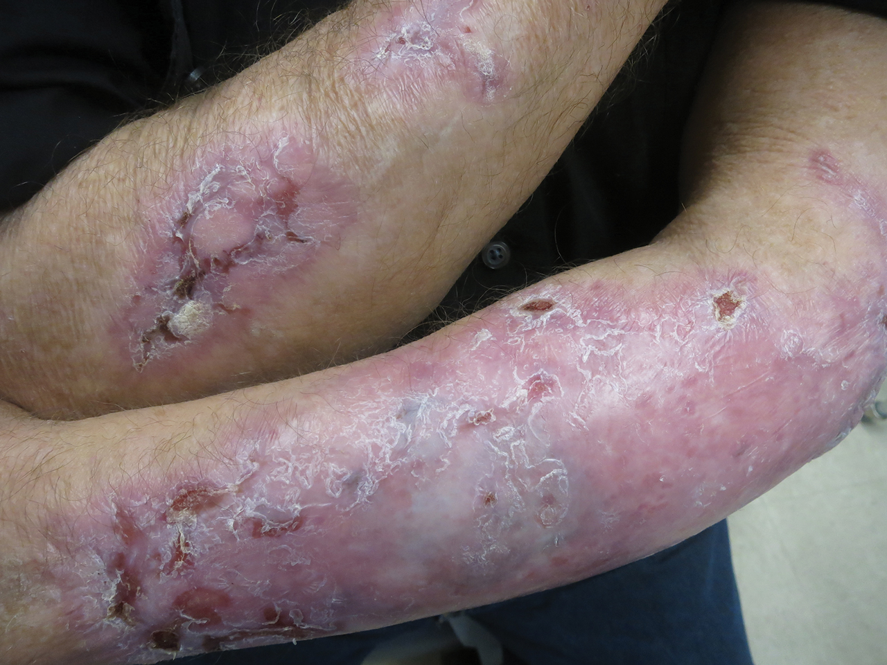
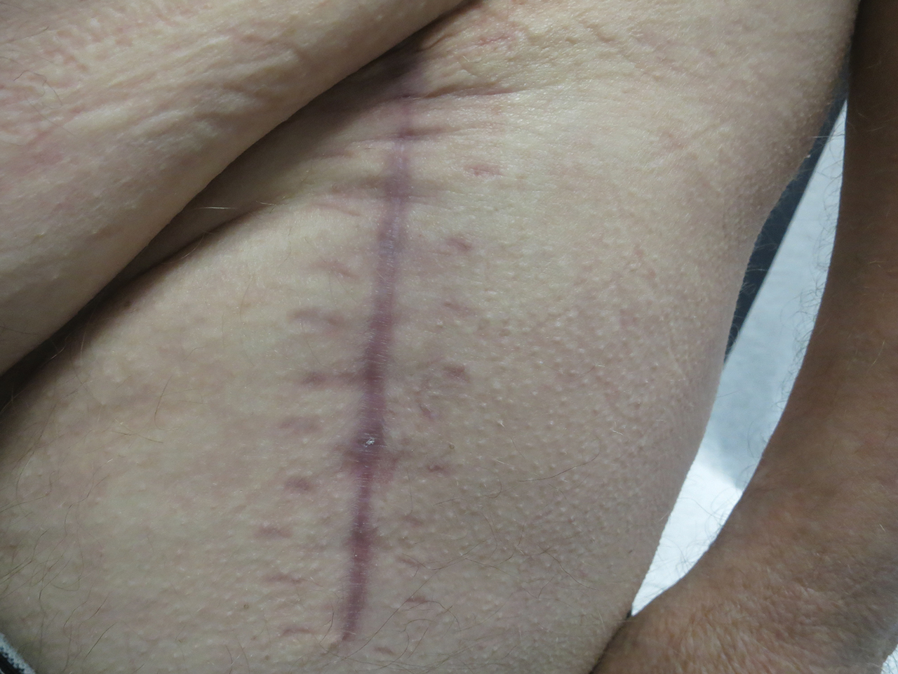
Our report is novel in that it supports a combination of IVIG and dapsone as a viable presurgical therapy for patients with EBA, and this treatment also may be applicable for other primary immunobullous disorders.2,5 Our case was particularly challenging in that the severity of the patient’s bullous disease precluded him from an elective orthopedic joint replacement due to the risk for wound dehiscence and surgical site infection.2 We determined that IVIG and dapsone would be the most optimal combination therapy to facilitate superior disease control and concurrently allow for appropriate wound healing without impairing the host immune response. This report is unique from a clinical perspective in that a balance was successfully achieved between immune suppression, with avoidance of associated side effects, and disease activity.
- Ahmed AR, Gürcan HM. Treatment of epidermolysis bullosa acquisita with intravenous immunoglobulin in patients non-responsive to conventional therapy: clinical outcome and post-treatment long-term follow-up [published online August 8, 2011]. J Eur Acad Dermatol Venereol. 2012;26:1074-1083.
- Rubin J, Touloei K, Favreau T, et al. Mohs surgery in patients immunobullous diseases: should prednisone be increased prior to surgery? J Clin Aesthet Dermatol. 2014;7:45-46.
- Ishii N, Hamada T, Dainichi T, et al. Epidermolysis bullosa acquisita: what’s new? J Dermatol. 2010;37:220-230.
- Mosqueira CB, Furlani Lde A, Xavier AF, et al. Intravenous immunoglobulin for treatment of severe acquired bullous epidermolysis refractory to conventional immunosuppressive therapy. An Bras Dermatol. 2010;85:521-524.
- Ludwig RJ. Clinical presentation, pathogenesis, diagnosis, and treatment of EBA. ISRN Dermatology. 2013;2013:812029.
To the Editor:
Evidence-based recommendations for optimal medical management of patients with immunobullous diseases prior to elective surgery are sparse.1,2 There is an uncertain balance between the use of immunomodulators and immunosuppressants, and implementation of these agents is heavily weighted against an increased infection risk from both active disease with denuded skin and suboptimal wound healing due to iatrogenic immunosuppression.1-5 Historically, clinical management of epidermolysis bullosa acquisita (EBA) seldomly has resulted in substantial disease resolution.1,3,4 Herein, we describe a case of recalcitrant EBA that was treated with a combination of intravenous immunoglobulin (IVIG) and dapsone, which resulted in a favorable clinical response and successful hip arthroplasty without cutaneous complications.
A 66-year-old man presented to an outside clinic with nonhealing ulcers on the oral mucosa, hands, groin, and feet. He was treated with systemic steroids after a histologic examination suggested bullous pemphigoid, but the lesions did not exhibit any appreciable improvement after several months of treatment. Despite the lack of improvement, the patient was continued on systemic steroids with a waxing and waning disease course.
Within a year, the patient presented to an orthopedist at our institution with severe left hip pain that had been limiting his mobility and had become unresponsive to conservative therapy. Radiologic investigations suggested advanced osteoarthritis and avascular necrosis of the left hip. Surgical intervention was delayed, as his orthopedist expressed concern that the extent of the body surface area affected by cutaneous denudation placed him at an unacceptable risk for infection. The orthopedic surgeon then referred the patient to our clinic for evaluation of the lesions. Physical examination revealed numerous crusted erosions in various stages of healing on the oral mucosa, palms, groin, and soles. Repeat biopsy of a denuded ulcer on the patient’s arm was obtained by our providers (nearly 1 year after the first biopsy by the outside physician). Histologic examination showed a pauci-immune subepidermal blister without acantholysis, which in combination with the clinical presentation of tense bullae on trauma-prone surfaces led to a favored diagnosis of EBA.
The patient began trials of several immunomodulatory and immunosuppressive agents, both in isolation and in combination, including systemic steroids, mycophenolate mofetil, four 1000-mg infusions of rituximab, and dapsone. Although results were suboptimal, dapsone 150 mg once daily for 3 months yielded the greatest clinical improvement with subsequent granulation and/or re-epithelialization of the chronic ulcers. After discussion during our department’s Grand Rounds, it was determined that the patient should undergo a trial of IVIG infusions, which were initiated with a loading dose of 2000 mg/kg over 5 consecutive days, followed by once-monthly maintenance infusion doses of 1200 mg/kg for 4 consecutive months. While receiving IVIG, he was maintained on a once-daily dose of dapsone 150 mg
Following this treatment regimen, he was noted to have marked improvement with only few scattered healing erosions. Upon completion of his last IVIG infusion, his cutaneous and mucosal manifestations of EBA were greatly minimized, demonstrating the best level of control that had been achieved during the disease course (Figure 1). This therapy completely cleared the cutaneous and mucosal ulcerations, thus permitting the patient to undergo a total left hip arthroplasty without complications (Figure 2).
Our report is novel in that it supports a combination of IVIG and dapsone as a viable presurgical therapy for patients with EBA, and this treatment also may be applicable for other primary immunobullous disorders.2,5 Our case was particularly challenging in that the severity of the patient’s bullous disease precluded him from an elective orthopedic joint replacement due to the risk for wound dehiscence and surgical site infection.2 We determined that IVIG and dapsone would be the most optimal combination therapy to facilitate superior disease control and concurrently allow for appropriate wound healing without impairing the host immune response. This report is unique from a clinical perspective in that a balance was successfully achieved between immune suppression, with avoidance of associated side effects, and disease activity.
To the Editor:
Evidence-based recommendations for optimal medical management of patients with immunobullous diseases prior to elective surgery are sparse.1,2 There is an uncertain balance between the use of immunomodulators and immunosuppressants, and implementation of these agents is heavily weighted against an increased infection risk from both active disease with denuded skin and suboptimal wound healing due to iatrogenic immunosuppression.1-5 Historically, clinical management of epidermolysis bullosa acquisita (EBA) seldomly has resulted in substantial disease resolution.1,3,4 Herein, we describe a case of recalcitrant EBA that was treated with a combination of intravenous immunoglobulin (IVIG) and dapsone, which resulted in a favorable clinical response and successful hip arthroplasty without cutaneous complications.
A 66-year-old man presented to an outside clinic with nonhealing ulcers on the oral mucosa, hands, groin, and feet. He was treated with systemic steroids after a histologic examination suggested bullous pemphigoid, but the lesions did not exhibit any appreciable improvement after several months of treatment. Despite the lack of improvement, the patient was continued on systemic steroids with a waxing and waning disease course.
Within a year, the patient presented to an orthopedist at our institution with severe left hip pain that had been limiting his mobility and had become unresponsive to conservative therapy. Radiologic investigations suggested advanced osteoarthritis and avascular necrosis of the left hip. Surgical intervention was delayed, as his orthopedist expressed concern that the extent of the body surface area affected by cutaneous denudation placed him at an unacceptable risk for infection. The orthopedic surgeon then referred the patient to our clinic for evaluation of the lesions. Physical examination revealed numerous crusted erosions in various stages of healing on the oral mucosa, palms, groin, and soles. Repeat biopsy of a denuded ulcer on the patient’s arm was obtained by our providers (nearly 1 year after the first biopsy by the outside physician). Histologic examination showed a pauci-immune subepidermal blister without acantholysis, which in combination with the clinical presentation of tense bullae on trauma-prone surfaces led to a favored diagnosis of EBA.
The patient began trials of several immunomodulatory and immunosuppressive agents, both in isolation and in combination, including systemic steroids, mycophenolate mofetil, four 1000-mg infusions of rituximab, and dapsone. Although results were suboptimal, dapsone 150 mg once daily for 3 months yielded the greatest clinical improvement with subsequent granulation and/or re-epithelialization of the chronic ulcers. After discussion during our department’s Grand Rounds, it was determined that the patient should undergo a trial of IVIG infusions, which were initiated with a loading dose of 2000 mg/kg over 5 consecutive days, followed by once-monthly maintenance infusion doses of 1200 mg/kg for 4 consecutive months. While receiving IVIG, he was maintained on a once-daily dose of dapsone 150 mg
Following this treatment regimen, he was noted to have marked improvement with only few scattered healing erosions. Upon completion of his last IVIG infusion, his cutaneous and mucosal manifestations of EBA were greatly minimized, demonstrating the best level of control that had been achieved during the disease course (Figure 1). This therapy completely cleared the cutaneous and mucosal ulcerations, thus permitting the patient to undergo a total left hip arthroplasty without complications (Figure 2).
Our report is novel in that it supports a combination of IVIG and dapsone as a viable presurgical therapy for patients with EBA, and this treatment also may be applicable for other primary immunobullous disorders.2,5 Our case was particularly challenging in that the severity of the patient’s bullous disease precluded him from an elective orthopedic joint replacement due to the risk for wound dehiscence and surgical site infection.2 We determined that IVIG and dapsone would be the most optimal combination therapy to facilitate superior disease control and concurrently allow for appropriate wound healing without impairing the host immune response. This report is unique from a clinical perspective in that a balance was successfully achieved between immune suppression, with avoidance of associated side effects, and disease activity.
- Ahmed AR, Gürcan HM. Treatment of epidermolysis bullosa acquisita with intravenous immunoglobulin in patients non-responsive to conventional therapy: clinical outcome and post-treatment long-term follow-up [published online August 8, 2011]. J Eur Acad Dermatol Venereol. 2012;26:1074-1083.
- Rubin J, Touloei K, Favreau T, et al. Mohs surgery in patients immunobullous diseases: should prednisone be increased prior to surgery? J Clin Aesthet Dermatol. 2014;7:45-46.
- Ishii N, Hamada T, Dainichi T, et al. Epidermolysis bullosa acquisita: what’s new? J Dermatol. 2010;37:220-230.
- Mosqueira CB, Furlani Lde A, Xavier AF, et al. Intravenous immunoglobulin for treatment of severe acquired bullous epidermolysis refractory to conventional immunosuppressive therapy. An Bras Dermatol. 2010;85:521-524.
- Ludwig RJ. Clinical presentation, pathogenesis, diagnosis, and treatment of EBA. ISRN Dermatology. 2013;2013:812029.
- Ahmed AR, Gürcan HM. Treatment of epidermolysis bullosa acquisita with intravenous immunoglobulin in patients non-responsive to conventional therapy: clinical outcome and post-treatment long-term follow-up [published online August 8, 2011]. J Eur Acad Dermatol Venereol. 2012;26:1074-1083.
- Rubin J, Touloei K, Favreau T, et al. Mohs surgery in patients immunobullous diseases: should prednisone be increased prior to surgery? J Clin Aesthet Dermatol. 2014;7:45-46.
- Ishii N, Hamada T, Dainichi T, et al. Epidermolysis bullosa acquisita: what’s new? J Dermatol. 2010;37:220-230.
- Mosqueira CB, Furlani Lde A, Xavier AF, et al. Intravenous immunoglobulin for treatment of severe acquired bullous epidermolysis refractory to conventional immunosuppressive therapy. An Bras Dermatol. 2010;85:521-524.
- Ludwig RJ. Clinical presentation, pathogenesis, diagnosis, and treatment of EBA. ISRN Dermatology. 2013;2013:812029.
Practice Points
- Treatment of epidermolysis bullosa acquisita (EBA) is difficult, and most treatment regimens are based on anecdotal reports.
- Systemic corticosteroids have been the mainstay of therapy for severe or extensive disease but impose an increased risk for postoperative complications including surgical site infections.
- A steroid-sparing regimen of intravenous immunoglobulin and systemic dapsone may be used when rapid clearance of EBA is needed prior to elective surgery.
What is your diagnosis? - September 2019
Erosive protein-losing enteropathy secondary to disseminated histoplasmosis
This patient was treated with amphotericin B and transitioned to oral itraconazole with frequent blood level monitoring to ensure absorption. His symptoms improved gradually. Small-bowel enteroscopy 3 weeks after presentation showed a normal duodenum and healing, superficial ulcers in the proximal jejunum (Figure F, G). Blood albumin levels had recovered to 3.1 g/dL (normal, 3.5–5.0 g/dL).
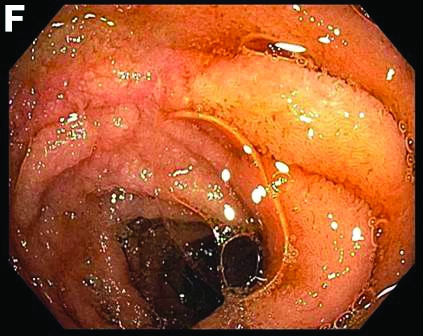
Protein-losing enteropathy (PLE) is a rare syndrome characterized by loss of serum proteins in the gastrointestinal (GI) tract, resulting in significant hypoproteinemia and consequent edema.1 PLE can also result in ascites, pleural and pericardial effusions, and, in prolonged cases, malnutrition. There are a variety of causes of PLE that can be broadly grouped into erosive GI disorders, disorders of increased GI mucosal permeability, and disorders of increased interstitial pressure. The clinical presentation depends on the underlying etiology, but commonly includes generalized edema owing to hypoproteinemia and resulting reduced oncotic pressure. GI symptoms are not frequently observed. The initial step in evaluating a patient with symptoms concerning for PLE is to rule out more common causes of hypoproteinemia, such as renal or hepatic disease, and malnutrition. To confirm enteric protein loss, alpha 1-antitrypsin clearance with a 24-hour stool collection is commonly and reliably used. Treatment of PLE is centered on treating the underlying cause while monitoring and treating malnutrition, including micronutrient deficiencies.
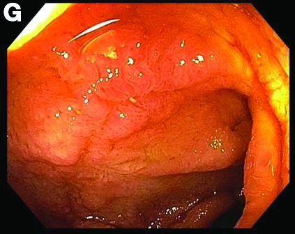
Fungal infections are a rare cause of PLE, but important to recognize as a potential complication of tumor necrosis factor–therapy, because these medications are commonly used for a variety of autoimmune diseases.2 Although histoplasmosis is an uncommon cause of GI inflammation, disseminated histoplasmosis causing PLE has been previously reported.3 In our patient, Histoplasma capsulatum infection caused diffuse GI ulcers, which allowed protein loss in the GI tract (erosive PLE). Antifungal treatment resulted in healing of intestinal ulcers and correction of hypoalbuminemia, thereby confirming the diagnosis of PLE and obviating the need for a confirmatory alpha 1-antitrypsin clearance study.
References
1. Umar SB, DiBaise JK. Protein-losing enteropathy: case illustrations and clinical review. Am J Gastroenterol. 2010;105:43-9.
2. Tsiodras S, Samonis G, Boumpas DT. et al. Fungal infections complicating tumor necrosis factor alpha blockade therapy. Mayo Clin Proc. 2008;83:181-94.
3. Kok J, Chen SC, Anderson L, et al. Protein-losing enteropathy and hypogammaglobulinaemia as first manifestations of disseminated histoplasmosis coincident with Nocardia infection. J Med Microbiol. 2010;59:610-3.
Erosive protein-losing enteropathy secondary to disseminated histoplasmosis
This patient was treated with amphotericin B and transitioned to oral itraconazole with frequent blood level monitoring to ensure absorption. His symptoms improved gradually. Small-bowel enteroscopy 3 weeks after presentation showed a normal duodenum and healing, superficial ulcers in the proximal jejunum (Figure F, G). Blood albumin levels had recovered to 3.1 g/dL (normal, 3.5–5.0 g/dL).
Protein-losing enteropathy (PLE) is a rare syndrome characterized by loss of serum proteins in the gastrointestinal (GI) tract, resulting in significant hypoproteinemia and consequent edema.1 PLE can also result in ascites, pleural and pericardial effusions, and, in prolonged cases, malnutrition. There are a variety of causes of PLE that can be broadly grouped into erosive GI disorders, disorders of increased GI mucosal permeability, and disorders of increased interstitial pressure. The clinical presentation depends on the underlying etiology, but commonly includes generalized edema owing to hypoproteinemia and resulting reduced oncotic pressure. GI symptoms are not frequently observed. The initial step in evaluating a patient with symptoms concerning for PLE is to rule out more common causes of hypoproteinemia, such as renal or hepatic disease, and malnutrition. To confirm enteric protein loss, alpha 1-antitrypsin clearance with a 24-hour stool collection is commonly and reliably used. Treatment of PLE is centered on treating the underlying cause while monitoring and treating malnutrition, including micronutrient deficiencies.
Fungal infections are a rare cause of PLE, but important to recognize as a potential complication of tumor necrosis factor–therapy, because these medications are commonly used for a variety of autoimmune diseases.2 Although histoplasmosis is an uncommon cause of GI inflammation, disseminated histoplasmosis causing PLE has been previously reported.3 In our patient, Histoplasma capsulatum infection caused diffuse GI ulcers, which allowed protein loss in the GI tract (erosive PLE). Antifungal treatment resulted in healing of intestinal ulcers and correction of hypoalbuminemia, thereby confirming the diagnosis of PLE and obviating the need for a confirmatory alpha 1-antitrypsin clearance study.
References
1. Umar SB, DiBaise JK. Protein-losing enteropathy: case illustrations and clinical review. Am J Gastroenterol. 2010;105:43-9.
2. Tsiodras S, Samonis G, Boumpas DT. et al. Fungal infections complicating tumor necrosis factor alpha blockade therapy. Mayo Clin Proc. 2008;83:181-94.
3. Kok J, Chen SC, Anderson L, et al. Protein-losing enteropathy and hypogammaglobulinaemia as first manifestations of disseminated histoplasmosis coincident with Nocardia infection. J Med Microbiol. 2010;59:610-3.
Erosive protein-losing enteropathy secondary to disseminated histoplasmosis
This patient was treated with amphotericin B and transitioned to oral itraconazole with frequent blood level monitoring to ensure absorption. His symptoms improved gradually. Small-bowel enteroscopy 3 weeks after presentation showed a normal duodenum and healing, superficial ulcers in the proximal jejunum (Figure F, G). Blood albumin levels had recovered to 3.1 g/dL (normal, 3.5–5.0 g/dL).
Protein-losing enteropathy (PLE) is a rare syndrome characterized by loss of serum proteins in the gastrointestinal (GI) tract, resulting in significant hypoproteinemia and consequent edema.1 PLE can also result in ascites, pleural and pericardial effusions, and, in prolonged cases, malnutrition. There are a variety of causes of PLE that can be broadly grouped into erosive GI disorders, disorders of increased GI mucosal permeability, and disorders of increased interstitial pressure. The clinical presentation depends on the underlying etiology, but commonly includes generalized edema owing to hypoproteinemia and resulting reduced oncotic pressure. GI symptoms are not frequently observed. The initial step in evaluating a patient with symptoms concerning for PLE is to rule out more common causes of hypoproteinemia, such as renal or hepatic disease, and malnutrition. To confirm enteric protein loss, alpha 1-antitrypsin clearance with a 24-hour stool collection is commonly and reliably used. Treatment of PLE is centered on treating the underlying cause while monitoring and treating malnutrition, including micronutrient deficiencies.
Fungal infections are a rare cause of PLE, but important to recognize as a potential complication of tumor necrosis factor–therapy, because these medications are commonly used for a variety of autoimmune diseases.2 Although histoplasmosis is an uncommon cause of GI inflammation, disseminated histoplasmosis causing PLE has been previously reported.3 In our patient, Histoplasma capsulatum infection caused diffuse GI ulcers, which allowed protein loss in the GI tract (erosive PLE). Antifungal treatment resulted in healing of intestinal ulcers and correction of hypoalbuminemia, thereby confirming the diagnosis of PLE and obviating the need for a confirmatory alpha 1-antitrypsin clearance study.
References
1. Umar SB, DiBaise JK. Protein-losing enteropathy: case illustrations and clinical review. Am J Gastroenterol. 2010;105:43-9.
2. Tsiodras S, Samonis G, Boumpas DT. et al. Fungal infections complicating tumor necrosis factor alpha blockade therapy. Mayo Clin Proc. 2008;83:181-94.
3. Kok J, Chen SC, Anderson L, et al. Protein-losing enteropathy and hypogammaglobulinaemia as first manifestations of disseminated histoplasmosis coincident with Nocardia infection. J Med Microbiol. 2010;59:610-3.
A 34-year-old man with a medical history of psoriasis, on adalimumab, presented with a 2-week history of progressively worsening abdominal pain, nausea, vomiting, melenic diarrhea, subjective fevers, and generalized weakness. One week into the illness, he developed progressive bilateral extremity and scrotal swelling.
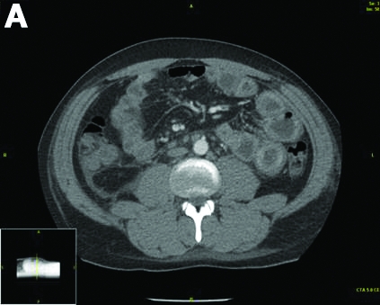
His vital signs included a temperature of 36.8°C, heart rate of 104 beats per minute, respiratory rate of 18 breaths per minute, and a blood pressure of 114/71 mm Hg. The physical examination was notable for a well-nourished appearance, diffuse abdominal tenderness to palpation without distension, organomegaly, or rigidity, and pitting lower extremity edema.
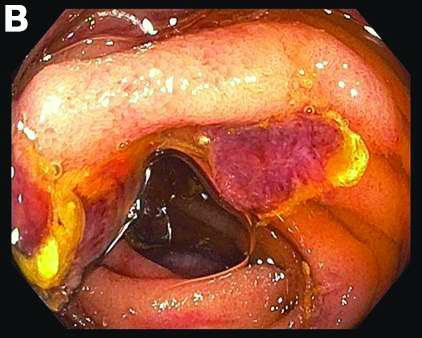
Laboratory evaluation showed hemoglobin 10.3 g/dL (normal, 13.5–17.5 g/dL), leukocytes 10 × 109/L (normal, 3.5–10.5 × 10⁹/L), platelets 212 × 109/L (normal, 150–450 × 109/L), sodium 131 mmol/L (normal, 135–145 mmol/L), creatinine 1 mg/dL (normal, 0.8–1.3 mg/dL), albumin 1.8 g/dL (normal, 3.5–5.0 g/dL), and C-reactive protein 53 mg/L (normal, less than 8 mg/L). Liver chemistries were all normal.
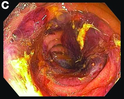
Urinalysis was unremarkable with normal urine protein levels. The enteric pathogen panel by polymerase chain reaction was negative. Computed tomography (CT) of the abdomen and pelvis showed marked circumferential wall thickening with mural enhancement of multiple loops of jejunum (Figure A).
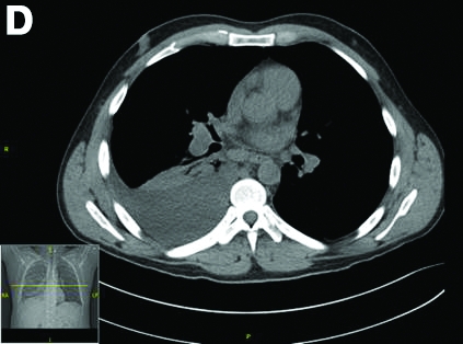
Small-bowel enteroscopy showed diffuse erosions in the entire duodenum and many oozing superficial ulcers with edematous and erythematous mucosa in the proximal jejunum (Figures B, C).
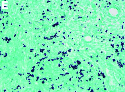
CT scan of the chest showed right lower lobe consolidation associated with a large right pleural effusion, and mediastinal, bilateral, hilar and abdominal lymphadenopathy (Figure D). Endobronchial ultrasound-guided transbronchial biopsy of lymph nodes was positive for oval-shaped organisms exhibiting narrow-based budding on GMS stain (Figure E).
Based on the clinical scenario and images, what is the most likely diagnosis?
Idiopathic Bilateral Auricular Ossificans
To the Editor:
A 60-year-old man with a history of basal cell carcinoma, rosacea, and seborrheic dermatitis presented for a routine skin examination. The patient mentioned incidentally that both of his ears were “rock hard” and had been so for the last 10 to 20 years. He was not experiencing hearing abnormalities and denied any history of external ear trauma, frostbite injury of the ears, or history of endocrinopathy. Physical examination revealed normal-appearing skin on the bilateral ears (Figure 1), and palpation confirmed a bonelike consistency of the auricles with sparing of the earlobes. The differential diagnosis included osteoma cutis, ectopic calcification, tophaceous gout, localized scleroderma, and relapsing polychondritis. Incisional biopsy of the left posterior auricle to the level of cartilage revealed a histologically normal epidermis and dermis, with small fragments of cartilage calcification and lacunar bone formation (Figure 2). A radiograph of the skull showed faint calcification of the auricular cartilage, but no other osseous abnormalities were observed (Figure 3). Further laboratory workup including a comprehensive metabolic panel; complete blood cell count; and thyroid stimulating hormone, parathyroid hormone, and cortisol levels were normal. Based on these findings, a diagnosis of bilateral idiopathic auricular ossificans was made. Therapy was not pursued because the patient was asymptomatic, but referral to otolaryngology would have been considered if hearing impairment had occurred.

with auricular ossificans. Palpation of the auricle revealed a
bonelike consistency.
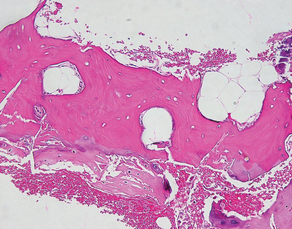
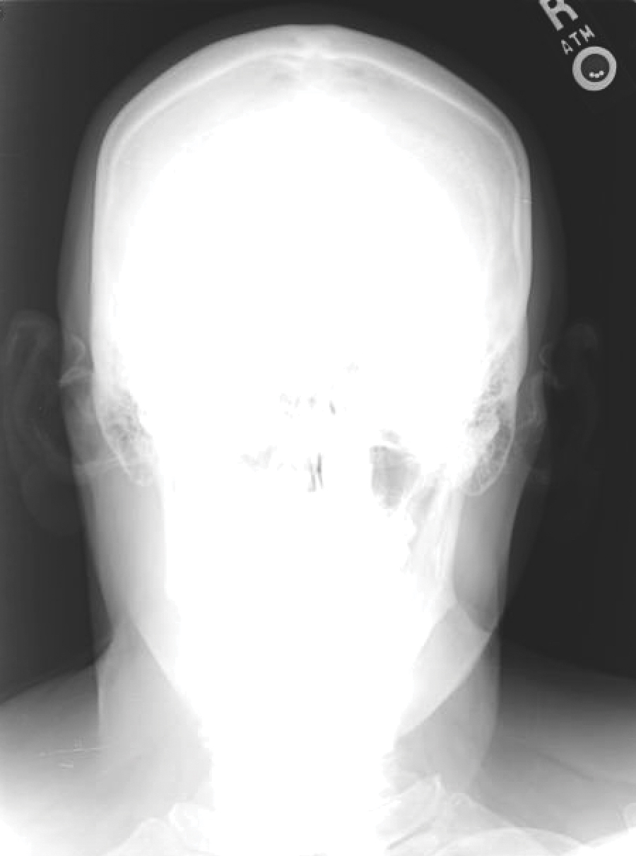
Auricular ossificans, which is characterized by the replacement of external ear cartilage by bone, is a rare condition with as few as 22 histologically proven cases documented in the literature.1,2 One case was reported in 2012 with consistent clinical and radiographic findings, but the patient declined biopsy.3 Similar to our patient, many pathologically documented cases have been determined to be idiopathic after workup, with identifiable triggering factors including cold injury, trauma, perichondritis, Addison disease, diabetes, and postpartum hypopituitarism.1 The male-to-female ratio is 18 to 5, the average age at diagnosis is 57 years, and as many as 70% of cases have demonstrated bilateral involvement.1,2
The majority of cases of auricular ossificans are asymptomatic at presentation with an insidious onset of the disease process over several years. Physical examination of the ear generally reveals a petrified auricle with sparing of the lobule and an otherwise normal clinical appearance. Radiographs demonstrate calcification, sometimes exactly mimicking the pattern seen in normal bone. Biopsy and histologic analysis show not only calcified cartilage but also actual lamellar bone formation.4 Depending on the precipitating factors, laboratory workup may uncover underlying metabolic abnormalities but often is unremarkable. Expert opinion generally recommends against extensive workup, which should be guided by the clinical presentation and the physician’s judgement.5 An insufficient number of patients with auricular ossificans have been definitively identified to provide clearer evidence-based recommendations.
Most patients present without pain or hearing abnormalities and do not require treatment. In one case, involvement of both the auricle and external ear canal resulted in intractable cerumen impaction that caused conductive hearing loss, eventually requiring resection of the ossified external ear cartilage and tragus.6 The most common reason for treatment has been discomfort impairing sleep, with surgical intervention including conchal reduction or wedge resection.5 A combined paucity of cases and poor understanding of the pathophysiologic mechanisms behind auricular ossificans limits current therapeutic options. Fortunately, this process appears to be benign in the majority of patients and generally represents a phenomenon that is much more interesting to the clinician than it is vexing to the patient.
- Calderon-Komaromy A, Cordoba S, Tardio JC, et al. Bilateral ossification of the auricular cartilage [published online November 28, 2014]. Actas Dermosifiliogr. 2015;106:433-435.
- Chang KH, Kim DK, Kim JH, et al. Idiopathic acquired ectopic auricular ossification: a case report and review of the literature. Ear Nose Throat J. 2011;90:424-427.
- Buikema KE, Adams EG. A rare case of petrified ear [published online October 15, 2012]. Case Rep Dermatol Med. 2012;2012:410601.
- Mastronikolis NS, Zampakis P, Kalogeropoulou C, et al. Bilateral ossification of the auricles: an unusual entity and review of the literature. Head Face Med. 2009;5:17.
- High WA, Larson MJ, Hoang MP. Idiopathic bilateral auricular ossificans: a case report and review of the literature. Arch Pathol Lab Med. 2004;128:1432-1434.
- Manni JJ, Berénos-Riley LC. Ossification of the external ear: a case report and review of the literature [published online June 18, 2005]. Eur Arch Otorhinolaryngol. 2005;262:961-964.
To the Editor:
A 60-year-old man with a history of basal cell carcinoma, rosacea, and seborrheic dermatitis presented for a routine skin examination. The patient mentioned incidentally that both of his ears were “rock hard” and had been so for the last 10 to 20 years. He was not experiencing hearing abnormalities and denied any history of external ear trauma, frostbite injury of the ears, or history of endocrinopathy. Physical examination revealed normal-appearing skin on the bilateral ears (Figure 1), and palpation confirmed a bonelike consistency of the auricles with sparing of the earlobes. The differential diagnosis included osteoma cutis, ectopic calcification, tophaceous gout, localized scleroderma, and relapsing polychondritis. Incisional biopsy of the left posterior auricle to the level of cartilage revealed a histologically normal epidermis and dermis, with small fragments of cartilage calcification and lacunar bone formation (Figure 2). A radiograph of the skull showed faint calcification of the auricular cartilage, but no other osseous abnormalities were observed (Figure 3). Further laboratory workup including a comprehensive metabolic panel; complete blood cell count; and thyroid stimulating hormone, parathyroid hormone, and cortisol levels were normal. Based on these findings, a diagnosis of bilateral idiopathic auricular ossificans was made. Therapy was not pursued because the patient was asymptomatic, but referral to otolaryngology would have been considered if hearing impairment had occurred.
with auricular ossificans. Palpation of the auricle revealed a
bonelike consistency.
Auricular ossificans, which is characterized by the replacement of external ear cartilage by bone, is a rare condition with as few as 22 histologically proven cases documented in the literature.1,2 One case was reported in 2012 with consistent clinical and radiographic findings, but the patient declined biopsy.3 Similar to our patient, many pathologically documented cases have been determined to be idiopathic after workup, with identifiable triggering factors including cold injury, trauma, perichondritis, Addison disease, diabetes, and postpartum hypopituitarism.1 The male-to-female ratio is 18 to 5, the average age at diagnosis is 57 years, and as many as 70% of cases have demonstrated bilateral involvement.1,2
The majority of cases of auricular ossificans are asymptomatic at presentation with an insidious onset of the disease process over several years. Physical examination of the ear generally reveals a petrified auricle with sparing of the lobule and an otherwise normal clinical appearance. Radiographs demonstrate calcification, sometimes exactly mimicking the pattern seen in normal bone. Biopsy and histologic analysis show not only calcified cartilage but also actual lamellar bone formation.4 Depending on the precipitating factors, laboratory workup may uncover underlying metabolic abnormalities but often is unremarkable. Expert opinion generally recommends against extensive workup, which should be guided by the clinical presentation and the physician’s judgement.5 An insufficient number of patients with auricular ossificans have been definitively identified to provide clearer evidence-based recommendations.
Most patients present without pain or hearing abnormalities and do not require treatment. In one case, involvement of both the auricle and external ear canal resulted in intractable cerumen impaction that caused conductive hearing loss, eventually requiring resection of the ossified external ear cartilage and tragus.6 The most common reason for treatment has been discomfort impairing sleep, with surgical intervention including conchal reduction or wedge resection.5 A combined paucity of cases and poor understanding of the pathophysiologic mechanisms behind auricular ossificans limits current therapeutic options. Fortunately, this process appears to be benign in the majority of patients and generally represents a phenomenon that is much more interesting to the clinician than it is vexing to the patient.
To the Editor:
A 60-year-old man with a history of basal cell carcinoma, rosacea, and seborrheic dermatitis presented for a routine skin examination. The patient mentioned incidentally that both of his ears were “rock hard” and had been so for the last 10 to 20 years. He was not experiencing hearing abnormalities and denied any history of external ear trauma, frostbite injury of the ears, or history of endocrinopathy. Physical examination revealed normal-appearing skin on the bilateral ears (Figure 1), and palpation confirmed a bonelike consistency of the auricles with sparing of the earlobes. The differential diagnosis included osteoma cutis, ectopic calcification, tophaceous gout, localized scleroderma, and relapsing polychondritis. Incisional biopsy of the left posterior auricle to the level of cartilage revealed a histologically normal epidermis and dermis, with small fragments of cartilage calcification and lacunar bone formation (Figure 2). A radiograph of the skull showed faint calcification of the auricular cartilage, but no other osseous abnormalities were observed (Figure 3). Further laboratory workup including a comprehensive metabolic panel; complete blood cell count; and thyroid stimulating hormone, parathyroid hormone, and cortisol levels were normal. Based on these findings, a diagnosis of bilateral idiopathic auricular ossificans was made. Therapy was not pursued because the patient was asymptomatic, but referral to otolaryngology would have been considered if hearing impairment had occurred.
with auricular ossificans. Palpation of the auricle revealed a
bonelike consistency.
Auricular ossificans, which is characterized by the replacement of external ear cartilage by bone, is a rare condition with as few as 22 histologically proven cases documented in the literature.1,2 One case was reported in 2012 with consistent clinical and radiographic findings, but the patient declined biopsy.3 Similar to our patient, many pathologically documented cases have been determined to be idiopathic after workup, with identifiable triggering factors including cold injury, trauma, perichondritis, Addison disease, diabetes, and postpartum hypopituitarism.1 The male-to-female ratio is 18 to 5, the average age at diagnosis is 57 years, and as many as 70% of cases have demonstrated bilateral involvement.1,2
The majority of cases of auricular ossificans are asymptomatic at presentation with an insidious onset of the disease process over several years. Physical examination of the ear generally reveals a petrified auricle with sparing of the lobule and an otherwise normal clinical appearance. Radiographs demonstrate calcification, sometimes exactly mimicking the pattern seen in normal bone. Biopsy and histologic analysis show not only calcified cartilage but also actual lamellar bone formation.4 Depending on the precipitating factors, laboratory workup may uncover underlying metabolic abnormalities but often is unremarkable. Expert opinion generally recommends against extensive workup, which should be guided by the clinical presentation and the physician’s judgement.5 An insufficient number of patients with auricular ossificans have been definitively identified to provide clearer evidence-based recommendations.
Most patients present without pain or hearing abnormalities and do not require treatment. In one case, involvement of both the auricle and external ear canal resulted in intractable cerumen impaction that caused conductive hearing loss, eventually requiring resection of the ossified external ear cartilage and tragus.6 The most common reason for treatment has been discomfort impairing sleep, with surgical intervention including conchal reduction or wedge resection.5 A combined paucity of cases and poor understanding of the pathophysiologic mechanisms behind auricular ossificans limits current therapeutic options. Fortunately, this process appears to be benign in the majority of patients and generally represents a phenomenon that is much more interesting to the clinician than it is vexing to the patient.
- Calderon-Komaromy A, Cordoba S, Tardio JC, et al. Bilateral ossification of the auricular cartilage [published online November 28, 2014]. Actas Dermosifiliogr. 2015;106:433-435.
- Chang KH, Kim DK, Kim JH, et al. Idiopathic acquired ectopic auricular ossification: a case report and review of the literature. Ear Nose Throat J. 2011;90:424-427.
- Buikema KE, Adams EG. A rare case of petrified ear [published online October 15, 2012]. Case Rep Dermatol Med. 2012;2012:410601.
- Mastronikolis NS, Zampakis P, Kalogeropoulou C, et al. Bilateral ossification of the auricles: an unusual entity and review of the literature. Head Face Med. 2009;5:17.
- High WA, Larson MJ, Hoang MP. Idiopathic bilateral auricular ossificans: a case report and review of the literature. Arch Pathol Lab Med. 2004;128:1432-1434.
- Manni JJ, Berénos-Riley LC. Ossification of the external ear: a case report and review of the literature [published online June 18, 2005]. Eur Arch Otorhinolaryngol. 2005;262:961-964.
- Calderon-Komaromy A, Cordoba S, Tardio JC, et al. Bilateral ossification of the auricular cartilage [published online November 28, 2014]. Actas Dermosifiliogr. 2015;106:433-435.
- Chang KH, Kim DK, Kim JH, et al. Idiopathic acquired ectopic auricular ossification: a case report and review of the literature. Ear Nose Throat J. 2011;90:424-427.
- Buikema KE, Adams EG. A rare case of petrified ear [published online October 15, 2012]. Case Rep Dermatol Med. 2012;2012:410601.
- Mastronikolis NS, Zampakis P, Kalogeropoulou C, et al. Bilateral ossification of the auricles: an unusual entity and review of the literature. Head Face Med. 2009;5:17.
- High WA, Larson MJ, Hoang MP. Idiopathic bilateral auricular ossificans: a case report and review of the literature. Arch Pathol Lab Med. 2004;128:1432-1434.
- Manni JJ, Berénos-Riley LC. Ossification of the external ear: a case report and review of the literature [published online June 18, 2005]. Eur Arch Otorhinolaryngol. 2005;262:961-964.
Practice Points
- Auricular ossificans is a rare condition characterized by the replacement of external ear cartilage by bone.
- The majority of cases are asymptomatic at presentation with an insidious onset of the disease process over several years.
- Most patients present without pain or hearing abnormalities and do not require treatment.
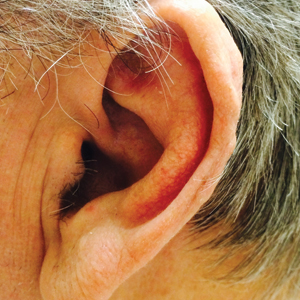
Severe lipodystrophy linked to pembrolizumab
according to a case study.
A 47-year-old woman received pembrolizumab to treat metastatic melanoma and developed immune-related generalized acquired lipodystrophy. The condition has persisted 12 months after she stopped taking pembrolizumab.
Julie Delyon, MD, PhD, of Saint-Louis Hospital in Paris, and colleagues described this case in the British Journal of Dermatology.
The patient was diagnosed with BRAF-mutated, stage IV melanoma with bone and lymph node metastases. She received pembrolizumab at 2 mg/kg every 3 weeks as first-line treatment. She achieved a complete response and was still in remission at last follow-up.
The patient was obese at baseline, with a body mass index of 40 kg/m2, but she did not have diabetes, hepatic steatosis, or dyslipidemia.
Within 2 months of starting pembrolizumab, the patient observed “some major changes to her physical appearance,” according to Dr. Delyon and colleagues. At 10 months, the patient had severe lipodystrophy.
The patient experienced a change in fat distribution – namely, severe peripheral lipoatrophy and an accumulation of trunk fat. Imaging revealed a decrease in subcutaneous adipose tissue, which contrasted with an increase in visceral fat observed from baseline.
The patient also developed “moderate hirsutism, facial and limb atrophy, and prominent forearm and leg muscles and veins,” according to the authors. She had impaired glucose tolerance with insulin resistance, reduced concentrations of leptin and adiponectin, hypertriglyceridemia, a low level of HDL cholesterol, and hepatic steatosis.
Analyses of subcutaneous fat revealed adipose tissue atrophy with edema, lipophages, and CD3+/CD4+ T-cell infiltration of the fat and vessel walls. This suggested that the lipodystrophy had an autoimmune origin, according to the authors.
The patient had no family history of autoimmune disease or lipodystrophy. She tested negative for mutations in 23 genes associated with generalized lipodystrophy.
The patient also tested negative for HIV, antinuclear antibodies, native anti-DNA, and anti-insulin receptor antibodies. There were no signs of panniculitis, and the authors noted that “there were no arguments in favor of hyperthyroidism, Cushing syndrome, or acromegaly.”
In an attempt to reverse the lipodystrophy, the researchers stopped pembrolizumab treatment. The patient was treated for diabetes and hypertriglyceridemia as well. She could not receive corticosteroids because of the risk of severe metabolic complications, and she didn’t receive metreleptin because it wasn’t available. The patient still had lipodystrophy 12 months after stopping pembrolizumab.
Dr. Delyon and colleagues wrote that this case suggests pembrolizumab, and perhaps other anti–programmed death 1 therapies, may cause lipodystrophy with severe metabolic complications. “The long-term side effects of such metabolic adverse events, although rare, are unknown and will probably become a topic of utmost importance, considering the increasing rate of remission following ICIs and their use in the adjuvant setting.”
Two coauthors reported relationships with Merck, which markets pembrolizumab as Keytruda. The authors also reported relationships with Bristol-Myers Squibb, Pierre Fabre, Takeda, Innate Pharma, LEO Pharma, Roche, GlaxoSmithKline, Novartis, and Amgen.
SOURCE: Delyon J et al. Br J Dermatol. 2019 May 11. doi: 10.1111/bjd.18124.
according to a case study.
A 47-year-old woman received pembrolizumab to treat metastatic melanoma and developed immune-related generalized acquired lipodystrophy. The condition has persisted 12 months after she stopped taking pembrolizumab.
Julie Delyon, MD, PhD, of Saint-Louis Hospital in Paris, and colleagues described this case in the British Journal of Dermatology.
The patient was diagnosed with BRAF-mutated, stage IV melanoma with bone and lymph node metastases. She received pembrolizumab at 2 mg/kg every 3 weeks as first-line treatment. She achieved a complete response and was still in remission at last follow-up.
The patient was obese at baseline, with a body mass index of 40 kg/m2, but she did not have diabetes, hepatic steatosis, or dyslipidemia.
Within 2 months of starting pembrolizumab, the patient observed “some major changes to her physical appearance,” according to Dr. Delyon and colleagues. At 10 months, the patient had severe lipodystrophy.
The patient experienced a change in fat distribution – namely, severe peripheral lipoatrophy and an accumulation of trunk fat. Imaging revealed a decrease in subcutaneous adipose tissue, which contrasted with an increase in visceral fat observed from baseline.
The patient also developed “moderate hirsutism, facial and limb atrophy, and prominent forearm and leg muscles and veins,” according to the authors. She had impaired glucose tolerance with insulin resistance, reduced concentrations of leptin and adiponectin, hypertriglyceridemia, a low level of HDL cholesterol, and hepatic steatosis.
Analyses of subcutaneous fat revealed adipose tissue atrophy with edema, lipophages, and CD3+/CD4+ T-cell infiltration of the fat and vessel walls. This suggested that the lipodystrophy had an autoimmune origin, according to the authors.
The patient had no family history of autoimmune disease or lipodystrophy. She tested negative for mutations in 23 genes associated with generalized lipodystrophy.
The patient also tested negative for HIV, antinuclear antibodies, native anti-DNA, and anti-insulin receptor antibodies. There were no signs of panniculitis, and the authors noted that “there were no arguments in favor of hyperthyroidism, Cushing syndrome, or acromegaly.”
In an attempt to reverse the lipodystrophy, the researchers stopped pembrolizumab treatment. The patient was treated for diabetes and hypertriglyceridemia as well. She could not receive corticosteroids because of the risk of severe metabolic complications, and she didn’t receive metreleptin because it wasn’t available. The patient still had lipodystrophy 12 months after stopping pembrolizumab.
Dr. Delyon and colleagues wrote that this case suggests pembrolizumab, and perhaps other anti–programmed death 1 therapies, may cause lipodystrophy with severe metabolic complications. “The long-term side effects of such metabolic adverse events, although rare, are unknown and will probably become a topic of utmost importance, considering the increasing rate of remission following ICIs and their use in the adjuvant setting.”
Two coauthors reported relationships with Merck, which markets pembrolizumab as Keytruda. The authors also reported relationships with Bristol-Myers Squibb, Pierre Fabre, Takeda, Innate Pharma, LEO Pharma, Roche, GlaxoSmithKline, Novartis, and Amgen.
SOURCE: Delyon J et al. Br J Dermatol. 2019 May 11. doi: 10.1111/bjd.18124.
according to a case study.
A 47-year-old woman received pembrolizumab to treat metastatic melanoma and developed immune-related generalized acquired lipodystrophy. The condition has persisted 12 months after she stopped taking pembrolizumab.
Julie Delyon, MD, PhD, of Saint-Louis Hospital in Paris, and colleagues described this case in the British Journal of Dermatology.
The patient was diagnosed with BRAF-mutated, stage IV melanoma with bone and lymph node metastases. She received pembrolizumab at 2 mg/kg every 3 weeks as first-line treatment. She achieved a complete response and was still in remission at last follow-up.
The patient was obese at baseline, with a body mass index of 40 kg/m2, but she did not have diabetes, hepatic steatosis, or dyslipidemia.
Within 2 months of starting pembrolizumab, the patient observed “some major changes to her physical appearance,” according to Dr. Delyon and colleagues. At 10 months, the patient had severe lipodystrophy.
The patient experienced a change in fat distribution – namely, severe peripheral lipoatrophy and an accumulation of trunk fat. Imaging revealed a decrease in subcutaneous adipose tissue, which contrasted with an increase in visceral fat observed from baseline.
The patient also developed “moderate hirsutism, facial and limb atrophy, and prominent forearm and leg muscles and veins,” according to the authors. She had impaired glucose tolerance with insulin resistance, reduced concentrations of leptin and adiponectin, hypertriglyceridemia, a low level of HDL cholesterol, and hepatic steatosis.
Analyses of subcutaneous fat revealed adipose tissue atrophy with edema, lipophages, and CD3+/CD4+ T-cell infiltration of the fat and vessel walls. This suggested that the lipodystrophy had an autoimmune origin, according to the authors.
The patient had no family history of autoimmune disease or lipodystrophy. She tested negative for mutations in 23 genes associated with generalized lipodystrophy.
The patient also tested negative for HIV, antinuclear antibodies, native anti-DNA, and anti-insulin receptor antibodies. There were no signs of panniculitis, and the authors noted that “there were no arguments in favor of hyperthyroidism, Cushing syndrome, or acromegaly.”
In an attempt to reverse the lipodystrophy, the researchers stopped pembrolizumab treatment. The patient was treated for diabetes and hypertriglyceridemia as well. She could not receive corticosteroids because of the risk of severe metabolic complications, and she didn’t receive metreleptin because it wasn’t available. The patient still had lipodystrophy 12 months after stopping pembrolizumab.
Dr. Delyon and colleagues wrote that this case suggests pembrolizumab, and perhaps other anti–programmed death 1 therapies, may cause lipodystrophy with severe metabolic complications. “The long-term side effects of such metabolic adverse events, although rare, are unknown and will probably become a topic of utmost importance, considering the increasing rate of remission following ICIs and their use in the adjuvant setting.”
Two coauthors reported relationships with Merck, which markets pembrolizumab as Keytruda. The authors also reported relationships with Bristol-Myers Squibb, Pierre Fabre, Takeda, Innate Pharma, LEO Pharma, Roche, GlaxoSmithKline, Novartis, and Amgen.
SOURCE: Delyon J et al. Br J Dermatol. 2019 May 11. doi: 10.1111/bjd.18124.
FROM THE BRITISH JOURNAL OF DERMATOLOGY