User login
Reactive aggressive disorder in children with ADHD is looking for a name
NEW YORK – according to Robert L. Findling, MD.
Emphasizing the reactive component to this behavioral problem, he said: “They look okay until someone bumps into them at school. They do not have a mood disorder. They have a disorder of reactivity.”
The hurdle is that there is no accepted terminology to encourage clinicians to identify and initiate treatment in children with this behavior. The term conduct disorder has been used in the past, but Dr. Findling said that care delivered for conduct disorder is not reimbursable. This may be among the reasons that aggressive reactive behavior of ADHD is overlooked – even though treatment is likely to improve long-term outcome.
“I wish I had a magic label for this, but I don’t,” Dr. Findling said. However, he maintained that most clinicians who work with ADHD children are familiar with this type of behavior. Indeed, clinicians “grapple with this day to day. We all see these kids, and they are oftentimes the most impaired kids in our practices,” he said at a pediatric psychopharmacology update held by the American Academy of Child and Adolescent Psychiatry.
This behavior should not be confused with the aggression associated with mood disorders, such as disruptive mood dysregulation disorder (DMDD) or bipolar disease, according to Dr. Findling. Children with DMDD, for example, are chronically irritable or angry. Although bipolar disorder patients with aggressive behavior are not necessarily angry between episodes, they also have persistent mood disturbances.
In contrast, preadolescent children with ADHD who have episodes of aggression, a symptom far more common among males than females, do not otherwise exhibit disturbances in mood. In addition, the episodes of impulsive, reactive aggression are provoked. They require a perceived insult, threat, or similar trigger.
While many of these children continue to have episodes of impulsive aggressive behavior even on treatment effective for other ADHD symptoms, Dr. Findling said, “The good news is that there are treatments for aggression.” In addition to psychosocial support aimed at reducing aggressive behavior, once the diagnosis has been made, these include adjusting ADHD treatments to better target symptoms of episodic aggression. If needed, therapies known to treat aggression, such as atypical antipsychotics, anticonvulsants, or lithium also are options.
Dr. Findling did review one older double-blind study that associated methylphenidate with a reduction in aggression in children with conduct disorder, but said he believes that there is no guarantee for a response from any treatment. Rather, he recommended empirical strategies for symptom management and keeping in mind the benefit-to-risk relationship when considering treatments that impose a high burden of adverse events.
However, the first step to treatment is recognizing the problem.
“In my opinion, what is missing is the nosology for these kids,” Dr. Findling said. An evidence-based label will help increase awareness of the problem and encourage more extensive clinical study, he said.
“These children are not rare and they are really impaired. It is heartbreaking, because when you talk to them when they are still little, they know what people think of them. They know their teachers don’t like them. They know their parents think they’re bad. They know their peers are scared of them, and they cannot make friends,” he said. However, there is a potential for reversing these problems if treatment is initiated early.
“As you watch them get older, you watch them scarring over,” he added.
Dr. Findling reported financial ties with numerous pharmaceutical companies.
SOURCE: Findling RL. Psychopharmacology Update Institute
REPORTING FROM THE PSYCHOPHARMACOLOGY UPDATE INSTITUTE
Characterize duration when seeking etiology of tantrums in children
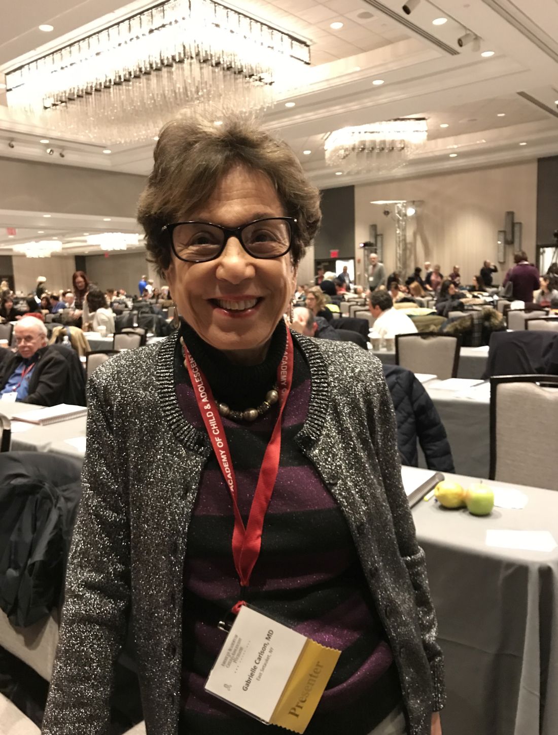
NEW YORK – Although explosive outbursts or tantrums accompany nearly every psychiatric illness that affects children, the specific features may help identify an etiology, according to Gabrielle A. Carlson, MD.
“There are two components of irritability,” explained Dr. Carlson, professor of psychiatry and pediatrics, Stony Brook (N.Y.) University Medical Center. “One is how often the child loses his or her temper, and the other is what they do when they lose their temper.”
To be useful in identifying the source, the characterization of explosive outbursts must be undertaken in the context of the patient’s history and the duration and types of tantrum-related behaviors, particularly aggressive behavior toward others, according to Dr. Carlson.
Presenting a diagnostic algorithm relevant to children with frequent explosive outbursts, Dr. Carlson suggested that pathways differ for young children and adolescents. Yet, the first step – which is evaluating whether or not irritability is a feature of the patient’s disposition when not in the midst of a tantrum – is common to both groups.
In young children with new onset of explosive outbursts, stressors in school, such as bullying, or family, such as abuse, represent an appropriate initial focus. In adolescents, initial attention should be paid to the potential role of mood disorders, particularly depression, mania, or anxiety, according to Dr. Carlson.
For most patients and most etiologies, tantrums follow a trigger and then resolve quickly. When tantrums do not resolve quickly in patients who remain generally irritable even when they are not having a tantrum, there is an increased likelihood of disruptive mood dysregulation disorder (DMDD).
Relative to tantrums associated with attention deficit hyperactive disorder (ADHD), oppositional defiant disorder (ODD), or affective disorders, explosive outbursts associated with DMDD are also more likely to include aggression toward others.
Physical restraint to safeguard the patient or others during a tantrum is uncommon in most conditions associated with tantrums, with the exception of DMDD. Greater aggression tracks with greater DMDD severity. According to data presented by Dr. Carlson, 92% of a clinical sample of DMDD patients exhibited physical aggression, compared with none of those in a community sample.
Tantrums lasting more than 30 minutes were observed in 60% of the clinic sample, versus only 12.5% of the community sample.
Explosive outbursts “are not an uncommon or trivial problem,” according to Dr. Carlson, who cited data suggesting that 70% of children between the ages of 5 and 12 years hospitalized for a psychiatric diseases are referred for an explosive outburst.
She believes that a systematic approach toward characterizing the tantrum will be helpful in understanding the underlying etiology and appropriate treatment. Using such tools as the Irritability and Rages Inventory or the Affective Reactivity Index Child Form, clinicians should seek to evaluate the frequency of tantrums, the duration, and the patient’s symptom burden between tantrums.
If explosive outbursts are rare, they are unlikely to be due to DMDD or affective disorders, such as bipolar disease. If frequent in a patient with chronic psychopathology, those who are generally “fine until frustrated” are the ones more likely to have ADHD or even oppositional defiant disorder (ODD).
The less common profile, which is rage that does not completely resolve, suggests DMDD, a condition that Dr. Carlson described with the mnemonic OI VEY to convey key features. The letters stand for Outbursts that are frequent, Irritable mood in the absence of an outburst, Very chronic (more than 1 per year), Explained by other co-existing conditions, such as mania, and Young (starts between ages 6 and 10 years).
Although tantrums are the way in which children with a broad array of psychiatric conditions express frustration, Dr. Carlson said it is not clear if the mechanisms for irritability and explosive outbursts are shared across conditions. Despite the guidance she offered for linking specific tantrum features with DMDD, she also reiterated that tantrums cannot be considered a symptom specific to any single etiology. The difference between etiologies for irritable children having a tantrum “is not how they feel, the difference is what they do,” Dr. Carlson suggested.
Dr. Carlson reported no relevant financial relationships.
REPORTING FROM THE PSYCHOPHARMACOLOGY UPDATE INSTITUTE
Brain structural changes on MRI predict sudden death in epilepsy
WASHINGTON – It has long been hypothesized that sudden unexpected death in epilepsy (SUDEP) is the result of damage to areas of the brain that control breathing and heart rate, but two studies presented at the American Epilepsy Society annual meeting identified specific areas where structural changes correlate with SUDEP, suggesting the potential for screening.
In one of two studies that evaluated MRI images in patients who subsequently died of SUDEP and compared them to patients with epilepsy or normal healthy individuals, brainstem volume loss was not only greater in those with SUDEP but there was a correlation between greater volume loss and a shorter period of survival, reported Susanne G. Mueller, MD, a radiologist affiliated with the Center for Imaging of Neurodegenerative Diseases at the San Francisco Veterans Affairs Medical Center.
“This is the first evidence that brainstem damage, one of the mechanisms that has been shown to cause SUDEP in animals, could also play a role in people with epilepsy,” Dr. Mueller reported. Although more work is needed, volume loss observed on MRI “could be used as a potential biomarker to assess the individual SUDEP risk in epilepsy patients.”
In one of two populations studied, investigators analyzed MRI scans from 27 SUDEP patients taken prior to death. Focusing on brainstem areas involved in autonomic function, deformation morphometry generated profile maps that isolated areas of volume loss. The changes suggested that focal epilepsy produced structural changes in the mesencephalic region of the lower brainstem.
Damage to nuclei involved in control of heart rate variability and other autonomic functions would be consistent with increased risk of SUDEP.
“We can just report what we see on imaging. These data do not tell us the cause of SUDEP, but they do show correlations that are consistent with current theories,” Dr. Mueller explained.
In a second study that compared 18 patients with focal epilepsy to 11 controls, greater volume loss in patients with epilepsy correlated negatively with heart rate variability, a relationship not seen in the controls. The volume loss in periventricular gray and medulla oblongata nuclei was most closely associated with heart rate variability, which is considered a surrogate for altered autonomic function, Dr. Mueller reported.
Similar conclusions were reached in a different study conducted at a separate institution. In this study, MRI scans from 237 patients with epilepsy and 110 healthy controls were evaluated. Four of the epilepsy patients subsequently died of SUDEP.

“These are all areas that can be involved in autonomic function potentially involved in SUDEP,” Mr. George said. Like Dr. Mueller, Mr. George cautioned that clinically viable algorithms that would allow MRI to assess SUDEP risk may be years away, but these studies provide preliminary evidence that structural changes on MRI could eventually serve as a SUDEP biomarker.
There was limited overlap between the areas of structural change potentially associated with SUDEP in the studies presented by Dr. Mueller and Mr. George, but Dr. Mueller said that SUDEP might not stem from a single epilepsy-induced brain abnormality. She reported that more MRI scans to trace structural changes in SUDEP patients are likely to identify more areas of interest. Acquiring a large number of MRI scans is a challenge, but Dr. Mueller envisions a registry where routine scans could be submitted. This would permit this research to be conducted on a larger scale.
“There is a similar initiative to collect MRI brain images of patients with Alzheimer’s disease,” said Dr. Mueller, noting that this provides a precedent for the type of research needed in epilepsy. If a similar program could be undertaken in epilepsy, Dr. Mueller believes it might substantially accelerate the effort to understand and recognize risk of SUDEP.
Mr. George’s study was funded by FACES (Finding a CURE for Epilepsy/Seizures). Dr. Mueller’s study was funded by grants from UCSF, the Epilepsy Foundation, and the National Institutes of Health.
Dr. Mueller and Mr. George reported no potential conflicts of interest related to this topic.
SOURCE: Mueller S et al., AES 2017 abstract 3.205 and George A et al., AES 2017 abstract 3.214
WASHINGTON – It has long been hypothesized that sudden unexpected death in epilepsy (SUDEP) is the result of damage to areas of the brain that control breathing and heart rate, but two studies presented at the American Epilepsy Society annual meeting identified specific areas where structural changes correlate with SUDEP, suggesting the potential for screening.
In one of two studies that evaluated MRI images in patients who subsequently died of SUDEP and compared them to patients with epilepsy or normal healthy individuals, brainstem volume loss was not only greater in those with SUDEP but there was a correlation between greater volume loss and a shorter period of survival, reported Susanne G. Mueller, MD, a radiologist affiliated with the Center for Imaging of Neurodegenerative Diseases at the San Francisco Veterans Affairs Medical Center.
“This is the first evidence that brainstem damage, one of the mechanisms that has been shown to cause SUDEP in animals, could also play a role in people with epilepsy,” Dr. Mueller reported. Although more work is needed, volume loss observed on MRI “could be used as a potential biomarker to assess the individual SUDEP risk in epilepsy patients.”
In one of two populations studied, investigators analyzed MRI scans from 27 SUDEP patients taken prior to death. Focusing on brainstem areas involved in autonomic function, deformation morphometry generated profile maps that isolated areas of volume loss. The changes suggested that focal epilepsy produced structural changes in the mesencephalic region of the lower brainstem.
Damage to nuclei involved in control of heart rate variability and other autonomic functions would be consistent with increased risk of SUDEP.
“We can just report what we see on imaging. These data do not tell us the cause of SUDEP, but they do show correlations that are consistent with current theories,” Dr. Mueller explained.
In a second study that compared 18 patients with focal epilepsy to 11 controls, greater volume loss in patients with epilepsy correlated negatively with heart rate variability, a relationship not seen in the controls. The volume loss in periventricular gray and medulla oblongata nuclei was most closely associated with heart rate variability, which is considered a surrogate for altered autonomic function, Dr. Mueller reported.
Similar conclusions were reached in a different study conducted at a separate institution. In this study, MRI scans from 237 patients with epilepsy and 110 healthy controls were evaluated. Four of the epilepsy patients subsequently died of SUDEP.
“These are all areas that can be involved in autonomic function potentially involved in SUDEP,” Mr. George said. Like Dr. Mueller, Mr. George cautioned that clinically viable algorithms that would allow MRI to assess SUDEP risk may be years away, but these studies provide preliminary evidence that structural changes on MRI could eventually serve as a SUDEP biomarker.
There was limited overlap between the areas of structural change potentially associated with SUDEP in the studies presented by Dr. Mueller and Mr. George, but Dr. Mueller said that SUDEP might not stem from a single epilepsy-induced brain abnormality. She reported that more MRI scans to trace structural changes in SUDEP patients are likely to identify more areas of interest. Acquiring a large number of MRI scans is a challenge, but Dr. Mueller envisions a registry where routine scans could be submitted. This would permit this research to be conducted on a larger scale.
“There is a similar initiative to collect MRI brain images of patients with Alzheimer’s disease,” said Dr. Mueller, noting that this provides a precedent for the type of research needed in epilepsy. If a similar program could be undertaken in epilepsy, Dr. Mueller believes it might substantially accelerate the effort to understand and recognize risk of SUDEP.
Mr. George’s study was funded by FACES (Finding a CURE for Epilepsy/Seizures). Dr. Mueller’s study was funded by grants from UCSF, the Epilepsy Foundation, and the National Institutes of Health.
Dr. Mueller and Mr. George reported no potential conflicts of interest related to this topic.
SOURCE: Mueller S et al., AES 2017 abstract 3.205 and George A et al., AES 2017 abstract 3.214
REPORTING FROM AES 2017
Key clinical point: MRI-detected damage to brain areas mediating autonomic function is implicated in sudden unexpected death in epilepsy (SUDEP).
Major finding: Two separate studies drew the conclusion that brain structural difference seen on MRI may become an effective screening tool for identifying epilepsy patients at high risk for SUDEP.
Data source: Prospective studies.
Disclosures: Dr. Mueller’s study was funded by grants from UCSF, the Epilepsy Foundation, and the National Institutes of Health. Mr. George’s study was funded by FACES (Finding a CURE for Epilepsy/Seizures). Dr. Mueller and Mr. George reported no potential conflicts of interest related to this topic.
Source: Mueller S et al., AES 2017 abstract 3.205 and George A et al., AES 2017 abstract 3.214
Seizures captured on a smartphone found diagnostic for epilepsy
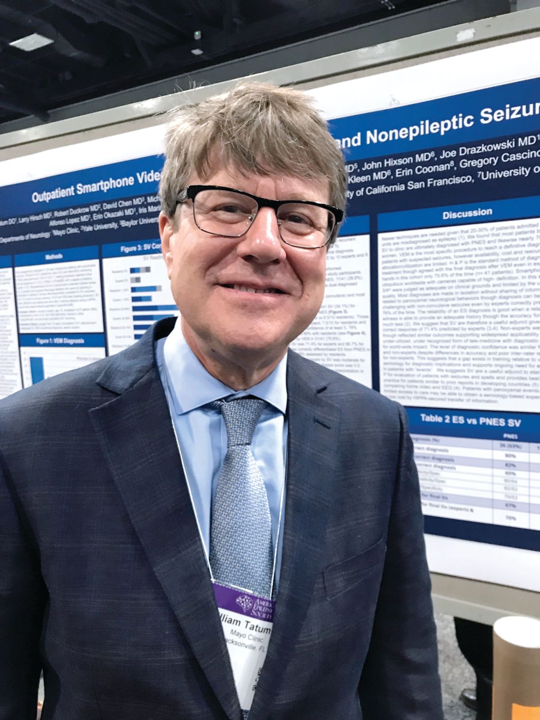
WASHINGTON – Smartphone videos brought to the clinic by patients are valid tools for the diagnosis of epilepsy, according to a prospective blinded and multicenter study of 41 consecutive videos presented at the American Epilepsy Society annual meeting.
“These findings have global implications, because they suggest that smartphone videos are a cost effective tool that can accelerate the time to diagnosis even in places where video-EEG monitoring is not readily available,” reported William Tatum, DO, professor of neurology, Mayo Clinic, Jacksonville, Fla.
In addition to submitting a smartphone video, all patients in this study underwent a history and physical (H&P) and were evaluated with video-EEG monitoring. The smartphone videos underwent review by 10 epilepsy experts and 8 general neurology residents blinded to the video EEG findings. The latter group was selected to test the value of smartphone video in clinicians with general knowledge but no special expertise.
The final diagnosis was made on the basis of all the clinical information, including the video-EEG, which Dr. Tatum characterized as the gold standard for the diagnosis of epilepsy. Based on the video-EEG, 11 of the 41 patients (26.8%) had seizures, 26 (63.4%) had psychogenic nonepileptic seizures (PNES), 3 (7.4%) had physiologic nonepileptic events (PhysNEE), and 1 (2.4%) had both PhysNEE and PNES.
On the basis of the blinded smartphone video alone, the median correct diagnosis was 71.4% for experts and 66.7% for residents. Although this difference was not significant, Dr. Tatum reported that there was substantially less inter-rater variability among experts.
“Overall, smartphone video review correctly differentiated epilepsy from PNES in 68% of the videos evaluated by experts and 58% assessed by residents,” Dr. Tatum reported.
For experts, the smartphone video assessment yielded a specificity of 43% and sensitivity of 83% for epilepsy. For PNES, these figures were 80% and 54%, respectively. Among residents, the sensitivities and specificities for epilepsy (32% and 83%) and PNES (82% and 53%) were similar. Dr. Tatum noted that H&P predicted the definitive diagnosis in 75.6% of cases.
The rate of correct diagnoses with blinded smartphone video analysis in this study was respectable, but Dr. Tatum suggested that smartphone video should be an adjunctive tool that is reviewed in the context of H&P, which would be expected to further boost accuracy. Although he acknowledged that smartphone videos plus H&P will not completely supplant the need for video-EEG monitoring to reach a definitive diagnosis in all cases, he believes that it is accurate in many, and it accelerates the time to diagnosis.
“The median duration of the smartphone review was about a minute and a half. The median duration of H&P was 60 minutes, but the median time to a result with video-EEG was 2.54 days,” said Dr. Tatum, noting that this difference was highly significant (P lees than .001). If a diagnosis can be reached without video-EEG, it would also be expected to greatly reduce costs.
On a yes-no basis, 78% of those who evaluated the smartphone videos judged them to be adequate for a diagnosis. An analysis of those considered to be poor quality by expert and resident viewers was presented as a separate report. The most common reasons that smartphone videos were considered to have inadequate quality, according to this analysis, were inadequate duration, insufficient lighting, and poor audio. In other words, essentially all of the problems stemmed from inadequate technique, not technical limitations, reported Erin E. Coonan, an undergraduate research intern working with Dr. Tatum.
“We think that disseminating information to the general public about how to take an adequate quality smartphone video could increase the quality of these videos when they are brought to the clinic,” Ms. Coonan reported. She and Dr. Tatum believe that patient-submitted smartphone videos will be increasingly common tool in clinical medicine, making information about proper technique valuable.
Most U.S. adults now carry smartphones, and these are becoming increasingly common even in resource-poor areas of the world, according to Dr. Tatum. He said that clinical medicine should embrace this technology.
“The cost of a smartphone video is essentially zero, but our data suggest that they can be a useful adjunctive diagnostic tool,” Dr. Tatum said.
The presenters reported no potential conflicts of interest related to these studies.
SOURCE: Tatum W et al., AES 2017 abstract 3.161 and Coonan E et al., AES 2017 abstract 3.070
Key clinical point: Videos taken with a smartphone can contribute to the accurate diagnosis of epilepsy, according to results of a blinded study.
Major finding: Experts correctly differentiated epileptic seizures from psychogenic nonepileptic seizures with smartphone video in 68% of cases.
Data source: A multicenter, prospective blinded trial of 41 consecutive videos.
Disclosures: The presenters reported no potential conflicts of interest related to this topic.
Source: Tatum W et al., AES 2017 abstract 3.161 and Coonan E et al., AES 2017 abstract 3.070
Efficacy of neurostimulation for epilepsy underestimated with patient reports
WASHINGTON – The benefit of implanting a responsive brain stimulator for the control of refractory epilepsy may be grossly underestimated without relying on an objective measure of baseline seizure activity rather than patient reports, according to a study presented at the annual meeting of the American Epilepsy Society.
In a retrospective evaluation at one center, the efficacy of the Responsive Neurostimulation System (RNS) came nowhere near that observed in the pivotal clinical trial until objective measures of seizure activity were analyzed, reported Michael Young, DO, a neurophysiology fellow in the department of neurology at the University of California, Irvine (UCI).
In this study, investigators evaluated seizure frequency in the first 2 months after RNS implantation with the ECoG component of the RNS device. They assessed change in seizure frequency relative to this baseline at 3, 6, and 12 months, and also compared the reduction in seizures against the patient self-report of baseline seizure activity.
The differences were large. On patient report, the reduction in seizure activity at month 3 was just 10%, compared with 85% when measured on ECoG.
“Our results with the RNS compare favorably to the pivotal trial only when using the ECoG seizure frequency baseline. The reason for this discrepancy is due to underreporting of seizures by patients and consequently a falsely low seizure frequency,” Dr. Young explained at the meeting.
The RNS system has been implanted for refractory focal or partial seizures in adult patients at UCI since 2015. The device is indicated for adjunctive use in patients not adequately controlled on at least two antiepileptic medications. Twelve patients have been treated, but two were excluded from this analysis because they had surgical resection at the time of the RNS implantation and one because of an infection related to the implantation.
In general, patients treated at UCI had characteristics similar to those in the pivotal trial, which was published more than 3 years ago (Epilepsia. 2014;55[3]:432-41). In that 191-patient trial, the reduction in seizure frequency at the end of 5 months of blinded analysis with RNS was 37.9% versus 17.3% for a sham procedure. Progressive further reductions in seizure activity were observed during an extended open-label follow-up.
In the UCI analysis, the mean reduction in seizure frequency at 12 months was 56% relative to the patient-reported baseline but 78% on the basis of the ECoG analysis. Although only four of the nine patients have 12 or more months of follow-up, three were considered to be responders to RNS whether evaluated in relation to the patient-reported baseline seizure activity or in relation to ECoG. The responder rate at 3 months on the basis of patient-reported baseline activity, however, was only 56%, compared with 100% based on ECoG.
“The big issue is underreporting of seizures by patients,” Dr. Young explained. He cited numerous other studies demonstrating the same phenomenon. He noted that noncompliance is only one reason patients underreport. In many cases, patients are simply unaware of seizure activity.
Based on these data, “we think ECoG may be a more objective way to track patient response to RNS,” Dr. Young said. He acknowledged that the number of patients limits this study and suggested that larger studies are needed to confirm the findings.
Dr. Young reported having no potential conflicts of interest related to this topic.
SOURCE: Young M et al. AES abstract 3.109.
REPORTING FROM AES 2017
Key clinical point:
Major finding: At 3 months after implantation, seizure activity was reduced 10% by patient report but 85% by objective measurement.
Data source: Retrospective study of nine patients implanted with the Responsive Neurostimulation System.
Disclosures: Dr. Young reported having no potential conflicts of interest related to this topic.
Source: Young M et al. AES abstract 3.109.
MRI-guided focused ultrasound shows promise for subcortical epilepsy
WASHINGTON – MRI-guided focused ultrasound (FUS) is now being employed on an experimental basis to treat deep subcortical lesions, such as hypothalamic hamartoma, to control intractable epilepsy, according to an expert summary of a “hot topic” presented at the American Epilepsy Society annual meeting.
“If the risk of FUS is as low as we expect, it could change our paradigm,” reported Nathan B. Fountain, MD, director of the F.E. Dreifuss Comprehensive Epilepsy Program at the University of Virginia, Charlottesville.

FUS has been used clinically for the treatment of uterine fibroids since 2004, according to an overview provided by Dr. Fountain. Clinical studies of MRI-guided FUS for lesions in the brain began in 2009. The approval of MRI-guided FUS thalamotomy for essential tumor in 2016 was based on a pivotal trial led by Jeffrey Elias, MD, a colleague of Dr. Fountain’s at the University of Virginia (N Engl J Med. 2016;375:730-9). Many of the principles for treating subcortical lesions causing epilepsy are the same as those for treating essential tremor.
Under MRI guidance, FUS is delivered via a helmet with 1,024 transducers. These focus sound waves to a highly targeted area of the brain, resulting in thermal ablation. The treatment is noninvasive in the sense that no craniotomy is involved. It can be delivered without anesthesia. When used to treat essential tremor in awake patients, MRI-guided FUS confirms the target when the tremor resolves.
“There is no injury to the brain as far as we can tell,” reported Dr. Fountain, referring to the tremor studies.
Because the thermal ablation is delivered by sound waves, this approach appears to be safer to structures surrounding the lesion than would be anticipated with energy delivered by radiation. For treatment of lesions in the hypothalamus, where surrounding tissue is responsible for important brain functions, the apparent low risk of collateral damage is a major potential advantage, according to Dr. Fountain.
Although Dr. Fountain conceded that the term “subcortical” is not commonly used to describe epilepsy lesions, he considers it appropriate to explain the role of MRI-guided FUS. Without technical advancements, this tool is not appropriate for the cortical lesions that are responsible for the majority of epileptic seizures. Rather, lesions must be positioned deep in the skull to be in the “envelope” where energy can be concentrated. Lesions in the temporal or hippocampal areas of the brain, for example, will not be suitable without technical advances.
Due to its position in the brain, “hypothalamic hamartoma is the prototype lesion,” Dr. Fountain reported. Importantly, these and other lesions within the envelope where energy can be targeted are the most difficult to treat with other options. Due to the need to transverse much of the brain to reach these areas, open surgery is often not practical. Even though Dr. Fountain acknowledged that MRI-guided stereotactic laser has been proposed for these types of lesions, the laser must also transverse vulnerable structures of the brain that can be avoided with MR-guided FUS.
Results on the first patient in a planned pediatric treatment series with MRI-guided FUS were presented at the AES annual meeting by Travis Tierney, MD, PhD, a neurosurgeon associated with Nicklaus Children’s Hospital in Miami. According to the data presented by Dr. Tierney, the 21-year-old patient was treated for a hypothalamic hamartoma. She was rendered seizure free and had no complications.
An adult series is now recruiting candidates, according to Dr. Fountain. He reported that adults of at least 18 years of age with intractable epilepsy due to subcortical lesions in the central envelope suitable for MRI-guided FUS are eligible if they have at least three seizures per month while taking at least two antiepileptic drugs. He encouraged referrals.
“The primary outcome will be just to demonstrate that a lesion can be created,” Dr. Fountain said. He reported that the planned enrollment of 15 subjects would not be sufficient to draw conclusions about efficacy “unless, of course, we eliminate everyone’s seizures – and that would be useful – but that is still a secondary outcome,”
There are a number of applications in neurology beyond treatment of tremors and epilepsy that are also being considered for MRI-guided FUS, Dr. Fountain reported. This could include, for example, clot lysis in stroke, but he indicated that there are a number of reasons to be particularly optimistic about its potential role in the treatment intractable epilepsy due to subcortical lesions. This strategy seems feasible in a condition with limited treatment options.
WASHINGTON – MRI-guided focused ultrasound (FUS) is now being employed on an experimental basis to treat deep subcortical lesions, such as hypothalamic hamartoma, to control intractable epilepsy, according to an expert summary of a “hot topic” presented at the American Epilepsy Society annual meeting.
“If the risk of FUS is as low as we expect, it could change our paradigm,” reported Nathan B. Fountain, MD, director of the F.E. Dreifuss Comprehensive Epilepsy Program at the University of Virginia, Charlottesville.
FUS has been used clinically for the treatment of uterine fibroids since 2004, according to an overview provided by Dr. Fountain. Clinical studies of MRI-guided FUS for lesions in the brain began in 2009. The approval of MRI-guided FUS thalamotomy for essential tumor in 2016 was based on a pivotal trial led by Jeffrey Elias, MD, a colleague of Dr. Fountain’s at the University of Virginia (N Engl J Med. 2016;375:730-9). Many of the principles for treating subcortical lesions causing epilepsy are the same as those for treating essential tremor.
Under MRI guidance, FUS is delivered via a helmet with 1,024 transducers. These focus sound waves to a highly targeted area of the brain, resulting in thermal ablation. The treatment is noninvasive in the sense that no craniotomy is involved. It can be delivered without anesthesia. When used to treat essential tremor in awake patients, MRI-guided FUS confirms the target when the tremor resolves.
“There is no injury to the brain as far as we can tell,” reported Dr. Fountain, referring to the tremor studies.
Because the thermal ablation is delivered by sound waves, this approach appears to be safer to structures surrounding the lesion than would be anticipated with energy delivered by radiation. For treatment of lesions in the hypothalamus, where surrounding tissue is responsible for important brain functions, the apparent low risk of collateral damage is a major potential advantage, according to Dr. Fountain.
Although Dr. Fountain conceded that the term “subcortical” is not commonly used to describe epilepsy lesions, he considers it appropriate to explain the role of MRI-guided FUS. Without technical advancements, this tool is not appropriate for the cortical lesions that are responsible for the majority of epileptic seizures. Rather, lesions must be positioned deep in the skull to be in the “envelope” where energy can be concentrated. Lesions in the temporal or hippocampal areas of the brain, for example, will not be suitable without technical advances.
Due to its position in the brain, “hypothalamic hamartoma is the prototype lesion,” Dr. Fountain reported. Importantly, these and other lesions within the envelope where energy can be targeted are the most difficult to treat with other options. Due to the need to transverse much of the brain to reach these areas, open surgery is often not practical. Even though Dr. Fountain acknowledged that MRI-guided stereotactic laser has been proposed for these types of lesions, the laser must also transverse vulnerable structures of the brain that can be avoided with MR-guided FUS.
Results on the first patient in a planned pediatric treatment series with MRI-guided FUS were presented at the AES annual meeting by Travis Tierney, MD, PhD, a neurosurgeon associated with Nicklaus Children’s Hospital in Miami. According to the data presented by Dr. Tierney, the 21-year-old patient was treated for a hypothalamic hamartoma. She was rendered seizure free and had no complications.
An adult series is now recruiting candidates, according to Dr. Fountain. He reported that adults of at least 18 years of age with intractable epilepsy due to subcortical lesions in the central envelope suitable for MRI-guided FUS are eligible if they have at least three seizures per month while taking at least two antiepileptic drugs. He encouraged referrals.
“The primary outcome will be just to demonstrate that a lesion can be created,” Dr. Fountain said. He reported that the planned enrollment of 15 subjects would not be sufficient to draw conclusions about efficacy “unless, of course, we eliminate everyone’s seizures – and that would be useful – but that is still a secondary outcome,”
There are a number of applications in neurology beyond treatment of tremors and epilepsy that are also being considered for MRI-guided FUS, Dr. Fountain reported. This could include, for example, clot lysis in stroke, but he indicated that there are a number of reasons to be particularly optimistic about its potential role in the treatment intractable epilepsy due to subcortical lesions. This strategy seems feasible in a condition with limited treatment options.
EXPERT ANALYSIS FROM AES 2017
Promising add-on therapy for neonatal seizures found active in safety study
WASHINGTON – presented at the annual meeting of the American Epilepsy Society.
“This is an early-phase trial, but it did associate bumetanide with an additional reduction in seizure burden relative to phenobarbital alone,” reported Janet S. Soul, MD, director of the fetal-neonatal neurology program at Boston Children’s Hospital. She added, “The adverse events observed were not substantially different in the group that received the experimental agent.”
Of the 111 neonates with documented seizures enrolled at four participating hospitals, 43 proceeded to randomization if their seizures proved to be refractory to standard doses of phenobarbital. After randomization, the next dose of phenobarbital was administered either with placebo or with 0.1, 0.2, or 0.3 mg/kg of bumetanide. Seizure burden was evaluated at 0-2, 2-4, and 0-4 hours after study-drug administration and compared with the burden during the 2 hours before treatment.
All three doses were active, reducing the seizure burden by a median of 41%-75% in a dose-dependent manner. Whether assessed in the first 2 hours or the first 4 hours, the efficacy of bumetanide was significantly greater in those with the greatest, relative to the least, baseline seizure burden (P = .01 for hours 0-2; P = .04 for hours 0-4). The median seizure burden during the baseline period was higher in the 27 children randomized to bumetanide (114 minutes) relative to those randomized to placebo (33 minutes), although researchers attributed this to random effects in a small study.
The evidence of antiseizure activity from bumetanide as an add-on to phenobarbital is consistent with its mechanism of action, which is blockading the chloride transporter NKCC1. In the immature neurons of neonates, NKCC1 is highly expressed, and there is basic scientific evidence that this impairs the efficacy of gamma-aminobutyric acid–receptor agonists like phenobarbital, according to Dr. Soul. The hypothesis driving the study of bumetanide is that blockading NKCC1 would improve the efficacy of phenobarbital while adding its own antiseizure effects, which together could potentially provide synergistic benefit.
The efficacy and the safety of this study are somewhat discordant with a previously published study evaluating bumetanide in 14 neonates with hypoxic-ischemic encephalopathy (HIE) seizures (Lancet Neurol 2015;14:469-77). Even though there were seizure reductions in five children in this other series, which did not include a control arm, there were three cases of hearing loss considered potentially related to bumetanide. The authors of that study concluded that efficacy was not shown.
There were also three cases of hearing loss in the randomized trial presented by Dr. Soul, but one occurred in the placebo group. Although the potential for ototoxicity “still needs to be addressed” in the next set of studies, Dr. Soul noted that hearing loss in children with epilepsy is common and has numerous potential etiologies. Based on these data, she concluded, “All serious adverse events were related to severe HIE with multiorgan dysfunction and/or withdrawal of care for poor prognosis.”
Among nonserious adverse events, diuresis was the only one found significantly more common in the bumetanide group (P = .02).
Phenobarbital has been a standard in the treatment of neonatal seizures for several decades despite the substantial proportion of children who do not achieve an adequate response, according to Dr. Soul. She noted that bumetanide is one of several agents being evaluated as an adjunctive agent. For example, a phase 2 crossover trial with levetiracetam is now underway. She suggested that there is reason for optimism about gaining new treatments for neonates in an area in which she believes there are unmet needs.
“I think we may see a phase 2 trial with bumetanide within a year or 2,” Dr. Soul said. If bumetanide moves forward, she expects its role to be primarily for the treatment of acute seizures caused by HIE, stroke, or hemorrhage. She is less optimistic about its benefit for seizures caused by other etiologies, such as brain malformations.
SOURCE: Soul J Abstract 2.426
REPORTING FROM AES 2017
Key clinical point: Bumetanide is associated with antiseizure activity as add-on therapy to phenobarbital for neonatal seizures.
Major finding: Relative to pretreatment, there was greater reduction in seizure burden (P = .01) at 4 hours in those with the highest seizure burden.
Data source: Randomized, double-blind phase 1/2 trial.
Disclosures: Dr. Soul reports no potential conflicts of interest related to this topic.
Source: Soul J et al. Abstract 2.426
Fenfluramine trials in Dravet syndrome yield highly positive results
WASHINGTON – The oral experimental agent fenfluramine, also known as ZX008, has been associated with a high degree of efficacy and good tolerability for the adjunctive treatment of Dravet syndrome, according to combined results of the first patients enrolled in two phase III trials.
“For me, a highlight of this study is the finding that 45% of patients on the higher dose achieved at least a 75% reduction from baseline in monthly convulsive seizures. This is a life-changing improvement,” reported Joseph Sullivan, MD, director of the pediatric epilepsy center at the University of California, San Francisco. He presented the results at the annual meeting of the American Epilepsy Society.
“If fenfluramine is approved as an adjunctive agent, it is likely to be introduced as the second or third medication in an effort to gain adequate symptom control,” Dr. Sullivan speculated.
Three phase 3 trials with fenfluramine are underway. The data presented at the American Epilepsy Society meeting were based on the first 119 patients who had participated in either of the two identical trials conducted in Europe and North America. The data from these two trials has now been combined, and the outcomes in the remaining patients in these two trials will be presented at a later time along with results from a third phase 3 study.
Patients between the ages of 2 and 18 years with a clinical diagnosis of Dravet syndrome were eligible for the European and North American trials if they were not controlled on current therapy, which could include multiple agents. However, patients had to be on stable therapies prior to enrollment for at least 4 weeks. Once enrolled, they were observed for 6 weeks prior to randomization.
After randomization to placebo, 0.2 mg/kg fenfluramine, or 0.8 mg/kg fenfluramine, patients completed a 2-week titration before they reached their maintenance dose. They were then evaluated over an additional 12-week treatment period. There were three withdrawals over the course of treatment in the placebo group, none in the lower-dose fenfluramine group, and six in the higher-dose fenfluramine group.
The primary endpoint was change in mean monthly convulsive seizure frequency from the observation period. When compared with placebo, these reductions were 63.9% (P less than .001) in the 0.8-mg/kg group and 33.7% (P = .019) in the 0.2-mg/kg group. When expressed as the median percent reduction in convulsive seizures from the observation period per 28 days, the reductions were 72.4% for the 0.8-mg/kg dose (P less than .001 vs. placebo), 37.6% for the 0.2-mg/kg group (P = .185 vs. placebo), and 17.4% for placebo.
Other efficacy measures supported the relative advantage of fenfluramine. For example, 70% and 41% of the patients in the 0.8-mg/kg and 0.2-mg/kg groups, respectively, versus 8% of placebo patients, had at least a 50% reduction in seizure frequency. Median seizure-free intervals for the three groups were 20.5, 14, and 9 days, respectively. Seizure activity was reduced to one or no seizures over the treatment period in 25% of the 0.8-mg/kg group, 12.8% of the 0.2-mg/kg group, and 0% of the placebo group.
The most common adverse events on the 0.8-mg/kg dose of fenfluramine, compared with placebo, were decreased appetite (37.5% vs. 5%) and lethargy (17.5% vs. 5%). The proportion of patients with weight loss was also greater on 0.8 mg/kg (5%) and 0.2 mg/kg (12.8%) versus placebo (0%). Diarrhea was more common in the 0.2-mg/kg group (30.8%) than in the 0.8-mg/kg group (17.5%) or in the placebo group (7.5%).
Although monitored closely, cardiotoxicity was not observed in this study. Concern about potential cardiotoxic effects was generated by the increased risk of valvular disease observed in patients taking fenfluramine with phentermine (fen-phen) for weight loss in the 1990s. This combination was withdrawn from the market in 1997.
“The potential for cardiotoxicity will continue to be monitored closely, but these initial results were reassuring,” reported Dr. Sullivan, who noted that a history of cardiovascular or cerebrovascular disease were exclusion criteria from this study.
Application for regulatory approval is not anticipated until all the phase 3 trial data are available, but Dr. Sullivan said that the results so far suggest that fenfluramine as an adjunctive agent “may represent a significant advance over existing treatment options for Dravet syndrome.”
The studies are funded by Zogenix. Dr. Sullivan reported financial relationships with Epygenix and Zogenix.
REPORTING FROM AES 2017
Key clinical point:
Major finding: For the highest dose, the mean reduction in convulsive seizure frequency was 63.9% (P less than .001) versus placebo over a 14-week treatment period.
Data source: An analysis of the first 119 patients with Dravet syndrome enrolled in two ongoing randomized, double-blind, multicenter, phase 3 trials.
Disclosures: The studies are funded by Zogenix. The presenter reported financial relationships with Epygenix and Zogenix.
Source: L Lagae et al. AES 2017 Abstract 2.434
Continuous bedside monitoring improved safety of intracranial stereotactic EEG
WASHINGTON – After a life-threatening event prompted a trial of continuous monitoring of patients during intracranial stereotactic electroencephalogram (EEG), the rate of adverse events and missed seizures went to zero, according to a single-center analysis presented at the annual meeting of the American Epilepsy Society.
“After initiating a full-time, bedside sitter, none of the major events we saw in the presitter period, which included unrecognized tonic-clonic seizures or patient removal of electrodes, was observed,” reported Brad Kamitaki, MD, who is completing an epilepsy fellowship at Columbia University, New York.

Continuous observation of patients undergoing intracranial EEG has been described as mandatory in guidelines from the National Association of Epilepsy Centers (Epilepsia. 2010 Nov;51[11]:2322-33), but the type of monitoring, such as bedside sitter versus closed circuit video, has not been specified, according to Dr. Kamitaki. Although continuous bedside monitoring might offer the best opportunity to capture seizures and reduce the risk of adverse events, there are few comparative data.
In this study, the rate of adverse events was evaluated after a full-time, bedside sitter was initiated and compared with the rate observed prior to this step. There were 13 adult patients each in the presitter and sitter groups. All patients were admitted to an epilepsy unit for intracranial stereotactic EEG evaluation. Video monitoring by nursing staff was in place in the presitter period and continued to be active during the sitter periods.
There were 63 seizures captured in the presitter group and 53 in the sitter group. Of these, 21 were unrecognized in the presitter group versus 8 in the sitter group (P = .03). While most of the missed seizures were focal unaware in both groups (19 and 8, respectively), two focal-to-bilateral tonic-clonic seizures were missed in the presitter group versus zero in the sitter group.
In addition, there were two seizure-related adverse events in the presitter group versus none in the sitter group. Both of the adverse events, occurring in separate patients, were inappropriate electrode removals attributed to peri-ictal confusion during a focal unaware seizure. One required surgical reimplantation.
The greater mean time to nursing response after EEG onset of a seizure in the presitter group, compared with that of the sitter group, fell just short of statistical significance (77.1 vs. 56.7 seconds; P = .06), but the mean response time after clinical onset was significantly shorter in the sitter group (58.8 vs. 37.7 seconds; P = .02), according to Dr. Kamitaki.
Overall, the study “supports the likelihood that continuous bedside monitoring reduces the risk of adverse events,” Dr. Kamitaki said. “We did not look at what this costs, but concern about cost at our center was the reason that this program was not continued.”
A nursing assistant trained to recognize seizure activity performed the continuous bedside monitoring. The sitter remained beside the patient’s bed over a 24-hour period, leaving only if patient visitors obviated the need for a sitter. The monitoring was typically maintained over several days.
“Not all patients liked having a bedside sitter there at all times,” conceded Dr. Kamitaki, who acknowledged that other, less labor-intensive strategies might provide similar protection against adverse events. For example, a dedicated observer of multiple patients through video monitoring also might be effective, although formal studies are needed to evaluate how this compares with the current system of video monitors in a nursing station that do not have a dedicated observer.
“These data associate continuous bedside monitoring with fewer unrecognized seizures, quicker nursing response, and a reduced risk of seizure-related adverse events, but each center needs to decide whether it is practical,” Dr. Kamitaki said.
Dr. Kamitaki reported having no potential conflicts of interest related to this topic.
WASHINGTON – After a life-threatening event prompted a trial of continuous monitoring of patients during intracranial stereotactic electroencephalogram (EEG), the rate of adverse events and missed seizures went to zero, according to a single-center analysis presented at the annual meeting of the American Epilepsy Society.
“After initiating a full-time, bedside sitter, none of the major events we saw in the presitter period, which included unrecognized tonic-clonic seizures or patient removal of electrodes, was observed,” reported Brad Kamitaki, MD, who is completing an epilepsy fellowship at Columbia University, New York.
Continuous observation of patients undergoing intracranial EEG has been described as mandatory in guidelines from the National Association of Epilepsy Centers (Epilepsia. 2010 Nov;51[11]:2322-33), but the type of monitoring, such as bedside sitter versus closed circuit video, has not been specified, according to Dr. Kamitaki. Although continuous bedside monitoring might offer the best opportunity to capture seizures and reduce the risk of adverse events, there are few comparative data.
In this study, the rate of adverse events was evaluated after a full-time, bedside sitter was initiated and compared with the rate observed prior to this step. There were 13 adult patients each in the presitter and sitter groups. All patients were admitted to an epilepsy unit for intracranial stereotactic EEG evaluation. Video monitoring by nursing staff was in place in the presitter period and continued to be active during the sitter periods.
There were 63 seizures captured in the presitter group and 53 in the sitter group. Of these, 21 were unrecognized in the presitter group versus 8 in the sitter group (P = .03). While most of the missed seizures were focal unaware in both groups (19 and 8, respectively), two focal-to-bilateral tonic-clonic seizures were missed in the presitter group versus zero in the sitter group.
In addition, there were two seizure-related adverse events in the presitter group versus none in the sitter group. Both of the adverse events, occurring in separate patients, were inappropriate electrode removals attributed to peri-ictal confusion during a focal unaware seizure. One required surgical reimplantation.
The greater mean time to nursing response after EEG onset of a seizure in the presitter group, compared with that of the sitter group, fell just short of statistical significance (77.1 vs. 56.7 seconds; P = .06), but the mean response time after clinical onset was significantly shorter in the sitter group (58.8 vs. 37.7 seconds; P = .02), according to Dr. Kamitaki.
Overall, the study “supports the likelihood that continuous bedside monitoring reduces the risk of adverse events,” Dr. Kamitaki said. “We did not look at what this costs, but concern about cost at our center was the reason that this program was not continued.”
A nursing assistant trained to recognize seizure activity performed the continuous bedside monitoring. The sitter remained beside the patient’s bed over a 24-hour period, leaving only if patient visitors obviated the need for a sitter. The monitoring was typically maintained over several days.
“Not all patients liked having a bedside sitter there at all times,” conceded Dr. Kamitaki, who acknowledged that other, less labor-intensive strategies might provide similar protection against adverse events. For example, a dedicated observer of multiple patients through video monitoring also might be effective, although formal studies are needed to evaluate how this compares with the current system of video monitors in a nursing station that do not have a dedicated observer.
“These data associate continuous bedside monitoring with fewer unrecognized seizures, quicker nursing response, and a reduced risk of seizure-related adverse events, but each center needs to decide whether it is practical,” Dr. Kamitaki said.
Dr. Kamitaki reported having no potential conflicts of interest related to this topic.
AT AES 2017
Key clinical point: compared with closed circuit video monitoring.
Major finding: The missed seizures and unintentional electrode removals that occurred without continuous bedside monitoring did not occur in the course of monitoring.
Data source: Retrospective study of 26 patients.
Disclosures: Dr. Kamitaki reported having no potential conflicts of interest related to this topic.
Analysis strengthens association between epilepsy onset, menarche
WASHINGTON – The high rate of new onset epilepsy during the period surrounding menarche has been strengthened in a data analysis that suggests surging hormones may be a treatable trigger of epileptogenic activity, according to a new analysis of the Epilepsy Birth Control Registry (EBCR) presented at the annual meeting of the American Epilepsy Society.
“The evidence connecting neuroactive sexual maturation hormones with the onset of epilepsy and seizure activity is becoming strong enough that we can at least conceptualize how hormonal interventions might be used for prevention or treatment,” reported Andrew G. Herzog, MD, director of the neuroendocrine unit at Beth Israel Deaconess Hospital, Boston.

Confirming previous observations, new onset epilepsy was more likely to occur in the year of menarche than in any other year, and the rate, 8.3%, was approximately four times greater than an expected rate of 2.1% (P less than .0001), Dr. Herzog reported. However, the association was even stronger when a cluster analysis was performed to widen the window in which sexual maturation hormones begin to surge.
“Menarche is a single event along a continuum of sexual maturation that involves hormonal surges that begin much earlier,” Dr. Herzog explained. He suggested that adrenarche is a better term to capture the relationship between increasing hormone levels and risk of new onset epilepsy. Adrenarche describes a period in which sex steroids released by the adrenal cortex drive puberty and secondary sexual characteristics, such as growth of pubic hair.
In the cluster analysis, the most common period of new onset epilepsy occurred in a span stemming from 2 years before the onset of menarche to 6 year after onset. Almost half of new onset epilepsy in the EBCR occurred in this 8-year period, and it was more than double the rate that would have been expected if new onset epilepsy were distributed evenly by age (49.3% vs. 18.9%; P less than .0001).
“This implicates the onset of puberty and the massive increase in neuroactive steroids that modulate neurohormonal activity and seizures,” said Dr. Herzog, who noted that some neuroactive steroids increase 10-fold during this period. He suggested that the fact that there is also an increased rate of new onset epilepsy in males during the same period does not weaken this association but is likely related to the same phenomenon of neuroactive steroid release.
These data are consistent with a wide variety of other evidence from the EBCR that has linked hormones involved in sexual maturation with change in epilepsy risk, according to Dr. Herzog. He noted, for example, that his group has shown that the release of unopposed estrogen in anovulatory cycles experienced by adolescent girls in the early years of menstruation produces a higher rate of seizures than does ovulatory cycles in which both estrogen and progesterone are released. This is consistent with evidence that estrogen is associated with increased and progesterone with reduced risk of seizure activity.
The relationship between steroid release and risk of new onset epilepsy or seizure activity in patients who already have epilepsy is becoming sufficiently strong that Dr. Herzog believes that efforts should now turn to considering how this information might lead to new interventions. Although clinical trials are not near, he suggested that it might make sense to pursue medications that inhibit neuroactive hormones to prevent seizures in girls at high risk for new onset epilepsy or treat seizures suspected of being hormone related.
Dr. Herzog reports no potential conflicts of interest related to this topic. The study was partially funded by Lundbeck.
WASHINGTON – The high rate of new onset epilepsy during the period surrounding menarche has been strengthened in a data analysis that suggests surging hormones may be a treatable trigger of epileptogenic activity, according to a new analysis of the Epilepsy Birth Control Registry (EBCR) presented at the annual meeting of the American Epilepsy Society.
“The evidence connecting neuroactive sexual maturation hormones with the onset of epilepsy and seizure activity is becoming strong enough that we can at least conceptualize how hormonal interventions might be used for prevention or treatment,” reported Andrew G. Herzog, MD, director of the neuroendocrine unit at Beth Israel Deaconess Hospital, Boston.
Confirming previous observations, new onset epilepsy was more likely to occur in the year of menarche than in any other year, and the rate, 8.3%, was approximately four times greater than an expected rate of 2.1% (P less than .0001), Dr. Herzog reported. However, the association was even stronger when a cluster analysis was performed to widen the window in which sexual maturation hormones begin to surge.
“Menarche is a single event along a continuum of sexual maturation that involves hormonal surges that begin much earlier,” Dr. Herzog explained. He suggested that adrenarche is a better term to capture the relationship between increasing hormone levels and risk of new onset epilepsy. Adrenarche describes a period in which sex steroids released by the adrenal cortex drive puberty and secondary sexual characteristics, such as growth of pubic hair.
In the cluster analysis, the most common period of new onset epilepsy occurred in a span stemming from 2 years before the onset of menarche to 6 year after onset. Almost half of new onset epilepsy in the EBCR occurred in this 8-year period, and it was more than double the rate that would have been expected if new onset epilepsy were distributed evenly by age (49.3% vs. 18.9%; P less than .0001).
“This implicates the onset of puberty and the massive increase in neuroactive steroids that modulate neurohormonal activity and seizures,” said Dr. Herzog, who noted that some neuroactive steroids increase 10-fold during this period. He suggested that the fact that there is also an increased rate of new onset epilepsy in males during the same period does not weaken this association but is likely related to the same phenomenon of neuroactive steroid release.
These data are consistent with a wide variety of other evidence from the EBCR that has linked hormones involved in sexual maturation with change in epilepsy risk, according to Dr. Herzog. He noted, for example, that his group has shown that the release of unopposed estrogen in anovulatory cycles experienced by adolescent girls in the early years of menstruation produces a higher rate of seizures than does ovulatory cycles in which both estrogen and progesterone are released. This is consistent with evidence that estrogen is associated with increased and progesterone with reduced risk of seizure activity.
The relationship between steroid release and risk of new onset epilepsy or seizure activity in patients who already have epilepsy is becoming sufficiently strong that Dr. Herzog believes that efforts should now turn to considering how this information might lead to new interventions. Although clinical trials are not near, he suggested that it might make sense to pursue medications that inhibit neuroactive hormones to prevent seizures in girls at high risk for new onset epilepsy or treat seizures suspected of being hormone related.
Dr. Herzog reports no potential conflicts of interest related to this topic. The study was partially funded by Lundbeck.
AT AES 2017
Key clinical point:
Major finding: In an 8-year period surrounding menarche, the rate of new onset epilepsy is more than double that expected (49.3% vs. 18.9%; P less than .0001).
Data source: Retrospective study of participants in the Epilepsy Birth Control Registry.
Disclosures: Dr. Herzog reports no potential conflicts of interest related to this topic. The study was partially funded by Lundbeck.