User login
Fake malaria drugs may be less common than we thought

Photo courtesy of the
London School of Hygiene
& Tropical Medicine
Investigations into antimalarial drug quality conducted in Cambodia and Tanzania uncovered no evidence of fake medicines.
Previous reports had suggested that up to a third of antimalarials might be falsified, or do not contain the stated active pharmaceutical ingredient.
The new research revealed no falsified drugs in either country, but it did unearth substandard antimalarial drugs, or genuine medicines that do not have the correct amount of the active ingredient.
These findings were published in 2 articles in the American Journal of Tropical Medicine and Hygiene.
“Although there have been alarming reports about the prevalence of fake antimalarials, our study provides ample data showing that the quality of drugs is not so bad based on comprehensive sampling and analysis presented here,” said Harparkash Kaur, PhD, of the London School of Hygiene & Tropical Medicine in the UK.
“The lack of falsified medicines in Cambodia and Tanzania are reassuring, but the presence of substandard medicines is definitely a concern.”
The researchers analyzed 2028 antimalarials from Tanzania and Cambodia at 3 independent laboratories in the UK and US. They classified drugs as acceptable, falsified, or substandard.
In Tanzania, the researchers used an “overt sampling” system, telling vendors they were going to analyze the quality of their medicines.
In Cambodia, the researchers used overt sampling as well as a “mystery client” approach, where actors pretended to be patients with malaria, or their relatives, and bought the medicines offered to them.
Both studies used a randomized approach to sampling of drug outlets, which differs from previous studies that mostly used non-representative methods for selecting drugs for analysis.
Neither study unearthed falsified drugs, but substandard drugs were found in 31% of samples in Cambodia and 12% of samples in Tanzania.
“Falsified medicines have received much attention globally, but substandard drugs are far more prevalent and of great concern,” said Shunmay Yeung, MBBS, PhD, also of the London School of Hygiene & Tropical Medicine.
“Not only do they leave patients with malaria undertreated, which could be fatal, but they may also contribute to the development of resistance to [artemisinin-based combination therapies], the most effective drugs for malaria. Generally, the fact that no falsified antimalarials were identified reflects the positive impact of the [countries’ efforts] to control drug quality.” ![]()
EBV-CTLs produce durable responses in EBV-LPD
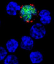
among uninfected cells (blue)
Image courtesy of NIH/
Benjamin Chaigne-Delalande
PHILADELPHIA—Cytotoxic T lymphocytes designed to target Epstein-Barr virus (EBV-CTLs) can elicit durable responses in patients
with EBV–associated lymphoproliferative disorder (EBV-LPD), according to data presented at the AACRAnnual Meeting 2015.
Results from two trials showed that EBV-CTLs derived from a patient’s transplant donor could produce a response rate of 62%, and EBV-CTLs derived from third-party donors could produce a response rate of 61%.
Study investigators noted that, with the achievement of complete response (CR), remission proved durable. And, unlike with chemotherapy, partial responses (PRs) to EBV-CTLs were durable as well.
The team presented these results as abstract CT107.*
“The purpose of our clinical trials was to see if giving T cells from a normal-immune individual that were expanded in culture and stimulated to respond to multiple proteins from the Epstein-Barr virus could provide a safe and effective treatment,” said Richard J. O’Reilly, MD, of Memorial Sloan Kettering Cancer Center in New York.
“The good news from our two clinical trials is that EBV-CTLs generated from either the patient’s transplant donor or from the bank of normal donor T cells developed at Memorial Sloan Kettering put aggressive EBV-LPD that had failed to respond to rituximab into long-lasting remission in more than 60% of patients.”
In the first trial, 26 patients with EBV-LPD received EBV-CTLs generated from their transplant donor. Thirteen of these patients had previously received rituximab, and 16 had high-risk disease.
Thirteen patients in this trial received HLA-matched, EBV-CTLs from the Memorial Sloan Kettering Cancer Center bank of EBV-CTLs generated from third-party, healthy donors. All 13 patients had high-risk disease, and 12 had received prior rituximab.
Dr O’Reilly noted that good results were observed with EBV-CTLs from both sources in this trial. And because EBV-CTLs from the bank are available immediately, he and his team used only EBV-CTLs from the bank when treating the 18 patients enrolled in the second trial.
Among the 39 patients enrolled in the first trial, 23 had a CR, none had a PR, and 2 had stable disease.
For patients who received EBV-CTLs from their primary donor, the combined rate of CR and PR was 62% (16 CRs). For patients who received third-party EBV-CTLs, the combined rate of CR and PR was 54% (7 CRs).
Sixteen of the patients who achieved a CR are still doing well, Dr O’Reilly said. Eight of these patients are alive more than 5 years after receiving EBV-CTLs, and 1 is alive more than 10 years after treatment.
Among the 18 patients enrolled in the second trial, 9 had a CR, 3 had a PR, and 1 had stable disease. The combined rate of CR and PR was 67%.
All of the patients who achieved a CR in this trial continue to do well, and the investigators will be following them long-term, Dr O’Reilly said.
He also noted that toxicities with EBV-CTLs were minimal, and there were no treatment-related deaths. None of the patients developed cytokine release syndrome or graft-vs-host disease requiring systemic therapy.
“The EBV-CTLs work well for the majority of recipients,” Dr O’Reilly said. “However, the responses became clinically evident only after the T cells expanded in vivo, which took about 7 to 14 days. We are rigorously pursuing the development of biomarkers or other tests to predict response earlier.”
Memorial Sloan Kettering Cancer Center has entered into an option agreement with Atara Biotherapeutics to further develop EBV-CTLs for clinical use. However, the data presented at AACR were accrued prior to that agreement.
Last month, the US Food and Drug Administration granted breakthrough therapy designation to EBV-CTLs generated from third-party donors for the treatment of patients with rituximab-refractory EBV-LPD. ![]()
*Information in the abstract differs from that presented at the meeting.
New mAb can overcome resistance to other mAbs

Photo courtesy of the
University of Southampton
A newly developed monoclonal antibody (mAb) can reverse resistance to other mAbs in chronic lymphocytic leukemia (CLL) and mantle cell lymphoma (MCL), according to research published in Cancer Cell.
Investigators found that some cancer cells draw mAbs inside themselves, making them invisible to immune cells.
But a mAb called BI-1206 can prevent this process and enhance cancer killing by binding to a molecule called FcγRIIB.
In preclinical experiments, BI-1206 was able to overcome resistance to mAbs such as rituximab.
“With more monoclonal antibody treatments being developed, there is an urgent need to understand how tumors become resistant to them and develop ways to overcome it,” said study author Mark Cragg, PhD, of the University of Southampton in the UK.
“Not only does BI-1206 appear to be able to reverse resistance to a range of monoclonal antibodies, it is also effective at directly killing cancer cells itself.”
In the Cancer Cell paper, BI-1206 is referred to as 6G11. The investigators found that 6G11 can block rituximab internalization and has “potent antitumor activity” in vitro. 6G11 was also well-tolerated and did not prompt cytokine storm.
In a mouse model of CLL, 6G11 enhanced rituximab-mediated depletion of primary CLL cells and improved responses when compared to rituximab alone.
In mice engrafted with cells from patients with CLL that was refractory to rituximab, ofatumumab, and/or alemtuzumab, 6G11 alone depleted CLL cells but did not improve overall response rates compared to rituximab alone. However, 6G11 in combination with rituximab did improve overall response rates compared to rituximab alone.
In a mouse model of MCL, neither 6G11 nor rituximab alone improved long-term survival. However, 30% of mice treated with both drugs survived tumor-free out to 100 days.
Combining 6G11 with obinutuzumab significantly improved splenic tumor cell depletion in mice with CLL. And more than 90% of mice that received 6G11 and alemtuzumab had a complete response to the treatment.
The investigators said these data suggest 6G11 can overcome mAb resistance for multiple targets. They said the drug will be tested in patients with CLL and non-Hodgkin lymphoma in an early stage clinical trial. ![]()
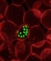
Mutation linked to drug-resistant malaria in Africa

Image by Ute Frevert
and Margaret Shear
New research provides genetic evidence that malaria parasites in Africa are developing resistance to antimalarial drugs.
Researchers found that Plasmodium falciparum parasites with a mutation in the gene ap2mu were less sensitive to both artemisinin and quinine.
A study published in 2013 suggested a link between a mutation in ap2mu and low levels of malaria parasites remaining in the blood of Kenyan children after treatment with artemisinin.
However, further research was needed to confirm that these genetic characteristics represented an early step toward resistance.
In the new study, published in Antimicrobial Agents and Chemotherapy, researchers genetically altered the malaria parasite to mutate ap2mu in the same way that had been observed in Kenya.
The team found the altered parasite was significantly less susceptible to treatment, requiring 32% more drug to be killed by artemisinin. The genetically altered parasite was also 42.4% less susceptible to quinine.
Earlier this year, a different research group discovered mutations in the gene kelch13, which were linked to reduced susceptibility to artemisinin combination treatment in South East Asia.
Historically, resistance to antimalarial medicines has emerged in South East Asia and then spread to Africa. But these new findings suggest a different route to drug resistance may be developing independently in Africa.
“Our findings could be a sign of much worse things to come for malaria in Africa,” said study author Colin Sutherland, PhD, of the London School of Hygiene & Tropical Medicine in the UK.
“The malaria parasite is constantly evolving to evade our control efforts. We’ve already moved away from using quinine to treat cases as the malaria parasite has become more resistant to it, but if further drug resistance were to develop against our most valuable malaria drug, artemisinin, we would be facing a grave situation.”
“We now know that the gene ap2mu is an important factor in determining how well our drugs kill malaria parasites. We will be conducting laboratory and field studies to more accurately measure the impact of mutations in the ap2mu gene. We hope our findings will help [us] understand resistance of malaria to drugs and potentially be an important tool for monitoring malaria treatment in the future.” ![]()
Explaining drug-resistant malaria
infecting a red blood cell
Image courtesy of St. Jude
Children’s Research Hospital
Researchers say they have identified a molecular mechanism responsible for making malaria parasites resistant to artemisinins, the leading class of antimalarial drugs.
The team found that a kinase, Plasmodium falciparum phosphatidylinositol-3-kinase (PfPI3K), and its lipid product, phosphatidylinositol-3-phosphate (PI3P), play key roles in artemisinin resistance.
So targeting PfPI3K or PI3P could potentially treat resistant Plasmodium falciparum malaria.
Alassane Mbengue, PhD, of the University of Notre Dame in Indiana, and his colleagues described this research in a letter to Nature.
“We observed that levels of [PI3P] were higher in artemisinin-resistant P falciparum than artemisinin-sensitive strains,” Dr Mbengue said. “This lipid is produced by an enzyme called PfPI3K. We found that artemisinins block this kinase from producing PI3P lipids. We also discovered that the amount of the kinase present in the parasite is controlled by the gene PfKelch13.”
“Mutation in the gene increases the kinase levels, which, in turn, increases PI3P lipid levels. The higher the level of PI3P lipids present in the parasite, the greater the level of artemisinin resistance. We also studied the lipid levels in parasites without the gene mutation and observed that when PI3P lipid levels were increased artificially, the parasites still became proportionately resistant.”
Specifically, the researchers found that increased PfPI3K was associated with the C580Y mutation in PfKelch13. The mutation reduced polyubiquitination of PfPI3K and its binding to PfKelch13, which limited proteolysis of PfPI3K and led to increased levels of both PfPI3K and PI3P.
The team found that PI3P levels were predictive of artemisinin resistance in clinical and engineered parasites. And although increases in PI3P levels induced artemisinin resistance in the absence of PfKelch13 mutations, PI3P levels were still responsive to regulation by PfKelch13.
Dr Mbengue and his colleagues said the next step for this research is to identify drugs that can kill P falciparum by preventing PfPI3K from making PI3P or disrupting the production of the kinase itself. ![]()
Tumor sequencing fails to paint the whole picture

Image courtesy of NIGMS
Many of the genetic alterations revealed by sequencing a cancer patient’s tumor DNA are not actually associated with the cancer, according to a study published in Science Translational Medicine.
In fact, these alterations are inherited germline mutations already present in an individual’s normal cells.
To make this discovery, researchers compared DNA from tumors and normal cells in 815 patients with hematologic and solid tumor malignancies.
Almost half of the patients analyzed using tumor-only approaches had genetic alterations in their tumors that were also present in their normal cells, which suggests the alterations were “false-positive” changes not specific to the tumor.
As personalized medicine is predicated on tailoring treatments to the genetic makeup of a patient’s tumor, the high rate of false-positives uncovered in this study has implications for the accuracy of the approach when it relies on tumor-only sequencing, the researchers said.
“We knew from our pioneering whole-exome analyses of cancer patients that a significant number of the genetic alterations that were thought to be associated with tumors were also present in the inherited germline DNA,” said Siân Jones, PhD, of Personal Genome Diagnostics in Baltimore, Maryland.
“By comparing tumor DNA to DNA from normal tissue, we were able to separate out those genetic alterations that are truly tumor-specific. Accurately identifying tumor-specific alterations is essential to realizing the potential of personalized medicine to achieve better treatment outcomes.”
The researchers identified 382 genetic alterations that were potentially tumor-specific by first detecting all of the genetic changes in patients’ tumors and then eliminating those that were well-known germline alterations.
However, when the remaining alterations were compared to the genomic profiles of the patients’ germline DNA, an average of 249, or 65%, turned out to be false-positive changes that were already present in the normal cells.
The researchers also looked at the alterations in actionable genes, which have been identified as potential targets for approved or investigational cancer therapies. They found that almost half of the tumor samples (48%) had at least one false-positive mutation in an actionable gene.
The use of false-positive findings to guide personalized treatment decisions could result in a substantial number of patients receiving therapies that are not optimized for their cancer, according to the researchers. Therefore, it seems sequencing normal DNA alongside tumor DNA is essential.
In addition to selecting personalized therapies for cancer patients, sequencing normal DNA can increase the overall understanding of cancer, including finding cancer predisposition due to germline genome changes, said Victor Velculescu, MD, PhD, of Johns Hopkins University School of Medicine in Baltimore, Maryland.
In this study, the germline analyses revealed changes in cancer-related genes in 3% of the patients who had no known signs of genetically linked cancer.
“These analyses can help us find alterations in cancer-predisposing genes in ways that weren’t previously appreciated,” Dr Velculescu said.
He also acknowledged, however, that there are challenges to implementing tumor-normal DNA analyses in a clinical setting. These include the additional work and costs of sequencing and analyzing a patient’s normal tissue along with his or her tumor tissue.
Costs for tumor gene sequencing begin at several thousand dollars, which would increase if sequencing was done on DNA from normal tissue as well. And health insurance may not fully cover normal-tissue genetic sequencing. ![]()
Technique helps determine DLBCL subtypes

Photo by Darren Baker
A new technique can quickly and easily differentiate the two major subtypes of diffuse large B-cell lymphoma (DLBCL), researchers have reported in The Journal of Molecular Diagnostics.
The method is based on a reverse transcriptase multiplex ligation-dependent probe amplification (RT-MLPA) assay and 14 gene signatures.
It proved as accurate as the gold standard technology for identifying germinal center B-cell-like (GCB) and activated B-cell-like (ABC) DLBCL.
So the researchers believe this RT-MLPA-based assay could help clinicians evaluate a patient’s prognosis and choose the optimal therapy for a person with DLBCL.
“Differences in the progression of the disease and clinical outcomes can, at least in part, be explained by the heterogeneity of lymphoma, which can be classified into two major subtypes with different outcomes,” said study author Philippe Ruminy, PhD, of the University of Rouen in France.
“Unfortunately, these lymphomas are morphologically undistinguishable in routine diagnosis, which is a major problem for the development of targeted therapies. Furthermore, array-based gene expression profiling (GEP), which is considered the gold standard for discriminating these tumors, remains poorly transposable to routine diagnosis, and the surrogate immunohistochemical (IHC) algorithms that have been proposed are often considered poorly reliable.”
With this in mind, Dr Ruminy and his colleagues tested the RT-MLPA-based assay alongside GEP techniques in samples from 259 patients with de novo DLBCL.
The team used a series of 195 patients from a single institution to train and validate the new technique. Of these patients, 115 had a classification determined with a Veracode DASL assay, 100 had an IHC classification, and 135 had survival data. A second series of 64 patients that were previously analyzed by Affymetrix U133+2 GEP arrays served as an external validation cohort.
When the researchers looked at 50 patients randomly selected from the training cohort, they found that the RT-MLPA-based assay classified 90% of the cases within the expected subtypes (20 GCB and 25 ABC). The remaining 10% (1 GCB and 4 ABC) were considered unclassified.
In the external validation cohort, the RT-MLPA-based assay classified 80% of cases within the expected subtypes (24 ABC and 28 GCB). However, 13.8% were considered unclassified (3 GCB and 6 ABC), and 4 were misclassified.
The researchers also found the RT-MLPA-based assay was sensitive for analyzing archived formalin-fixed, paraffin-embedded (FFPE) tissue samples. A comparison of samples from paired frozen and FFPE biopsies showed that the technique correctly classified 89.3% of 28 cases.
“Because RT-MLPA requires only short cDNA fragments for the correct binding and ligation of the gene-specific oligonucleotide probes, it is less affected by the use of low RNA concentrations and RNA degradation,” Dr Ruminy said.
“It could thus be used for the retrospective analysis of archival collections and for the inclusion of patients in prospective clinical trials, because only a few institutions routinely collect frozen biopsy material.”
To evaluate the prognostic value of the assay, the researchers looked at survival in 135 treated DLBCL patients who were diagnosed between 2001 and 2011.
Patients determined to have the ABC subtype by the RT-MLPA-based assay had significantly worse progression-free survival and overall survival than patients with the GCB subtype. And the expression of several individual genes within the MLPA signature was significantly associated with prognosis (ie, high LMO2, high BCL6, and low TNFRSF13B expression).
“The robust and cost-effective RT-MLPA assay can yield results within one day and requires reagents costing less than $5 per sample,” Dr Ruminy said. “Since RT-MLPA utilizes materials and equipment that are standard in many laboratories, the process can easily be implemented for routine use.” ![]()
Photo by Darren Baker
A new technique can quickly and easily differentiate the two major subtypes of diffuse large B-cell lymphoma (DLBCL), researchers have reported in The Journal of Molecular Diagnostics.
The method is based on a reverse transcriptase multiplex ligation-dependent probe amplification (RT-MLPA) assay and 14 gene signatures.
It proved as accurate as the gold standard technology for identifying germinal center B-cell-like (GCB) and activated B-cell-like (ABC) DLBCL.
So the researchers believe this RT-MLPA-based assay could help clinicians evaluate a patient’s prognosis and choose the optimal therapy for a person with DLBCL.
“Differences in the progression of the disease and clinical outcomes can, at least in part, be explained by the heterogeneity of lymphoma, which can be classified into two major subtypes with different outcomes,” said study author Philippe Ruminy, PhD, of the University of Rouen in France.
“Unfortunately, these lymphomas are morphologically undistinguishable in routine diagnosis, which is a major problem for the development of targeted therapies. Furthermore, array-based gene expression profiling (GEP), which is considered the gold standard for discriminating these tumors, remains poorly transposable to routine diagnosis, and the surrogate immunohistochemical (IHC) algorithms that have been proposed are often considered poorly reliable.”
With this in mind, Dr Ruminy and his colleagues tested the RT-MLPA-based assay alongside GEP techniques in samples from 259 patients with de novo DLBCL.
The team used a series of 195 patients from a single institution to train and validate the new technique. Of these patients, 115 had a classification determined with a Veracode DASL assay, 100 had an IHC classification, and 135 had survival data. A second series of 64 patients that were previously analyzed by Affymetrix U133+2 GEP arrays served as an external validation cohort.
When the researchers looked at 50 patients randomly selected from the training cohort, they found that the RT-MLPA-based assay classified 90% of the cases within the expected subtypes (20 GCB and 25 ABC). The remaining 10% (1 GCB and 4 ABC) were considered unclassified.
In the external validation cohort, the RT-MLPA-based assay classified 80% of cases within the expected subtypes (24 ABC and 28 GCB). However, 13.8% were considered unclassified (3 GCB and 6 ABC), and 4 were misclassified.
The researchers also found the RT-MLPA-based assay was sensitive for analyzing archived formalin-fixed, paraffin-embedded (FFPE) tissue samples. A comparison of samples from paired frozen and FFPE biopsies showed that the technique correctly classified 89.3% of 28 cases.
“Because RT-MLPA requires only short cDNA fragments for the correct binding and ligation of the gene-specific oligonucleotide probes, it is less affected by the use of low RNA concentrations and RNA degradation,” Dr Ruminy said.
“It could thus be used for the retrospective analysis of archival collections and for the inclusion of patients in prospective clinical trials, because only a few institutions routinely collect frozen biopsy material.”
To evaluate the prognostic value of the assay, the researchers looked at survival in 135 treated DLBCL patients who were diagnosed between 2001 and 2011.
Patients determined to have the ABC subtype by the RT-MLPA-based assay had significantly worse progression-free survival and overall survival than patients with the GCB subtype. And the expression of several individual genes within the MLPA signature was significantly associated with prognosis (ie, high LMO2, high BCL6, and low TNFRSF13B expression).
“The robust and cost-effective RT-MLPA assay can yield results within one day and requires reagents costing less than $5 per sample,” Dr Ruminy said. “Since RT-MLPA utilizes materials and equipment that are standard in many laboratories, the process can easily be implemented for routine use.”
Team uncovers new role for macrophages

pseudopodia to engulf particles
Macrophages can substitute for dendritic cells as primers of T-cell-dependent immune responses, according to research published in Proceedings of the National Academy of Sciences.
The study showed that macrophages that function as a first line of defense in the innate immune system can also present antigens to T cells, a previously unknown role for macrophages in the induction of adaptive immune responses.
“It has been assumed until now that the dendritic cells are considered to be essentially the only cell type responsible for antigen presentation in the immune system,” said Dr Thomas Brocker, of Ludwig-Maximilians-Universität München in Germany.
“We have now discovered that macrophages can also do this job. Not only that, in certain situations, they can be more effective than dendritic cells.”
Dendritic cells present antigens to cytotoxic T lymphocytes (CTLs) if they have been directly infected, but they can also capture and display antigens from other cells. This type of indirect antigen presentation is referred to as cross-presentation.
“So, theoretically, dendritic cells could be responsible for the induction of all CTL-based responses, regardless of whether they are themselves infected or not,” Dr Brocker said. “But the significance of cross-presentation is hotly debated in the literature.”
Dr Brocker and his team used several antigens that were specifically targeted to and processed by macrophages but could not be taken up directly by dendritic cells. They were able to demonstrate that each antigen induced a normal immune response in a mouse model system, and even in a mouse strain that lacked dendritic cells altogether.
Further experiments showed the targeted macrophages were actually able to prime a more comprehensive immune reaction than cross-presenting dendritic cells. They activated T cells specific for all antigen-binding sites (epitopes) presented, whereas cross-presentation by dendritic cells stimulates only those T cells that recognize immunodominant epitopes.
“Macrophages naturally function as filters,” Dr Brocker noted. “They gobble everything up that might be harmful to the organism. And our study shows that, in contrast to cross-priming dendritic cells, they are capable of producing and presenting all T-cell-priming epitopes we tested. Macrophages therefore induce a complete immune response. These observations indicate that the significance of cross-presentation by dendritic cells has been overrated.”
He added that these findings are relevant for the development of immunization strategies.
“Preclinical trials are already underway with vaccines that are designed to activate specific sets of dendritic cells,” Dr Brocker said. “But the weak epitopes are important for a broadly directed immune response, because they can potentially recognize mutant variants of viruses, for instance.”
“Cross-priming dendritic cells fail to induce weakly antigenic epitopes, as our study shows. Our results indicate that it may make more sense to manipulate macrophages directly because they stimulate a wider-ranging immune response.”
RCOG releases new VTE guidelines

Photo by Nina Matthews
The Royal College of Obstetricians and Gynaecologists (RCOG) has released new guidelines for treating and preventing venous thromboembolism (VTE) during pregnancy, birth, and following delivery.
“These updated guidelines provide new evidence about risk factors for thrombosis in pregnancy and strategies that should be employed to reduce the chances of a thrombosis occurring,” said Andrew Thomson, MD, cochair of the RCOG guidelines committee.
“Furthermore, the guidelines provide updated information on the way women with a suspected thrombosis should be investigated and treated.”
VTE is uncommon in pregnancy or in the first 6 weeks postnatally, and the absolute risk of VTE is around 1 in 1000 pregnancies. It can occur at any stage in pregnancy, but the time of the highest risk is the first 6 weeks following birth, when the risk increases 20-fold.
Risk factors include previous VTE or thrombophilia, obesity, increased maternal age, immobility and long-distance travel, admission to hospital during pregnancy, and other comorbidities such as heart disease, inflammatory bowel disease, and pre-eclampsia.
Additional risk factors occurring during the first trimester of pregnancy include hyperemesis gravidarum, ovarian hyperstimulation, and in vitro fertilization pregnancy. Caesarean section is also a risk factor.
The guidelines emphasize that all women should undergo a thorough assessment for VTE in early pregnancy or prepregnancy and again intrapartum or immediately postpartum.
Any woman with risk factors should be considered for prophylactic low-molecular-weight heparin. The duration of treatment depends on the number of risk factors a woman has. It may be offered both antenatally and after the baby is born.
In addition, women with previous VTE must be offered prepregnancy counseling. A prospective management plan for VTE should also be made, including appropriate treatment to be offered as early as possible and a careful history documented.
The guidance on treating VTE focuses on the acute management of the condition and highlights the signs and symptoms, including leg pain and swelling, lower abdominal pain, shortness of breath, chest pain, coughing blood, and collapse.
Any woman presenting with signs and symptoms suggestive of VTE should be tested for the condition immediately and offered treatment with low-molecular-weight heparin.
All hospitals should have a protocol for the diagnosis of suspected VTE, with the involvement of a multidisciplinary team of obstetricians, radiologists, physicians, and hematologists.
“This guidance provides clinicians with accurate, scientific-based guidelines on the risk factors for VTE, as well as on how to prevent and treat the condition,” said guideline author Catherine Nelson-Piercy, MBBS, of Guy’s and St. Thomas’ NHS Foundation Trust in London, UK.
“It is vital that VTE is discussed with all women who are at risk, and the reasons for individual treatment recommendations must also be explained.”
A new treatment approach for ALK- ALCL?

Investigators say they have uncovered a new approach for treating ALK-negative anaplastic large cell lymphoma (ALCL).
Results of genomic analyses indicated that many cases of ALK-negative ALCL may be driven by alterations in the JAK/STAT3 pathway, and in vivo
experiments showed the disease can be inhibited by compounds that target this pathway.
The investigators believe these compounds could be more effective than current therapies.
“Current therapies for this form of lymphoma fail to work in the majority of cases,” said Raul Rabadan, PhD, of Columbia University in New York, New York.
“However, now that we know the mutations that drive a significant percentage of cases, we can envision a new, personalized genomic approach to the treatment of ALK-negative ALCL.”
Dr Rabadan and his colleagues recounted their discovery of the mutations in Cancer Cell.
The team had sequenced the exomes and RNA of cancer cells from 155 patients with ALCL and 74 control subjects with other types of lymphoma.
Results revealed mutations in either JAK1 or STAT3 in about 20% of the 88 patients with ALK-negative ALCL. Of that 20%, 38% had mutations in both genes.
The investigators also detected the presence of several novel gene fusions (NFκB2-ROS1, NFκB2-TYK2, NCOC2-ROS1, and PABPC4-TYK2), some of which appear to activate the JAK/STAT3 pathway (NFκB2-ROS1 and NFκB2-TYK2).
Patients with these fusions did not have JAK1 or STAT3 mutations, which suggests the fusions are an independent cause of ALK-negative ALCL.
To confirm whether JAK1 and STAT3 mutations can cause ALK-negative ALCL, the investigators induced these mutations in normal human cells. The mutations did, in fact, lead to diseased cells.
Finally, the team tested JAK/STAT3 pathway inhibitors—ruxolitinib and PUH71—in mouse models of ALK-negative ALCL. And they found that both drugs significantly inhibited tumor growth.
“Our findings demonstrate that drugs targeting the JAK/STAT3 pathway offer a viable therapeutic strategy in a subset of patients with ALCL,” Dr Rabadan said.
“A couple of JAK/STAT3 inhibitors have been approved by the FDA for the treatment of psoriasis and rheumatoid arthritis, and several more are currently in clinical trials. These could be tested in patients whose genetic profile matches those we identified in our study.”